oxytocin modulates hippocampal perfusion in people at ... · uncorrected proof article open...
TRANSCRIPT

UNCORRECTED PROOF
ARTICLE OPEN
Oxytocin modulates hippocampal perfusion in people atclinical high risk for psychosisCathy Davies1, Yannis Paloyelis 2, Grazia Rutigliano1, Marco Cappucciati1, Andrea De Micheli1,3, Valentina Ramella-Cravaro 1,Umberto Provenzani1,4, Mathilde Antoniades3,5, Gemma Modinos 2,5, Dominic Oliver1, Daniel Stahl 6, Silvia Murguia7,Fernando Zelaya2, Paul Allen5,8, Sukhi Shergill5, Paul Morrison5, Steve Williams2, David Taylor9, Philip McGuire3,5,10 andPaolo Fusar-Poli1,3,4,10
Preclinical and human studies suggest that hippocampal dysfunction is a key factor in the onsetQ1 of psychosis. People at ClinicalHigh Risk for psychosis (CHR-P) present with a clinical syndrome that can include social withdrawal and have a 20–35% risk ofdeveloping psychosis in the next 2 years. Recent research shows that resting hippocampal blood flow is altered in CHR-P individualsand predicts adverse clinical outcomes, such as non-remission/transition to frank psychosis. Previous work in healthy malesindicates that a single dose of intranasal oxytocin has positive effects on social function and marked effects on resting hippocampalblood flow. The present study examined the effects of intranasal oxytocin on hippocampal blood flow in CHR-P individuals. In adouble-blind, placebo-controlled, crossover design, 30 CHR-P males were studied using pseudo-continuous Arterial Spin Labellingon 2 occasions, once after 40IU intranasal oxytocin and once after placebo. The effects of oxytocin on left hippocampal blood flowwere examined in a region-of-interest analysis of data acquired at 22–28 and at 30–36minutes post-intranasal administration.Relative to placebo, administration of oxytocin was associated with increased hippocampal blood flow at both time points (p= .0056; p= .034), although the effect at the second did not survive adjustment for the effect of global blood flow. These dataindicate that oxytocin can modulate hippocampal function in CHR-P individuals and therefore merits further investigation as acandidate novel treatment for this group.
Neuropsychopharmacology (2019) 0:1–10; https://doi.org/10.1038/s41386-018-0311-6
INTRODUCTIONAt present, there is a lack of effective treatments for individuals atClinical High Risk of Psychosis (CHR-P [1]). Recent studies suggestthat existing interventions do not significantly impact ontransition to psychosis [2], attenuated positive [3] or negativesymptoms [4], or social and functional outcomes [5]. Noveltreatments for this population are therefore needed [6].A substantial body of scientific work places aberrant hippo-
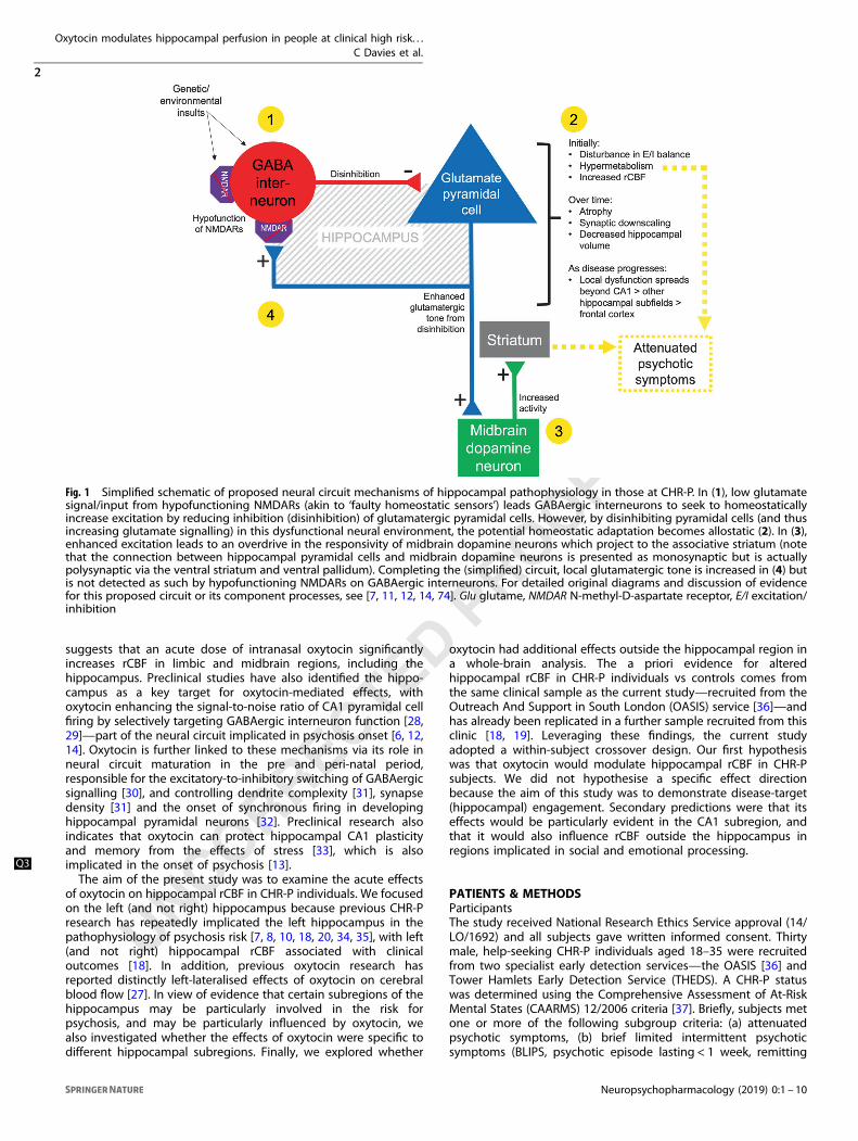
campal structure and function at the core of neurobiologicalmechanisms underlying the onset of psychosis [7]. Evidence frompost-mortem, neuroimaging and preclinical research suggests thatthe onset of attenuated psychotic symptoms may be driven bydysregulated glutamate neurotransmission in the Cornu Ammonis1 (CA1) region of the hippocampus, which is thought to lead tohypermetabolism and altered (increased) blood flow [7–10].Enhanced glutamatergic tone in CA1 induces allostatic adapta-tions [11] in γ-aminobutyric acid (GABA)-ergic neurotransmission,with consequent disinhibition of pyramidal neurons (Fig. 1) [12].These changes may lead to disturbed neural excitation/inhibition
balance, and via polysynaptic projection pathways to themidbrain/striatum, to midbrain hyperdopaminergia [13, 14]. Asthe CHR-P state progresses to the first episode of psychosis, thefunctional perturbations once localised to (particularly the left)CA1 may spread to extra-hippocampal regions such as the frontalcortex [7, 8], and excitotoxic as well as atrophic processesculminate in hippocampal volume loss -structural changes-beginning in CA1 [15–17]. These findings are consistent withevidence that CHR-P individuals show increased resting regionalcerebral blood flow (rCBF) in the hippocampus relative to controls[18, 19], and normalisation (reduction) of left hippocampal rCBF isassociated with remission from the CHR-P state [18]. HippocampalrCBF in CHR-P individuals has also been correlated with corticalGABA levels [20]. Q2
The neuropeptide oxytocin is a key modulator of social, sexualand emotional processes [21], including hypothalamic-pituitary-adrenal axis regulation [22], emotion recognition [23], socialmemory [24] and reward, and possesses anxiolytic [21, 22] andprosocial properties [25, 26]. Previous work in healthy males [27]
Received: 21 October 2018 Revised: 27 November 2018 Accepted: 18 December 2018
1Early Psychosis: Interventions & Clinical-detection (EPIC) Lab, Department of Psychosis Studies, Institute of Psychiatry, Psychology & Neuroscience, King’s College London,London, UK; 2Department of Neuroimaging, Institute of Psychiatry, Psychology & Neuroscience, King’s College London, London, UK; 3National Institute for Health Research (NIHR)Biomedical Research Centre (BRC), South London and Maudsley NHS Foundation Trust, London, UK; 4Department of Brain and Behavioral Sciences, University of Pavia, Pavia, Italy;5Department of Psychosis Studies, Institute of Psychiatry, Psychology & Neuroscience, King’s College London, London, UK; 6Department of Biostatistics and Health Informatics,Institute of Psychiatry, Psychology & Neuroscience, King’s College London, London, UK; 7Tower Hamlets Early Detection Service (THEDS), East London NHS Foundation Trust,London, UK; 8Department of Psychology, University of Roehampton, London, UK; 9Institute of Pharmaceutical Science, King’s College London, London, UK and 10Outreach AndSupport in South London (OASIS) Service, South London and Maudsley NHS Foundation Trust, London, UKCorrespondence: Paolo Fusar-Poli ([email protected])
www.nature.com/npp
© American College of Neuropsychopharmacology 2019
UNCORRECTED PROOF
suggests that an acute dose of intranasal oxytocin significantlyincreases rCBF in limbic and midbrain regions, including thehippocampus. Preclinical studies have also identified the hippo-campus as a key target for oxytocin-mediated effects, withoxytocin enhancing the signal-to-noise ratio of CA1 pyramidal cellfiring by selectively targeting GABAergic interneuron function [28,29]—part of the neural circuit implicated in psychosis onset [6, 12,14]. Oxytocin is further linked to these mechanisms via its role inneural circuit maturation in the pre and peri-natal period,responsible for the excitatory-to-inhibitory switching of GABAergicsignalling [30], and controlling dendrite complexity [31], synapsedensity [31] and the onset of synchronous firing in developinghippocampal pyramidal neurons [32]. Preclinical research alsoindicates that oxytocin can protect hippocampal CA1 plasticityand memory from the effects of stress [33], which is alsoimplicated in the onset of psychosis [13].Q3
The aim of the present study was to examine the acute effectsof oxytocin on hippocampal rCBF in CHR-P individuals. We focusedon the left (and not right) hippocampus because previous CHR-Presearch has repeatedly implicated the left hippocampus in thepathophysiology of psychosis risk [7, 8, 10, 18, 20, 34, 35], with left(and not right) hippocampal rCBF associated with clinicaloutcomes [18]. In addition, previous oxytocin research hasreported distinctly left-lateralised effects of oxytocin on cerebralblood flow [27]. In view of evidence that certain subregions of thehippocampus may be particularly involved in the risk forpsychosis, and may be particularly influenced by oxytocin, wealso investigated whether the effects of oxytocin were specific todifferent hippocampal subregions. Finally, we explored whether
oxytocin had additional effects outside the hippocampal region ina whole-brain analysis. The a priori evidence for alteredhippocampal rCBF in CHR-P individuals vs controls comes fromthe same clinical sample as the current study—recruited from theOutreach And Support in South London (OASIS) service [36]—andhas already been replicated in a further sample recruited from thisclinic [18, 19]. Leveraging these findings, the current studyadopted a within-subject crossover design. Our first hypothesiswas that oxytocin would modulate hippocampal rCBF in CHR-Psubjects. We did not hypothesise a specific effect directionbecause the aim of this study was to demonstrate disease-target(hippocampal) engagement. Secondary predictions were that itseffects would be particularly evident in the CA1 subregion, andthat it would also influence rCBF outside the hippocampus inregions implicated in social and emotional processing.
PATIENTS & METHODSParticipantsThe study received National Research Ethics Service approval (14/LO/1692) and all subjects gave written informed consent. Thirtymale, help-seeking CHR-P individuals aged 18–35 were recruitedfrom two specialist early detection services—the OASIS [36] andTower Hamlets Early Detection Service (THEDS). A CHR-P statuswas determined using the Comprehensive Assessment of At-RiskMental States (CAARMS) 12/2006 criteria [37]. Briefly, subjects metone or more of the following subgroup criteria: (a) attenuatedpsychotic symptoms, (b) brief limited intermittent psychoticsymptoms (BLIPS, psychotic episode lasting < 1 week, remitting
Fig. 1 Simplified schematic of proposed neural circuit mechanisms of hippocampal pathophysiology in those at CHR-P. In (1), low glutamatesignal/input from hypofunctioning NMDARs (akin to ‘faulty homeostatic sensors’) leads GABAergic interneurons to seek to homeostaticallyincrease excitation by reducing inhibition (disinhibition) of glutamatergic pyramidal cells. However, by disinhibiting pyramidal cells (and thusincreasing glutamate signalling) in this dysfunctional neural environment, the potential homeostatic adaptation becomes allostatic (2). In (3),enhanced excitation leads to an overdrive in the responsivity of midbrain dopamine neurons which project to the associative striatum (notethat the connection between hippocampal pyramidal cells and midbrain dopamine neurons is presented as monosynaptic but is actuallypolysynaptic via the ventral striatum and ventral pallidum). Completing the (simplified) circuit, local glutamatergic tone is increased in (4) butis not detected as such by hypofunctioning NMDARs on GABAergic interneurons. For detailed original diagrams and discussion of evidencefor this proposed circuit or its component processes, see [7, 11, 12, 14, 74]. Glu glutame, NMDAR N-methyl-D-aspartate receptor, E/I excitation/inhibition
Oxytocin modulates hippocampal perfusion in people at clinical high risk. . .C Davies et al.
2
Neuropsychopharmacology (2019) 0:1 – 10
1234567890();,:

UNCORRECTED PROOF
without treatment), or (c) either schizotypal personality disorder orfirst-degree relative with psychosis [37], all coupled withfunctional decline. Individuals were excluded if there was ahistory of previous psychotic disorder (with the exception of BLIPS,some of whom may meet acute and transient psychotic disordercriteria [38]) or manic episode, exposure to antipsychotics,neurological disorder or current substance-use disorder, estimatedIQ < 70, acute intoxication on the day of scanning, and anycontraindications to magnetic resonance imaging (MRI) orintranasal oxytocin or placebo. History of Axis I disorder(s) wasnot an exclusion criterion due to the transdiagnostic nature of theCHR-P state and the high prevalence of such diagnoses withinthese populations [39].Q4
Design, materials, procedureWe used a randomised, double-blind, 40 IU intranasal oxytocin vsplacebo single-dose challenge in a crossover design (1-week washout). During each challenge, subjects underwent an MRI scanwhich started at 1130 h to minimise potential effects of diurnalvariation in oxytocin or vasopressin [27]. Anxiety was measuredusing the State-Trait Anxiety Inventory (STAI) prior to each scan(and prior to intranasal administration) so that pre-scan anxietyscore could be included in statistical models as a covariate. Fordescriptive purposes, we also collected information on medicationhistory, use of alcohol (Alcohol Use Disorders IdentificationTest, tobacco and cannabis, functioning using the GlobalFunctioning Role and Social scales [40] and later transition status.Intranasal administration followed recommended guidelines anda protocol adopted by a previous study conducted at our institute[27]. Briefly, participants self-administered one puff (4 IU) ofintranasal oxytocin or matched placebo every 30 s, alternatingbetween nostrils, until 40 IU had been administered (Supplemen-tary Materials and Methods). During the scan, participantswere asked to maintain their gaze on a centrally-placed fixationcross.
MRI acquisition and image processingAll scans were conducted on a General Electric Discovery MR750 3Tesla system (General Electric, Chicago, USA) using a 32-channelhead coil. Measurement of Cerebral Blood Flow (CBF) was carriedout using a 3D pseudo-continuous Arterial Spin Labeling (3D-pCASL) sequence during two consecutive runs: 22–28 (run 1) and30–36 (run 2) min post-intranasal administration. The timing of thetwo runs was selected based on previous findings of thespatiotemporal profile of oxytocin-induced cerebral blood flowchanges in healthy males, which demonstrated sustained effectsover a ~20–73min period (post-intranasal administration) [27]. Foreach subject, we also computed a mean (average) CBF map fromthe CBF maps for runs 1 and 2. ASL data were preprocessed usingthe Automatic Software for ASL Processing (ASAP) 2.0 toolbox [41]running in Statistical Parametric Mapping version 12 (SPM12;https://www.fil.ion.ucl.ac.uk/spm/) on Matlab R2017a. 3D-pCASLacquisition parameters and image preprocessing procedures wereconducted in line with previous studies and are detailed in theSupplementary Materials and Methods.
Statistical analysisStatistical analyses were performed in STATA SE14.2.
Pre-scan anxiety scoresFor pre-scan anxiety (STAI) scores, missing data were imputedusing next-observation-carried-backward (Supplementary Materi-als and Methods). Differences in pre-scan anxiety scores in theoxytocin vs placebo conditions was assessed using a paired t-test.In line with previous CHR-P studies [18, 19] and because anxietyhas been demonstrated to have systematic effects on CBF [42](including rCBF specifically in the hippocampus [43]), all analysesincluded mean-centred pre-scan anxiety as a covariate.Q5
Global cerebral blood flow (CBF)To measure global CBF signal, we used the ASAP toolbox toextract average CBF values from a grey matter mask for eachsubject. The ICBM-152 mask was obtained from the DARTELtoolbox in SPM and thresholded to contain voxels with a >.25probability of being grey matter. To ascertain whether global CBFwas significantly different in the oxytocin relative to placeboconditions, we conducted repeated-measures analyses of covar-iance (RM-ANCOVA) in STATA for run 1, run 2, and the mean of theruns (separately), using pre-scan anxiety as covariate (Supplemen-tary Materials and Methods). All subsequent analyses wereconducted with and without global CBF as covariate.
Hippocampal ROI rCBFEffects of oxytocin on hippocampal rCBF were determined using aregion-of-interest (ROI) approach. A left hippocampal ROI wasdefined anatomically in MNI space using the cytoarchitectonicprobabilistic atlas [44] as implemented in the Anatomy toolbox[45] in SPM (Figs. 2a, b). The ROI mask was composed of regionsCA1, CA2, CA3, dentate gyrus, and subiculum. Mean rCBF valuesfor the ROI were extracted for each subject using ASAP toolboxand entered into RM-ANCOVAs in STATA (Supplementary Materi-als and Methods). Our primary analyses tested for effects in eachof the two runs separately. However, due to the low signal-to-noise ratio inherent in ASL data, we also conducted an analysis ofthe mean effect across the two runs, which can help to reducenoise if the effects are stable [27]. We contained the family-wiseerror (FWE) rate at α= .05 using the Hochberg procedure, which isa 'sharper' and more powerful version of the Bonferroniadjustment and which allows non-independence between statis-tical tests [46]. Original p values (two-tailed) are reportedalongside indication of Hochberg correction survival (i.e., whetheror not they remain significant (survive) after accounting for FWE).Effect sizes are reported as omega-squared (ω2).
Exploratory/supplemental analysesWe used analogous procedures to those described directly aboveto extract mean rCBF values for each hippocampal subregion,using separate masks for left CA1, CA2, CA3, dentate gyrus, andsubiculum (Fig. 3a; and Supplementary Materials and Methods).No multiplicity correction was applied as subregion analyses wereexploratory. Finally, for completeness we examined whole-braineffects in runs 1, 2 and the mean of the runs—separately—usingpaired t-tests (second-level analysis) in SPM, with pre-scan anxietyand with/without global CBF signal as nuisance covariates. Weconducted a whole-brain search using cluster level inference(cluster forming threshold: p < .005; cluster reported as significantat p < .05 using FWE correction in SPM). Analyses were restrictedusing the explicit ICBM mask again thresholded to contain voxelswith >.25 probability of being grey matter.
RESULTSSample characteristicsDemographic and clinical characteristics of the sample arepresented in Table 1. All participants completed the study withno drop-outs. No adverse side effects were clinically observed.One subject was removed due to protocol violations, leaving asample of N= 29. There was a significant difference in pre-scan(pre-intranasal administration) anxiety scores in the oxytocin vsplacebo condition (oxytocin [mean ± SE]= 37.4 ± 1.9; placebo=33.4 ± 1.7; t(28)= 2.46, p= .020), which may have arisen bychance or due to slightly more individuals receiving treatmentorder oxytocin > placebo (N= 15) vs placebo > oxytocin (N= 14).
Global CBFThere was no significant difference in global grey matter CBF values(ml/100 g/min) in the oxytocin relative to the placebo condition in
Oxytocin modulates hippocampal perfusion in people at clinical high risk. . .C Davies et al.
3
Neuropsychopharmacology (2019) 0:1 – 10

UNCORRECTED PROOF
the mean of both runs (oxytocin [marginal mean ± SE]= 52.91 ± 0.91;placebo= 50.23 ± 0.91; F(1,27)= 4.00, p= .056) or run 1 (oxytocin=52.89 ± 0.94; placebo= 50.44 ± 0.94; F(1,27)= 3.14, p= .088), but asignificant difference was observed in run 2 (oxytocin= 52.94 ± 0.90;placebo= 50.03 ± 0.90; F(1,27)= 4.75, p= .038).
Hippocampal rCBFrCBF values for all runs were log transformed due to deviationsfrom distributional assumptions for parametric tests. Compared toplacebo, oxytocin administration was associated with increasedhippocampal rCBF in run 1 (F(1,27)= 9.06, p= .0056; ω2= .223),run 2 (F(1,27)= 4.96, p= .034; ω2= .124) and the mean of the 2runs (F(1,27)= 7.31, p= .012; ω2= .184), all of which survivedHochberg multiplicity correction (Figure S1). After controlling forglobal signal effects, oxytocin administration was associated withincreased hippocampal rCBF in run 1 (F(1,26)= 7.68, p= .010; ω2
= .198) which survived multiplicity correction (Fig. 2c). The effectswere no longer evident in run 2 (F(1,26)= 0.44, p= .51; Fig. 2c) orin the mean of the runs (F(1,26)= 3.27, p= .082). Exclusion ofparticipants taking antidepressants (N= 8) and benzodiazepines(N= 1) in sensitivity analyses made no material change to theunadjusted effects on hippocampal rCBF (Supplementary Materi-als and Methods).
Exploratory/supplemental analysesHippocampal subregions. Hippocampal subregion effects wereexplored in run 1 only. Relative to placebo, oxytocin administra-tion was associated with increased rCBF in all hippocampalsubregions, including CA1 (F(1,27)= 9.44, p= .0048; ω2= .232),CA2 (F(1,27)= 9.33, p= .0050; ω2= .229), CA3 (F(1,27)= 6.83,p= .014; ω2= .172), subiculum (F(1,27)= 7.61, p= .010; ω2= .191)
and particularly the dentate gyrus (F(1,27)= 10.11, p= .0037;ω2= .246)(Figure S2). After controlling for global CBF effects,oxytocin administration was associated with increased rCBF in CA1(F(1,26)= 7.29, p= .012; ω2= .189), CA2 (F(1,26)= 6.32, p= .018;ω2= .165), subiculum (F(1,26)= 6.03, p= .021; ω2= .157) anddentate gyrus (F(1,26)= 7.40, p= .011; ω2= .192), but no differ-ence was found in CA3 (F(1,26)= 3.20, p= .086)(Fig. 3b). As notedabove, these results were not corrected for multiple comparisons.
Whole-brain. Since there was no significant difference in globalsignal, unadjusted whole-brain results (including for the mean ofthe runs) are reported in Table 2 (see Table S1 and SupplementaryMaterial for global-signal adjusted results). In run 1, oxytocinadministration was associated with increased perfusion in a largepredominantly left-lateralised cluster spanning the cerebellum,hippocampus, parahippocampal gyrus and visual cortex, with apeak in the cerebellum (pFWE < .05). There were no regions whereperfusion decreased after oxytocin. In run 2, oxytocin wasassociated with increased perfusion in a large left-hemispherecluster spanning the thalamus, parahippocampal gyrus, hippo-campus, and fusiform gyrus, with a peak in the parahippocampalgyrus (pFWE < .05), and in a separate right-hemisphere cluster witha peak in the superior parietal lobule (pFWE < .05).
DISCUSSIONThis is the first study to investigate the neurophysiological effectsof oxytocin in CHR-P individuals. The key finding was that a singledose of intranasal oxytocin increased resting cerebral perfusion inthe hippocampus, a region critically implicated in the pathophy-siology of the CHR-P state and the later onset of psychosis. This
Fig. 2 rCBF Effects in Left Hippocampus. a ROI mask for the left hippocampus (yellow) overlaid on a standard brain template, and (b) overlaidon a representative subject-level cerebral blood flow map in normalised space, and (c) bar charts showing mean hippocampal rCBF in theoxytocin and placebo conditions in run 1 and run 2 after adjustment for global effects
Oxytocin modulates hippocampal perfusion in people at clinical high risk. . .C Davies et al.
4
Neuropsychopharmacology (2019) 0:1 – 10

UNCORRECTED PROOF
Fig. 3 rCBF in Left Hippocampal Subregions. a ROI masks for left hippocampal subregions: dentate gyrus (pink), subiculum (yellow), CA1(cyan), CA2 (blue), and CA3 (green) displayed on a standard brain template, and (b) bar charts showing mean hippocampal subregion rCBF inthe oxytocin and placebo conditions in run 1 after adjustment for global effects
Oxytocin modulates hippocampal perfusion in people at clinical high risk. . .C Davies et al.
5
Neuropsychopharmacology (2019) 0:1 – 10

UNCORRECTED PROOF
finding is consistent with the only previous study of the effects ofoxytocin on rCBF, which found left-lateralised increases in a largelimbic cluster which included the hippocampus [27]. Our analysisof hippocampal subregions indicated that the largest effects ofoxytocin were in the dentate gyrus and CA1 (although theseanalyses were exploratory and require confirmation and replica-tion), while whole-brain analysis showed that oxytocin alsomodulated perfusion in the thalamus, parietal cortex andcerebellum.Altered (increased) cerebral blood flow represents a core
pathophysiological mechanism for psychosis onset [7–9] and isone of the few neuroimaging findings to have been replicated inindependent CHR-P samples [8, 18, 19]. Increased hippocampalactivity is also a key feature of preclinical models of psychosis [8,12] and is thought to drive subcortical dopamine dysfunction [14].Increases in hippocampal rCBF may therefore represent a disease-modifying target [7]. In view of this literature, our analyses focusedon the hippocampal region. The left side (alone) was selectedbecause previous CHR-P research has repeatedly implicated theleft hippocampus in the pathophysiology of psychosis risk [7, 8,10, 18, 20, 34, 35], with left (and not right) hippocampal blood flow
associated with clinical outcomes (i.e., remission from a CHR-Pstate vs non-remission/transition to psychosis) [18]. In addition,previous oxytocin research has reported distinctly left-lateralisedeffects of oxytocin on cerebral blood flow [27].We found that the effect of oxytocin on hippocampal rCBF in
run 2 became non-significant after controlling for global signal.This may have reflected poorer signal-to-noise ratio in run 2 thanrun 1, as inspection of the raw data suggested there was greatervariance in both hippocampal rCBF and global CBF values.Another possibility is that it was related to the much morepronounced effects of oxytocin on global rCBF during run 2 thanin run 1. A final consideration is that our findings were influencedby the time course and dose-response effects of oxytocin, whichmay follow an inverted U-shaped curve [47, 48].In exploratory analyses, we found that oxytocin increased rCBF
in all of the hippocampal subregions that were examined, with thelargest effects in the dentate gyrus and in CA1 (other subregionsare discussed in the Supplementary Material). Previous neuroima-ging research in CHR-P individuals suggests that CA1 is a key locusof dysfunction [8, 15]. The CA1 region plays an integral role insocial and autobiographical memory [49], and CHR-P individualsshow impairments in these domains [50]. In healthy individuals,oxytocin enhances social learning [51] and memory [24]. CA1dysfunction is also at the centre of pathophysiological processesimplicated in the onset of psychosis [7]; transition and/or non-remission from a CHR-P state is associated with enhanced CA1perfusion and hypermetabolism [8, 18] and a gradual decline inCA1 volume [15]. Compared to other hippocampal subregions,CA1 has the highest number of GABAergic interneurons [15, 52]and an N-methyl-D-aspartate receptor (NMDAR) expression profilewhich confers enhanced susceptibility to glutamatergic alterationsand excitotoxicity [53] – key features of the proposed neuralcircuit underlying psychosis onset [7]. In preclinical studies,oxytocin modulates this neural circuit by targeting GABAergicinterneuron function and enhancing the signal-to-noise ratio ofCA1 pyramidal cell firing [28, 29].In terms of the dentate gyrus, CHR-P individuals whose
symptoms had remitted were recently shown to have a long-itudinal reduction in left hippocampal perfusion [18], whichreference to a cytoarchitectonic atlas [44, 45] indicates had itspeak coordinate in the left dentate gyrus. Another study reportedreduced dentate gyrus volumes in CHR-P patients vs controls [17].The dentate gyrus is thought to function as a computationalpattern separator, with dysfunction here mechanistically linked toNMDAR hypofunction [54] and generation of spurious associationsthat may contribute to the onset of psychotic symptoms [55].Patients with first-episode psychosis show deficits in patternseparation, which can be recreated in healthy volunteers usingketamine (NMDAR antagonist) challenge [54]. Interestingly,oxytocin is thought to exert its facilitatory effects on socialrecognition and behaviour via oxytocin receptors in the dentategyrus, which recruit pattern separation circuits to minimiseinterference between similar social memories -at least inpreclinical models [56]. In rats, activation of oxytocin receptorsdrives GABA release in the dentate gyrus in an action potential-dependent manner [57], and exogenous oxytocin has stimulatoryeffects on cell proliferation and adult neurogenesis, even underconditions of stress and elevated glucocorticoids, which alsoappears to be specific to the dentate gyrus [58]. These findingsfurther demonstrate that oxytocin engages key pathophysiologi-cal circuits that are associated with the onset of psychosis.We also investigated the effects of oxytocin at the whole-brain
level. We found that oxytocin was associated with increasedperfusion in large clusters spanning the hippocampus, para-hippocampal gyrus and fusiform gyrus, as well as the cerebellum,and in run 2, the thalamus. Effects in these regions are consistentwith previous work on (a) the effects of oxytocin on perfusion inhealthy individuals [27], (b) high levels of oxytocin pathway gene
Table 1. Participant demographic and clinical characteristics
Variable Total sample(N= 30)
Demographic Age, years; mean (SD) 23.2 (4.7)
Age range, years 18–35
Sex, male/female 30/0
Ethnicity (White/Black/Asian/Mixed) 16/6/4/4
Handedness, right/left 26/4
Education, years; mean (SD) 13.2 (1.9)
Clinical CHR-P Subtypea (BLIPS/APS/GRD) 6/23/1
CAARMS attenuated positivesymptomsb; mean (SD)
11.7 (3.3)
Transition to psychosis (yes/no)c 4/26
Baseline anxiety scored; mean (SD) 35.6 (8.7)
GF social score; mean (SD) 6.8 (1.5)
GF role score; mean (SD) 7.0 (1.7)
Current antidepressant medication(yes/no)
8/22
Current antipsychotic medication(yes/no)
0/30
Current benzodiazepine medication(yes/no)
1/29
Substance Use Current smoker (yes/no) 17/13
Cigarettes/day; mean (SD) 9.8 (6.0)
Cannabis usee; median (range) 2 (0–4)
Alcohol, AUDIT total; mean (SD) 7.2 (7.7)
aComprehensive Assessment of At-Risk Mental States (CAARMS) subgroup;BLIPS brief limited intermittent psychotic symptoms, APS attenuatedpsychotic symptoms, GRD genetic risk and deteriorationbSum of the global (severity) ratings for positive subscale items (P1-P4) ofthe CAARMScThe 4 transitions occurred within 26 months but the follow up is stillongoingdMean of pre-scan anxiety scores across conditions as measured by theState Trait Anxiety Inventory (STAI)eCannabis use: 0= never, 1= experimental use (tried occasionally), 2=occasional use (small quantities from time to time), 3=moderate use(moderate quantities regularly / large amounts occasionally), 4= severeuse (frequently used large quantities, often to intoxication/debilitation).AUDIT alcohol use disorders identification test, CHR-P clinical high risk forpsychosis, GF global functioning (role and social) scale
Oxytocin modulates hippocampal perfusion in people at clinical high risk. . .C Davies et al.
6
Neuropsychopharmacology (2019) 0:1 – 10

UNCORRECTED PROOF
expression and mRNA in the hippocampus, parahippocampalgyrus and thalamus (preprint [59]), and (c) the role of theseregions in emotion processing and social cognition [21, 60, 61].Increased perfusion was observed -albeit as part of a large cluster-in the left posterior hippocampus (including CA1 and dentategyrus) at the whole-brain level across all runs, despite notsurviving adjustment for global signal effects. The left-lateralisedtemporal lobe findings are in line with previous oxytocin work [27,47, 60, 62] and predominantly left-hemisphere ROIs used in CHR-Pneuroimaging studies [34, 63].A separate healthy control group was not included in the
current study because two previous independent CHR-P samplesrecruited from the same clinical service - the OASIS - have shownthat hippocampal perfusion is altered in CHR-P individuals vscontrols. These studies were large and the findings replicated,thus providing a priori evidence of hippocampal rCBF alterationsin CHR-P individuals. Thus, we have used ROIs based on previousstudies that reflect validated CHR-P vs control differences.However, future studies that include a parallel group of healthyvolunteers would allow examination of the specificity andpotential differential effects of oxytocin in CHR-P vs normativesamples, as well as aiding the interpretation of the direction(increase vs decrease) of cerebral blood flow effects. Because weonly tested one relatively mid-to-high range dose of oxytocin(40IU, which may be sufficient to cross-react with vasopressinreceptors to give a vasopressin-like effect [64, 65]), we were notable to evaluate whether lower doses would show different effects
(i.e., reduction of hippocampal perfusion). Given that previousstudies have reported increased hippocampal perfusion in peopleat CHR-P [8, 18, 19], it may well be that a reduction in perfusion isthe ultimate therapeutic target. These investigations were notpossible in the current study, which was primarily an acutechallenge to demonstrate disease-target engagement, but theyprovide the first evidence that intranasal oxytocin can altercerebral blood flow in CHR-P individuals in target brain regions.Furthermore, while initial evidence of direct nose-to-braintransport has recently emerged [66], the exact mechanism bywhich it enters the brain is not fully understood, and differences innasal anatomy and administration technique could influence theamount of oxytocin that reaches the brain. Our crossover (andcounterbalanced order) design helped to control for this, butfuture research could use novel devices which may provide amore consistent and optimised delivery of oxytocin. Althoughnone of the CHR-P participants were taking antipsychoticmedication, a minority were taking antidepressants (N= 8) orbenzodiazepines (N= 1), which could have affected the results.However, excluding these subjects did not alter the main results.We also excluded female subjects due to sexual dimorphism inoxytocinergic function [48, 60]. We did not include specificbehavioural or symptom data because the study was designed(and therefore powered) to investigate the neurophysiologicalbasis for the effects of oxytocin and to primarily show disease-target engagement. Finally, findings in CHR-P subjects can beinfluenced by sampling biases that modulate the level of risk for
Table 2. Effects of oxytocin vs placebo on whole-brain CBF (without adjustment for global CBF effects)
Cluster Description Hemis-phere
k P(FWE-corr) Peakcoordinates
Peak description
x y z
Run 1, Oxytocin > Placebo
Left cerebellum, visual cortex, parahippocampal gyrus, hippocampus,fusiform gyrus, lingual gyrus; right cuneus, calcarine gyrus, visual cortex,cerebellum
Left 3904 <.05 −26 −32 −36 Cerebellum (culmen)
−20 −46 −28 Cerebellum (culmen)
−24 −72 −2 Lingual gyrus
Run 1, Placebo > Oxytocin
None
Run 2, Oxytocin > Placebo
Left cerebellum, fusiform gyrus, parahippocampal gyrus, hippocampus,lingual gyrus, thalamus; right cerebellum
Left 3117 <.05 −36 −44 −8 Parahippocampal gyrus
−2 −80 −34 Cerebellum (pyramis)
−16 −58 −12 Cerebellum (culmen)
Right superior parietal lobule, precuneus, calcarine gyrus, cuneus, visualcortex; left visual cortex
Right 2394 <.05 22 −60 68 Superior parietal lobule
8 −48 74 Postcentral gyrus
4 −82 28 Cuneus
Run 2, Placebo > Oxytocin
None
Mean of the runs, Oxytocin > Placebo
Left cerebellum, parahippocampal gyrus, hippocampus, fusiform gyrus,thalamus, lingual gyrus, visual cortex; right cuneus, visual cortex,cerebellum
Left 5348 <.05 −26 −32 −36 Cerebellum (culmen)
−26 −48 20 White matter
−30 −48 10 White matter
Mean of the runs, Placebo > Oxytocin
None
k number of voxels in the cluster, pFWE FWE-corrected p-value
Oxytocin modulates hippocampal perfusion in people at clinical high risk. . .C Davies et al.
7
Neuropsychopharmacology (2019) 0:1 – 10

UNCORRECTED PROOF
psychosis [67], but the level of risk in subjects from our local CHR-Pclinic [36] has remained stable over recent years [68].CHR-P individuals show deficits in social cognition [69] and
altered neural responses during social and emotion processingfMRI tasks [70], which may contribute to a reduction in social andoccupational functioning. Because oxytocin can have prosocialeffects in healthy volunteers [23, 25] and in patients withschizophrenia [71], and modulates brain activation during socialand emotion processing fMRI paradigms [72], this suggests that itmight -subject to future research- be useful as a novel treatmentin CHR-P subjects. However, while our results are promising inshowing that oxytocin can engage brain regions stronglyimplicated in the onset of psychosis, they do not tell us abouteffects on symptoms, functioning, social cognition or any otherCHR-P presentation, which limits the clinical interpretability of ourfindings. These outcomes remain important avenues for futureresearch and we envisage that this study will provide theneurophysiological evidence in support of future longer-termclinical trials that can provide clinical validation. Furthermore,oxytocin has a good side effect profile; it is safe and well tolerated[73], and none of our participants reported adverse effects. Atpresent, there are no licensed pharmacological treatments for thisgroup, and although psychological interventions have beenrecommended, there is limited evidence that these are effective[2, 3]. Developing effective treatments for CHR-P subjects thusrepresents an unmet clinical need.
CONCLUSIONSThe present study indicates that a single dose of oxytocin cansignificantly modulate hippocampal perfusion in people at CHR forpsychosis. This suggests that oxytocin merits further investigationas a candidate novel treatment for this group.
FUNDING AND DISCLOSUREThis work was supported by the National Institute for HealthResearch (NIHR) Biomedical Research Centre (BRC) at SouthLondon and Maudsley NHS Foundation Trust and King’s CollegeLondon (PFP, PM, DS); by a Brain & Behaviour ResearchFoundation NARSAD Award (grant number 22593 to PFP); andby the Department of Psychosis Studies, Institute of Psychiatry,Psychology & Neuroscience, King’s College London. DO issupported by the UK Medical Research Council (MR/N013700/1)and is a King’s College London member of the MRC DoctoralTraining Partnership in Biomedical Sciences. The views expressedare those of the authors and not necessarily those of the NHS, theNIHR or the Department of Health and Social Care. The fundershad no influence on the design, collection, analysis andinterpretation of the data, writing of the report and decision tosubmit this article for publication. PFP has received advisoryconsultancy fees from Lundbeck outside of this work. The authorsdeclare no competing interests.
ACKNOWLEDGEMENTSThe authors wish to thank the study volunteers for their participation and membersof the OASIS and THEDS clinical teams.
ADDITIONAL INFORMATIONSupplementary Information accompanies this paper at (https://doi.org/10.1038/s41386-018-0311-6).
Publisher’s note: Springer Nature remains neutral with regard to jurisdictional claimsin published maps and institutional affiliations.
REFERENCES1. Fusar-Poli P. The clinical high-risk state for psychosis (CHR-P), Version II. Schizophr
Bull. 2017;43:44–7.2. Davies C, Cipriani A, Ioannidis JPA, Radua J, Stahl D, Provenzani U, et al. Lack of
evidence to favor specific preventive interventions in psychosis: a network meta-analysis. World Psychiatry. 2018;17:196–209. https://doi.org/10.1002/wps.20526.
3. Davies C, Radua J, Cipriani A, Stahl D, Provenzani U, McGuire P, et al. Efficacy andacceptability of interventions for attenuated positive psychotic symptoms inindividuals at clinical high risk of psychosis: a network meta-analysis. FrontPsychiatry. 2018. https://www.frontiersin.org/article/10.3389/fpsyt.2018.00187/full
4. Devoe J, Peterson A, Addington J. Negative symptom interventions in youth atrisk of psychosis: a systematic review and network meta-analysis. Schizophr Bull.2018. https://doi.org/10.1093/schbul/sbx139.
5. Devoe DJ, Peterson A, Addington J. Interventions and social functioning in youthat risk of psychosis: a systematic review and meta-analysis. Early Interv Psychiatry.2018. http://academic.oup.com/schizophreniabulletin/article/doi/10.1093/schbul/sbx139/4563824/Negative-Symptom-Interventions-in-Youth-at-Risk-of
6. Millan MJ, Andrieux A, Bartzokis G, Cadenhead K, Dazzan P, Fusar-Poli P, et al.Altering the course of schizophrenia: progress and perspectives. Nat Rev DrugDiscov. 2016. http://www.nature.com/doifinder/10.1038/nrd.2016.28
7. Lieberman JA, Girgis RR, Brucato G, Moore H, Provenzano F, Kegeles L, et al.Hippocampal dysfunction in the pathophysiology of schizophrenia: a selectivereview and hypothesis for early detection and intervention. Mol Psychiatry.2018;23:1764–72. http://www.nature.com/doifinder/10.1038/mp.2017.249
8. Schobel SA, Chaudhury NH, Khan UA, Paniagua B, Styner MA, Asllani I, et al.Imaging patients with psychosis and a mouse model establishes a spreadingpattern of hippocampal dysfunction and implicates glutamate as a driver. Neuron .2013;78:81–93.
9. Schobel SA, Lewandowski NM, Corcoran CM, Moore H, Brown T, Malaspina D,et al. Differential targeting of the CA1 subfield of the hippocampal formation byschizophrenia and related psychotic disorders. Arch Gen Psychiatry.2009;66:938–46.
10. Bossong MG, Antoniades M, Azis M, Samson C, Quinn B, Bonoldi I, et al. Asso-ciation of hippocampal glutamate levels with adverse outcomes in individuals atclinical high risk for psychosis. JAMA Psychiatry. 2018. http://archpsyc.jamanetwork.com/article.aspx? https://doi.org/10.1001/jamapsychiatry.2018.3252
11. Krystal JH, Anticevic A, Yang GJ, Dragoi G, Driesen NR, Wang XJ, et al. Impairedtuning of neural ensembles and the pathophysiology of schizophrenia: a trans-lational and computational neuroscience perspective. Biol Psychiatry [Internet].2017;81:874–85. https://doi.org/10.1016/j.biopsych.2017.01.004.
12. Lisman JE, Coyle JT, Green RW, Javitt DC, Benes FM, Heckers S, et al. Circuit-basedframework for understanding neurotransmitter and risk gene interactions inschizophrenia. Trends Neurosci. 2008;31:234–42.
13. Grace AA, Gomes FV. The circuitry of dopamine system regulation and its disruptionin schizophrenia: insights into treatment and prevention. Schizophr Bull. 2018.https://academic.oup.com/schizophreniabulletin/advance-article/doi/10.1093/schbul/sbx199/4827886
14. Modinos G, Allen P, Grace AA, McGuire P. Translating the MAM model of psy-chosis to humans. Trends Neurosci. 2015;38:129–38.
15. Ho NF, Holt DJ, Cheung M, Iglesias JE, Goh A, Wang M, et al. Progressive declinein hippocampal CA1 volume in individuals at ultra-high-risk for psychosis who donot remit: findings from the longitudinal youth at risk study. Neuropsycho-pharmacology. 2017;42:1361–70. https://doi.org/10.1038/npp.2017.5.
16. Ho NF, Iglesias JE, Sum MY, Kuswanto CN, Sitoh YY, De Souza J, et al. Progressionfrom selective to general involvement of hippocampal subfields in schizophrenia.Mol Psychiatry. 2017;22:142–52.
17. Vargas T, Dean DJ, Osborne KJ, Gupta T, Ristanovic I, Ozturk S, et al. Hippocampalsubregions across the psychosis spectrum. Schizophr Bull [Internet].2017;44:1091–9. http://academic.oup.com/schizophreniabulletin/advance-article/doi/10.1093/schbul/sbx160/4762478
18. Allen P, Chaddock CA, Egerton A, Howes OD, Bonoldi I, Zelaya F, et al. Restinghyperperfusion of the hippocampus, midbrain, and basal ganglia in people athigh risk for psychosis. Am J Psychiatry. 2016;173:392–9.
19. Allen P, Azis M, Modinos G, Bossong MG, Bonoldi I, Samson C, et al. Increasedresting hippocampal and basal ganglia perfusion in people at ultra high risk forpsychosis: replication in a second cohort. Schizophr Bull. 2017. http://fdslive.oup.com/www.oup.com/pdf/production_in_progress.pdf
20. Modinos G, Şimşek F, Azis M, Bossong M, Bonoldi I, Samson C, et al. PrefrontalGABA levels, hippocampal resting Q6perfusion and the risk of psychosis. Neu-ropsychopharmacology. 2018:1–8.
21. Meyer-Lindenberg A, Domes G, Kirsch P, Heinrichs M. Oxytocin and vasopressinin the human brain: social neuropeptides for translational medicine. Nat RevNeurosci. 2011;12:524–38. https://doi.org/10.1038/nrn3044.
Oxytocin modulates hippocampal perfusion in people at clinical high risk. . .C Davies et al.
8
Neuropsychopharmacology (2019) 0:1 – 10

UNCORRECTED PROOF
22. Smith AS, Tabbaa M, Lei K, Eastham P, Butler MJ, Linton L, et al. Local oxytocintempers anxiety by activating GABAA receptors in the hypothalamic para-ventricular nucleus. Psychoneuroendocrinology . 2016;63:50–8.
23. Domes G, Heinrichs M, Michel A, Berger C, Herpertz SC. Oxytocin improves “mind-reading” in humans. Biol Psychiatry. 2007;61:731–3.
24. Guastella AJ, Mitchell PB, Mathews F. Oxytocin enhances the encoding of positivesocial memories in humans. Biol Psychiatry. 2008;64:256–8.
25. Kosfeld M, Heinrichs M, Zak PJ, Fischbacher U, Fehr E. Oxytocin increases trust inhumans. Nature. 2005;435:673–7. http://www.ncbi.nlm.nih.gov/pubmed/15931222
26. Feifel D, Shilling PD, MacDonald K. A review of oxytocin’s effects on the positive,negative, and cognitive domains of schizophrenia. Biol Psychiatry.2015;79:222–33. https://doi.org/10.1016/j.biopsych.2015.07.025.
27. Paloyelis Y, Doyle OM, Zelaya FO, Maltezos S, Williams SC, Fotopoulou A, et al. Aspatiotemporal profile of in vivo cerebral blood flow changes following intranasaloxytocin in humans. Biol Psychiatry. 2016;79:693–705. https://doi.org/10.1016/j.biopsych.2014.10.005.
28. Owen SF, Tuncdemir SN, Bader PL, Tirko NN, Fishell G, Tsien RW. Oxytocinenhances hippocampal spike transmission by modulating fast-spiking inter-neurons. Nature. 2013;500:458–62. http://www.ncbi.nlm.nih.gov/pubmed/23913275
29. Zaninetti M, Raggenbass M. Oxytocin receptor agonists enhance inhibitorysynaptic transmission in the rat hippocampus by activating interneurons instratum pyramidale. Eur J Neurosci. 2000;12:3975–84.
30. Leonzino M, Busnelli M, Antonucci F, Verderio C, Mazzanti M, Chini B. The timingof the excitatory-to-inhibitory GABA switch is regulated by the oxytocin receptorvia KCC2. Cell Rep. 2016;15:96–103. https://doi.org/10.1016/j.celrep.2016.03.013.
31. Ripamonti S, Ambrozkiewicz MC, Guzzi F, Gravati M, Biella G, Bormuth I, et al.Transient oxytocin signaling primes the development and function of excitatoryhippocampal neurons. Elife . 2017;6:1–31.
32. Crépel V, Aronov D, Jorquera I, Represa A, Ben-Ari Y, Cossart R. A parturition-associated nonsynaptic coherent activity pattern in the developing hippo-campus. Neuron . 2007;54:105–20.
33. Lee S, Park S, Chung C, Kim JJ, Choi S. Oxytocin protects hippocampal memoryand plasticity from uncontrollable stress. Nat Publ Gr. 2015. https://doi.org/10.1038/srep18540
34. Shakory S, Watts JJ, Hafizi S, Da Silva T, Khan S, Kiang M, et al. Hippocampalglutamate metabolites and glial activation in clinical high risk and first episodepsychosis. Neuropsychopharmacology. 2018;43:2249–55. http://www.nature.com/articles/s41386-018-0163-0
35. Wood SJ, Kennedy D, Phillips LJ, Seal ML, Yücel M, Nelson B, et al. Hippocampalpathology in individuals at ultra-high risk for psychosis: a multi-modal magneticresonance study. Neuroimage. 2010;52:62–8. https://doi.org/10.1016/j.neuroimage.2010.04.012.
36. Fusar-Poli P, Byrne M, Badger S, Valmaggia LR, McGuire PK. Outreach and supportin South London (OASIS), 2001–2011: ten years of early diagnosis and treatmentfor young individuals at high clinical risk for psychosis. Eur Psychiatry.2013;28:315–26. http://linkinghub.elsevier.com/retrieve/pii/S0924933812000983
37. Yung aR, Yuen HP, Phillips LJ, Francey S, McGorry PD. Mapping the onset ofpsychosis: The comprehensive assessment of at risk mental states (CAARMS).Schizophr Res. 2005;60:30–1.
38. Fusar-Poli P, Cappucciati M, De Micheli A, Rutigliano G, Bonoldi I, Tognin S, et al.Diagnostic and prognostic significance of brief limited intermittent psychoticsymptoms (BLIPS) in individuals at ultra high risk. Schizophr Bull. 2017;43:48–56.https://academic.oup.com/schizophreniabulletin/article-lookup/doi/10.1093/schbul/sbw151
39. Fusar-Poli P, Nelson B, Valmaggia L, Yung AR, McGuire PK. Comorbid depressiveand anxiety disorders in 509 individuals with an at-risk mental state: Impact onpsychopathology and transition to psychosis. Schizophr Bull. 2014;40:120–31.
40. Cornblatt BA, Auther AM, Niendam T, Smith CW, Zinberg J, Bearden CE, et al.Preliminary findings for two new measures of social and role functioning in theprodromal phase of schizophrenia. Schizophr Bull. 2007;33:688–702.
41. Mato Abad V, García-Polo P, O’Daly O, Hernández-Tamames JA, Zelaya F. ASAP(Automatic Software for ASL Processing): a toolbox for processing arterial spinlabeling images. Magn Reson Imaging. 2016;34:334–44. https://doi.org/10.1016/j.mri.2015.11.002.
42. Mathew R, Wilson W. Anxiety and cerebral blood flow. Am J Psychiatry. 1990;(July):838–49.
43. Hasler G, Fromm S, Alvarez RP, Luckenbaugh DA, Drevets WC, Grillon C. Cerebralblood flow in immediate and sustained anxiety. J Neurosci. 2007;27:6313–9.http://www.jneurosci.org/cgi/doi/10.1523/JNEUROSCI.5369-06.2007
44. Amunts K, Kedo O, Kindler M, Pieperhoff P, Mohlberg H, Shah NJ, et al.Cytoarchitectonic mapping of the human amygdala, hippocampal region andentorhinal cortex: Intersubject variability and probability maps. Anat Embryol.2005;210:343–52.
45. Eickhoff SB, Stephan KE, Mohlberg H, Grefkes C, Fink GR, Amunts K, et al. A newSPM toolbox for combining probabilistic cytoarchitectonic maps and functionalimaging data. Neuroimage . 2005;25:1325–35.
46. Hochberg Y. A sharper bonferroni procedure for multiple tests of significance.Biometrika . 1988;75:800–2.
47. Spengler FB, Schultz J, Scheele D, Essel M, Maier W, Heinrichs M, et al. Kineticsand dose dependency of intranasal oxytocin effects on amygdala reactivity. BiolPsychiatry. 2017;82:885–94. https://doi.org/10.1016/j.biopsych.2017.04.015.
48. Rilling JK, DeMarco AC, Hackett PD, Chen X, Gautam P, Stair S, et al. Sex differ-ences in the neural and behavioral response to intranasal oxytocin and vaso-pressin during human social interaction. Psychoneuroendocrinology.2014;39:237–48. https://doi.org/10.1016/j.psyneuen.2013.09.022.
49. Bartsch T, Dohring J, Rohr A, Jansen O, Deuschl G. CA1 neurons in the humanhippocampus are critical for autobiographical memory, mental time travel, andautonoetic consciousness. Proc Natl Acad Sci. 2011;108:17562–7. http://www.pnas.org/cgi/doi/10.1073/pnas.1110266108
50. Valli I, Tognin S, Fusar-Poli P, Mechelli A. Episodic memory dysfunction in indi-viduals at high-risk of psychosis: a systematic review of neuropsychological andneurofunctional studies. Curr Pharm Des. 2012;18:443–58.
51. Hu J, Qi S, Becker B, Luo L, Gao S, Gong Q, et al. Oxytocin selectively facilitateslearning with social feedback and increases activity and functional connectivity inemotional memory and reward processing regions. Hum Brain Mapp.2015;36:2132–46.
52. Konradi C, Yang CK, Zimmerman EI, Lohmann KM, Gresch P, Pantazopoulos H,et al. Hippocampal interneurons are abnormal in schizophrenia. Schizophr Res.2011;131:165–73. https://doi.org/10.1016/j.schres.2011.06.007.
53. Small SA, Schobel SA, Buxton RB, Witter MP, Barnes CA. A pathophysiologicalframework of hippocampal dysfunction in ageing and disease. Nat Rev Neurosci.2011;12:585–601. https://doi.org/10.1038/nrn3085.
54. Kraguljac NV, Carle M, Frölich MA, Tran S, Yassa MA, White DM, et al. Mnemonicdiscrimination deficits in first-episode psychosis and a ketamine model suggestsdentate gyrus pathology linked to N-methyl-D-aspartate receptor hypofunction.Biol Psychiatry Cogn Neurosci Neuroimaging. 2018;3:231–8. https://doi.org/10.1016/j.bpsc.2017.02.005.
55. Tamminga Ca, Southcott S, Sacco C, Wagner AD, Ghose S. Glutamate dysfunctionin hippocampus: Relevance of dentate gyrus and CA3 signaling. Schizophr Bull.2012;38:927–35.
56. Raam T, McAvoy KM, Besnard A, Veenema A, Sahay A. Hippocampal oxytocinreceptors are necessary for discrimination of social stimuli. Nat Commun.2017;8:2001 http://www.nature.com/articles/s41467-017-02173-0
57. Harden SW, Frazier CJ. Oxytocin depolarizes fast-spiking hilar interneurons andinduces GABA release onto mossy cells of the rat dentate gyrus. Hippocampus .2016;26:1124–39.
58. Leuner B, Caponiti JM, Gould E. Oxytocin stimulates adult neurogenesis evenunder conditions of stress and elevated glucocorticoids. Hippocampus .2012;22:861–8.
59. Quintana DS, Rokicki J, Meer D van der, Alnaes D, Kaufmann T, Palomera AC, et al.Oxytocin gene networks in the human brain: a gene expression and large-scalefMRI meta-analysis study. bioRxiv. 2017. https://www.biorxiv.org/content/early/2017/12/18/149526
60. Wigton R, Radua J, Allen P, Averbeck B, Meyer-Lindenberg A, McGuire PK, et al.Neurophysiological effects of acute oxytocin administration: systematic reviewand meta-analysis of placebo-controlled imaging studies. J Psychiatry Neu-rosci. 2015;40:E1–22. http://www.pubmedcentral.nih.gov/articlerender.fcgi?artid=4275335&tool=pmcentrez&rendertype=abstract
61. Adolphs R. Cognitive neuroscience: Cognitive neuroscience of human socialbehaviour. Nat Rev Neurosci. 2003;4:165–78.
62. Rocchetti M, Radua J, Paloyelis Y, Xenaki LA, Frascarelli M, Caverzasi E, et al.Neurofunctional maps of the 'maternal brain' and the effects of oxytocin: amultimodal voxel-based meta-analysis. Psychiatry Clin Neurosci. 2014;68:733–51.
63. Merritt K, Egerton A, Kempton MJ, Taylor MJ, McGuire PK. Nature of glutamatealterations in schizophrenia a meta-analysis of proton magnetic resonancespectroscopy studies. JAMA Psychiatry. 2016;73:665–74.
64. Galbusera A, De Felice A, Stefano G, Bassetto G, Maschietto M, Nishimori K, et al.Intranasal oxytocin and vasopressin modulate divergent brainwide functionalsubstrates. Neuropsychopharmacology. 2016. http://www.nature.com/doifinder/10.1038/npp.2016.283
65. Heinrichs M, von Dawans B, Domes G. Oxytocin, vasopressin, and human socialbehavior. Front Neuroendocrinol. 2009;30:548–57. https://doi.org/10.1016/j.yfrne.2009.05.005.
66. Beard R, Singh N, Grundschober C, Gee AD, Tate EW. High-yielding18F radio-synthesis of a novel oxytocin receptor tracer, a probe for nose-to-brain oxytocinuptake in vivo. Chem Commun. 2018;54:8120–3. http://xlink.rsc.org/?DOI=C8CC01400K
Oxytocin modulates hippocampal perfusion in people at clinical high risk. . .C Davies et al.
9
Neuropsychopharmacology (2019) 0:1 – 10

UNCORRECTED PROOF
67. Fusar-Poli P, Schultze-Lutter F, Cappucciati M, Rutigliano G, Bonoldi I, Stahl D, et al.The dark side of the moon: meta-analytical impact of recruitment strategies onrisk enrichment in the clinical high risk state for psychosis. Schizophr Bull. 2015.http://schizophreniabulletin.oxfordjournals.org.ep.fjernadgang.kb.dk/content/early/2015/11/19/schbul.sbv162.long
68. Fusar-Poli P, Palombini E, Davies C, Oliver D, Bonoldi I, Ramella-Cravaro V, et al.Why transition risk to psychosis is not declining at the OASIS ultra high riskservice: the hidden role of stable pretest risk enrichment. Schizophr Res.2018;192:385–90. http://linkinghub.elsevier.com/retrieve/pii/S0920996417303535
69. Van Donkersgoed RJM, Wunderink L, Nieboer R, Aleman A, Pijnenborg GHM.Social cognition in individuals at ultra-high risk for psychosis: a meta-analysis.PLoS One. 2015;10:1–16.
70. Brüne M, Özgürdal S, Ansorge N, von Reventlow HG, Peters S, Nicolas V, et al. AnfMRI study of 'theory of mind' in at-risk states of psychosis: comparison withmanifest schizophrenia and healthy controls. Neuroimage. 2011;55:329–37.https://doi.org/10.1016/j.neuroimage.2010.12.018.
71. Pedersen CA, Gibson CM, Rau SW, Salimi K, Smedley KL, Casey RL, et al. Intranasaloxytocin reduces psychotic symptoms and improves Theory of Mind and socialperception in schizophrenia. Schizophr Res. 2011;132:50–3. https://doi.org/10.1016/j.schres.2011.07.027.
72. Grace SA, Rossell SL, Heinrichs M, Kordsachia C, Labuschagne I. Oxytocin andbrain activity in humans: a systematic review and coordinate-based meta-analysisof functional MRI studies. Psychoneuroendocrinology. 2018;96:6–24. https://doi.org/10.1016/j.psyneuen.2018.05.031.
73. MacDonald E, Dadds MR, Brennan JL, Williams K, Levy F, Cauchi AJ. A review ofsafety, side-effects and subjective reactions to intranasal oxytocin in humanresearch. Psychoneuroendocrinology. 2011;36:1114–26. https://doi.org/10.1016/j.psyneuen.2011.02.015.
74. Krystal JH, Anticevic A. Toward illness phase-specific pharmacotherapy for schi-zophrenia. Biol Psychiatry. 2015;78:738–40. https://doi.org/10.1016/j.biopsych.2015.08.017.
Open Access This article is licensed under a Creative CommonsAttribution 4.0 International License, which permits use, sharing,
adaptation, distribution and reproduction in anymedium or format, as long as you giveappropriate credit to the original author(s) and the source, provide a link to the CreativeCommons license, and indicate if changes were made. The images or other third partymaterial in this article are included in the article’s Creative Commons license, unlessindicated otherwise in a credit line to the material. If material is not included in thearticle’s Creative Commons license and your intended use is not permitted by statutoryregulation or exceeds the permitted use, you will need to obtain permission directlyfrom the copyright holder. To view a copy of this license, visit http://creativecommons.org/licenses/by/4.0/.
© The Author(s) 2019
Oxytocin modulates hippocampal perfusion in people at clinical high risk. . .C Davies et al.
10
Neuropsychopharmacology (2019) 0:1 – 10

UNCORRECTED PROOF
AOP
QUERY FORM
NPP
Manuscript ID [Art. Id: 311]
Author
Editor
Publisher
Journal: NPP
Author :- The following queries have arisen during the editing of your manuscript. Please answer by makingthe requisite corrections directly in the e.proofing tool rather than marking them up on the PDF. This willensure that your corrections are incorporated accurately and that your paper is published as quickly aspossible.
QueryNo.
Description Author’s Response
AQ1 Affiliations has been reordered. Please check.
AQ2 Please check your article carefully, coordinate with any co-authors and enter all finaledits clearly in the eproof, remembering to save frequently. Once corrections aresubmitted, we cannot routinely make further changes to the article.
AQ3 Note that the eproof should be amended in only one browser window at any one time;otherwise changes will be overwritten.
AQ4 Author surnames have been highlighted. Please check these carefully and adjust if thefirst name or surname is marked up incorrectly. Note that changes here will affectindexing of your article in public repositories such as PubMed. Also, carefully check thespelling and numbering of all author names and affiliations, and the correspondingemail address(es).
AQ5 Please note that after the paper has been formally accepted you can only provideamended Supplementary Information files for critical changes to the scientific content,not for style. You should clearly explain what changes have been made if you doresupply any such files.
AQ6 Please provide volume no. in reference no. 20; 42.