p75ntr-dependent rac1 activation requires receptor...
TRANSCRIPT

Jour
nal o
f Cel
l Sci
ence
RESEARCH ARTICLE
p75NTR-dependent Rac1 activation requires receptor cleavageand activation of an NRAGE and NEDD9 signaling cascade
Michele Zeinieh1, Amir Salehi2, Vijidha Rajkumar1 and Philip A. Barker1,*
ABSTRACT
The p75 neurotrophin receptor (p75NTR, also known as tumor
necrosis factor receptor superfamily member 16) is implicated in
diverse cellular events, but fundamental aspects of its signaling
mechanisms remain unclear. To address this, we have established
a novel bioassay to characterize signaling cascades activated by
p75NTR. We show that in COS7 cells, p75NTR expression causes
a large increase in cell surface area that relies on the activation of
Rac1, and we demonstrate that the p75NTR-dependent COS7
phenotype is dependent on ADAM17- and c-secretase-dependent
cleavage of p75NTR and generation of the p75NTR intracellular
domain (p75NTRICD). We show that the p75NTR adaptor protein
NRAGE (also known as MAGED1) acts downstream of the
p75NTRICD in this cascade and, through a yeast two-hybrid
screen, identify NEDD9, a Cas family adaptor protein, as a novel
NRAGE-binding partner that mediates p75NTR-dependent Rac1
activation and cell spreading. Our results demonstrate a crucial role
for p75NTR cleavage in small GTPase activation and define a novel
Rac1 activation pathway involving the p75NTRICD, NRAGE and
NEDD9.
KEY WORDS: NGF, ADAM17, Rac1, RhoA, Cytoskeleton, CasL,
NEDD9
INTRODUCTIONThe p75 neurotrophin receptor (p75NTR, also known as tumor
necrosis factor receptor superfamily member 16), a member of
the tumor necrosis factor receptor (TNFR) superfamily,
participates in an array of cellular events that include apoptosis,
survival signaling, differentiation, neurite outgrowth and growth
cone collapse (reviewed in Schecterson and Bothwell., 2010).
Some of these are activated by the neurotrophin binding to
p75NTR and, in others, p75NTR acts as an accessory
receptor and signaling component for other ligand-binding
receptors (reviewed in Reichardt., 2006). Several studies have
demonstrated the importance of small GTPases, most notably
RhoA and Rac1 (Yamashita et al., 1999, Yamashita et al., 2002,
Harrington et al., 2002, Yamashita and Tohyama., 2003,
Domeniconi et al., 2005, Harrington et al., 2008, Coulson et al.,
2008, Park et al., 2010, Sun et al., 2012), in p75NTR signaling
cascades but the precise mechanisms by which these and other
downstream elements are activated remain uncertain.
In neuronal growth inhibition, a receptor complex containing
p75NTR, Nogo (also known as reticulon-4) receptor and Lingo1
responds to myelin-based inhibitors (MBIs), and p75NTR
functions as the receptor component that induces RhoA
activation (Yamashita et al., 2002, Domeniconi et al., 2005,
Harrington et al., 2008, Park et al., 2010, Sun et al., 2012).
Kalirin-9, which is a dual Rho and Rac guanine exchange factor
(GEF), and Rho GDP-dissociation inhibitor (Rho-GDI) share a
binding site on p75NTR, and it has been proposed that MBIs shift
the p75NTR binding preference from kalirin-9 to Rho-GDI
(Harrington et al., 2008). This, in turn, acts to inhibit Rho-GDI
activity and promote RhoA action. p75NTR can also mediate
Rac1 regulation. In some settings, this links the activated receptor
to the JNK signaling cascade and promotes apoptosis (Harrington
et al., 2002), and in others, p75NTR collaborates with Par3 to
localize Rac1 to the axon–glial interface and thereby promote
myelination (Tep et al., 2012).
p75NTR undergoes a two-step cleavage event, known as
regulated intramembrane proteolysis (RIP), in which the
extracellular juxtamembrane domain is cleaved by ADAM17,
followed by cleavage of the transmembrane domain through the
c-secretase complex. These sequential cleavage events release the
intracellular domain (ICD) from its transmembrane tether. The
untethered p75NTRICD fragment that is generated has been
implicated in cell migration (Wang et al., 2008), enhancement of
pro-survival signaling (Ceni et al., 2010, Matusica et al., 2013),
induction of apoptosis (Kenchappa et al., 2006, Kenchappa et al.,
2010) and changes in cell morphology (Domeniconi et al., 2005).
p75NTR is not catalytically active and therefore downstream
signaling events rely on its interaction with cytosolic adaptor
proteins. Previous studies have identified NRAGE (neurotrophin
receptor interacting MAGE homolog, also known as MAGED1)
as a p75NTR interactor that activates a JNK and caspase-3-
dependent apoptotic pathway in vitro and in vivo (Salehi et al.,
2000, Salehi et al., 2002, Bronfman et al., 2003, Bertrand et al.,
2008). Interestingly, NRAGE has also been shown to play a role
in regulating homotypic cell adhesion (Xue et al., 2005) and in
cell movements that include epithelial to mesenchymal transitions
(EMT) in mammary epithelia (Kumar et al., 2011).
Taken together, available data suggest that p75NTR and the
soluble p75NTRICD fragment can participate in a wide array of
functions in different contexts. The mechanisms that allow for the
activation of distinct signaling paths remain uncertain, and
specific signaling cascades that are selectively activated by the
p75NTRICD have not been identified. This is, in part, due to a lack
of reliable in vitro assays for analyzing p75NTR and p75NTRICD
signaling cascades. Here, we describe a novel COS7-based cell
spreading assay that provides a robust output for analyzing a
1Department of Neurology and Neurosurgery, Montreal Neurological Institute,McGill University, 3801 University Street, Montreal, Quebec H3A 2B4, Canada.2Department of Pathology, McGill University, 3801 University Street, Montreal,Quebec H3A 2B4, Canada.
*Author for correspondence ([email protected])
Received 28 February 2014; Accepted 21 November 2014
� 2015. Published by The Company of Biologists Ltd | Journal of Cell Science (2015) 128, 447–459 doi:10.1242/jcs.152173
447

Jour
nal o
f Cel
l Sci
ence
subset of p75NTR-dependent signaling events. Using this assay,we show that p75NTR-dependent Rac1 activation only occurs
after ADAM17-dependent cleavage of p75NTR and liberationof the p75NTRICD. We demonstrate that activation of Rac1 bythe p75NTRICD relies on an NRAGE-dependent pathway and,through yeast-two hybrid screening, we identify NEDD9 as a
novel NRAGE cofactor that is also required for p75NTR-dependent Rac1 activation and cell spreading. Our resultsdemonstrate a crucial role for p75NTR cleavage in small
GTPase activation and define a novel Rac1 activation pathwayinvolving the p75NTRICD, NRAGE and NEDD9.
RESULTSp75NTR induces COS7 cell spreadingTo gain insight into the role of p75NTR cleavage and proximal
signaling events, we established a bioassay based loosely on aCOS7 contractility assay used previously to study semaphorin-induced signaling events (Takahashi et al., 1999, Takahashi andStrittmatter, 2001, Zanata et al., 2002, Mitsui et al., 2002). COS7
cells were transfected with EGFP together with p75NTRconstructs (Fig. 1A,B), then plated at low density on glasscoverslips that had been pre-coated with laminin. After
24 hours, cells were fixed, left non-permeabilized and exposedto wheat germ agglutinin conjugated to Cy3 to visualize cellsurfaces. Fig. 1C,D shows that overexpression of full-length
wild-type p75NTR or the p75NTRICD resulted in significant
COS7 cell spreading, giving a mean cell surface area almostdouble that of control cells. Interestingly, overexpression of a
cleavage-resistant form of p75NTR did not promote cellspreading, suggesting that COS7 cell spreading might providea bioassay for p75NTR signaling activities that are dependent onreceptor cleavage.
p75NTR acts through Rac1 to promote cell spreadingThe Rho family of small GTPases plays a prominent role in
transducing signals from plasma membrane receptors to the actincytoskeleton, and p75NTR has previously been shown to activateRhoA and Rac1. Because Rac1 is necessary for fibroblast and
macrophage cell spreading (Wells et al., 2004, Guo et al., 2006),we asked whether Rac1 was required for the cell spreadinginduced by p75NTR overexpression. To do this, COS7 cells were
transfected with RacN17, a dominant-negative Rac1 isoform, andwith full-length p75NTR or with the p75NTRICD. Fig. 2A,Bshows that cell spreading evoked by the p75NTR constructs wasblocked in cells expressing RacN17.
As an alternative approach, we asked whether NSC 23766, aRac1 inhibitor that blocks the interaction of Rac1 with its cognateGEFs, reduced the amount of cell spreading induced by p75NTR.
COS7 cells transfected with full-length p75NTR or thep75NTRICD were plated on glass coverslips precoated withlaminin and, after 24 hours, were treated with NSC 23766
(100 mM) for 6 hours. Fig. 2C,D shows that the Rac1 inhibitor
Fig. 1. p75NTRICD mediates COS7cell spreading. (A) Schematicshowing the different p75NTRconstructs overexpressed in COS7cells: full-length p75 (p75FL),cleavage-resistant p75 (p75CR), inwhich the transmembrane domain(shown in black) is replaced with thetransmembrane domain of Fas that isunable to be cleaved, and p75intracellular domain (p75ICD).(B) Western blot showing theoverexpression of the differentp75NTR constructs in COS7 cellswith and without epoxomicin to showthe cleavage products.(C) Representative spreading assayof COS7 cells overexpressing EGFPalone (CT) or the different p75NTRconstructs (p75FL, CR and ICD).Scale bar: 25 mm. (D) Quantificationof the spreading assay showing asignificant increase in cell area inp75FL- and p75ICD-expressing cellscompared with that of the control(CT) and p75CR cells. Values areexpressed as the percent change incell area compared to that of controlsand are shown as the mean6s.e.m.(n53 independent experiments);**P,0.01.
RESEARCH ARTICLE Journal of Cell Science (2015) 128, 447–459 doi:10.1242/jcs.152173
448

Jour
nal o
f Cel
l Sci
ence
Fig. 2. p75NTR acts through Rac1 to induce cell spreading. (A) Representative spreading assay of COS7 cells overexpressing EGFP (CT), full-length p75(FL) or p75ICD with or without dominant-negative Rac (RacN17). (B) Quantification of the spreading assay in A showing a significant decrease in the cell area ofp75FL and p75ICD cells after overexpressing RacN17, but not in that of CT cells. Values are expressed as the percent change in cell area compared to that ofcontrols and are shown as the mean6s.e.m. (n55 independent experiments). (C) Representative spreading assay of COS7 cells expressing EGFP (CT), p75FLor p75ICD with or without the Rac inhibitor NSC 23766 for 6 hours. Scale bars: 25 mm. (D) Quantification of the spreading assay in C showing a significantdecrease in the cell area of p75FL- and p75ICD-expressing cells treated with NSC 23766 that was not observed in CTcells. Values are expressed as the percentchange in cell area compared to that of controls and are shown as the mean6s.e.m. (n53 independent experiments). (E) Quantification of the spreading assayperformed with cells overexpressing p75FL and ICD with or without dominant-negative RhoA (RhoAN19), showing no effect on cell spreading. Values areexpressed as the percent change in cell area compared to that of controls and are shown as the mean6s.e.m. (n52 independent experiments); *P,0.05;**P,0.01; n.s., not significant.
RESEARCH ARTICLE Journal of Cell Science (2015) 128, 447–459 doi:10.1242/jcs.152173
449

Jour
nal o
f Cel
l Sci
ence
effectively blocked cell spreading evoked by full-length p75NTRand by the p75NTRICD.
In parallel experiments, we tested whether a dominant-negativeform of RhoA (N19) altered spreading induced by full-length
p75NTR or the p75NTRICD, but we found that it had no effect inthis assay (Fig. 2E). Taken together, these data demonstrate that
Rac1, but not RhoA, plays a crucial role in evoking p75NTR-induced cell spreading in COS7 cells.
Fig. 3. See next page for legend.
RESEARCH ARTICLE Journal of Cell Science (2015) 128, 447–459 doi:10.1242/jcs.152173
450

Jour
nal o
f Cel
l Sci
ence
ADAM17-mediated p75NTR cleavage is a prerequisite forcell spreadingp75NTR undergoes a two-step cleavage event, known as
regulated intramembrane proteolysis (RIP), to generate thep75NTRICD. To determine whether RIP-dependent generationof the p75NTRICD is required for the changes in COS7
cell morphology, we first determined whether GM6001 andBB94, metalloprotease inhibitors capable of targeting ADAMproteases, blocked the cell spreading induced by overexpressionof full-length p75NTR. COS7 cells transfected with full-length
p75NTR or the p75NTRICD were plated on glass coverslips pre-coated with laminin and, after 24 hours, were treated withGM6001 (10 mM) and BB94 (200 nM) for 1 hour. Fig. 3A,B
shows that GM6001 and BB94 sharply reduced spreading inCOS7 cells expressing full-length p75NTR and that thesecompounds had no effect on cell spreading induced by the
p75NTRICD. This suggests that p75NTR cleavage mediated bycell-surface ADAMs plays a crucial role in generating the cellspreading phenotype.
We have previously shown that ADAM17 mediates the initialextracellular cleavage of p75NTR (Kommaddi et al., 2011, Ceniet al., 2010), and therefore characterized its effect on cellspreading. Fig. 3C shows that ADAM17 mRNA is readily
detected in COS7 cells and its levels are not altered byoverexpression of full-length p75NTR, the cleavage-resistantform of p75NTR or the p75NTRICD. To determine whether
ADAM17 is required for the effect of p75NTR on cell spreading,small interfering RNA (siRNA) was used to reduce endogenousADAM17 levels in COS7 cells overexpressing full-length
p75NTR and p75NTRICD. ADAM17 siRNA effectively reducedADAM17 expression in COS7 cells, and accumulation of thep75NTRICD was blocked when ADAM17 levels were suppressed(Fig. 3D). To test the effect of ADAM17 knockdown on
p75NTR-induced cell spreading, COS7 cells were transfectedwith ADAM17 siRNA or with control siRNA, together withp75NTR+EGFP or EGFP alone, and assessed for cell area
48 hours later. ADAM17 knockdown effectively blocked cellspreading induced by p75NTR overexpression but had no effecton spreading induced by the p75NTRICD (Fig. 3E,F). Taken
together, these data demonstrate that by initiating p75NTRcleavage and allowing generation of the p75NTRICD, ADAM17plays a crucial role in elaborating the cell spreading phenotype
induced by p75NTR overexpression.
The p75NTRICD drives cell spreadingThe p75NTRICD is generated in COS7 cells when the full-length
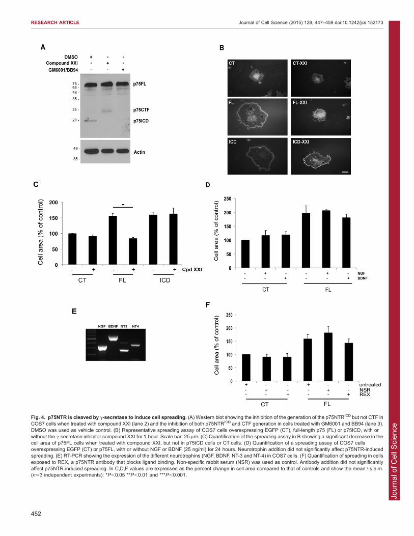
receptor is overexpressed. The production of this fragment isblocked by the metalloprotease inhibitors GM6001 and BB94 aswell as by compound XXI, a c-secretase inhibitor (Fig. 4A).Consistent with previous results, c-secretase inhibition resulted in
accumulation of a 25-kDa C-terminal fragment (CTF) of p75NTR(Fig. 4A, lane 2). Several studies have suggested that theCTF and the p75NTRICD have distinct biological activities
(Domeniconi et al., 2005, Kenchappa et al., 2006, Underwoodet al., 2008, Kenchappa et al., 2010), and we therefore addressedwhether the CTF could mediate p75NTR-dependent cell
spreading. COS7 cells overexpressing p75FL, p75NTRICD or anEGFP control were plated on glass coverslips and treated withcompound XXI (10 mM) or vehicle for 1 hour. Fig. 4B,C shows
that compound XXI had no effect on cells expressing EGFP aloneor expressing the p75NTRICD yet it blocked the spreading of cellsexpressing full-length p75NTR. Because compound XXI causesaccumulation of the p75NTR CTF while preventing p75NTRICD
accumulation, this indicates that the p75NTRICD is the relevantfragment required for p75NTR-dependent cell spreading in COS7cells. Therefore, the p75NTRICD, generated through sequential
ADAM17- and c-secretase-dependent p75NTR cleavage, isrequired to drive changes in COS7 cell shape.
p75NTR binds to the four neurotrophins present in mammals
and in our next experiments, we addressed whether these ligandshave an impact on the COS7 spreading assay. The addition of nervegrowth factor (NGF) or brain-derived neurotrophic factor (BDNF)
(each at 25 ng/ml), for 1 hour or for 24 hours, had no effecton p75NTR cell spreading (Fig. 4D). To determine whetherendogenous neurotrophins might play a role, we used RT-PCR toestablish whether neurotrophins or their receptors are expressed in
COS7 cells. Neither p75NTR nor the Trk receptor mRNA weredetected (data not shown) but mRNA encoding each of the fourneurotrophins was present in this line (Fig. 4E). The capacity of
full-length p75NTR to drive cell spreading was not significantlychanged when cells were maintained in REX (Fig. 4F), an antibodydirected against the p75NTR extracellular domain that blocks
ligand binding (Clary et al., 1994). We conclude that neurotrophinbinding to p75NTR is not required for the cell spreading phenotypeobserved in this line and that constitutive, rather than ligand-induced, p75NTR cleavage drives the cell spreading phenotype.
NRAGE acts downstream of p75NTR to mediate cellspreadingThe p75NTRICD does not have intrinsic enzymatic activity andtherefore relies on interaction with cytosolic binding proteins toelicit downstream effects. One of these adaptors, termed NRAGE,
mediates morphological changes in transformed humanmammary epithelial cells and U2OS cells (Xue et al., 2005,Kumar et al., 2011), and we therefore asked whether NRAGE is
required for p75NTR-dependent COS7 cell spreading. Afterconfirming that NRAGE siRNAs suppressed NRAGE proteinexpression in COS7 cells (Fig. 5A), we asked whether NRAGEdepletion altered cell spreading induced by overexpression of
full-length p75NTR or the p75NTRICD. NRAGE knockdown hadno effect on the area occupied by control cells but it eliminatedthe increase in cell spreading induced by p75NTR or the
p75NTRICD (Fig. 5B,C). NRAGE could mediate these effectsby binding to the p75NTRICD and playing a direct role indownstream signaling or by facilitating the generation or
maintenance of the p75NTRICD. However, levels of full-length
Fig. 3. p75NTR cleavage by ADAM17 is necessary for cell spreading.(A) Representative spreading assay of COS7 cells overexpressing EGFP (CT),full-length p75 (FL) and p75ICD with or without the broad matrixmetalloprotease inhibitors GM6001 and BB94 (GM/BB) for 1 hour.(B) Quantification of the spreading assay in A showing a significant decrease inthe cell area of p75FL cells when treated with GM6001 and BB94, but not inp75ICD cells or CT cells. Values are expressed as the percent change in cellarea compared to that of controls and are shown as the mean6s.e.m. (n53independent experiments). (C) Western blot showing a decrease in ADAM17expression in COS7 cells expressing ADAM17 siRNA (Ad17si) as well as areduction in p75ICD generation upon ADAM17 knockdown. (D) Representativespreading assay of COS7 cells overexpressing EGFP (CT), p75FL or p75ICDwith or without ADAM17 siRNA. Non-specific siRNA (NSsi) was used ascontrol. Scale bars: 25 mm. (E) Quantification of the spreading assay in Dshowing a significant decrease in the cell area of p75FL cells after ADAM17knockdown, but not in p75ICD cells or CT cells. Values are expressed as thepercent change in cell area compared to that of controls and are shown as themean6s.e.m. (n53 independent experiments); *P,0.05. (F) RT-PCR ofADAM17 mRNA expression in control COS7 cells or in cells overexpressingthe different p75NTR constructs.
RESEARCH ARTICLE Journal of Cell Science (2015) 128, 447–459 doi:10.1242/jcs.152173
451
Jour
nal o
f Cel
l Sci
ence
Fig. 4. p75NTR is cleaved by c-secretase to induce cell spreading. (A) Western blot showing the inhibition of the generation of the p75NTRICD but not CTF inCOS7 cells when treated with compound XXI (lane 2) and the inhibition of both p75NTRICD and CTF generation in cells treated with GM6001 and BB94 (lane 3).DMSO was used as vehicle control. (B) Representative spreading assay of COS7 cells overexpressing EGFP (CT), full-length p75 (FL) or p75ICD, with orwithout the c-secretase inhibitor compound XXI for 1 hour. Scale bar: 25 mm. (C) Quantification of the spreading assay in B showing a significant decrease in thecell area of p75FL cells when treated with compound XXI, but not in p75ICD cells or CT cells. (D) Quantification of a spreading assay of COS7 cellsoverexpressing EGFP (CT) or p75FL, with or without NGF or BDNF (25 ng/ml) for 24 hours. Neurotrophin addition did not significantly affect p75NTR-inducedspreading. (E) RT-PCR showing the expression of the different neurotrophins (NGF, BDNF, NT-3 and NT-4) in COS7 cells. (F) Quantification of spreading in cellsexposed to REX, a p75NTR antibody that blocks ligand binding. Non-specific rabbit serum (NSR) was used as control. Antibody addition did not significantlyaffect p75NTR-induced spreading. In C,D,F values are expressed as the percent change in cell area compared to that of controls and show the mean6s.e.m.(n53 independent experiments); *P,0.05 **P,0.01 and ***P,0.001.
RESEARCH ARTICLE Journal of Cell Science (2015) 128, 447–459 doi:10.1242/jcs.152173
452

Jour
nal o
f Cel
l Sci
ence
p75NTR or the p75NTRICD were unchanged by NRAGEknockdown (data not shown), ruling out the latter possibility.
We therefore focused our efforts on the hypothesis that NRAGElinks the p75NTRICD to downstream signaling partners.
NRAGE interacts physically and functionally with NEDD9To identify NRAGE-interacting proteins that could link thep75NTRICD to Rac1 activity, we performed a cytosolic yeast two-hybrid screen, using an NRAGE–SOS fusion protein as bait with
a human fetal brain library of cDNAs fused to a myristoylatedmembrane-localization signal as a source of potential bindingpartners. Interestingly, four of the positive clones to emerge from
this screen encoded distinct overlapping regions of NEDD9, amember of the Cas family (Fig. 6A). NEDD9 is a key player inthe regulation of cell shape and cell migration and has recently
emerged as a key player directing EMT in melanoma, lung andbreast cancer (Kim et al., 2006, Izumchenko et al., 2009, Miaoet al., 2013, Kondo et al., 2012, Little et al., 2014).
All four of the NRAGE-binding clones contained a NEDD9
fragment that started at amino acid 637 and ended at the
C-terminus of the protein (amino acid 834), and Fig. 6B showsthat a GST–NEDD9 fusion protein containing this 198-amino-
acid fragment was capable of associating with NRAGE expressedby in vitro translation, indicating that NRAGE directly binds tothis NEDD9 fragment. To identify the minimal region within
NEDD9 required for NRAGE binding, pulldowns wereperformed using progressively smaller fragments of NEDD9and, from this, a fragment of NEDD9 stretching from amino acids704 to 765 emerged as the NRAGE interaction domain (Fig. 6B).
Examination of the primary sequence of this 62-amino-acidregion revealed a putative helix-loop-helix domain. To test therelevance of the helical domains in this region, we disrupted each
of the two helices by substituting proline residues in helix 1(G722P) or in helix 2 (G744P). The G722P substitution had noeffect on the NRAGE–NEDD9 interaction, whereas the G744P
mutation abolished the interaction between the two proteins(Fig. 6C). Taken together, these data indicate that the helix-loop-helix present in NEDD9 is required to bind to NRAGE and thatthe second helix of this domain plays a crucial role in their
association.
Fig. 5. NRAGE acts downstream of p75NTR in cell spreading.(A) Western blot showing NRAGE knockdown in COS7 cells usingthree different siRNAs. Only NRAGE siRNA 1 was used insubsequent experiments. NS, nonspecific siRNA. (B) Representativespreading assay of COS7 cells overexpressing EGFP (CT), full-length p75 (FL) or p75ICD with or without NRAGE siRNA (NRsi).Nonspecific siRNA (NSsi) was used as control. Scale bar: 25 mm.(C) Quantification of spreading assay showing a significant decreasein the cell area of p75FL and p75ICD cells after NRAGE depletion butnot in that of CTcells. Values are expressed as the percent change incell area compared to that of controls and are shown as themean6s.e.m. (n53 independent experiments); *P,0.05.
RESEARCH ARTICLE Journal of Cell Science (2015) 128, 447–459 doi:10.1242/jcs.152173
453

Jour
nal o
f Cel
l Sci
ence
To determine whether the NRAGE–NEDD9 interactionoccurred in mammalian cells and to identify the domain in
NRAGE that bound to NEDD9, we fused amino acids 637–834of NEDD9 to GST and then expressed this with FLAG-taggedNRAGE, or with several FLAG-tagged NRAGE deletionmutants (shown schematically in Fig. 7A), in HEK293T cells
and performed pull downs. Full-length NRAGE showed robustbinding to the GST–NEDD9 fusion protein but not to GST alone(Fig. 7B). The main structural motifs present in this section of
NRAGE are the interspersed repeat domain, the MAGEhomology domain and a putative coiled-coil domain.Screening the NRAGE deletion mutants revealed that the
NEDD9 interaction relied on a region within amino acids 666–775 that is C-terminal to the MAGE homology domain. Deletionof the coiled-coil domain or the C-terminal 48 amino acids
reduced, but did not completely abrogate, the NRAGE–NEDD9interaction. Therefore, much of this 110-amino-acid fragmentof NRAGE is required to sustain a robust interaction withNEDD9.
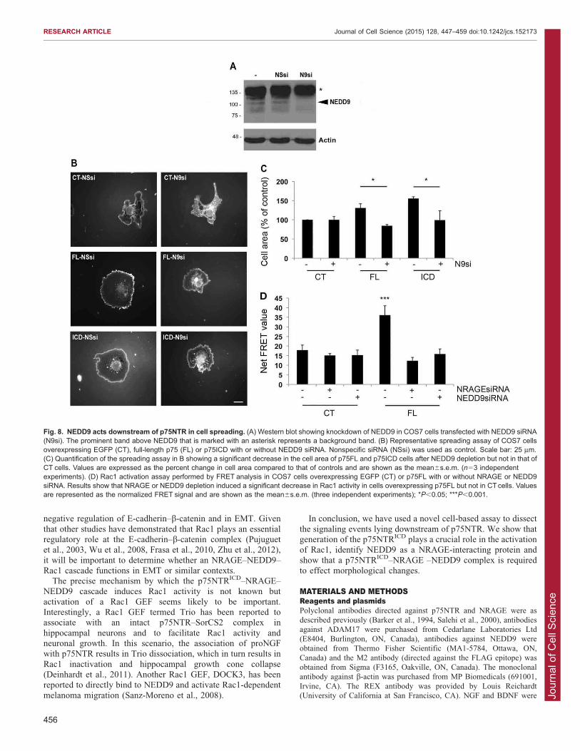
We then asked whether NEDD9 is required for p75NTR-induced spreading, using NEDD9-specific siRNA to reduce itslevels in COS7 cells (Fig. 8A). Interestingly, NEDD9 knockdown
had no effect on cells transfected with EGFP alone but stronglyinhibited cell spreading mediated by overexpression of full-lengthp75NTR or by the p75NTRICD (Fig. 8B,C). We conclude that
NEDD9 is a downstream effector of a p75NTR–NRAGE pathwaythat mediates the cell spreading phenotype.
Finally, to determine whether p75NTR activates Rac1 through
an NRAGE- and NEDD9-dependent pathway, we assessed Rac1activity in COS7 cells using the fluorescence resonance energytransfer (FRET) biosensor described by Hodgson and colleagues(2010). p75NTR overexpression produced a robust increase in
Rac1 activity and, significantly, the p75NTR-dependent increasewas blocked when NRAGE or NEDD9 were depleted usingsiRNA (Fig. 8D). Taken together, these findings demonstrate that
NRAGE and NEDD9 function downstream of p75NTR toactivate Rac1.
DISCUSSIONHere, we show that p75NTR induces cell spreading throughactivation of the small GTPase Rac1. We demonstrate thatp75NTR must be cleaved in a proteolytic process involving
ADAM17 and c-secretase for this effect to be manifested, andshow that the p75NTRICD is the relevant signaling moiety in thiscontext. We demonstrate that NRAGE participates downstream
of the p75NTRICD in producing the cell spreading phenotype andidentify NEDD9 as a novel NRAGE-binding protein thatparticipates in this cascade. Thus, our data suggest that
p75NTR-dependent cell spreading is dependent on generationof the p75NTRICD, which in turn drives NRAGE- and NEDD9-dependent activation of Rac1. Although generation of the
p75NTRICD, NRAGE signaling and Rac1 activation have allbeen proposed to play important roles downstream of p75NTR(Salehi et al., 2002, Harrington et al., 2002, Domeniconi et al.,2005, Kenchappa et al., 2006, Bertrand et al., 2008, Wang et al.,
2008, Kenchappa et al., 2010, Ceni et al., 2010, Tep et al., 2012,Matusica et al., 2013), this is the first study that links these eventsin a single cascade.
RIP is a conserved and well-established mechanism that affectsthe function of diverse membrane-anchored proteins, such asAPP, Notch and Delta. RIP of p75NTR is characterized by the
dual cleavage of the receptor by ADAM17 and c-secretase, which
Fig. 6. NRAGE interacts with the C-terminal domain of NEDD9.(A) Schematic showing the structure of NEDD9, which contains an SH3 domain,a substrate-binding domain, a serine-rich domain and a helix loop helix (HLH)domain. The four clones identified contain overlapping regions of the HLHspanning amino acids 637–834. (B) Different GST–NEDD9 fusion proteinscontaining the 198-amino-acid region were generated, and their interaction withNRAGE produced by in vitro translation was analyzed. In the schematic in B, theHLH region is represented in light gray. Regions outside of the HLH region arerepresented in dark gray. The interaction of NRAGE with GST–NEDD9 fusionswas detected by NRAGE immunoblot. ! and2 indicate the presence or absenceof interaction, respectively. The lower panel shows a Coomassie-Blue-stainedgel that confirms the expression of the different GST–NEDD9 mutants.(C) Introduction of two proline residues in helix 1 and helix 2 of the NEDD9 HLHidentifies helix 2 of NEDD9 as the NRAGE-binding domain. In the schematic inC, the HLH region is represented in light gray, and regions outside of the HLHregion are represented in dark gray. In the lower panel, the interaction of NRAGEwith GST–NEDD9 fusions was detected by immunoblotting for NRAGE.
RESEARCH ARTICLE Journal of Cell Science (2015) 128, 447–459 doi:10.1242/jcs.152173
454

Jour
nal o
f Cel
l Sci
ence
then releases the p75NTRICD into the cytosol (Kanning et al.,2003, Zampieri et al., 2005). Our data demonstrate that ADAM17
is responsible for the initial cleavage of p75NTR in COS7 cellsand show that this is a prerequisite for subsequent c-secretase-dependent proteolysis and release of the p75NTRICD. Becauseknockdown of ADAM17 inhibited production of the p75NTRICD
and prevented cell spreading in cells transfected with full-lengthp75NTR, but did not block cell spreading of cells transfected withthe p75NTRICD, we conclude that the p75NTRICD is the active
signaling component required for Rac1 activation and cellspreading.
In previous work, we have shown that NGF-dependent TrkA
(also known as NTRK1) activation activates an Erk-dependentsignaling pathway to generate the p75NTRICD, which in turnfacilitates NGF-dependent Akt signaling and cell survival
(Kommaddi et al., 2011). Consistent with this, another recentstudy has shown that p75NTR cleavage facilitates Akt signalingand Erk signaling in sympathetic neurons (Matusica et al., 2013).Other works have shown that p75NTR cleavage is necessary for
BDNF-induced sympathetic neuron death, for myelin-associated-glycoprotein-induced growth cone collapse (Kenchappa et al.,2006; Domeniconi et al., 2005) and for glioma migration (Wang
et al., 2008). Here, we show that the addition of exogenousneurotrophin has no effect on p75NTR-induced cell spreading,suggesting that this effect might be ligand independent. In
sympathetic neurons, expression of ADAM17 mRNA is induced
through a p75NTR-dependent signaling cascade but we foundthat, in COS7 cells, p75NTR overexpression does not alter
ADAM17 mRNA levels. All four neurotrophins are expressed inCOS7 cells, and it is conceivable that they bind to p75NTR andinduce its cleavage in this setting. However, we found thatincubation with REX, an antibody that blocks neurotrophin
binding to p75NTR, had no effect on cell spreading. Therefore,consistent with our previous results (Ceni et al., 2010), weconclude that ligand binding to p75NTR is not required for the
cleavage of the receptor. Determining the precise mechanismsthat regulate p75NTR cleavage under physiologicalcircumstances remains an interesting challenge.
NEDD9 (also called HEF1 or CasL) belongs to the Cas familyof adaptor proteins that also includes p130Cas, Efs and HEPL(also known as BCAR1, Sin and CASS4, respectively). NEDD9
plays important roles in cell migration and cell adhesion(reviewed in Bouton et al., 2001, Guerrero et al., 2012), canstabilize focal adhesions and induce cell spreading (Bradshawet al., 2011, Zhong et al., 2012, Baquiran et al., 2013), and has
emerged as pro-metastasis factor in melanoma (Kim et al., 2006,Ahn et al., 2012), breast cancer (Izumchenko et al., 2009, Littleet al., 2014), lung cancer (Miao et al., 2013, Kondo et al., 2012)
and glioblastoma (Natarajan et al., 2006). This is the first study toestablish a link between NRAGE and NEDD9 but it is interestingto note that other works have implicated NRAGE (Kumar et al.,
2011) and NEDD9 (Kong et al., 2011, Ahn et al., 2012) in the
Fig. 7. NEDD9 interacts with the C-terminal domain of NRAGE.(A) Schematic showing the different NRAGE deletion mutants thatwere generated. ISP, interspersed repeat domain; MHD, Magehomology domain; CCD, coiled-coil domain. 2 to +++ represent theinteraction avidity from B. (B) The interaction of NRAGE fragmentswith GST–NEDD9 fusion proteins was detected using NRAGEimmunoblotting. The lower panel shows a Coomassie-Blue-stainedgel that confirms the expression of GST and GST–NEDD9. FL, full-length p75.
RESEARCH ARTICLE Journal of Cell Science (2015) 128, 447–459 doi:10.1242/jcs.152173
455
Jour
nal o
f Cel
l Sci
ence
negative regulation of E-cadherin–b-catenin and in EMT. Given
that other studies have demonstrated that Rac1 plays an essentialregulatory role at the E-cadherin–b-catenin complex (Pujuguetet al., 2003, Wu et al., 2008, Frasa et al., 2010, Zhu et al., 2012),
it will be important to determine whether an NRAGE–NEDD9–Rac1 cascade functions in EMT or similar contexts.
The precise mechanism by which the p75NTRICD–NRAGE–NEDD9 cascade induces Rac1 activity is not known but
activation of a Rac1 GEF seems likely to be important.Interestingly, a Rac1 GEF termed Trio has been reported toassociate with an intact p75NTR–SorCS2 complex in
hippocampal neurons and to facilitate Rac1 activity andneuronal growth. In this scenario, the association of proNGFwith p75NTR results in Trio dissociation, which in turn results in
Rac1 inactivation and hippocampal growth cone collapse(Deinhardt et al., 2011). Another Rac1 GEF, DOCK3, has beenreported to directly bind to NEDD9 and activate Rac1-dependent
melanoma migration (Sanz-Moreno et al., 2008).
In conclusion, we have used a novel cell-based assay to dissect
the signaling events lying downstream of p75NTR. We show thatgeneration of the p75NTRICD plays a crucial role in the activationof Rac1, identify NEDD9 as a NRAGE-interacting protein and
show that a p75NTRICD–NRAGE –NEDD9 complex is requiredto effect morphological changes.
MATERIALS AND METHODSReagents and plasmidsPolyclonal antibodies directed against p75NTR and NRAGE were as
described previously (Barker et al., 1994, Salehi et al., 2000), antibodies
against ADAM17 were purchased from Cedarlane Laboratories Ltd
(E8404, Burlington, ON, Canada), antibodies against NEDD9 were
obtained from Thermo Fisher Scientific (MA1-5784, Ottawa, ON,
Canada) and the M2 antibody (directed against the FLAG epitope) was
obtained from Sigma (F3165, Oakville, ON, Canada). The monoclonal
antibody against b-actin was purchased from MP Biomedicals (691001,
Irvine, CA). The REX antibody was provided by Louis Reichardt
(University of California at San Francisco, CA). NGF and BDNF were
Fig. 8. NEDD9 acts downstream of p75NTR in cell spreading. (A) Western blot showing knockdown of NEDD9 in COS7 cells transfected with NEDD9 siRNA(N9si). The prominent band above NEDD9 that is marked with an asterisk represents a background band. (B) Representative spreading assay of COS7 cellsoverexpressing EGFP (CT), full-length p75 (FL) or p75ICD with or without NEDD9 siRNA. Nonspecific siRNA (NSsi) was used as control. Scale bar: 25 mm.(C) Quantification of the spreading assay in B showing a significant decrease in the cell area of p75FL and p75ICD cells after NEDD9 depletion but not in that ofCT cells. Values are expressed as the percent change in cell area compared to that of controls and are shown as the mean6s.e.m. (n53 independentexperiments). (D) Rac1 activation assay performed by FRET analysis in COS7 cells overexpressing EGFP (CT) or p75FL with or without NRAGE or NEDD9siRNA. Results show that NRAGE or NEDD9 depletion induced a significant decrease in Rac1 activity in cells overexpressing p75FL but not in CT cells. Valuesare represented as the normalized FRET signal and are shown as the mean6s.e.m. (three independent experiments); *P,0.05; ***P,0.001.
RESEARCH ARTICLE Journal of Cell Science (2015) 128, 447–459 doi:10.1242/jcs.152173
456

Jour
nal o
f Cel
l Sci
ence
obtained from Alomone laboratories (Jerusalem, Israel). Compound XXI,
ilomastast (GM6001) and epoxomicin were from Calbiochem (San
Diego, CA); batimastat (BB94) and NSC 23766 were from Tocris
Bioscience (Ellisville, MO). Laminin was purchased from BD
Biosciences (Mississauga, ON, Canada). Poly-D-lysine (PDL) was
obtained from Sigma (Oakville, ON, Canada). Rhodamine-conjugated
wheat germ agglutinin (WGA) was purchased from Vector Laboratories
(Burlington, ON, Canada) and horseradish-peroxidase-conjugated
secondary antibodies were purchased from Jackson ImmunoResearch
(West Grove, PA). All cell culture reagents were from Wisent
Bioproducts (Saint Bruno, QC, Canada). Dako anti-fading mounting
medium was purchased from Cedarlane Laboratories Ltd (Burlington,
ON, Canada). The Rac biosensor was obtained from Addgene
(Cambridge, MA) and was as described previously (Hodgson et al.,
2010). Glutathione plasmids encoding full-length p75NTR, cleavage-
resistant p75NTR and the p75NTRICD were as described previously
(Kommaddi et al., 2011). Plasmids encoding RacN17 and RhoAN19
were kindly provided by Dr Peter McPherson (McGill University, QC,
Canada).
Cell culture and transfectionCOS7 cells and HEK293T cells were maintained in Dulbecco’s modified
Eagle’s medium supplemented with 10% fetal bovine serum, 2 mM L-
glutamine and 100 mg/ml penicillin-streptomycin, under 5% CO2 at
37 C. COS7 cells were co-transfected with EGFP cDNA (0.5 mg) in the
absence or presence of plasmids encoding the various p75NTR isoforms
(2 mg) using the calcium phosphate transfection method. For experiments
involving dominant-negative Rac (RacN17) or RhoA (RhoAN19), COS7
cells were co-transfected with p75NTR plasmids in the absence or
presence of plasmids encoding the dominant-negative GTPases, with
EGFP (0.5 mg) co-transfected in all cases. Cells transfected with the
different constructs were maintained for 48 hours at 37 C before either
plating on coverslips for the spreading assay or lysing in Laemmli sample
buffer for analysis by immunoblotting.
NRAGE, NEDD9 and ADAM17 siRNA sequences directed against the
respective simian mRNAs were designed using the Invitrogen Stealth
RNAiTM siRNAs prediction algorithm (specific sequences available on
request). For knockdown experiments, cells were transfected with
p75NTR constructs using the calcium phosphate transfection and,
48 hours later, were transfected with the different siRNAs in antibiotic-
free medium using Lipofectamine 2000 (Invitrogen, Carlsbad, CA), as
per the manufacturer’s instructions. A non-specific siRNA was used for
control knockdowns. Cells were maintained for 48 hours at 37 C then
plated on coverslips for the spreading assay or lysed in Laemmli sample
buffer for analysis by immunoblotting.
Cell spreading assayGlass coverslips (12 mm, Fisherbrand, Fisher Scientific, Ottawa, ON,
Canada) were coated for 30 minutes with poly-D-lysine (0.5 mg/ml),
washed with sterile water, coated with laminin (0.5 mg/ml) for 2 hours at
37 C and again washed with sterile water. Transfected COS7 cells were
plated on coverslips (4000 cells/slip) then incubated at 37 C under 5%
CO2 for 24 hours. Medium was then removed and replaced with 4%
paraformaldehyde in PBS for 15 minutes at 37 C. After washing with
PBS (three 5-minute washes), cells were incubated with Rhodamine-
tagged WGA (5 mg/ml) in PBS for 10 minutes and then washed with PBS
(two 5-minute washes) and with water (one 5-minute wash). Coverslips
were mounted in anti-fading mounting media (Dako), and kept at 4 C
until imaging was performed. Imaging was performed using a 406objective (NA 1.4) on a Zeiss Axio observer fluorescent inverted
microscope equipped with Xenon illumination, and images were captured
using Zen software (Zeiss) with an AxioCam MRm Rev.3 camera. The
cell surface area of GFP-expressing cells was quantified with the NIH
ImageJ software. At least 100 cells were counted per condition in each
experiment.
In the case of treatment with GM6001, BB94 and compound XXI, cells
were treated with the different compounds for 1 hour prior to fixation and
staining. For NGF or BDNF, cells were serum starved for 2 hours then
treated with the different neurotrophins (25 ng/ml) for 1 hour or 24 hours
prior to fixation and staining. In the case of the REX treatment, cells were
treated with the REX antibody (1:100) for 24 hours prior to fixation and
staining.
RNA extraction and RT-PCRmRNA was extracted from COS7 cells using Qiagen RNeasy Mini Kit
according to the manufacturer’s instructions (Valencia, CA). cDNA was
produced using the Ominscript RT kit (Qiagen) with random hexamers
(GE Healthcare, Mississauga, ON, Canada) as primers. PCR was then
performed using the GoTaq green master mix reagent (Fisher, Ottawa,
ON, Canada) with primers targeting the simian neurotrophins NGF,
BDNF, NT-3 and NT-4, as well as the neurotrophin receptors, p75NTR,
TrkA, TrkB and TrkC. In the case of ADAM17 mRNA expression, COS7
cells were transfected with p75FL, CR or ICD, or left untransfected, and
mRNA was extracted as described above. PCR was then performed using
GoTaq green master mix reagent with primers against simian ADAM17.
The primer sequences are available upon request. The PCR run was
performed at an annealing temperature of 55 C for 35 cycles in the case
of neurotrophins and neurotrophin receptors and 28 cycles for ADAM17.
Actin was used as an internal control. PCR products were run on a 1.5%
agarose gel and bands were visualized with ethidium bromide.
Yeast two-hybrid screeningNRAGE-interacting proteins were identified through a cytosolic yeast
two-hybrid screen. For this, the NRAGE open reading frame was cloned
into the pSOS vector to produce a NRAGE–SOS fusion that was then
expressed in a cdc25H yeast strain. Screening was performed using a
library of human fetal brain cDNAs cloned into the pMyr vector, which
anchors fusion proteins to the yeast plasma membrane, as per the
manufacturer’s instructions (Stratagene/Agilent, CA). From 2.56106
clones analyzed, 70 supported growth of the cdc25H yeast strain at 37 C
and four of these contained distinct fragments of human NEDD9 cDNAs.
The longest of these encoded amino acids 637–834, which represents the
terminal 198 amino acids of NEDD9.
In vitro translation and pulldown experimentsThe 198-amino-acid NEDD9 fragment obtained from the yeast two-
hybrid screen was cloned into a pGEX4-1 to produce a GST fusion
protein. This region of NEDD9 contained a putative helix-loop-helix, and
deletion and site-directed mutants of this region were produced using
PCR overlap. Full-length NRAGE was produced using an in vitro
transcription-translation kit (Promega, WI) and pulldowns were
performed using the GST–NEDD9 fragments using the methodology
described previously (Salehi et al., 2000). Levels of NRAGE or its
deletion fragments that associated with the GST–NEDD9 fragment were
determined by immunoblotting with anti-NRAGE antisera.
To identify the region of NRAGE that bound to NEDD9, the GST fusion
containing the 198-amino-acid NEDD9 fragment (amino acids 637–834)
was transferred to a mammalian expression vector and co-expressed with
FLAG-tagged NRAGE or FLAG-tagged NRAGE deletion mutants in
HEK293T cells, using the calcium phosphate transfection. Cells were lysed
in NP40 buffer (10 mM Tris-HCl pH 8.0, 150 mM NaCl, 1% NP40, 10%
glycerol) after 24 hours and then incubated with 20 ml glutathione-
conjugated beads (GE Healthcare, Baie d’Urfe, QC, Canada) for 1 hour at
4 C. Beads were washed three times in NP40 buffer, resuspended in
Laemmli sample buffer, and incubated at 100 C for 5 minutes. Levels of
NRAGE or its deletion fragments that associated with the GST–NEDD9
fragment were determined by immunoblotting with M2.
Western blot analysis and immunoblottingCells were harvested in 26Laemmli sample buffer and boiled for 5 minutes
prior to loading on SDS-PAGE gels. In the case of ADAM17 detection,
BB94 (0.2 mM) was added to the 26Laemmli sample buffer before lysing
the cells. For p75NTRICD detection, cells were pre-treated with epoxomicin
(1 mM) for 6 hours before lysis to block proteasomal degradation of the
ICD, as described previously (Ceni et al., 2010). After SDS-PAGE and
transfer to nitrocellulose, membranes were rinsed in PBS then blocked in
RESEARCH ARTICLE Journal of Cell Science (2015) 128, 447–459 doi:10.1242/jcs.152173
457

Jour
nal o
f Cel
l Sci
ence
TBST (10 mM Tris-HCl pH 8.0, 150 mM NaCl, 2% Tween 20) that was
supplemented with 5% (w/v) dried skimmed-milk powder. Primary and
secondary antibody incubation was performed in TBST containing 2.5%
(w/v) dried skimmed-milk powder blocking solution, with primary
incubations performed overnight at 4 C and secondary incubations
performed for 1 hour at room temperature. Membranes were extensively
washed in TBST after each incubation. Immunoreactive bands were
detected using enhanced chemiluminescence solution kit (Perkin-Elmer
Life Sciences, Norwalk, CT), as per the manufacturer’s instructions.
Rac activity using FRETRac activity was assessed using the Rac FRET biosensors described
previously (Hodgson et al., 2010). Briefly, the Rac FLAIR biosensor
consists of a dual chain in which the donor CFP and the acceptor YFP are on
different chains. The ECFP is replaced by CyPet (CyPet–Rac1) and EYFP
is replaced by pYPet–PBD (PAK binding domain). Once Rac1 is activated,
PBD interacts with Rac1 and this interaction results in a FRET signal.
COS7 cells were transfected or not with p75FL, together with CyPet–
Rac1 and pYPet–PBD (2 mg) using calcium phosphate transfection and
were then incubated for 48 hours at 37 C under 5% CO2. In order to correct
for donor and acceptor bleed through, cells were transfected with either of
the biosensors (CyPet–Rac1 or pYPet–PBD). For NRAGE and NEDD9
siRNA transfection, cells were transfected or not with p75FL as well as the
Rac biosensors and, 48 hours later, NRAGE and NEDD9 were knocked
down with NRAGE and NEDD9 siRNA transfected into cells using
Lipofectamine 2000 in antibiotic-free medium. Cells were kept for
48 hours at 37 C under 5% CO2, then plated on glass coverslips precoated
with poly-D-lysine for 30 minutes and laminin for 2 hours at a density of
10,000 cells/ml. Cells were kept for 24 hours at 37 C under 5% CO2, then
fixed with 4% paraformaldehyde for 15 minutes at 37 C. They were then
washed with PBS (two 5-minute washes) and sterile water (one 5-minute
wash) and mounted on slides. Slides were kept at 4 C until imaging.
Imaging was performed using a 406 objective (NA 1.4) on a Zeiss
Axio observer fluorescent inverted microscope equipped with Xenon
illumination, and images were captured using Zen software (Zeiss) with
an AxioCam MRm Rev.3 camera. Images were taken for the three
channels YFP, CFP and FRET. CFP and FRET were imaged at the same
exposure time. For emission ratio imaging, the following filter sets
(Chroma Technology) were used: CFP, 430/24 (excitation) and 470/20
(emission); FRET, 430/24 (excitation) and 535/30 (emission); YFP, 500/
20 (excitation) and 540/40 (emission). FRET analysis was done using
PixFRET plugin in NIH ImageJ software. This program takes into
consideration the bleed-through intensities from each chain i.e the donor
(CyPet–Rac1) and the acceptor (YPet–PBD), subtracts them from the
FRET intensity and calculates the normalized FRET (NFRET) value
according to the following formula:
NFRET~IFRET{BTDonor|IDonor{BTAcceptor|IAcceptor
ffiffiffiffiffiffiffiffiffiffiffiffiffiffiffiffiffiffiffiffiffiffiffiffiffiffiffiffiffiffiffiffiffiffiffiffiffiffiffi
IDonor|IAcceptorp
where I represents the intensity and BT represents the bleed through
(Feige et al., 2005).
Statistical analysisStatistical analyses were performed using one-way ANOVA and Tukey
post-hoc analysis. All values are expressed as the mean6s.e.m.
AcknowledgementsWe thank Vincent Soubannier for assistance with FRET and comments on themanuscript, Genevieve Dorval for help with transfections and Inna Ermeichouk forhelp in imaging and quantification.
Competing interestsThe authors declare no competing or financial interests.
Author contributionsP.A.B. conceived of the project, and P.A.B., A.S. and M.Z. designed the overallexperimental plan. M.Z. performed all of the experiments except for the yeasttwo-hybrid screen, NEDD9 and MAGE-D1 mutagenesis and related interaction
assays, which were performed by A.S. V.R. collected, analyzed and quantifiedmicroscopy data. M.Z. and P.A.B. wrote the manuscript.
FundingThis project was supported by the Canadian Institute of Health Research [grantnumber MOP62827].
ReferencesAhn, J., Sanz-Moreno, V. and Marshall, C. J. (2012). The metastasis geneNEDD9 product acts through integrin b3 and Src to promote mesenchymalmotility and inhibit amoeboid motility. J. Cell Sci. 125, 1814-1826.
Baquiran, J. B., Bradbury, P. and O’Neill, G. M. (2013). Tyrosine Y189 in thesubstrate domain of the adhesion docking protein NEDD9 is conserved withp130Cas Y253 and regulates NEDD9-mediated migration and focal adhesiondynamics. PLoS ONE 8, e69304.
Barker, P. A., Barbee, G., Misko, T. P. and Shooter, E. M. (1994). The low affinityneurotrophin receptor, p75LNTR, is palmitoylated by thioester formation throughcysteine 279. J. Biol. Chem. 269, 30645-30650.
Bertrand, M. J., Kenchappa, R. S., Andrieu, D., Leclercq-Smekens, M.,Nguyen, H. N., Carter, B. D., Muscatelli, F., Barker, P. A. and De Backer, O.(2008). NRAGE, a p75NTR adaptor protein, is required for developmentalapoptosis in vivo. Cell Death Differ. 15, 1921-1929.
Bouton, A. H., Riggins, R. B. and Bruce-Staskal, P. J. (2001). Functions of theadapter protein Cas: signal convergence and the determination of cellularresponses. Oncogene 20, 6448-6458.
Bradshaw, L. N., Zhong, J., Bradbury, P., Mahmassani, M., Smith, J. L.,Ammit, A. J. and O’Neill, G. M. (2011). Estradiol stabilizes the 105-kDaphospho-form of the adhesion docking protein NEDD9 and suppresses NEDD9-dependent cell spreading in breast cancer cells. Biochim. Biophys. Acta 1813,340-345.
Bronfman, F. C., Tcherpakov, M., Jovin, T. M. and Fainzilber, M. (2003). Ligand-induced internalization of the p75 neurotrophin receptor: a slow route to thesignaling endosome. J. Neurosci. 23, 3209-3220.
Ceni, C., Kommaddi, R. P., Thomas, R., Vereker, E., Liu, X., McPherson, P. S.,Ritter, B. and Barker, P. A. (2010). The p75NTR intracellular domain generatedby neurotrophin-induced receptor cleavage potentiates Trk signaling. J. Cell Sci.123, 2299-2307.
Clary, D. O., Weskamp, G., Austin, L. R. and Reichardt, L. F. (1994). TrkA cross-linking mimics neuronal responses to nerve growth factor. Mol. Biol. Cell 5, 549-563.
Coulson, E. J., May, L. M., Osborne, S. L., Reid, K., Underwood, C. K.,Meunier, F. A., Bartlett, P. F. and Sah, P. (2008). p75 neurotrophin receptormediates neuronal cell death by activating GIRK channels throughphosphatidylinositol 4,5-bisphosphate. J. Neurosci. 28, 315-324.
Deinhardt, K., Kim, T., Spellman, D. S., Mains, R. E., Eipper, B. A., Neubert, T. A.,Chao,M. V. and Hempstead, B. L. (2011). Neuronal growth cone retraction relieson proneurotrophin receptor signaling through Rac. Sci. Signal. 4, ra82.
Domeniconi, M., Zampieri, N., Spencer, T., Hilaire, M., Mellado, W., Chao, M. V.and Filbin, M. T. (2005). MAG induces regulated intramembrane proteolysis ofthe p75 neurotrophin receptor to inhibit neurite outgrowth. Neuron 46, 849-855.
Feige, J. N., Sage, D., Wahli, W., Desvergne, B. and Gelman, L. (2005).PixFRET, an ImageJ plug-in for FRET calculation that can accommodatevariations in spectral bleed-throughs. Microsc. Res. Tech. 68, 51-58.
Frasa, M. A., Maximiano, F. C., Smolarczyk, K., Francis, R. E., Betson, M. E.,Lozano, E., Goldenring, J., Seabra, M. C., Rak, A., Ahmadian, M. R. et al.(2010). Armus is a Rac1 effector that inactivates Rab7 and regulates E-cadherindegradation. Curr. Biol. 20, 198-208.
Guerrero, M. S., Parsons, J. T. and Bouton, A. H. (2012). Cas and NEDD9Contribute to Tumor Progression through Dynamic Regulation of theCytoskeleton. Genes Cancer 3, 371-381.
Guo, F., Debidda, M., Yang, L., Williams, D. A. and Zheng, Y. (2006). Geneticdeletion of Rac1 GTPase reveals its critical role in actin stress fiber formationand focal adhesion complex assembly. J. Biol. Chem. 281, 18652-18659.
Harrington, A. W., Kim, J. Y. and Yoon, S. O. (2002). Activation of Rac GTPaseby p75 is necessary for c-jun N-terminal kinase-mediated apoptosis.J. Neurosci. 22, 156-166.
Harrington, A. W., Li, Q. M., Tep, C., Park, J. B., He, Z. and Yoon, S. O. (2008).The role of Kalirin9 in p75/nogo receptor-mediated RhoA activation in cerebellargranule neurons. J. Biol. Chem. 283, 24690-24697.
Hodgson, L., Shen, F. and Hahn, K. (2010). Biosensors for characterizing thedynamics of rho family GTPases in living cells. Curr. Protoc. Cell Biol. Chapter14, Unit 14 11 1-26.
Izumchenko, E., Singh, M. K., Plotnikova, O. V., Tikhmyanova, N., Little, J. L.,Serebriiskii, I. G., Seo, S., Kurokawa, M., Egleston, B. L., Klein-Szanto, A.et al. (2009). NEDD9 promotes oncogenic signaling in mammary tumordevelopment. Cancer Res. 69, 7198-7206.
Kanning, K. C., Hudson, M., Amieux, P. S., Wiley, J. C., Bothwell, M. andSchecterson, L. C. (2003). Proteolytic processing of the p75 neurotrophinreceptor and two homologs generates C-terminal fragments with signalingcapability. J. Neurosci. 23, 5425-5436.
Kenchappa, R. S., Zampieri, N., Chao, M. V., Barker, P. A., Teng, H. K.,Hempstead, B. L. and Carter, B. D. (2006). Ligand-dependent cleavage of theP75 neurotrophin receptor is necessary for NRIF nuclear translocation andapoptosis in sympathetic neurons. Neuron 50, 219-232.
RESEARCH ARTICLE Journal of Cell Science (2015) 128, 447–459 doi:10.1242/jcs.152173
458

Jour
nal o
f Cel
l Sci
ence
Kenchappa, R. S., Tep, C., Korade, Z., Urra, S., Bronfman, F. C., Yoon, S. O.and Carter, B. D. (2010). p75 neurotrophin receptor-mediated apoptosis insympathetic neurons involves a biphasic activation of JNK and up-regulation oftumor necrosis factor-alpha-converting enzyme/ADAM17. J. Biol. Chem. 285,20358-20368.
Kim, M., Gans, J. D., Nogueira, C., Wang, A., Paik, J. H., Feng, B., Brennan, C.,Hahn, W. C., Cordon-Cardo, C., Wagner, S. N. et al. (2006). Comparativeoncogenomics identifies NEDD9 as a melanoma metastasis gene. Cell 125,1269-1281.
Kommaddi, R. P., Thomas, R., Ceni, C., Daigneault, K. and Barker, P. A. (2011).Trk-dependent ADAM17 activation facilitates neurotrophin survival signaling.FASEB J. 25, 2061-2070.
Kondo, S., Iwata, S., Yamada, T., Inoue, Y., Ichihara, H., Kichikawa, Y.,Katayose, T., Souta-Kuribara, A., Yamazaki, H., Hosono, O. et al. (2012).Impact of the integrin signaling adaptor protein NEDD9 on prognosis andmetastatic behavior of human lung cancer. Clin. Cancer Res. 18, 6326-6338.
Kong, C., Wang, C., Wang, L., Ma, M., Niu, C., Sun, X., Du, J., Dong, Z., Zhu, S.,Lu, J. et al. (2011). NEDD9 is a positive regulator of epithelial-mesenchymaltransition and promotes invasion in aggressive breast cancer. PLoS ONE 6,e22666.
Kumar, S., Park, S. H., Cieply, B., Schupp, J., Killiam, E., Zhang, F., Rimm,D. L. and Frisch, S. M. (2011). A pathway for the control of anoikis sensitivity byE-cadherin and epithelial-to-mesenchymal transition. Mol. Cell. Biol. 31, 4036-4051.
Little, J. L., Serzhanova, V., Izumchenko, E., Egleston, B. L., Parise, E., Klein-Szanto, A. J., Loudon, G., Shubina, M., Seo, S., Kurokawa, M. et al. (2014). Arequirement for Nedd9 in luminal progenitor cells prior to mammarytumorigenesis in MMTV-HER2/ErbB2 mice. Oncogene 33, 411-420.
Matusica, D., Skeldal, S., Sykes, A. M., Palstra, N., Sharma, A. and Coulson,E. J. (2013). An intracellular domain fragment of the p75 neurotrophin receptor(p75(NTR)) enhances tropomyosin receptor kinase A (TrkA) receptor function.J. Biol. Chem. 288, 11144-11154.
Miao, Y., Li, A. L., Wang, L., Fan, C. F., Zhang, X. P., Xu, H. T., Yang, L. H., Liu,Y. and Wang, E. H. (2013). Overexpression of NEDD9 is associated with alteredexpression of E-Cadherin, b-Catenin and N-Cadherin and predictive of poorprognosis in non-small cell lung cancer. Pathol. Oncol. Res. 19, 281-286.
Mitsui, N., Inatome, R., Takahashi, S., Goshima, Y., Yamamura, H. and Yanagi,S. (2002). Involvement of Fes/Fps tyrosine kinase in semaphorin3A signaling.EMBO J. 21, 3274-3285.
Natarajan, M., Stewart, J. E., Golemis, E. A., Pugacheva, E. N.,Alexandropoulos, K., Cox, B. D., Wang, W., Grammer, J. R. and Gladson,C. L. (2006). HEF1 is a necessary and specific downstream effector of FAK thatpromotes the migration of glioblastoma cells. Oncogene 25, 1721-1732.
Park, K. J., Grosso, C. A., Aubert, I., Kaplan, D. R. and Miller, F. D. (2010).p75NTR-dependent, myelin-mediated axonal degeneration regulates neuralconnectivity in the adult brain. Nat. Neurosci. 13, 559-566.
Pujuguet, P., Del Maestro, L., Gautreau, A., Louvard, D. and Arpin, M. (2003).Ezrin regulates E-cadherin-dependent adherens junction assembly throughRac1 activation. Mol. Biol. Cell 14, 2181-2191.
Reichardt, L. F. (2006). Neurotrophin-regulated signalling pathways. Philos.Trans. R. Soc. Lond. B Biol. Sci. 361, 1545-1564.
Salehi, A. H., Roux, P. P., Kubu, C. J., Zeindler, C., Bhakar, A., Tannis, L. L.,Verdi, J. M. and Barker, P. A. (2000). NRAGE, a novel MAGE protein, interactswith the p75 neurotrophin receptor and facilitates nerve growth factor-dependentapoptosis. Neuron 27, 279-288.
Salehi, A. H., Xanthoudakis, S. and Barker, P. A. (2002). NRAGE, a p75neurotrophin receptor-interacting protein, induces caspase activation and cell
death through a JNK-dependent mitochondrial pathway. J. Biol. Chem. 277,48043-48050.
Sanz-Moreno, V., Gadea, G., Ahn, J., Paterson, H., Marra, P., Pinner, S., Sahai,E. and Marshall, C. J. (2008). Rac activation and inactivation control plasticityof tumor cell movement. Cell 135, 510-523.
Schecterson, L. C. and Bothwell, M. (2010). Neurotrophin receptors: Old friendswith new partners. Dev. Neurobiol. 70, 332-338.
Sun, Y., Lim, Y., Li, F., Liu, S., Lu, J. J., Haberberger, R., Zhong, J. H. andZhou, X. F. (2012). ProBDNF collapses neurite outgrowth of primary neurons byactivating RhoA. PLoS ONE 7, e35883.
Takahashi, T. and Strittmatter, S. M. (2001). Plexina1 autoinhibition by the plexinsema domain. Neuron 29, 429-439.
Takahashi, T., Fournier, A., Nakamura, F., Wang, L. H., Murakami, Y., Kalb,R. G., Fujisawa, H. and Strittmatter, S. M. (1999). Plexin-neuropilin-1complexes form functional semaphorin-3A receptors. Cell 99, 59-69.
Tep, C., Kim, M. L., Opincariu, L. I., Limpert, A. S., Chan, J. R., Appel, B.,Carter, B. D. and Yoon, S. O. (2012). Brain-derived neurotrophic factor (BDNF)induces polarized signaling of small GTPase (Rac1) protein at the onset ofSchwann cell myelination through partitioning-defective 3 (Par3) protein. J. Biol.Chem. 287, 1600-1608.
Underwood, C. K., Reid, K., May, L. M., Bartlett, P. F. and Coulson, E. J. (2008).Palmitoylation of the C-terminal fragment of p75(NTR) regulates death signalingand is required for subsequent cleavage by gamma-secretase. Mol. Cell.Neurosci. 37, 346-358.
Wang, L., Rahn, J. J., Lun, X., Sun, B., Kelly, J. J., Weiss, S., Robbins, S. M.,Forsyth, P. A. and Senger, D. L. (2008). Gamma-secretase represents atherapeutic target for the treatment of invasive glioma mediated by the p75neurotrophin receptor. PLoS Biol. 6, e289.
Wells, C. M., Walmsley, M., Ooi, S., Tybulewicz, V. and Ridley, A. J. (2004).Rac1-deficient macrophages exhibit defects in cell spreading and membraneruffling but not migration. J. Cell Sci. 117, 1259-1268.
Wu, X., Tu, X., Joeng, K. S., Hilton, M. J., Williams, D. A. and Long, F. (2008).Rac1 activation controls nuclear localization of beta-catenin during canonicalWnt signaling. Cell 133, 340-353.
Xue, B., Wen, C., Shi, Y., Zhao, D. and Li, C. (2005). Human NRAGE disrupts E-cadherin/beta-catenin regulated homotypic cell-cell adhesion. Biochem.Biophys. Res. Commun. 336, 247-251.
Yamashita, T. and Tohyama, M. (2003). The p75 receptor acts as a displacementfactor that releases Rho from Rho-GDI. Nat. Neurosci. 6, 461-467.
Yamashita, T., Tucker, K. L. and Barde, Y. A. (1999). Neurotrophin binding to thep75 receptor modulates Rho activity and axonal outgrowth. Neuron 24, 585-593.
Yamashita, T., Higuchi, H. and Tohyama, M. (2002). The p75 receptortransduces the signal from myelin-associated glycoprotein to Rho. J. Cell Biol.157, 565-570.
Zampieri, N., Xu, C. F., Neubert, T. A. and Chao, M. V. (2005). Cleavage of p75neurotrophin receptor by alpha-secretase and gamma-secretase requiresspecific receptor domains. J. Biol. Chem. 280, 14563-14571.
Zanata, S. M., Hovatta, I., Rohm, B. and Puschel, A. W. (2002). Antagonisticeffects of Rnd1 and RhoD GTPases regulate receptor activity in Semaphorin3A-induced cytoskeletal collapse. J. Neurosci. 22, 471-477.
Zhong, J., Baquiran, J. B., Bonakdar, N., Lees, J., Ching, Y. W., Pugacheva, E.,Fabry, B. and O’Neill, G. M. (2012). NEDD9 stabilizes focal adhesions,increases binding to the extra-cellular matrix and differentially effects 2D versus3D cell migration. PLoS ONE 7, e35058.
Zhu, G., Wang, Y., Huang, B., Liang, J., Ding, Y., Xu, A. and Wu, W. (2012). ARac1/PAK1 cascade controls b-catenin activation in colon cancer cells.Oncogene 31, 1001-1012.
RESEARCH ARTICLE Journal of Cell Science (2015) 128, 447–459 doi:10.1242/jcs.152173
459