paper d | faraday discussions
TRANSCRIPT

Quantitative approaches to defining normaland aberrant protein homeostasis
Michele Vendruscolo* and Christopher M. Dobson*
Received 23rd March 2009, Accepted 17th April 2009
First published as an Advance Article on the web 28th July 2009
DOI: 10.1039/b905825g
Protein homeostasis refers to the ability of cells to generate and regulate the levels
of their constituent proteins in terms of conformations, interactions,
concentrations and cellular localisation. We discuss here an approach in which
physico-chemical properties of proteins and their environments are used to
understand the underlying principles governing this process, which is crucial in all
living systems. By adopting the strategy of characterising the origins of specific
diseases to inform us about normal biology, we are bringing together methods
and concepts from chemistry, physics, engineering, genetics and medicine. In
particular, we are using a combination of in vitro, in silico and in vivo approaches
to study protein homeostasis through the analysis of the effects that result from
its perturbation in a select group of specific proteins, from either amino acid
mutations, or changes in concentration and solubility, or interactions with other
molecules. By developing a coherent and quantitative description of such
phenomena, we are finding that it is possible to shed new light on how the
physical and chemical properties of the cellular components can provide an
understanding of the normal and aberrant behaviour of living systems. Through
such an approach it is possible to provide new insights into the origin and
consequences of the failure to maintain homeostasis that is associated with
neurodegenerative diseases, in particular, and the phenomenon of ageing, in
general, and hence provide a framework for the rational design of therapeutic
approaches.
Introduction
One of the essential characteristics of living systems is the ability of their molecularcomponents to self-assemble into functional structures.1,2 Equally important, however,is the way in which the processes leading to this organisation are balanced within thecellular environment through the mechanism of homeostasis.3–7 Of central importancein the study of this mechanism is to focus specifically on proteins, since these are themolecules that enable, regulate and control essentially all chemical processes on whichlife depends. In order to function, the large majority of our proteins need to fold intoa specific three-dimensional structure.4,8–10 Indeed, the wide variety of highly specificstructures that results from protein folding, and which serve to bring key functionalgroups into close proximity, has enabled living systems to develop astonishing diversityand selectivity in their underlying chemical processes by using a common set of justtwenty building blocks—the amino acids.11 Much research has addressed the funda-mental mechanism of protein folding through a combination of in vitro and in silicostudies, and we now have considerable understanding at a molecular level of the
Department of Chemistry, University of Cambridge, Lensfield Road, Cambridge, UK CB2 1EW.E-mail: [email protected]; [email protected]
PAPER www.rsc.org/faraday_d | Faraday Discussions
This journal is ª The Royal Society of Chemistry 2009 Faraday Discuss., 2009, 143, 277–291 | 277

fundamental principles underlying this complex process.8,12–14 The next challenge is torelate this information to events occurring in living systems.
As well as simply generating biological activity, however, we now know thatprotein folding in living systems takes place in a complex environment, and as a poly-peptide chain emerges from the ribosome on which it has been synthesised, it inter-acts with a wide range of ancillary molecules including molecular chaperones.4,15,16
Much less is known about such events at a molecular level and a primary objectivein protein science is to extend the studies of folding from the test tube to the cell, andto understand how this process takes place in the cellular environment. Moreover, itis clear that protein folding and unfolding are closely coupled to many other biolog-ical processes ranging from the trafficking of molecules to specific cellular locationsto the regulation of the growth and differentiation of cells.7,10 In addition, onlycorrectly folded proteins have the ability to remain soluble in crowded biologicalenvironments and to interact selectively with their natural partners.10,15 The mannerin which proteins are able to maintain such homeostasis is a subject of centralinterest in molecular biology.
Given the tremendous importance of protein folding, it is not surprising that thefailure of proteins to fold correctly, or to remain correctly folded, is the origin ofa wide variety of pathological conditions, including cystic fibrosis, a1-antitrypsindeficiency and Alzheimer’s disease.10,17–20 In many of these diseases proteins self-assemble in an aberrant manner into large molecular aggregates, including amyloidfibrils. Considerable attention has been devoted to exploring the nature and origin ofsuch disorders from a structural viewpoint and to understanding the manner inwhich the balance between normal and aberrant conformational transitions canbe perturbed.20 Several studies have involved in vitro studies coupled with computersimulations,20–23 and many others have been concerned with the goal of relatingprocesses studied in atomic detail in the test tube to their quantitative effects in livingsystems.24–26 Moreover, recent findings suggest that further developments in thisarea could have much more general relevance to understanding the way in whichwell-established physical and chemical principles can provide new insights into theapparent complexity of biology.27
The discovery of the common existence of amyloid and amyloid-like states is ofunique importance in understanding the nature of biological systems because itreveals that there is an alternative stable and highly ordered state, accessibleessentially to all proteins, in addition to the native one;10,28,29 this observation hasprofound implications in diverse fields ranging from medicine to materials science.Because the structural interactions within the amyloid state and the native stateare similar—although the latter are largely intramolecular whilst the former alsoinclude strong intermolecular contributions—the stability of the native and amyloidstates can be comparable.30 There is thus a competition between the two states thatresults in normal or aberrant biological behaviour depending on whether the nativeor the aggregated state is populated.10,28,29 More generally, the maintenance of thecorrect balance in the populations of different states of proteins, one facet of proteinhomeostasis, is of great significance, as even marginal alterations in such populationscan result in disease in the long term.7,27 Indeed, it has been recently realised that thelimit to the safe concentration of proteins in living systems is likely to be reachedwhen the amyloid state becomes more stable than the native state.27
It is therefore of great importance to complement the well-established character-isation of the structure, folding and stability of native states with a similar analysis ofthe structure, assembly and stability of other states—ranging from unfolded andpartially folded species, including natively unfolded states, to aggregated speciessuch as amyloid fibrils. This is one of the main thrusts of our own work, togetherwith the exploration of the effects of the balance between the normal and aberrantstates of proteins in living organisms such as the Drosophila model system, which webelieve will inform us on the origins of amyloid-related disease and hence moregenerally on the mechanism of protein homeostasis.
278 | Faraday Discuss., 2009, 143, 277–291 This journal is ª The Royal Society of Chemistry 2009
A conceptual framework for understanding protein homeostasis
It is becoming clear that the interplay between the various states of different proteinmolecules creates a highly complex system, whose behaviour determines whethera living organism functions in a normal or aberrant manner, and yet, as with othercomplex systems,31,32 may be determined by the combination of relatively simpleunderlying processes.3,6,33 This complexity6 underlies the phenomena now oftenreferred to as protein homeostasis or ‘‘proteostasis’’7 perhaps in a similar mannerthat, for example, individual organisms interact34 in an ecosystem. The investigationof this particular class of biological molecules could therefore potentially shed a greatdeal of light on more general questions of the design and evolution of biologicalmolecules and the environments in which they function. Such information lies atthe heart both of understanding the molecular aspects of the phenomenon of lifeand of rational approaches to molecular medicine.
In this paper we present a strategy for describing and understanding in a coherentmanner the behaviour of proteins in living systems, including their folding, misfold-ing and assembly processes. Our approach is primarily based on five technical andconceptual developments that have recently been made in protein science:
(1) The ability to describe quantitatively, by a combination of experimental andcomputational approaches, the often disordered and dynamic structures of themultiple states of proteins on which their biological behaviour depends.35–38
(2) New ideas about protein aggregation,10,28 including the finding that the abilityto assemble into stable and highly organised structures (e.g. amyloid fibrils) is not anunusual feature exhibited by a small group of peptides and proteins with specialsequence or structural properties, but rather a property shared by most, if not all,proteins;
(3) The discovery that specific aspects of protein behaviour, including theiraggregation propensities21,23,39,40 and the cellular toxicity associated with the aggre-gation process,24,41 can be predicted with a remarkable degree of accuracy from theknowledge of their amino acid sequences;
(4) The realisation that a wide variety of techniques originally devised for appli-cations in nanotechnology can be used to probe the nature of protein aggregationand assembly and of the structures that emerge;30,42–44 and
(5) The development of powerful approaches using model organisms for probingthe origins and progression of misfolding diseases by linking concepts and principlesemerging from in vitro studies to in vivo phenomena such as neurodegeneration.24
An analysis of these results, which span across a wide range of subjects fromneuroscience to nanoscience, reveals that the ability to keep proteins in their solubleform is absolutely central for the maintenance of cell homeostasis.
Protein solubility and biological complexity
Considerable advances have been made in recent years in the search for the chemicaland physical principles that underlie the complexity of biological phenomena. Webelieve that it is possible to describe, for example, processes such as folding andaggregation, at least in outline, in generic terms and link them to well-establishedand quantifiable concepts of chemistry and physics. In this context, it should thenbe possible to study in depth a relatively small number of carefully chosen proteins,and yet extract general principles from such studies. In particular, we believe thatthere are many common features underlying the diseases associated with amyloidformation. Thus, much of our research is focused on the development of this themeusing amyloid-related diseases as a paradigm of the way the ideas of chemistry andphysics can provide fundamental insight into both normal and aberrant behaviourand suggest novel therapeutic strategies.
Biological systems have evolved to be efficient by achieving astonishing levels ofmolecular crowding within cells and extracellular space, which is of the order of
This journal is ª The Royal Society of Chemistry 2009 Faraday Discuss., 2009, 143, 277–291 | 279

300 g l�1.10 Information can then be transmitted rapidly, largely by molecular diffu-sion, between different components enabling them to function efficiently.3 Moleculessuch as proteins remain soluble and able to avoid interaction with all but a relativelysmall and specific selection of other molecules, yet are composed of chemical speciesthat are often extremely hydrophobic and prone to self-assemble. We are beginningto think that the ability to maintain the solubility of its component molecules is ofmuch more general significance in biology than previously imagined.27 Thus theobservation that the sequence determines the solubility of proteins45 could be justas important as the fact that it determines their structure and the ability to fold.As we understand in increasing detail how sequences define solubility, it is becomingpossible to predict aspects of biology in ways that were previously unsuspected.
Multiple forms of protein structure
Much is understood about the nature of globular protein structures and about theprinciples by which isolated denaturated polypeptide chains are able to achievesuch states.4,8–10 A more complete knowledge of the behaviour of proteins in thecell has, however, been limited by the challenges involved in defining the structuresof native states in complex environments and of the highly dynamic structuralensembles that describe most of the additional forms of proteins that are now knownto be of biological importance, including natively unfolded states and partiallyfolded states involved in folding and in aggregation.
A detailed structural description of native and non-native states lies at the heart ofour ability to describe in a quantitative manner the complex behaviour of proteinswithin a cell. Powerful techniques are being developed to complement more estab-lished methods to overcome the challenges posed by the task of providing sucha description.30,42–44,46 Our own approach is based primarily on methods that directlycombine experimental and computational techniques.5,6,35 These procedures involvethe use of experimental data, largely derived from NMR spectroscopy, as restraintsin computer simulations.36,37 We have already used in this way a range of differenttypes of experimental data, for example distance measurements from paramagneticprobes introduced by mutagenesis,47,48 and structural and dynamical informationfrom hydrogen–deuterium exchange experiments.49–51 But a major breakthroughhas come recently through the discovery37,52–55 of ways in which chemical shiftdata can be used in this approach to generate structures of native states to anaccuracy comparable to that of conventional methods (Fig. 1). The use of chemicalshifts requires only a resolved and assigned spectrum and thus renders unnecessarythe measurement of large numbers of additional parameters such as interatomicdistances derived from nuclear Overhauser effects (NOEs). The latter measurementsare very challenging (and in practice virtually impossible) in highly dynamicalsystems and for the conformationally heterogeneous states populated, for example,along the folding and misfolding pathways.
These chemical shift techniques, particularly in conjunction with the measurementof residual dipolar couplings (RDCs), can be used to generate structural ensemblesof non-native states for which these parameters are often the only ones that can bereadily measured.56,57 These methods can be used to study a wider range of proteinstates, including highly dynamical ones such as low-populated conformationsinvolved in the folding and misfolding processes. Indeed we have already provideda proof of principle that the use of chemical shift restraints, here derived fromrelaxation dispersion techniques, can be used to characterise transient species.58
We believe that these computational approaches, as well as advances in experimentaltechniques that enable the systematic measurements of chemical shifts in non-nativestates of proteins,56 will enable us to increase very considerably the resolution towhich this type of non-native structure can be determined.
This approach should enable the characterisation of proteins whose structureshave proved elusive by conventional means, but which are crucial to understanding
280 | Faraday Discuss., 2009, 143, 277–291 This journal is ª The Royal Society of Chemistry 2009
the process of protein folding. Two recent studies59,60 have already demonstrated thisapproach by using as test cases calmodulin,59 a protein that plays an essential role insignal transduction pathways, and ubiquitin,60 a protein that is a key component indegradation pathways, that the use of molecular dynamics simulations with NMRrestraints36 enables the changes in structure and dynamics upon binding to bedescribed at nearly atomic level resolution, thus enabling analysis of the molecularmechanisms responsible for binding to be carried out. These studies have providedstrong support for the ‘‘equilibrium shift’’ model,61,62 according to which the confor-mations that proteins adopt in the bound state are already present, although withlow statistical weights, in the unbound states in solution. This mechanism enablesproteins such as calmodulin and ubiquitin to interact with large numbers of otherproteins in a selective and efficient manner.
Another crucial area in which the inclusion of experimental measurements inmolecular dynamics simulations is having a major impact is in the investigation ofthe structures of protein complexes, where even weak interactions can be detectedby NMR methods.63 In a first study,53 we have already established, using the caseof the structure of the cytotoxic endonuclease domain from bacterial toxin colicin(E9) in complex with its cognate immunity protein (Im9),64 that chemical shiftsenable the determination of protein complexes even when the complexes themselvesexhibit significant dynamics and the component proteins undergo conformationalrearrangements upon binding. It is also possible to determine the structure ofprotein–protein complexes using chemical shift information when the chemical shift
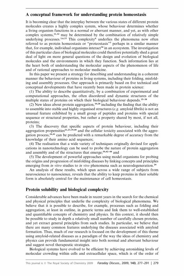
Fig. 1 Comparison between protein structures determined by X-ray crystallography (pink)and by the technique that we have recently introduced that uses only NMR chemical shift infor-mation (blue).37 Despite being only at the initial stages of development of the method, we arealready able to generate structures of globular proteins of up to 120 residues in length thatagree with the those determined by conventional methods with RMSD values of 1.2–1.8 �Afor the backbone atoms and 2.1–2.6 �A for the side-chain atoms.
This journal is ª The Royal Society of Chemistry 2009 Faraday Discuss., 2009, 143, 277–291 | 281

changes upon binding are relatively small and hence particularly difficult to computeaccurately. This result is a consequence of the well-known fact in NMR spectroscopythat the availability of a large number of restraints—in this case derived from chem-ical shifts—can provide enough information for high-resolution structure determi-nation, even if they are not accurately known individually.65 We have developeda computer code called CamDock to enable the structures of protein–proteincomplexes to be determined by combining advanced docking methods53 with theinformation provided by chemical shifts53 and residual dipolar couplings.66
It is then of very great importance to be able to relate the principles that emergefrom studies in the test tube to analogous events occurring in the cell. Interestingwork has been done using NMR spectroscopy in environments designed to mimicthe cellular milieu,67 but our ambition is to go further and ultimately to exploreprocesses taking place in the cellular environment itself.
One of the most fundamental, and yet so far elusive, aspects of protein foldingconcerns the way in which this process is initiated during or following biosynthesison the ribosome, i.e. the manner in which folding occurs in the cellular environ-ment.4,9,15,16 We have recently shown the feasibility of applying advanced NMRtechniques to obtain detailed structural insights into the conformations of nascentproteins during the process of their synthesis on the ribosome.38 In collaborationwith Dr John Christodoulou (UCL) we generated ribosome–nascent chain complexes(RNCs) by arresting RNA translation, and used this technique in the first instance tostudy a tandem immunoglobulin (Ig) domain repeat (Ig2) of an actin-bindingprotein.38 Analysis of the spectra of these RNCs selectively 15N/13C-labelled in thenascent chains reveals that the first Ig domain of the translation-arrested nascentchain is able to fold to a native-like state that remains tethered to the ribosome bythe second highly disordered Ig domain. This study is now being extended by studyingnascent chains of different lengths to probe the progressive development of structurein a growing nascent chain. We believe that this approach can open the door todescriptions at an atomistic level of detail of the process of co-translational folding,so as to characterise in detail the process by which proteins fold as the nascent chainemerges from the ribosomal exit tunnel.
In the immediate future, there are also great opportunities provided by theinclusion of NMR observables, including chemical shifts,37,52–54 residual dipolarcouplings68 and interatomic distances obtained by paramagnetic relaxation enhance-ment experiments,47,48 with molecular dynamics simulations to probe in detail thecrucial processes by which cellular components such as molecular chaperones,including Hsp70 and trigger factor, interact with nascent chains and help to promotecorrect folding and inhibiting misfolding and aggregation.4,15 This work has thepotential of opening up a vast range of new opportunities to explore the study ofthe fundamental mechanism of folding in environments directly relevant to livingsystems.
Molecular basis of protein aggregation
The structures, dynamics and interactions that stabilise protein aggregates are diffi-cult to study, since these species are often insoluble and resist crystallisation, thusmaking it very challenging to apply standard solution NMR spectroscopy andX-ray crystallography techniques.20,69 Interdisciplinary approaches appear to beparticularly suitable to address the problem of describing the structures of a varietyof protein assemblies.
Very considerable progress has been recently made to characterise quantitatively thephysical properties of fully formed amyloid fibrils and of their partially ordered proto-fibrillar precursors by bringing together solid-state NMR spectroscopy, cryo-electronmicroscopy and techniques of nanoscience. Advances in solid-state NMR methodsare making it possible to obtain interatomic distance information in states that areinsoluble and non-crystalline such as amyloid fibrils.69 In addition to information
282 | Faraday Discuss., 2009, 143, 277–291 This journal is ª The Royal Society of Chemistry 2009
about the structure of the amyloid fibrils formed by an 11-residue fragment of humantransthyretin, these approaches have provided great insight into the structures ofamyloid fibrils formed by several other peptides and proteins, including Het-S andAb.70–72
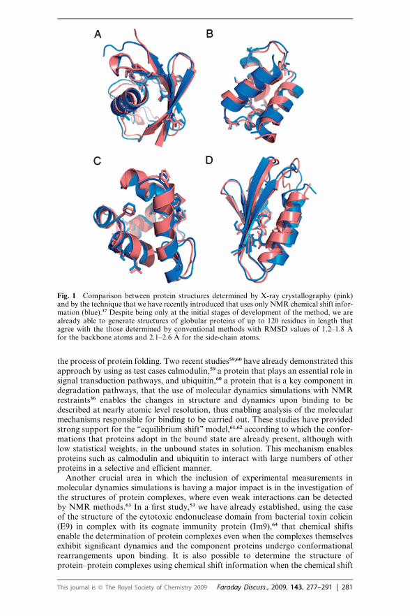
Together with the strategy mentioned above, nanoscience techniques are alsoemerging as powerful tools that can provide insight into the factors that stabiliseamyloid fibrils.30,42,44 In an initial study, by using atomic force microscopy (AFM)imaging we described the changes in the distribution of inter- versus intra-molecularbonding interactions associated with the transition of proteins from their nativeglobular structures into ordered supramolecular assemblies. This work revealsthat hydrogen bonded arrays of polypeptide chains exhibit material propertiesintermediate between those of hard materials such as steel and carbon nanotubesand softer biological fibres such as tubulin and actin (Fig. 2).30 AFM and othertechniques also generate information about the dimensions of the cross-b structuralcore of amyloid fibrils and of the less structured regions flanking them. Thesemeasurements provide unique insight into the kinetic and thermodynamic factorsresponsible for the stability of amyloid fibrils.
Much of our current understanding of the process of aggregation has beenobtained by light scattering and fluorescence measurements of the kinetics of theirgrowth.20,29 As the spectroscopic signals in these techniques do not exclusively arisefrom amyloid fibrils but also from other types of aggregates that may be present insolution, and because of the non-linear relationship between aggregate abundanceand signal intensity, the results can be difficult to interpret in a highly quantitativemanner. In order to overcome such problems we have shown that very accuratemeasurements of growth rates can be obtained by a strategy in which real-timemonitoring of the increase in mass of the fibrils themselves is carried out bymeasuring the variation in the frequency of a quartz crystal oscillator.42 The appli-cation of this quartz crystal microbalance technique for monitoring the kinetics ofaggregation of a series of proteins and under a variety of conditions is enablinga systematic analysis of the factors that can influence protein aggregation, particu-larly the mechanism by which molecular chaperones can inhibit fibril growth.42
Fig. 2 A clear relationship between bending rigidity and moment of inertia has revealed theexistence of universal mechanical properties of amyloid fibrils.30 Left: illustration for a set ofproteins of the values determined by atomic force microscopy (AFM) for the bending rigiditiesas a function of their cross-sectional moments of inertia (blue rectangles). For comparison,values for single-wall carbon nanotubes (SWNT), steel, actin, tubulin, rubber and elastin arealso shown. The green shaded region shows the range of elastic moduli for materials heldtogether by amphiphilic interactions. Right: comparison of the elastic moduli of fibrils from(blue squares, left to right) AFM measurements, full atom simulations, contribution of back-bone alone, and results from the Gaussian network theory, in which the stability of a structureis estimated from the contributions of its hydrogen bonds.
This journal is ª The Royal Society of Chemistry 2009 Faraday Discuss., 2009, 143, 277–291 | 283
Origins and consequences of aggregation in living systems
As we have discusses above, a wide range of human diseases is associated withprotein misfolding and aggregation.10,17–20 A powerful approach that has recentlybeen proposed to study these phenomena is based on the combination of in silico,in vitro and in vivo studies to investigate the factors responsible for the abnormalassembly of proteins into insoluble aggregates and the effects that these conforma-tional species have once released in the cellular environment.
Considerable progress has been made in characterising the major physico-chemical factors that promote the aggregation of polypeptide chains, and increas-ingly sophisticated computational approaches have been developed21,23,39,40 thatenable predictions to be made about a variety of features of the process of aggrega-tion of peptides and proteins. On this basis a method of predicting ‘‘aggregationpropensity profiles’’ has been established that enables the identification of regionswith a high intrinsic propensity for aggregation,21,23,39,40 providing a platform forfurther development of this approach.
In the case of fully folded proteins, we have shown that it is possible to take intoaccount the fact that the most amyloidogenic regions are protected from aggregationsince they are located in the structural core of the native state and hence they areunable to form inter-molecular interactions without at least a degree of unfolding.23
In essence, given the amino acid sequence of a protein, we have shown how it ispossible to combine the predictions of the intrinsic aggregation propensity profileswith those for folding into stable structures to determine new aggregation propensityprofiles of structured or partially structured proteins that account for the influenceof the structural context. We have provided a initial demonstration of the potentialof this approach through its application to the prediction of aggregation profiles fora range of peptides and proteins whose aggregation propensities have been charac-terized experimentally in particular detail, including the human prion protein, whichis involved in sporadic, inherited and infectious forms of Creutzfeld–Jakob disease.
We anticipate that, in addition to its relevance for understanding misfoldingdiseases, the insight provided by these studies will in time represent a significantcontribution to improving the biotechnological production of therapeutic peptidesand proteins, in drug discovery initiatives and for antibody production. The abilityto design rationally, and with increasing reliability, specific amino acid substitutionscapable of altering significantly the aggregation propensities of peptides and proteinswill enable us to investigate the physico-chemical factors responsible for theformation of amyloid fibrils and their oligomeric precursors.20,21
A range of strategies is being developed to combine in vivo approaches with in vitroand in silico methods to obtain a quantitative understanding of the molecular basis ofneurodegenerative and other misfolding diseases. In an initial study we have demon-strated the potential of this approach in the case of the Ab peptide, by showingthat the relative toxicity in Drosophila of its mutational variants can be predictedwith a remarkable 83% accuracy from their amino acid sequences (Fig. 3).24 Theadvantage of using Drosophila models for such studies is that the brevity of theirlifecycle, the power of the associated genetic tools, and the ease with which a rangeof toxicity-related phenotypes may be measured allows us to quantify the linksbetween the in vivo toxicity of protein aggregates and the fundamental chemicalproperties of peptides and proteins.24,41,73,74
The results discussed above were obtained by developing a method for predictingthe rate of formation of protofibrillar aggregates based on the physico-chemicalproperties of the amino acids comprising the sequences of the mutational variantsof the Ab42 peptide that we have investigated. It is also remarkable that, despitethe fact that the intrinsic aggregation propensities of typical protein sequencesvary by at least five orders of magnitude, we have been able to achieve profoundalterations in the pathogenic effects of Ab42 by increasing or decreasing its propen-sity to aggregate by less than 15%. This result suggests that proteins implicated in
284 | Faraday Discuss., 2009, 143, 277–291 This journal is ª The Royal Society of Chemistry 2009

misfolding diseases are likely to be extremely close to the limit of their solubilityunder normal physiological conditions and consequently the small alterations intheir concentration, environment or sequence, such as those that occur with geneticmutations or with increasing age, are likely to be the fundamental origin of thesehighly debilitating and increasingly common conditions.
The approach that we are developing is already enabling us to obtain accuratequantitative measurements of the relationships between the manifestations ofneuronal dysfunction in a complex organism, such as locomotor defects and reducedlifespans, and the fundamental physico-chemical factors that determine the propen-sities of peptides and proteins to aggregate into oligomeric species and protofibrils.Our research is aimed at demonstrating that, despite the presence within the cell ofmultiple regulatory mechanisms such as molecular chaperones and degradationsystems, it is the intrinsic, sequence-dependent propensity of the polypeptide chainsto aggregate to form protofibrillar aggregates that is the primary determinant of itspathological behaviour in living systems.
Thus, by using quantitative in vivo and in vitro techniques we are exploring thelinks between various conformational states and pathological effects. Our strategyis to use the results of in vitro biophysical methods, including NMR spectroscopy,fibril formation assays and amyloid staining, to deduce the events occurringin vivo. We have pioneered this strategy in the case of the Ab peptide to differentiatethe effects of the propensities to form either fibrillar or protofibrillar aggregates, byrationally designing mutations that alter either the fibrillar or the protofibrillarpropensities.24
Protein homeostasis in normal and aberrant biology
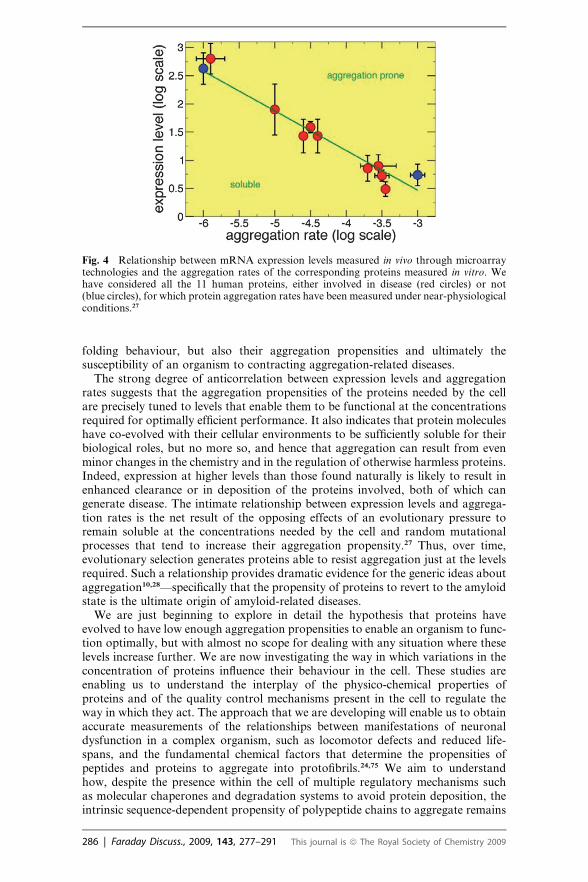
The fundamental connection between two aspects of proteins in the cell—their abun-dance and their solubility—is increasingly evident. A direct characterisation of thisrelationship is the very high correlation (97%) observed between the in vitro aggre-gation rates of a series of human proteins and the corresponding in vivo mRNAexpression levels.27 Thus, even relatively small alterations in protein abundanceand in vivo solubility can be linked to human disease, as described below.
The existence of a close relationship between mRNA expression levels and proteinaggregation rates (Fig. 4) provides a new perspective on the phenomenon of proteinaggregation and of its connections with misfolding diseases.27 In essence, theseresults suggest that the amino acid sequences of proteins determine not only their
Fig. 3 Rational design of the toxic effects of Ab42 mutants in a transgenic Drosophila modelof Alzheimer’s disease.24 The relative longevity (Stox, y-axis) of flies expressing a range of Ab42variants is predicted accurately by a score (Ztox, x-axis) for the propensity to form protofibrillaraggregates (r ¼ 0.83, p < 0.00001).
This journal is ª The Royal Society of Chemistry 2009 Faraday Discuss., 2009, 143, 277–291 | 285
folding behaviour, but also their aggregation propensities and ultimately thesusceptibility of an organism to contracting aggregation-related diseases.
The strong degree of anticorrelation between expression levels and aggregationrates suggests that the aggregation propensities of the proteins needed by the cellare precisely tuned to levels that enable them to be functional at the concentrationsrequired for optimally efficient performance. It also indicates that protein moleculeshave co-evolved with their cellular environments to be sufficiently soluble for theirbiological roles, but no more so, and hence that aggregation can result from evenminor changes in the chemistry and in the regulation of otherwise harmless proteins.Indeed, expression at higher levels than those found naturally is likely to result inenhanced clearance or in deposition of the proteins involved, both of which cangenerate disease. The intimate relationship between expression levels and aggrega-tion rates is the net result of the opposing effects of an evolutionary pressure toremain soluble at the concentrations needed by the cell and random mutationalprocesses that tend to increase their aggregation propensity.27 Thus, over time,evolutionary selection generates proteins able to resist aggregation just at the levelsrequired. Such a relationship provides dramatic evidence for the generic ideas aboutaggregation10,28—specifically that the propensity of proteins to revert to the amyloidstate is the ultimate origin of amyloid-related diseases.
We are just beginning to explore in detail the hypothesis that proteins haveevolved to have low enough aggregation propensities to enable an organism to func-tion optimally, but with almost no scope for dealing with any situation where theselevels increase further. We are now investigating the way in which variations in theconcentration of proteins influence their behaviour in the cell. These studies areenabling us to understand the interplay of the physico-chemical properties ofproteins and of the quality control mechanisms present in the cell to regulate theway in which they act. The approach that we are developing will enable us to obtainaccurate measurements of the relationships between manifestations of neuronaldysfunction in a complex organism, such as locomotor defects and reduced life-spans, and the fundamental chemical factors that determine the propensities ofpeptides and proteins to aggregate into protofibrils.24,75 We aim to understandhow, despite the presence within the cell of multiple regulatory mechanisms suchas molecular chaperones and degradation systems to avoid protein deposition, theintrinsic sequence-dependent propensity of polypeptide chains to aggregate remains
Fig. 4 Relationship between mRNA expression levels measured in vivo through microarraytechnologies and the aggregation rates of the corresponding proteins measured in vitro. Wehave considered all the 11 human proteins, either involved in disease (red circles) or not(blue circles), for which protein aggregation rates have been measured under near-physiologicalconditions.27
286 | Faraday Discuss., 2009, 143, 277–291 This journal is ª The Royal Society of Chemistry 2009
a major determinant of the pathologies associated with misfolding and aggregationin living systems. In addition to demonstrating that rational mutagenesis can be usedto alter systematically the toxicity of peptides and proteins, the transgenicDrosophila models that we are developing are enabling us to perform a quantitativeanalysis of the effects on the lifespan and mobility of Drosophila of other factorslikely to be relevant to the in vivo aggregation process, in particular molecularchaperones, small therapeutic molecules and antibodies, or antibody-like species,by co-expression techniques.
Towards a quantitative biology based on physico-chemical principles
Complex regulatory networks, involving primarily nucleic acids and proteins,orchestrate the cellular functions required to maintain protein homeostasis.3,33 Thesesame cellular functions are also, however, dependent on the basic chemistry of themolecules taking part in them. Therefore, the ‘‘chemical’’ and the ‘‘cellular’’ viewsof cell biology are closely related, as is revealed, for example, by the high correlationbetween expression levels and aggregation propensities. By this statement, we do notmean that gene regulation itself is not important, as there is a huge amount ofevidence that demonstrates its key role in protein homeostasis.76 What we are sayingis that very significant advances can be made by considering the ‘‘chemical view’’ ofprotein homeostasis. Studies are under way to explore the validity of the hypothesisthat we have formulated according to which the necessity of avoiding aggregationplays a major evolutionary role in ensuring that proteins can remain soluble inthe cell at the concentrations required for their function.
As an initial example, we discuss here the case of gene expression, which is theprocess through which the information contained in the DNA sequence of anorganism is converted into functional proteins.76 In response to the requirementsof a cell, each step in the process of gene expression is regulated by complex cellularmechanisms, from the transcription of DNA into mRNA to the post-translationalmodification of proteins. The conversion of the information stored in DNA intoproteins takes place through several phases that are highly regulated in responseto the functional requirements of proteins by the cell. A detailed knowledge of themechanisms of regulation can be used to rationalise and ultimately predict the
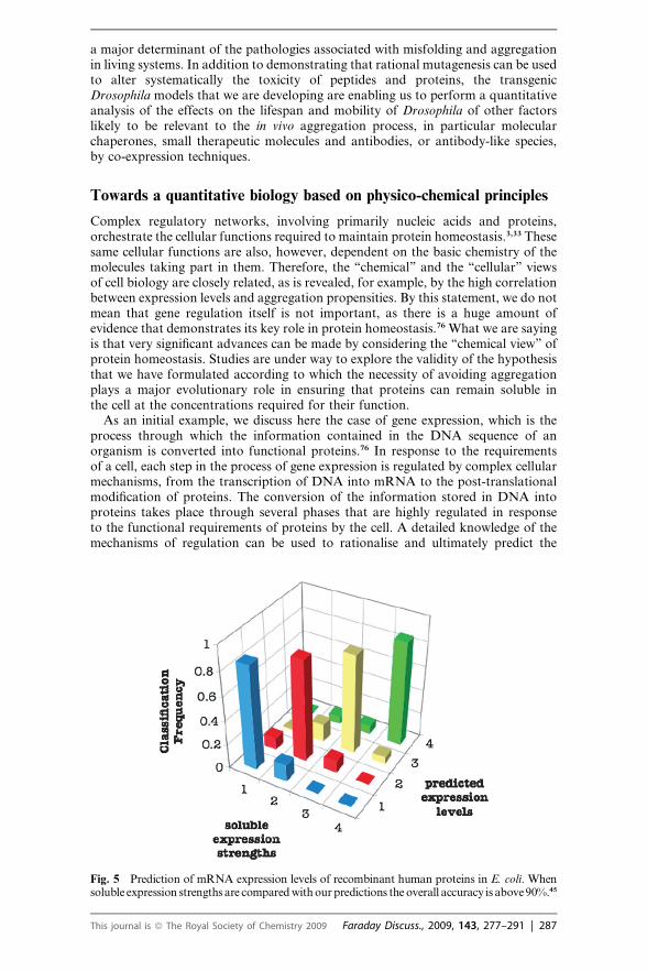
Fig. 5 Prediction of mRNA expression levels of recombinant human proteins in E. coli. Whensoluble expression strengths are compared with our predictions the overall accuracy is above 90%.45
This journal is ª The Royal Society of Chemistry 2009 Faraday Discuss., 2009, 143, 277–291 | 287
outcome of the gene expression process. For example, the study of cis-regulatorymotifs encoded in DNA sequences has reported an accuracy of about 70% for theprediction of expression patterns, while the correlation between the frequency oftranslational codons and gene expression levels is estimated to be around 60%.77
As an alternative to the strategy of exploiting the knowledge of the cellular regu-latory processes to predict gene expression, we have proposed an approach, whichhas been prompted by the observation that proteins, once expressed, must remainsoluble and avoid misfolding and aggregation in order to function optimally andto avoid cellular damage.27 Since, as discussed above, we have established quantita-tive methods for predicting aggregation rates of proteins from the knowledge of thechemical properties of their sequences, we are in a position to investigate the rela-tionship between these properties and the levels of expression of the genes. Ourresults indicate that it is possible to predict mRNA expression levels in E. coliwith an accuracy of 90% or better from the knowledge of the sequences of thecorresponding proteins (Fig. 5).45
Conclusions
Quantitative methods in molecular biology are providing unprecedented insightsinto the molecular mechanisms by which protein homeostasis is maintained in thecell. By drawing primarily on results from our own research we have discusseda variety of strategies based on the physico-chemical properties of proteins thatappear to be particularly promising for increasing our ability to describe their behav-iour in vivo and to suggest rational approaches to modulate it.
Acknowledgements
The results that we have described from our own research have been generatedthrough a series of interdisciplinary collaborations. We would like in particular toacknowledge the contributions from Dr Andrea Cavalli, Dr Damian Crowther,Dr Anthony Fitzpatrick, Dr Bob Griffin, Dr Thomas Jahn, Dr Tuomas Knowles,Dr David Lomas, Dr Leila Luheshi, Dr Rinaldo Montalvao, Dr Gian GaetanoTartaglia, Dr Mark Welland and Dr Helen Saibil. Our research is supported bythe Royal Society, the Wellcome Trust, the Leverhulme Trust, the Alzheimer’sResearch Trust, the European Community, the European Molecular BiologyOrganisation, the Biotechnology and Biological Sciences Research Council andthe Medical Research Council.
References
1 P. Aloy, B. Bottcher, H. Ceulemans, C. Leutwein, C. Mellwig, S. Fischer, A. C. Gavin,P. Bork, G. Superti-Furga, L. Serrano and R. B. Russell, Structure-based assembly ofprotein complexes in yeast, Science, 2004, 303, 2026–2029.
2 C. V. Robinson, A. Sali and W. Baumeister, The molecular sociology of the cell, Nature,2007, 450, 973–982.
3 L. H. Hartwell, J. J. Hopfield, S. Leibler and A. W. Murray, From molecular to modularcell biology, Nature, 1999, 402, C47–C52.
4 F. U. Hartl and M. Hayer-Hartl, Protein folding—Molecular chaperones in the cytosol:from nascent chain to folded protein, Science, 2002, 295, 1852–1858.
5 M. Vendruscolo, J. Zurdo, C. E. MacPhee and C. M. Dobson, Protein folding andmisfolding: a paradigm of self-assembly and regulation in complex biological systems,Philos. Trans. R. Soc. London, Ser. A, 2003, 361, 1205–1222.
6 M. Vendruscolo and C. M. Dobson, Towards complete descriptions of the free-energylandscapes of proteins, Philos. Trans. R. Soc. London, Ser. A, 2005, 363, 433–450.
7 W. E. Balch, R. I. Morimoto, A. Dillin and J. W. Kelly, Adapting proteostasis for diseaseintervention, Science, 2008, 319, 916–919.
8 A. R. Fersht, Structure and Mechanism in Protein Science: A Guide to Enzyme Catalysis andProtein Folding, W.H. Freeman, New York, 1999.
288 | Faraday Discuss., 2009, 143, 277–291 This journal is ª The Royal Society of Chemistry 2009
9 M. J. Gething and J. Sambrook, Protein folding in the cell, Nature, 1992, 355, 33–45.10 C. M. Dobson, Protein folding and misfolding, Nature, 2003, 426, 884–890.11 C. M. Dobson, Chemical space and biology, Nature, 2004, 432, 824–828.12 K. A. Dill and H. S. Chan, From Levinthal to pathways to funnels, Nat. Struct. Biol., 1997,
4, 10–19.13 C. M. Dobson, A. Sali and M. Karplus, Protein folding: A perspective from theory and
experiment, Angew. Chem., Int. Ed., 1998, 37, 868–893.14 J. N. Onuchic and P. G. Wolynes, Theory of protein folding, Curr. Opin. Struct. Biol., 2004,
14, 70–75.15 J. Frydman, Folding of newly translated proteins in vivo: The role of molecular chaperones,
Annu. Rev. Biochem., 2001, 70, 603–647.16 B. Bukau, J. Weissman and A. Horwich, Molecular chaperones and protein quality control,
Cell, 2006, 125, 443–451.17 R. W. Carrell and D. A. Lomas, Conformational disease, Lancet, 1997, 350, 134–138.18 B. Caughey and P. T. Lansbury, Protofibrils, pores, fibrils, and neurodegeneration:
Separating the responsible protein aggregates from the innocent bystanders, Annu. Rev.Neurosci., 2003, 26, 267–298.
19 C. Haass and D. J. Selkoe, Soluble protein oligomers in neurodegeneration: lessons fromthe Alzheimer’s amyloid beta-peptide, Nat. Rev. Mol. Cell Biol., 2007, 8, 101–112.
20 F. Chiti and C. M. Dobson, Protein misfolding, functional amyloid, and human disease,Annu. Rev. Biochem., 2006, 75, 333–366.
21 F. Chiti, M. Stefani, N. Taddei, G. Ramponi and C. M. Dobson, Rationalization of theeffects of mutations on peptide and protein aggregation rates, Nature, 2003, 424, 805–808.
22 A. M. Fernandez-Escamilla, F. Rousseau, J. Schymkowitz and L. Serrano, Prediction ofsequence-dependent and mutational effects on the aggregation of peptides and proteins,Nat. Biotechnol., 2004, 22, 1302–1306.
23 G. G. Tartaglia, A. Pawar, S. Campioni, F. Chiti and M. Vendruscolo, Prediction ofaggregation-prone regions of structured proteins, J. Mol. Biol., 2008, 380, 425–436.
24 L. M. Luheshi, G. G. Tartaglia, A.-C. Brorsson, A. P. Pawar, I. E. Watson, F. Chiti,M. Vendruscolo, D. A. Lomas, C. M. Dobson and D. C. Crowther, Systematic in vivoanalysis of the intrinsic determinants of amyloid beta pathogenicity, PLoS Biol., 2007, 5, e290.
25 R. L. Wiseman, E. T. Powers, J. N. Buxbaum, J. W. Kelly and W. E. Balch, An adaptablestandard for protein export from the endoplasmic reticulum, Cell, 2007, 131, 809–821.
26 T. W. Mu, D. S. T. Ong, Y. J. Wang, W. E. Balch, J. R. Yates, L. Segatori and J. W. Kelly,Chemical and biological approaches synergize to ameliorate protein-folding diseases, Cell,2008, 134, 769–781.
27 G. G. Tartaglia, S. Pechmann, C. M. Dobson and M. Vendruscolo, Life on the edge: A linkbetween gene expression levels and aggregation rates of human proteins, Trends Biochem.Sci., 2007, 32, 204–206.
28 C. M. Dobson, Protein misfolding, evolution and disease, Trends Biochem. Sci., 1999, 24,329–332.
29 T. R. Jahn and S. E. Radford, Folding versus aggregation: Polypeptide conformations oncompeting pathways, Arch. Biochem. Biophys., 2008, 469, 100–117.
30 T. P. J. Knowles, A. W. Fitzpatrick, H. R. Mott, S. Meehan, M. Vendruscolo,C. M. Dobson and M. E. Welland, Mechanical properties reveal the dominance ofbackbone interactions in stabilising amyloid fibrils, Science, 2007, 318, 1900–1903.
31 A. L. Barab�asi and R. Albert, Emergence of scaling in random networks, Science, 1999, 286,509–512.
32 H. Jeong, B. Tombor, R. Albert, Z. N. Oltvai and A. L. Barabasi, The large-scaleorganization of metabolic networks, Nature, 2000, 407, 651–654.
33 A. C. Gavin, P. Aloy, P. Grandi, R. Krause, M. Boesche, M. Marzioch, C. Rau,L. J. Jensen, S. Bastuck, B. Dumpelfeld, A. Edelmann, M. A. Heurtier, V. Hoffman,C. Hoefert, K. Klein, M. Hudak, A. M. Michon, M. Schelder, M. Schirle, M. Remor,T. Rudi, S. Hooper, A. Bauer, T. Bouwmeester, G. Casari, G. Drewes, G. Neubauer,J. M. Rick, B. Kuster, P. Bork, R. B. Russell and G. Superti-Furga, Proteome surveyreveals modularity of the yeast cell machinery, Nature, 2006, 440, 631–636.
34 I. Volkov, J. R. Banavar, S. P. Hubbell and A. Maritan, Patterns of relative speciesabundance in rainforests and coral reefs, Nature, 2007, 450, 45–49.
35 M. Vendruscolo, E. Paci, C. M. Dobson and M. Karplus, Three key residues form a criticalcontact network in a protein folding transition state, Nature, 2001, 409, 641–645.
36 K. Lindorff-Larsen, R. B. Best, M. A. DePristo, C. M. Dobson and M. Vendruscolo,Simultaneous determination of protein structure and dynamics, Nature, 2005, 433, 128–132.
37 A. Cavalli, X. Salvatella, C. M. Dobson and M. Vendruscolo, Protein structuredetermination from NMR chemical shifts, Proc. Natl. Acad. Sci. U. S. A., 2007, 104,9615–9620.
This journal is ª The Royal Society of Chemistry 2009 Faraday Discuss., 2009, 143, 277–291 | 289
38 S. T. D. Hsu, P. Fucini, L. D. Cabrita, H. Launay, C. M. Dobson and J. Christodoulou,Structure and dynamics of a ribosome-bound nascent chain by NMR spectroscopy, Proc.Natl. Acad. Sci. U. S. A., 2007, 104, 16516–16521.
39 K. F. Dubay, A. P. Pawar, F. Chiti, J. Zurdo, C. M. Dobson and M. Vendruscolo,Prediction of the absolute aggregation rates of amyloidogenic polypeptide chains, J. Mol.Biol., 2004, 341, 1317–1326.
40 A. P. Pawar, K. F. DuBay, J. Zurdo, F. Chiti, M. Vendruscolo and C. M. Dobson,Prediction of ‘‘aggregation-prone’’ and ‘‘aggregation-susceptible’’ regions in proteinsassociated with neurodegenerative diseases, J. Mol. Biol., 2005, 350, 379–392.
41 D. C. Crowther, R. Page, D. Chandraratna and D. A. Lomas, A Drosophila model ofAlzheimer’s disease, Methods Enzymol., 2006, 412, 234–255.
42 T. P. J. Knowles, W. M. Shu, G. L. Devin, S. Meehan, S. Auer, C. M. Dobson andM. E. Welland, Kinetics and thermodynamics of amyloid formation from directmeasurements of fluctuations of fibril mass, Proc. Natl. Acad. Sci. U. S. A., 2007, 104,10016–10021.
43 T. P. J. Knowles, J. F. Smith, A. Craig, C. M. Dobson and M. E. Welland, Spatialpersistence of angular correlations in amyloid fibrils, Phys. Rev. Lett., 2006, 96, 238301.
44 J. F. Smith, T. P. J. Knowles, C. M. Dobson, C. E. MacPhee and M. E. Welland,Characterization of the nanoscale properties of individual amyloid fibrils, Proc. Natl.Acad. Sci. U. S. A., 2006, 103, 15806–15811.
45 G. G. Tartaglia, S. Pechmann, C. M. Dobson and M. Vendruscolo, A relationship betweenmRNA expression levels and protein solubility in E. coli, J. Mol. Biol., 2009, 388, 381–389.
46 H. J. Dyson and P. E. Wright, Intrinsically unstructured proteins and their functions, Nat.Rev. Mol. Cell Biol., 2005, 6, 197–208.
47 M. M. Dedmon, K. Lindorff-Larsen, J. Christodoulou, M. Vendruscolo and C. M. Dobson,Mapping long-range interactions in alpha-synuclein using spin- label NMR and ensemblemolecular dynamics simulations, J. Am. Chem. Soc., 2005, 127, 476–477.
48 K. Lindorff-Larsen, S. Kristjansdottir, K. Teilum, W. Fieber, C. M. Dobson, F. M. Poulsenand M. Vendruscolo, Determination of an ensemble of structures representing thedenatured state of the bovine acyl-coenzyme a binding protein, J. Am. Chem. Soc., 2004,126, 3291–3299.
49 R. B. Best and M. Vendruscolo, Determination of protein structures consistent with NMRorder parameters, J. Am. Chem. Soc., 2004, 126, 8090–8091.
50 R. B. Best and M. Vendruscolo, Structural interpretation of hydrogen exchange protectionfactors in proteins: Characterization of the native state fluctuations of C12, Structure, 2006,14, 97–106.
51 J. Gsponer, H. Hopearuoho, S. B. M. Whittaker, G. R. Spence, G. R. Moore, E. Paci,S. E. Radford and M. Vendruscolo, Determination of an ensemble of structuresrepresenting the intermediate state of the bacterial immunity protein Im7, Proc. Natl.Acad. Sci. U. S. A., 2006, 103, 99–104.
52 Y. Shen, O. Lange, F. Delaglio, P. Rossi, J. M. Aramini, G. H. Liu, A. Eletsky, Y. B. Wu,K. K. Singarapu, A. Lemak, A. Ignatchenko, C. H. Arrowsmith, T. Szyperski,G. T. Montelione, D. Baker and A. Bax, Consistent blind protein structure generationfrom NMR chemical shift data, Proc. Natl. Acad. Sci. U. S. A., 2008, 105, 4685–4690.
53 R. W. Montalvao, A. Cavalli, X. Salvatella, T. L. Blundell and M. Vendruscolo, Structuredetermination of protein–protein complexes using NMR chemical shifts: Case of anendonuclease colicin–immunity protein complex, J. Am. Chem. Soc., 2008, 130, 15990–15996.
54 P. Robustelli, A. Cavalli and M. Vendruscolo, Determination of protein structures fromsolid-state NMR chemical shifts, Structure, 2008, 16, 1764–1769.
55 D. S. Wishart, D. Arndt, M. Berjanskii, P. Tang, J. Zhou and G. Lin, CS23D: a web serverfor rapid protein structure generation using NMR chemical shifts and sequence data,Nucleic Acids Res., 2008, 36, W496–W502.
56 P. Vallurupalli, D. F. Hansen and L. E. Kay, Structures of invisible, excited protein statesby relaxation dispersion NMR spectroscopy, Proc. Natl. Acad. Sci. U. S. A., 2008, 105,11766–11771.
57 A. De Simone, B. Richter, X. Salvatella and M. Vendruscolo, Toward an accuratedetermination of free energy landscapes in solution states of proteins, J. Am. Chem. Soc.,2009, 131, 3810–3811.
58 D. M. Korzhnev, X. Salvatella, M. Vendruscolo, A. A. Di Nardo, A. R. Davidson,C. M. Dobson and L. E. Kay, Low-populated folding intermediates of Fyn SH3characterized by relaxation dispersion NMR, Nature, 2004, 430, 586–590.
59 J. Gsponer, J. Christodoulou, A. Cavalli, J. M. Bui, B. Richter, C. M. Dobson andM. Vendruscolo, A coupled equilibrium shift mechanism in calmodulin-mediated signaltransduction, Structure, 2008, 16, 736–746.
290 | Faraday Discuss., 2009, 143, 277–291 This journal is ª The Royal Society of Chemistry 2009

60 O. F. Lange, N. A. Lakomek, C. Fares, G. F. Schroder, K. F. A. Walter, S. Becker,J. Meiler, H. Grubmuller, C. Griesinger and B. L. de Groot, Recognition dynamics up tomicroseconds revealed from an RDC-derived ubiquitin ensemble in solution, Science,2008, 320, 1471–1475.
61 D. D. Boehr, D. McElheny, H. J. Dyson and P. E. Wright, The dynamic energy landscapeof dihydrofolate reductase catalysis, Science, 2006, 313, 1638–1642.
62 M. Vendruscolo and C. M. Dobson, Dynamic visions of enzymatic reactions, Science, 2006,313, 1586–1587.
63 E. R. P. Zuiderweg, Mapping protein–protein interactions in solution by NMRspectroscopy, Biochemistry, 2002, 41, 1–7.
64 C. Kleanthous, U. C. Kuhlmann, A. J. Pommer, N. Ferguson, S. E. Radford, G. R. Moore,R. James and A. M. Hemmings, Structural and mechanistic basis of immunity towardendonuclease colicins, Nat. Struct. Biol., 1999, 6, 243–252.
65 K. Wuthrich, Protein structure determination in solution by nuclear magnetic resonancespectroscopy, Science, 1989, 243, 45–50.
66 A. Grishaev, J. Wu, J. Trewhella and A. Bax, Refinement of multidomain protein structuresby combination of solution small-angle X-ray scattering and NMR data, J. Am. Chem. Soc.,2005, 127, 16621–16628.
67 P. Selenko, Z. Serber, B. Gade, J. Ruderman and G. Wagner, Quantitative NMR analysisof the protein G B1 domain in Xenopus laevis egg extracts and intact oocytes, Proc. Natl.Acad. Sci. U. S. A., 2006, 103, 11904–11909.
68 G. M. Clore and C. D. Schwieters, How much backbone motion in ubiquitin is required toaccount for dipolar coupling data measured in multiple alignment media as assessed byindependent cross-validation?, J. Am. Chem. Soc., 2004, 126, 2923–2938.
69 M. Baldus, ICMRBS founder’s medal 2006: Biological solid-state NMR, methods andapplications, J. Biomol. NMR, 2007, 39, 73–86.
70 C. Wasmer, A. Lange, H. Van Melckebeke, A. B. Siemer, R. Riek and B. H. Meier,Amyloid fibrils of the HET-s(218–289) prion form a beta solenoid with a triangularhydrophobic core, Science, 2008, 319, 1523–1526.
71 A. T. Petkova, W. M. Yau and R. Tycko, Experimental constraints on quaternary structurein Alzheimer’s beta-amyloid fibrils, Biochemistry, 2006, 45, 498–512.
72 N. Ferguson, J. Becker, H. Tidow, S. Tremmel, T. D. Sharpe, G. Krause, J. Flinders,M. Petrovich, J. Berriman, H. Oschkinat and A. R. Fersht, General structural motifs ofamyloid protofilaments, Proc. Natl. Acad. Sci. U. S. A., 2006, 103, 16248–16253.
73 M. B. Feany and W. W. Bender, A Drosophila model of Parkinson’s disease, Nature, 2000,404, 394–398.
74 J. Bilen and N. M. Bonini, Drosophila as a model for human neurodegenerative disease,Annu. Rev. Genet., 2005, 39, 153–171.
75 L. M. Luheshi, D. C. Crowther and C. M. Dobson, Protein misfolding and disease: from thetest tube to the organism, Curr. Opin. Chem. Biol., 2008, 12, 25–31.
76 M. Levine and R. Tjian, Transcription regulation and animal diversity, Nature, 2003, 424,147–151.
77 H. J. Bussemaker, B. C. Foat and L. D. Ward, Predictive modeling of genome-wide mRNAexpression: From modules to molecules, Annu. Rev. Biophys. Biomol. Struct., 2007, 36, 329–347.
This journal is ª The Royal Society of Chemistry 2009 Faraday Discuss., 2009, 143, 277–291 | 291