patrizia paterlini bréchot, md, ph.d. professor of cell...
TRANSCRIPT

Single cell genetic analysis
helps validating
cytopathological identification of CTCs
in patients with Clear Cells Renal Carcinoma
Patrizia Paterlini Bréchot, MD, Ph.D.
Professor of Cell Biology/Oncology
University Paris Descartes/INSERM

Disclosure
Inventor/coinventor of ISET patents
Founder and CSO of Rarecells
I do not receive conpensation from Rarecells
nor from any other company
I have a triple task:
Teaching (University Paris Descartes)
Developing and implementing new tests (Hôpital Necker)
Research activity (INSERM Unit)

Overview
Purpose
Technical note on ISET
Published results
New workflows
Molecular analyses of CRC
Molecular analysis of CTC from Kidney cancer

CS-CTC= EpCam and CK positive, CD45 negative
Toward a diagnostic use of CTC in predictive oncology
FDA clearence: clinical validity but clinical utility not demonstrated or demonstrated in M+ patients Sensitivity and specificity problems Reproducible data does not mean clinically useful data Underevaluation of CTC heterogeneity Confounding results for molecular analyses and theranostic application
Diagnosis of CTC: diagnosis of tumor
and clinical utility for patients

A. Sabile et al Am. J. Clin. Pathol. 112: 171-178, 1999
34 PAC 20 (59%) tumors with pos. cells
5-70% range of pos cells
27 HCC 5 (18%) tumors with pos. cells
1-50% range of pos. cells
HCC and PAC derived cell lines 95-99% pos. Cells
Tumor cells with epithelial markers in tumors: minority
Why Epithelial markers are used to
isolate CTC?
Epithelial to Mesenchymal transition
+ tumor heterogeneity

Tumor cells from Cervix Tumor cells from Ascitis
Tumor cells from Urine
Tumor cells from Blood
Diagnosis of CTC in Oncology

CTC- Circulating Tumor Cells
Paterlini Brechot , Cancer letters 2007, updated
Tumor cell
Proliferation Angiogenesis
Apoptotic CTC
Mesenchymal to Epithelial Transition
(MET)
Circulating Tumor
Microembolus (CTM)
Invasion
Epithelial- to Mesenchymal
Transition (EMT)
Circulating
Tumor Cell (CTC)
Inflammation
increases TC diffusion Early TC diffusion
at In situ carcinoma
stage
Stem CTCs concentrated
100 times in blood versus
tumor (animal model)
EMT essential for TC invasion
Stem character
Circulating Tumor cells are released into the
blood system long before the development
of metastasis,
Klein CA, Nature Reviews, 2009
Rhim A et al, Cell, 2012
Gerlinger et al, NEJM, 2012
Tumor cells heterogeneity in tumor
tissue

ISET Isolation by Size of Tumor/Trophoblastic cells
8
Leukocytes are the
smallest cells in the body
Blood treatment within 5-6 h after collection
Vona et al, Am J Pathol, 2000
Patented combination of
parameters allowing very
sensitive and rapid
isolation of CTC and CFTC
Storage for molecular analyses: 7 years
Open system allowing all types of analysis on CTCs

ISET is diagnostic and more sensitive than
CellSearch independent studies in vivo: % of positive patients
ISET (size+ cytopathology) versus
CellSearch (EpCAM +, CK+, CD45-) Type of cancer Number of patients Reference ISET
% of patients with
CTC
CellSearch % of patients with CEpC
(cut-off)
Non-Small Cell
Lung cancer
210 (non-metastatic
and metastatic)
Hofman V et al, Int J of
Cancer 2010 50% 39% (≥ 1)
21% (≥ 2)
20 (metastatic) Farace F et al, Br J of
Cancer, 2011 100% 45% (≥ 1)
25% (≥ 2)
15% (≥ 5)
40 (IIIA - IV) Krebs M et al, J. Thoracic
Onc. 2011 80% 23% (≥ 2)
Prostate cancer 20 (metastatic) Farace F et al, Br J of
Cancer, 2011 100% 90 % (≥ 1)
60 % (≥ 5)
Breast cancer 20 (metastatic) Farace F et al, Br J of
Cancer, 2011 85% 75% (≥ 1)
40% (≥ 5)
Pancreatic
cancer
54 (non-metastatic and
metastatic)
Khoja L et al, Br J of
Cancer, 2011 93% 40% (≥ 1)
ISET detects CTCs in higher numbers and in more patients CTM (Circulating Tumor Microemboli) are detected by ISET only

ISET - RARECELLS
Cytopathological
analysis is diagnostic
in Oncology
ISET allows the
diagnosis of CTC
2 x 10 mL of blood
ISET Enrichment/detection principle: Size, cytopathology
Work of process: Rapid, easy to use, efficient
15 minutes
Blood to be processed within 3 hours after collection
Low cost, filters and cells storable; multiplex tests
Scientific results: Sensitivity, Diagnostic specificity
ISET is diagnostic for CTC
Unparalleled sensitivity (1/10 ml)
Identifies the most malignant
CTCs (expressing Vimentin)
CTC
Pores
Identifies Circulating Tumor
Microemboli (CTM)
ISET allows molecular analysis of
CTCs
Circulating normal
epithelial cells
Circulating Tumor cells Vimentine + CTC
ISET isolates all types of
CTC (all types of cancer)
and all types of CRC
CTC microdissection
• Nuclear size equal or larger
than 16 microns,
• Irregularity of the nulear
countour
• Irregularity of the chromatin
• Presence of a visible cytoplasm
• High nuclear to cytoplasmic
ratio (>0.8)
CYTOPATHOLOGICAL DEFINITION
OF CTC
Aspiration controlled by a pump allows to
optimize filtration of all types of blood
samples
Fixed cells Fresh cells
Spot Lysis

SPECIFICITY
Heterogeneity of circulating rare cells
Tumor
Normal
Atypical
Hofman et al, Clinical Cancer Research, 2011

Hofman Clinical Cancer Research 2011
Preoperative CTC detection using the ISET method for patients with lung cancer is a new
prognostic biomarker
49% CNHC 208 patients 36% CTC

Diagnostic value of ICC for the detection of BRAFV600E mutation on CTC isolated
by ISET from metastatic melanoma patients
Hofman V et al , J Invest Dermatology, 2012
•BRAFV600E mutation searched in 98 tumour
tissues and CTCs by pyrosequencing (T)
and by ICC using the VE1 antibody (T,
CTC).
•87/98 (89%) patients with CTCs
•53/98 (54%) patients with BRAFV600E
mutation detected by pyrosequencing in T
•Consistent results (T, CTC)
•8/87 (8%) patients had CTCs positive by
ICC but no mutation detected on tumour
tissues.
•The ICC detection of BRAFV600E mutation
on CTCs isolated by ISET is a very
sensitive and specific method
Patient 1
Patient 2

EML4-ALK-gene rearrangement is
consistently found in CTC/CTM
isolated by ISET and in the tumorous
tissue Ilie M et al, Annals of Oncology, 2012
• CTCs isolated by ISET from 87 patients
with lung adenocarcinoma
• ALK break-apart FISH (Abbott)
• anti-ALK antibody (5A4 clone)
• Blindly on CTCs and corresponding
tumor tissues
• Five patients positive for ALK-gene
rearrangement and strong ALK ICC in
CTCs and T tissue.
• 82 patients negative for both ALK FISH
and ALK ICC in CTCs and T tissue

18 ALK-positive patients
14 ALK-negative patients
100 % consistant result (T, CTC)
100% of patients with detectable
CTCs by ISET
Single cell analysis: ALK-
rearranged CTCs expressed a
mesenchymal phenotype
contrasting with heterogenous
epithelial and mesenchymal
marker expressions in tumors
Representative example of vimentin/cytokeratins/
CD45/DAPI immunofluorescent staining of ALK-
rearranged CTCs in a ALK-positive patient

CTC challenges
Without decreasing the sensitivity
and keeping cells with intact morphology
CTC isolated unfixed for NGS, RNA analysis, culture, injecton into mice
CTC collection on a slide: high throughput immuno and FISH analysis
CTC stabilization and long storage for days

ISET* by Rarecells & CellCelector™ by ALS: powerful enrichment & selection of circulating rare cells
- WGA, genotyping, aneuploidy detection, mutation panel detection - Similar workflow to PGD
Dilution with Rarecells buffer for unfixed cells Filtration by Rarecells® Device (3 min)
Very Fast scanning, cell detection, selection and picking with CellCelector™
Blood drawing Filtration Sample preparation Cell selection
and picking
Single-cell genetic
characterization
Bright field or Immune Labeling : ex: EpCAM or Cytokeratin or Vimentin, etc..
Drawing a 10 mL-blood sample
* ISET: Isolation by SizE of Tumor/Trophoblastic Cells
15 µm
BF
CK

Sensitivity of isolation unfixed CRC
Number of micropipetted cells in 1 mL of blood Number of cells detected (A549) Recovery (A549)
1
1 100%
1 100%
1 100%
1 100%
1 100%
1 100%
1 100%
3 3 100%
4 3 75%
5
4 80%
5 100%
4 80%
4 80%
3 60%
5 100%
10
9 90%
8 80%
9 90%
10 100%
8 80%
Overall recovery 91%
Unpublished data
18


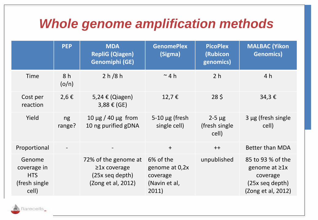
Whole genome amplification methods
PEP MDA RepliG (Qiagen) Genomiphi (GE)
GenomePlex (Sigma)
PicoPlex (Rubicon
genomics)
MALBAC (Yikon Genomics)
Time 8 h (o/n)
2 h /8 h ~ 4 h 2 h 4 h
Cost per reaction
2,6 € 5,24 € (Qiagen) 3,88 € (GE)
12,7 € 28 $ 34,3 €
Yield ng range?
10 µg / 40 µg from 10 ng purified gDNA
5-10 µg (fresh single cell)
2-5 µg (fresh single
cell)
3 µg (fresh single cell)
Proportional - - + ++ Better than MDA
Genome coverage in
HTS (fresh single
cell)
72% of the genome at ≥1x coverage
(25x seq depth) (Zong et al, 2012)
6% of the genome at 0,2x coverage (Navin et al, 2011)
unpublished 85 to 93 % of the genome at ≥1x
coverage (25x seq depth)
(Zong et al, 2012)

Feasibility of single cell NGS
by whole exome sequencing (Illumina)
Sample type number of reads
number of mapped reads
Percentage of coverage
Average sequencing
depth
Unfixed (Fresh) Single cell
51 600 412 45 5341 717 76 % 27
Fixed single cell 40 769 194 36 255 925 70 % 22
Fixed and microdissected
single cell
38 455 158 32 076 608 50 % 20
Genomic DNA 43 827 640 35 532 707 95 % 23
In collaboration with
22

CTC transfer on slides
Patient
Step 4 Staining and
cytopathology
10 min
Dilution with
Rarecells® Buffer
Step 1 –
Blood Collection
10 mL
Filtration by Rarecells® Device
Step 2- Filtration with Rarecells® System (15 min)
Blood to be processed within 5 hours of collection
Step 3- Transfer on slide
A549
leucocyte
Ongoing: sensitivity validation (1 tumor cell per mL of blood)

30 patients with CCRC
22 men et 8 women
Mean age 68,5
Two patients with metastasis
Tested by ISET before surgery
Mutation analysis: T-CTC- gDNA
VHL mutation analysis in CTCs
from patients with Clear Cells Renal Carcinoma
CTM from CCRC patients
• VHL gene
• Identified in 1993 by Latif et al.
• Tumor suppressor gene
• Position: 3p25.26 - 20Kb – 3 exons
• 2 ARNm de 4,5 et 4,7 Kb, two protein isoforms
• More than 500 mutations

EXON CODON MUTATION TYPE OF
MUTATION
CODON
INITIAL
CODON
MUTE CpG PROTEIN CHANGE
1
9 c.27G>T Tr GAC TAC Oui D9Y
18 c.53C>A Tr GCA GAA Non A18E
61 c.183C>G Tr CCC CCG Non P61P
65 c.194C>A Tr TCG TAG Non S65X
69 c.205-206delCG Fr CGC - - E69X
88 c.263G>A Ts TGG TAG Non W88X
92 c.275delA Fr GAC - - D92X
100 c.299delC Fr ACG - - T100X
109 c.327delC Fr ATC - - H109X
2
116 c.346C>G Tr CTT GTT Non L116V
118 c.353T>C Ts CTC CCC Non L118P
140 c.418delC Fr CTC - - L140X
145 c.435G>T Tr CAG CAT - Q145H
3
158 c.472delC Fr CTG - - L158X
163 c.486delC Fr TGC - - L163X
176 c.526A>T Tr AGG TGG Non R176W
183 c.548C>A Tr TCG TAG Non S183X
207 c.620C>T Ts GCA GTA Non A207V
VHL mutations found
Tr : Transversion; Ts : Transition; Fr : Frameshift
Résultats moléculaires
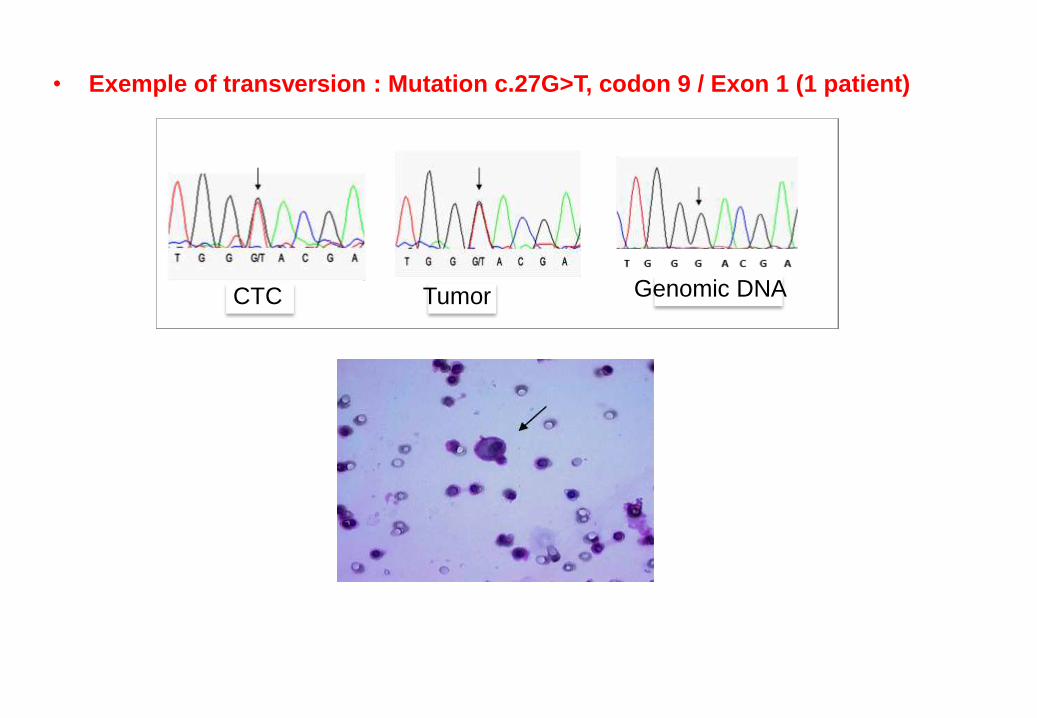
• Exemple of transversion : Mutation c.27G>T, codon 9 / Exon 1 (1 patient)
Résultats moléculaires
CTC Tumor Genomic DNA

Exemple of a transversion found in 2 patients
Mutation c.263G>A, codon 88 / Exon 1
Résultats moléculaires
CTC from patient CGN CTC from patient GRA
Tumor Genomic DNA CTC

VHL mutation analysis
can help cytopathological diagnosis of CTCs
in patients with Clear Cells Renal Carcinoma
Blind mutation analysis of VHL gene in
T, leucocytes and CTC isolated by ISET
Cytoapthology
30 patients with CCRC
25 with VHL mutation in tumor
20 patients with CTC
205 single cell analysis : 64 CTC
VHL mutations: 57/64 CTC
7 VHL-neg CTC from tumors neg for
VHL
VHL gene exon 2: T insertion in position 171 Mutation: GGTGTGGTCTCTTTAA (WT: GGTGTGGCTCTTTAA)
Ben Njima B et al, manuscript
in preparation

Acknowledgements
INSERM Unit 807
Dr Thierry Capiod
Dr Basma Ben Jima
Dr Ingrid Pfifer
Pr Patrizia Paterlini Bréchot
THANKS FOR YOUR ATTENTION!
Ms. Katia Hormigos
Dr Sophie Laget
Collaborations
Pr Paul Hofman, Patology Dpt, Nice
Dr. Marius Ilie, Patology DptNice
Pr. Nicole Brousse, Patology Dpt, Paris
Pr. Khaled Ben Romdhane, Tunis
Pr. Arnaud Mejean, Urology Dpt, Paris