ecg considerations with bundle branch … lead ecg variants, mimics and advanced skills 2 ecg...
TRANSCRIPT

CNEA / Key Choice 4/7/2012
www.cardionursing.com 1
2012
1C Y N T H I A W E B N E R D N P , R N , C C N S , C C R N - C M C K A R E N M A R Z L I N D N P , R N , C C N S , C C R N - C M C
12 LEAD ECG VARIANTS, MIMICS
AND ADVANCED SKILLS
2
ECG CONSIDERATIONS WITH
BUNDLE BRANCH BLOCKS

CNEA / Key Choice 4/7/2012
www.cardionursing.com 2
CONDUCTION SYSTEM REVIEW
• Left Bundle Branch • Left anterior fascicle
• Left posterior fascicle
• Right Bundle Branch
• Purkinje Network
• Purkinje Fibers
3
4
NORMAL DEPOLARIZATION
V1
V6
QRS .06-.10 sec
1
2

CNEA / Key Choice 4/7/2012
www.cardionursing.com 3
BUNDLE BRANCH BLOCK
• QRS complex is 0.12 sec or greater
• Incomplete BBB measures from 0.10 to 0.11
5
RIGHT BUNDLE BRANCH BLOCK
CAUSES
• CAD• Disease of right side of the heart• Cor pulmonale• Cardiomyopathy• Congenital lesions• A-S Defects• Pulmonic Stenosis• Pulmonary Embolism
6

CNEA / Key Choice 4/7/2012
www.cardionursing.com 4
RIGHT BUNDLE BRANCH BLOCK
V1 = rsR’
V6 = qRS
QRS = .12 sec or more
RIGHT BUNDLE BRANCH
BLOCK
8
• V1• Triphasic complex rsR’
pattern - positive• Or an M shaped R
wave with right peak taller
• Or a qR pattern
• V6• Triphasic complex• qRs with wide S waves• Positive
V1V6
rSR’ qRs
V1
R qR

CNEA / Key Choice 4/7/2012
www.cardionursing.com 5
9
LEFT BUNDLE BRANCH BLOCK
CAUSES
• Left Ventricular Hypertrophy
• MI
• CAD
• Aortic Stenosis
• Cardiomyopathy
• Hypertensive cardiomyopathy
10

CNEA / Key Choice 4/7/2012
www.cardionursing.com 6
11
LEFT BUNDLE BRANCH BLOCK
V1 = QS
V6 = wide R
QRS = .12 sec or more
V1 = rS
LEFT BUNDLE BRANCH
BLOCK• V1
• Wide QS or rS complex -negative
• Slick downstroke
• Nadir <0.06 sec
• V6• Wide R wave with no
initial septal q wave - -positive
12
V6
CNEA / Key Choice 4/7/2012
www.cardionursing.com 7
13
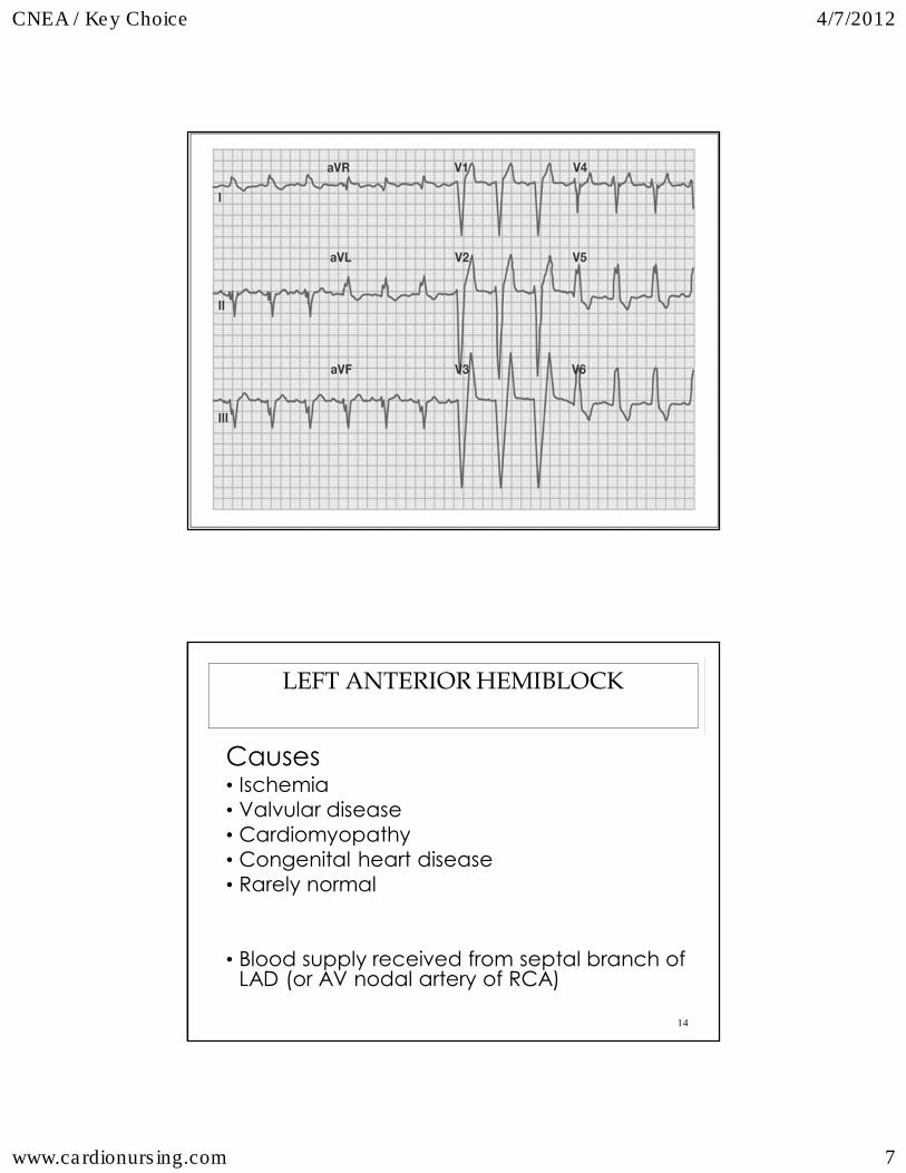
LEFT ANTERIOR HEMIBLOCK
Causes• Ischemia• Valvular disease• Cardiomyopathy• Congenital heart disease• Rarely normal
• Blood supply received from septal branch of LAD (or AV nodal artery of RCA)
14

CNEA / Key Choice 4/7/2012
www.cardionursing.com 8
LEFT ANTERIOR HEMIBLOCK
• Block of anterior –superior fascicle of the LBB
• Left axis deviation• - 30° to –75°• Become suspicious at -
30°• Definitive at – 40 to 45°• Common at -60 °
• Commonly seen in anterior wall MI• Low mortality if
isolated
• Left anterior hemiblock in association with RBBB during AMI• Associated with left
main occlusion and high mortality
15
Key for recognizing -60° Axis
- aVR most equiphasic limb lead
LEFT ANTERIOR HEMIBLOCK
• Lead 2, Lead 3 and aVF
• rS pattern
• Small r waves
• Slightly wide / deep S waves
• Increased limb lead voltage
• Lead 1 and aVL
• qR pattern
• Normal QRS duration 16

CNEA / Key Choice 4/7/2012
www.cardionursing.com 9
17
ST CHANGES WITH RBBB
• Normal direction of T wave (discordant) • In V1 – V3
• T wave inverted in leads with rSR’ pattern
• T wave upright in leads with S wave
• ST elevation is usually due to injury
• Measure QRS in easiest identified lead
• Determine the end of the QRS in any lead
2010
18
CNEA / Key Choice 4/7/2012
www.cardionursing.com 10
20
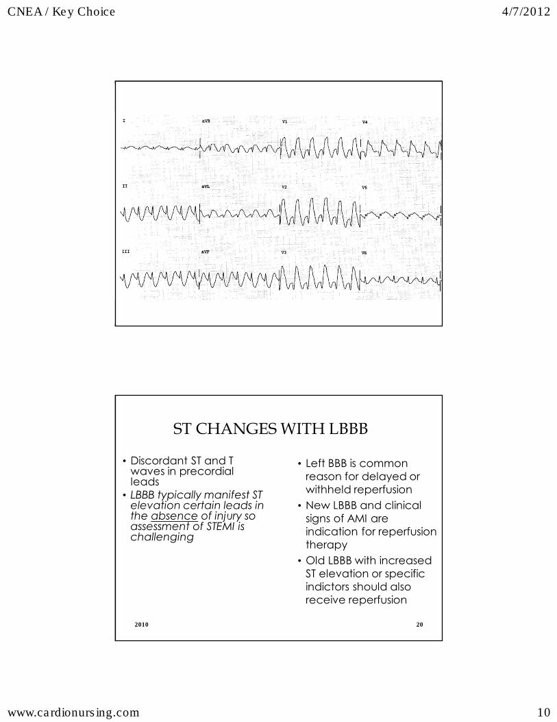
ST CHANGES WITH LBBB
• Discordant ST and T waves in precordial leads
• LBBB typically manifest ST elevation certain leads in the absence of injury so assessment of STEMI is challenging
• Left BBB is common reason for delayed or withheld reperfusion
• New LBBB and clinical signs of AMI are indication for reperfusion therapy
• Old LBBB with increased ST elevation or specific indictors should also receive reperfusion
2010

CNEA / Key Choice 4/7/2012
www.cardionursing.com 11
DIAGNOSTIC STRATEGIES FOR AMI
WITH LBBB
• √ Concordant ST elevation > 1 mm in leads where QRS is predominantly positive
• V5, V6, I, aVL, II
• √ Concordant ST depression > 1 mm in one or more leads in leads where QRS is predominantly negative
• V1 – V4
• √ Discordant ST elevation > 5 mm and disproportionate with the QRS voltage.

CNEA / Key Choice 4/7/2012
www.cardionursing.com 12
NORMAL VARIANTS
24
CNEA / Key Choice 4/7/2012
www.cardionursing.com 13
EARLY REPOLARIZATION
• Precordial ST elevation in most adults • Up to 90%
• Early repolarization most common normal variant• African American men < 50 years of age
25
? Increased Risk of Sudden Death
ST ELEVATION OF EARLY
REPOLORIZATION
• ST segments highest in V2-V3 • ST elevation up to 5 mm • J point elevation • Concavity, not convexity • Tall peaked asymmetric T waves • T waves are tall (not too tall) but not wide • ST elevation < 0.5 m in lead V5 and V6• T wave amplitude in V6 > V1 • No reciprocal ST depression • Less common after age 55
26
CNEA / Key Choice 4/7/2012
www.cardionursing.com 14
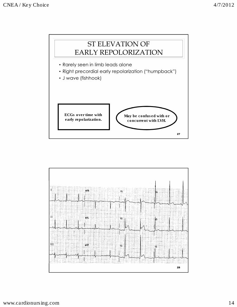
ST ELEVATION OF
EARLY REPOLORIZATION
• Rarely seen in limb leads alone
• Right precordial early repolarization (“humpback”)
• J wave (fishhook)
27
May be confused with orconcurrent with LVH.
ECGs over time with early repolarization.
28

CNEA / Key Choice 4/7/2012
www.cardionursing.com 15
29
PERSISTENT JUVENILE T WAVES
• Negative (inverted) T waves inV1-V3 (right precordial leads)
• Not “deep” inversions • Usually in young, healthy women • More common in African Americans
Note: Infants > 48 hours through childhood have inverted right precordial T waves. Progressive change to upright through late childhood.
30

CNEA / Key Choice 4/7/2012
www.cardionursing.com 16
RSR’ PATTERN
• V1 and V2
• r’ amplitude < r
• QRS interval < 0.11 seconds
• When r’ changes to R’ consider incomplete RBBB
31
? Fragmented QRS and non STEMI
ECG
MIMICS
32
CNEA / Key Choice 4/7/2012
www.cardionursing.com 17
LVH
• Common reason for false positive ST elevation
• Anatomic LVH may be present in absence of ECG criteria
• To ascribe ST elevation to LVH the ECG must meet the voltage criteria
33
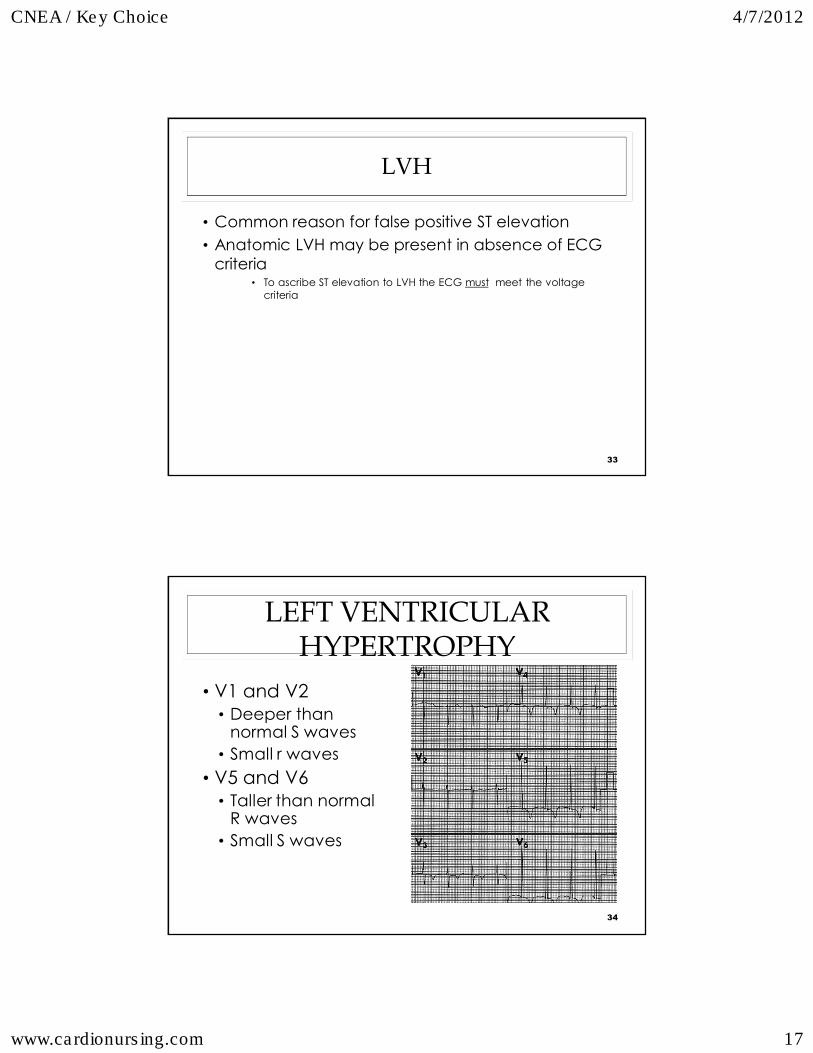
LEFT VENTRICULAR
HYPERTROPHY
• V1 and V2• Deeper than
normal S waves• Small r waves
• V5 and V6• Taller than normal
R waves• Small S waves
34

CNEA / Key Choice 4/7/2012
www.cardionursing.com 18
LVH VOLTAGE CRITERIA
• One or more voltage criteria
• Only applicable if QRS is < 120 ms
• Precordial lead voltage criteria • R-wave in V5 or V6 > 26 mm
• R-wave in V5 or V6 + S-wave in V1 > 35 mm
• Largest R-wave + largest S-wave in precordial leads > 45 mm
35
ST – T WAVE CHANGES
SECONDARY TO LVH
• ST elevation is generally discordant • ST elevation in V2 -V3 • ST elevation in lead III • ST depression in V4-V6
• Previously called strain pattern • Down sloping – not horizontal
Not due to LVH • ST elevation in lateral leads • ST depression in V2-V3
36

CNEA / Key Choice 4/7/2012
www.cardionursing.com 19
37
38

CNEA / Key Choice 4/7/2012
www.cardionursing.com 20
RIGHT VENTRICULAR
HYPERTROPHY• RV Hypertrophy
• Right Axis deviation is one of earliest signs
• Reverse R wave progression
• Dominant R wave in V1 and V2
• Deep S wave in V5 and V6
39
DIGITALIS EFFECT
• Sagging depression of ST segment in leads with positive QRS
• Reduced T wave amplitude
• Increased U wave amplitude
• Difficult to evaluate if hypertrophy or BBB
40
CNEA / Key Choice 4/7/2012
www.cardionursing.com 21
41
PAIN AND ECG
MIMICS
PERICARDITIS: ECG FINDINGS
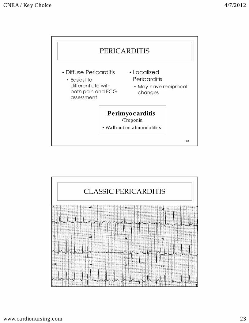
• Mimics: anteroinferior; inferolateral; antero-infero-lateral MI
• ST Elevation • ST elevation typically greatest in II and V5 (also I and V6)
• ST elevation may also be in V1 –V4; aVF, III and aVL (least)
• Upwardly concave ST segments
• ST elevation usually < 5 mm
42

CNEA / Key Choice 4/7/2012
www.cardionursing.com 22
PERICARDITIS: ECG FINDINGS
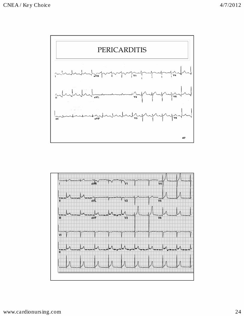
• Other ST changes • ST depression in aVR• Minimal depression V1, III, aVL may exist
• PR Segment depression • PR depression most common in II, aVF and V4 –
V6 • PR elevation > 0.5 mm in aVR
• Electrical Alternans• Voltage changes with pericardial effusion or
tamponade
43
STAGES OF PERICARDITIS • Stage I
• ST elevation • More concave • Lasts up to 2 weeks
• Stage II• ST to baseline • Decrease T wave amplitude • Lasts from days to several weeks
• Stage III • T wave inversion • Starts at end of second to third week
• Stage IV• Gradual resolution • T wave may stay inverted up to 3 months
44
CNEA / Key Choice 4/7/2012
www.cardionursing.com 23
PERICARDITIS
• Diffuse Pericarditis• Easiest to
differentiate with both pain and ECG assessment
• Localized Pericarditis • May have reciprocal
changes
45
Perimyocarditis •Troponin
• Wall motion abnormalities
CLASSIC PERICARDITIS
46
CNEA / Key Choice 4/7/2012
www.cardionursing.com 24
PERICARDITIS
47
48

CNEA / Key Choice 4/7/2012
www.cardionursing.com 25
ACUTE COR PULMONALE
• Transient ECG changes • ST or atrial tachycardia (or fib / flutter) • T wave inversion (or other ECG signs of
ischemia, injury, infarction) in both inferior and anteroseptal leads
• Elevated ST segments aVR and V1-V2• Prominent S waves in I and aVL• RAD or incomplete or complete RBBB • Widespread S waves• Prominent P waves in inferior leads(right atrial strain)
49
ACUTE COR PULMONALE
50

CNEA / Key Choice 4/7/2012
www.cardionursing.com 26
ACUTE COR PULMONALE
51
52
PULMONARY EMBOLISM
• Obstruction of blood flow to one or more arteries of the lung by a thrombus (other emboli – fat, air, amniotic fluid) lodged in a pulmonary vessel
• Lower lobes frequently affected due to increased perfusion

CNEA / Key Choice 4/7/2012
www.cardionursing.com 27
53
RISK FACTORS FOR PE • Stasis of blood
• Prolonged immobilization after surgical procedures• Plaster casts• Venous obstruction• Heart failure / Shock / Hypovolemia• Varicose veins• Obesity
• Hypercoagulability• Polycythemia vera• Sickle cell disease• Malignancy• Pregnancy• Recent trauma• Oral contraceptives
• Injury to the vascular endothelium• Central venous and arterial catheters• Phlebitis
54
PULMONARY EMBOLISM:
PATHOPHYSIOLOGY
• > 90% of thrombus develop in deep veins of iliofemoral system• Can also originate in the right side of the heart, pelvic veins, and
axillary or subclavian veins. • Another source is around indwelling catheters.
• Thrombus formation leads to platelet adhesiveness and release of serotonin (vasoconstrictor).
• Dislodgement of thrombus• Intravascular pressure changes (standing, massaging legs, fluid
challenge, valsalva maneuver).• Natural clot dissolution (7-10 days after development).
CNEA / Key Choice 4/7/2012
www.cardionursing.com 28
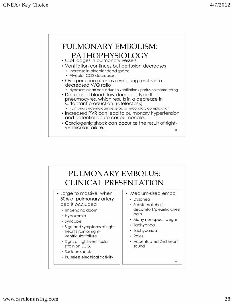
55
PULMONARY EMBOLISM:
PATHOPHYSIOLOGY• Clot lodges in pulmonary vessels• Ventilation continues but perfusion decreases
• Increase in alveolar dead space• Alveolar CO2 decreases
• Overperfusion of uninvolved lung results in a decreased V/Q ratio• Hypoxemia can occur due to ventilation / perfusion mismatching.
• Decreased blood flow damages type II pneumocytes, which results in a decrease in surfactant production. (atelectasis) • Pulmonary edema can develop as secondary complication
• Increased PVR can lead to pulmonary hypertension and potential acute cor pulmonale.
• Cardiogenic shock can occur as the result of right-ventricular failure.
56
PULMONARY EMBOLUS:
CLINICAL PRESENTATION
• Large to massive when 50% of pulmonary artery bed is occluded• Impending doom
• Hypoxemia
• Syncope
• Sign and symptoms of right heart strain or right-ventricular failure
• Signs of right-ventricular strain on ECG.
• Sudden shock
• Pulseless electrical activity
• Medium-sized emboli• Dyspnea
• Substernal chest discomfort/pleuritic chest pain
• Many non-specific signs
• Tachypnea
• Tachycarida
• Rales
• Accentuated 2nd heart sound
CNEA / Key Choice 4/7/2012
www.cardionursing.com 29
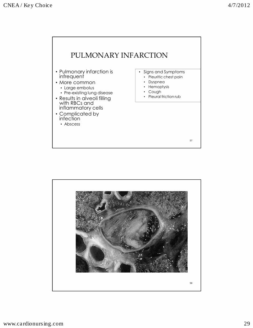
57
PULMONARY INFARCTION
• Pulmonary infarction is infrequent
• More common• Large embolus • Pre-existing lung disease
• Results in alveoli filling with RBCs and inflammatory cells
• Complicated by infection • Abscess
• Signs and Symptoms • Pleuritic chest pain• Dyspnea• Hemoptysis• Cough• Pleural friction rub
58
CNEA / Key Choice 4/7/2012
www.cardionursing.com 30
59
60
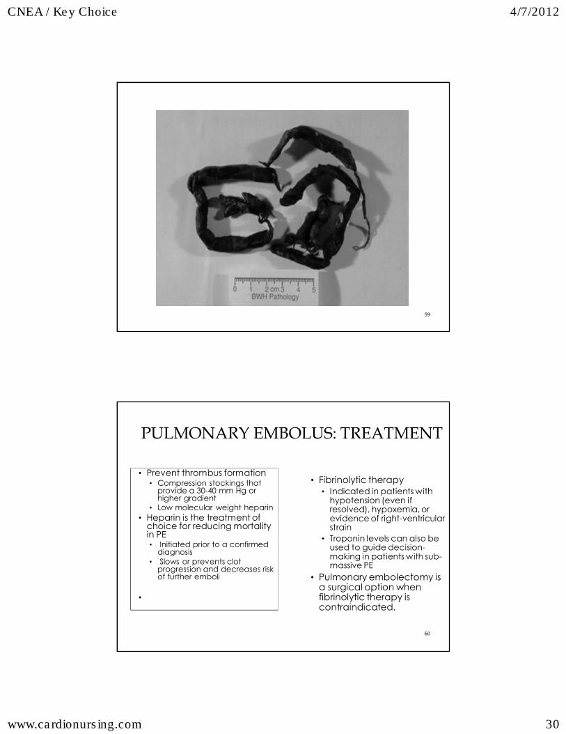
PULMONARY EMBOLUS: TREATMENT
• Prevent thrombus formation • Compression stockings that
provide a 30-40 mm Hg or higher gradient
• Low molecular weight heparin• Heparin is the treatment of
choice for reducing mortality in PE• Initiated prior to a confirmed
diagnosis• Slows or prevents clot
progression and decreases risk of further emboli
•
• Fibrinolytic therapy• Indicated in patients with
hypotension (even if resolved), hypoxemia, or evidence of right-ventricular strain
• Troponin levels can also be used to guide decision-making in patients with sub-massive PE
• Pulmonary embolectomy is a surgical option when fibrinolytic therapy is contraindicated.

CNEA / Key Choice 4/7/2012
www.cardionursing.com 31
61
PULMONARY EMBOLUS: TREATMENT
• Oxygen is indicated, even in the absence of hypoxemia
• Pulmonary vasodilators to help reduce pulmonary vascular resistance
• Treat right-ventricular failure with fluids and inotropes
• Obstructive Shock
• Warfarin• 3 to 6 months if there is
identifiable reversible risk factor
• Minimum of 6 six months if there is no identifiable risk factor
• Long term with recurrent PE or in patients with ongoing risk factors
• Surgical interruption of inferior vena cava with a filter
• Patients with contraindication to anticoagulants.
• Recurrent thromboembolism despite anticoagulant.
• Survivor of massive PE
ADDITIONAL MIMICS WITH DEEP SYMMETRICAL
T WAVE INVERSION
• Adams – Stokes attack
• Hypertrophic cardiomyopathy
• Central nervous system disease
• Post extrasystolic T wave change
62

CNEA / Key Choice 4/7/2012
www.cardionursing.com 32
ADDITIONAL MIMICS OF ST ELEVATION
• Hyperkalemia
• Intracranial bleed • Prolonged QT
• Prominent U wave
63
NON MI CAUSES OF Q WAVES
• LVH • RVH • Cor Pulmonale • Cardiomyopathy • LBBB • LAHB • WPW • Pulmonary Embolism
64

CNEA / Key Choice 4/7/2012
www.cardionursing.com 33
PAIN MIMICS
PATHOPHYSIOLOGY
• Intimal tear • False channel • Risk of rupture: outer
wall • Dissection – hematoma
– occlusion of vessels
• Ascending aorta • Descending aorta • Abdominal aorta

CNEA / Key Choice 4/7/2012
www.cardionursing.com 34
RISK FOR DISSECTION
• Hypertension is present in most patients with dissections
• Congenital disorders affecting connective tissue such as Marfan Syndrome or Ehlers-Danlos syndrome; also presence of bicuspid aortic valve
• 3rd trimester of pregnancy
• After procedures where aorta or the aortic branches have been cannulated.

CNEA / Key Choice 4/7/2012
www.cardionursing.com 35
CLASSIFICATION OF
DISSECTIONS
• Acute or chronic• Type A Dissections:
Dissections involving the ascending aorta.
• Type B Dissections:Dissections involving the descending thoracic aorta. These dissections begin distal to the left subclavian artery.
COMPLICATIONS OF DISSECTION
• Aortic regurgitation from retrograde dissection involving aortic valve or from aortic dilatation.
• MI from retrograde coronary artery dissection. • Cardiac tamponade from ascending aorta or aortic arch
rupture. • Intraplerual rupture from descending aortic dissection ruptures
into intrapleural space – most commonly left sided. • Retroperitoneal bleed from rupture of abdominal aorta
dissection. • Stroke from brachial artery compromise.• Paraplegia, reduced blood flow to kidneys, bowels, and lower
extremities from compromise of arterial branches.
CNEA / Key Choice 4/7/2012
www.cardionursing.com 36
CLINICAL PRESENTATION
Chest or back pain with variation in upper extremity blood pressure is key assessment finding in aortic dissection. Recurrent chest or back pain can indicate extension or rupture. The presence of aortic regurgitation in the setting of chest pain is also suspicious for aortic dissection.
DIAGNOSIS
• Chest Radiograph: An abnormality or the aorta can usually be seen as a widening of the aorta and mediastinum. Calcium present in the intima is separated from the outer border of the false channel.
• Transthoracic echocardiography and transesophageal echocardiography: A visible intimal flap separates the true and false lumens. Echocardiography is most helpful with type A dissections. Transesophageal echocardiography is very valuable and can be performed bedside.
• CT: CT is particularly helpful in evaluating dissections of the thoracic aorta.
• MRI: MRI is an excellent diagnostic tool but has limitation because it cannot be used emergently or in critically ill patients.
• Angiography is a definitive diagnostic tool and is required for patients having elective repair of the thoracic aorta.

CNEA / Key Choice 4/7/2012
www.cardionursing.com 37
TREATMENT OF DISSECTIONS
• Stop dissection • Control of hypertension
• Lowest possible pressure without compromising perfusion
• Velocity of ejection
• Rate of rise
• Beta-blocker in combination with sodium nitroprusside
TREATMENT OF DISSECTIONS
• Type A • Surgery required • Deep hypothermic circulatory arrest • Distal false channel not completely eliminated • Risk for bleeding • Replacement of aortic valve / reimplantation of
coronary arteries • Hypertension management • Surgical considerations similar for ascending
aortic aneurysms • Surgical mortality 15-20%
CNEA / Key Choice 4/7/2012
www.cardionursing.com 38
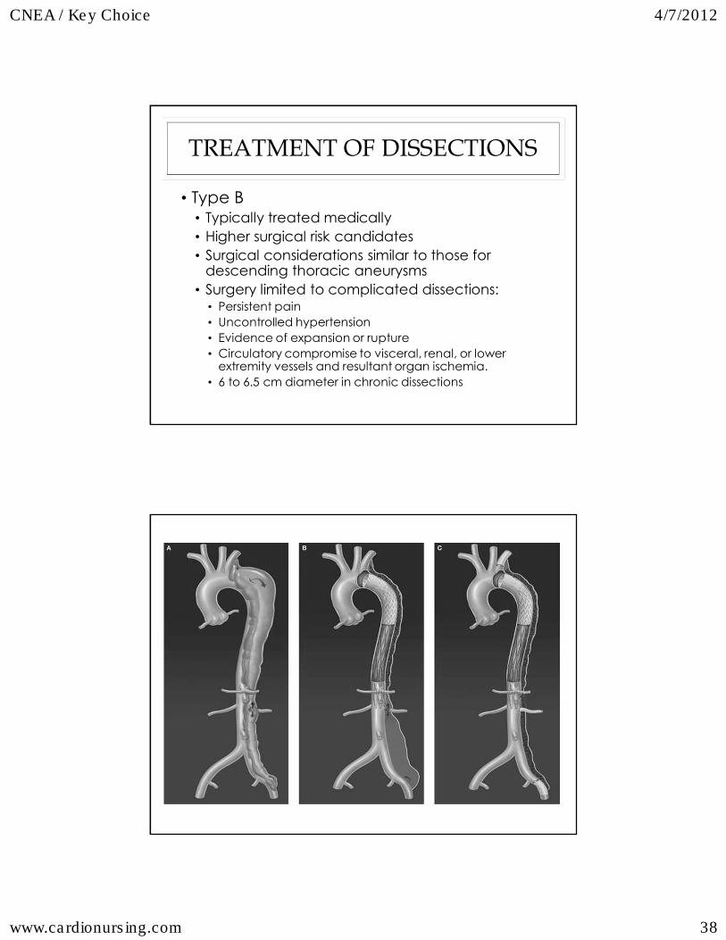
TREATMENT OF DISSECTIONS
• Type B • Typically treated medically • Higher surgical risk candidates • Surgical considerations similar to those for
descending thoracic aneurysms • Surgery limited to complicated dissections:
• Persistent pain • Uncontrolled hypertension• Evidence of expansion or rupture • Circulatory compromise to visceral, renal, or lower
extremity vessels and resultant organ ischemia. • 6 to 6.5 cm diameter in chronic dissections

CNEA / Key Choice 4/7/2012
www.cardionursing.com 39
ADDITIONAL ADVANCED ECG SKILLS
INJURY AND ISCHEMIA
Advanced Concepts in:
78
CNEA / Key Choice 4/7/2012
www.cardionursing.com 40
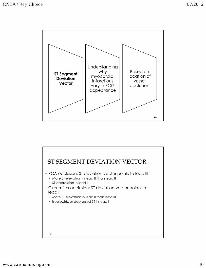
79
ST Segment Deviation Vector
Understanding why
myocardial infarctions
vary in ECG appearance
Based on location of
vessel occlusion
80
ST SEGMENT DEVIATION VECTOR
• RCA occlusion: ST deviation vector points to lead III• More ST elevation in lead III than lead II • ST depression in lead I
• Circumflex occlusion: ST deviation vector points to lead II • More ST elevation in lead II than lead III• Isoelectric or depressed ST in lead I

CNEA / Key Choice 4/7/2012
www.cardionursing.com 41
81
82
ST SEGMENT EVALUATION IN V4R
• ST elevation in V4R • Proximal occlusion of RCA prior to marginal branch
• Significant risk for advanced AV nodal block
• Isoelectric ST in V4R with upright T wave
• Isoelectric or depressed ST in V4R with negative T wave

CNEA / Key Choice 4/7/2012
www.cardionursing.com 42
83
ADDITIONAL ST / T WAVE
CHANGES WITH INFERIOR MI
• Extension into posterior wall • ST segment depression in the precordial leads
• ST deviation vector points to posterior wall
• Extension into lateral wall • ST elevation in leads V5, V6 (may also be leads I, and
aVL) • ST deviation vector points to lateral wall
• Usually caused by circumflex occlusion
84
ANTERIOR WALL MI
• Precordial ST elevation
• Location of ST segment deviation vector in limb leads helps predict the location of occlusion within the LAD
CNEA / Key Choice 4/7/2012
www.cardionursing.com 43
85
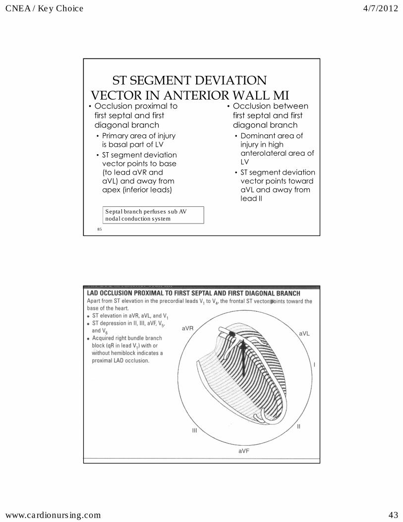
ST SEGMENT DEVIATION
VECTOR IN ANTERIOR WALL MI • Occlusion proximal to
first septal and first diagonal branch • Primary area of injury
is basal part of LV
• ST segment deviation vector points to base (to lead aVR and aVL) and away from apex (inferior leads)
• Occlusion between first septal and first diagonal branch • Dominant area of
injury in high anterolateral area of LV
• ST segment deviation vector points toward aVL and away from lead II
Septal branch perfuses sub AV nodal conduction system
86

CNEA / Key Choice 4/7/2012
www.cardionursing.com 44
87
88
ST SEGMENT DEVIATION
VECTOR IN ANTERIOR WALL MI• Occlusion distal to the
diagonal branch • ST segment deviation
vector points to apex • ST elevation in
inferior leads (II > III)
• Left Main Coronary Artery Occlusion • ST segment deviation
vector points to lead aVR • ST elevation in aVR • Less ST elevation in
V1 • ST depression in
leads V2 – V6 (posterior wall injury) – circumflex occlusion

CNEA / Key Choice 4/7/2012
www.cardionursing.com 45
89
90

CNEA / Key Choice 4/7/2012
www.cardionursing.com 46
91
LIMITATIONS OF ST SEGMENT
DEVIATION VECTOR ANALYSIS
• Previous MI / preexisting ST / T wave abnormalities
• Multi vessel disease
• Altered ventricular activation
• Dominant or small coronary arteries
• Coronary artery anomalies
92
ST DEVIATION SCORE
• Score > 12 mm: substantial amount of myocardium to benefit from reperfusion
• ST segment deviation may be better than ST segment deviation score
• Limb leads provide more global information
• Precordial leads provide more local information
CNEA / Key Choice 4/7/2012
www.cardionursing.com 47
93
RISK STRATIFICATION
• Severity of ischemia • Grade 1
• Peaked symmetrical T waves (hyperacute) • Grade 2
• ST elevation / no distortion of terminal part of QRS • Grade 3
• ST elevation with distortion of terminal part of QRS • J Point / R wave ratio > .5
• Grades of ischemia correlate with infarct size, LV function, and mortality (in hospital and late)
94
ADDITIONAL RISK
CONSIDERATIONS
• Significance of 2nd degree or more AV nodal block
• Significance of conduction disturbances in or below bundle of His • Existing versus acquired RBBB
• RBBB with LAHB
CNEA / Key Choice 4/7/2012
www.cardionursing.com 48
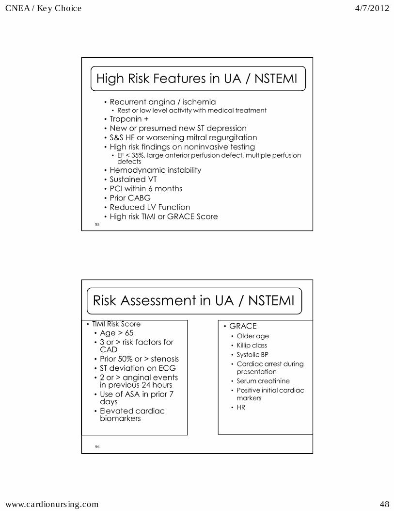
High Risk Features in UA / NSTEMI
95
• Recurrent angina / ischemia • Rest or low level activity with medical treatment
• Troponin + • New or presumed new ST depression • S&S HF or worsening mitral regurgitation • High risk findings on noninvasive testing
• EF < 35%, large anterior perfusion defect, multiple perfusion defects
• Hemodynamic instability • Sustained VT • PCI within 6 months • Prior CABG • Reduced LV Function • High risk TIMI or GRACE Score
Risk Assessment in UA / NSTEMI
96
• TIMI Risk Score • Age > 65 • 3 or > risk factors for
CAD • Prior 50% or > stenosis• ST deviation on ECG • 2 or > anginal events
in previous 24 hours • Use of ASA in prior 7
days • Elevated cardiac
biomarkers
• GRACE • Older age
• Killip class
• Systolic BP
• Cardiac arrest during presentation
• Serum creatinine
• Positive initial cardiac markers
• HR
CNEA / Key Choice 4/7/2012
www.cardionursing.com 49
97
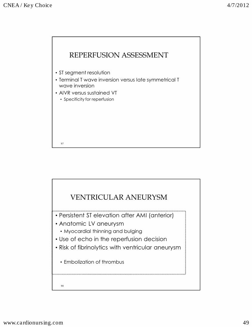
REPERFUSION ASSESSMENT
• ST segment resolution
• Terminal T wave inversion versus late symmetrical T wave inversion
• AIVR versus sustained VT • Specificity for reperfusion
98
VENTRICULAR ANEURYSM
• Persistent ST elevation after AMI (anterior)
• Anatomic LV aneurysm • Myocardial thinning and bulging
• Use of echo in the reperfusion decision
• Risk of fibrinolytics with ventricular aneurysm
• Embolization of thrombus

CNEA / Key Choice 4/7/2012
www.cardionursing.com 50
99
ST ELEVATION OF VENTRICULAR
ANEURYSM
• Most common in V1-V3 • Usually less than 3 mm elevation• Relatively unchanged from previous ECGs • Q waves are deep and well formed
• QS pattern in V1-V3 or very minimal r • QR pattern common in inferior aneurysm / RBBB
100

CNEA / Key Choice 4/7/2012
www.cardionursing.com 51
101
MYOCARDIAL RUPTURE
• Incidence • 10% MI deaths
• Definition • Myocardial leakage – hemipericardium – tamponade • Perceived sudden; often slow tear
• Associated Factors • Late fibrinolytics • Delayed hospital admission
• Septal involvement = VSD
102
MYOCARDIAL RUPTURE
• Post-infarction regional pericarditis precedes rupture
• Confirmation of rupture
T Wave Patterns in Post-infarction Regional Pericarditis
Persistently positive T waves 48 hours after an MI
Premature reversal of T wave inversion to positive ST segment
reelevation

CNEA / Key Choice 4/7/2012
www.cardionursing.com 52
103
10
4
Final Thought:
Mastery is not something that strikes in an instant, like a thunderbolt, but a gathering power that moves steadily through time,
like weather.
- John Champlain Gardner Jr. (1933-1982)