neuro-ophthalmic disease cases - optometry's … disease cases kelly a. malloy, od, faao, ......
TRANSCRIPT
5/5/2014
1
Neuro-Ophthalmic Disease Cases
Kelly A. Malloy, OD, FAAO, Diplomate
(Neuro-Ophthalmic Disease)
NOTHING TO DISCLOSE
CASE 1
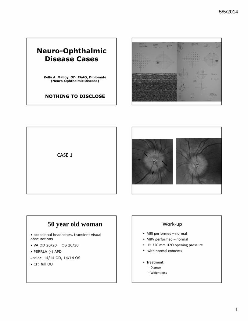
• occasional headaches, transient visual
obscurations
• VA OD 20/20 OS 20/20
• PERRLA (-) APD
• color: 14/14 OD, 14/14 OS
• CF: full OU
50 year old woman Work-up
• MRI performed – normal
• MRV performed – normal
• LP: 320 mm H2O opening pressure
• with normal contents
• Treatment:
– Diamox
– Weight loss

5/5/2014
2
Idiopathic Intracranial Hypertension
(or PTC)
• this is a diagnosis of exclusion, and the
following are necessary to make the diagnosis
Modified Dandy’s Diagnostic Criteria ( adults)
• Patient Must Be Awake & Alert
• Signs & Symptoms Of Increased Intracranial Pressure
• No Neurologic Signs Except CN VI Paresis
• CSF Opening Pressure > 200mm or 250 mm H20 &
Normal Composition of CSF
• Normal Neuro-Imaging (MRI, MRV)
IDIOPATHIC INTRACRANIAL HYPERTENSION
- EPIDEMIOLOGY
• 92% - Women
• Ages 11-58
• No Racial Bias
• 13/100,000 In Women - 10% Above Ideal
Body Weight
• 19/100,000 - 20% Above Ideal Body Weight
IDIOPATHIC INTRACRANIAL HYPERTENSION
- INVESTIGATIONS
• MRI and MRV
• LUMBAR PUNCTURE
• Opening pressure
• many controversial issues regarding this– what is the effect on the posture – lateral decubitus
position vs. lying flat (fluoroscopy)
– how accurate are the measurements
– what is truly a normal or abnormal opening pressure for adults and children
• analysis of CSF contents (cell count, glucose, protein, cytology, VDRL, etc.)

5/5/2014
3
IDIOPATHIC INTRACRANIAL
HYPERTENSION - TREATMENT
• WEIGHT LOSS
• CARBONIC ANHYDRASE INHIBITORS
– acetazolamide (Diamox)
• a much needed clinical trial is in the process to analyze the
effectiveness of this treatment
• SURGICAL TREATMENT (when needed)
– Lumboperitoneal Shunt
– Optic Nerve Sheath Fenestration
– Gastric Bypass Surgery
IDIOPATHIC INTRACRANIAL HYPERTENSION
- PROGNOSIS
• 49% Have Some Visual Loss (Corbett 1982;
Orcutt 1984)
• 25% Severe & Permanent Visual Loss (Folley
1955; Boddle 1974)
• 80% Improve In 8 Months (Corbett 1982)
• 10% Recurrence Rate
• ???????
Symptoms of Increased Intra-Cranial Pressure
• Headache
• Nausea
• Vomiting
• Diplopia (Abduction deficit – CN VI)
• Pulsatile tinnitus
• Transient Visual Obscurations (TVOs)
– Last few seconds (uni or bi-lateral)
– Transient optic nerve ischemia
Features of Papilledema
• Bilateral/Asymmetric (anatomic difference
in lamina)
• RARELY Unilateral
• ICP (Intracranial pressure) greater than 200 -
250 mm H2O (20 – 25 cm H20)
Features of edema
• Axoplasmic stasis in pre-laminar optic nerve
• Obscuration of retinal vessels coursing over the disc margin
• Paton’s lines temporally
• Extruded axoplasm (in chronic papilledema)

5/5/2014
4
Pattern of Edema
• Corresponds with NFL thickness
• Superior, Inferior > Nasal > Temp
• Superior and Inferior NFL swell first
• Last to swell is Temporal NFL
Spontaneous Venous Pulsation
• Presence of SVP means ICP normal
• (at that moment - can fluctuate)
• 10-20 % of normals may not have SVP
Is this papilledema?
Subtle Papilledema
vs.
Anomalous Disc ?
OCT use in differentiating papilledema from
pseudopapilledema
A/B scan ultrasound with 30
degree test
• A non-invasive method to determine if there is increased sub-arachnoid fluid in the optic nerve sheath
• A positive fluid sign, indicating increased sub-arachnoid fluid, is suggestive of papilledema
• Used in cases of anomalous disc vs. papilledema, especially when the patient has no symptoms of increased intracranial pressure
Source: e-medicine

5/5/2014
5
A/B scan ultrasound with 30
degree test
• A dynamic A-scan test that measures the width of the optic nerve in primary gaze and again after the patient shifts gaze 30 degrees from primary.
• In cases of increased ICP, the nerve and sheath are stretched as the globe turns 30 degrees, and the subarachnoid fluid is distributed over the extent of the nerve, resulting in measurements less than when in primary gaze.
• If nerve enlargement is due to infiltration or thickening of the optic nerve sheath itself, then the measurement will not change as the globe turns from primary position.
Young Woman above ideal body weight
Only symptom is occasional headaches
PATIENT A
PATIENT B
Young Woman above ideal body weight
Only symptom is occasional headaches
Results of A/B SCAN with 30 degree test
PATIENT A
Right Eye: 4.8 3.1
Left Eye: 5.5 2.7
primary gaze 30 degrees lateral
Results of A/B SCAN with 30 degree test
PATIENT B
primary gaze 30 degrees lateral
Right Eye: 2.5 2.5
Left Eye: 2.5 2.5

5/5/2014
6
CAUSES OF PAPILLEDEMA
• Brain Tumor or Spinal Cord Tumor
• Venous Sinus Thrombosis
• Arteriovenous Malformation
• Subdural or Subarachnoid Hemorrhage
• Meningitis
• Other Infectious / Inflammatory Etiology
• Idiopathic Intracranial Hypertension
(aka Pseudotumor Cerebri)
CASE 2
• 31 year-old, 5’2”, 270lb woman
• routine eye exam – referred for disc edema OU
• blurry VA x 2 mos (attributed to old CLs) ?TVOs
• denies any eye / head pain, diplopia
• denies nausea, vomiting, pulsatile tinnitus, fever
• Sys hx: asthma, depression, sinusitis, ? Hypothyroidism
• Meds: Effexor, Advair, recently d/c Ortho-Evra patch (weight-gain)
31 year old woman
• VA: OD 20/20
OS 20/20
• Color: OD 14/14 OS 14/14
• Pupils isocoric, (-)RAPD
• Ocular motilities: full
• SLE: unremarkable
• IOP: normal OU

5/5/2014
7
WORK-UP: (PTC is a diagnosis of exclusion)
• MRI of brain with gadolinium contrast
• MRV of head
• R/O mass, venous sinus thrombosis
• Neuro-Imaging needs to be done PRIOR to
Lumbar Puncture
• Lumbar Puncture:
•high opening pressure (460mm H2O)
• normal contents
MRV
CASE 3
46 year-old woman
• Decreased vision x 3-4 months OS > OD
• Transient visual obscurations
• Diplopia a few months ago, not now
• + nausea, vomiting, pulsatile tinnitus
• Recent vertigo, balance issues, dx with seizures
• Significant headaches, since decompression of Chiari malformation 13 years ago
• Scheduled for surgery for herniated disc in neck
• Last eye exam – 2 years prior: normal
Examination Results
• VA: OD 20/70 OS 20/100
• Color (Ishihara): OD 0/14 OS 0/14
• Pupils: PERRL (+ 0.3 log) APD OS
• Normal motility (-) abduction deficit
• Normal anterior segment exam
• IOP: OD 21 mm Hg OS 22 mm Hg
• BP: 140/90

5/5/2014
8
Day of Initial Presentation
10 days after initial presentation
Bilateral optic nerve sheath fenestrationV-P shunt
Progressive vision loss OU over the next 1-2 months
– ultimately to the level of NLP OU
IIH or PTC can have devastating vision loss!!
Idiopathic Intracranial Hypertension Treatment
Trial (IIHTT)
• ClinicalTrials.gov identifier: NCT01003639
• Information obtained from: ClinicalTrials.gov on March 26, 2010
• NORDIC (Neuro-Ophthalmology Research Disease Investigator Consortium
• A Multicenter, Double-blind, Randomized, Placebo-controlled Study
in Subjects With Idiopathic Intracranial Hypertension With Mild
Visual Loss
Weight-Reduction and/or Low Sodium Diet Plus Acetazolamide
vs
Weight-Reduction and/or Low Sodium Diet Plus Placebo
CASE 4
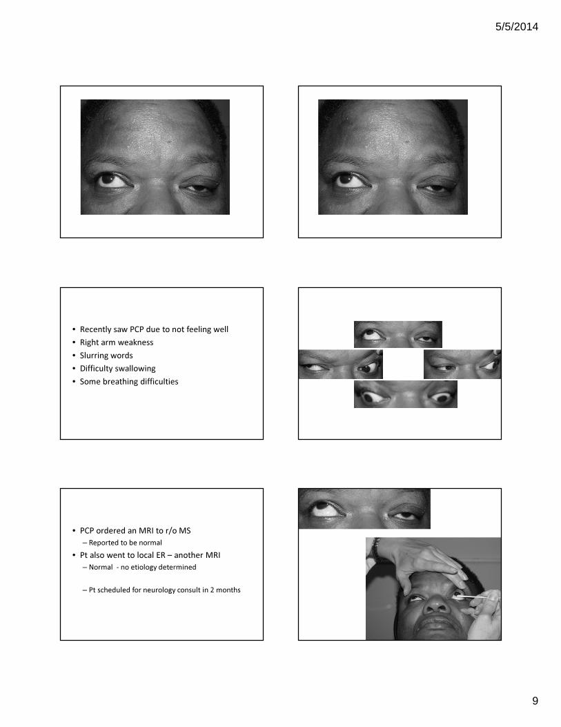
42 year old woman
• Noticed right eye has been pointing upward x 1 month
– Getting progressively worse
• Denies diplopia
• History of poor vision OD due to macular scar
5/5/2014
9
• Recently saw PCP due to not feeling well
• Right arm weakness
• Slurring words
• Difficulty swallowing
• Some breathing difficulties
• PCP ordered an MRI to r/o MS
– Reported to be normal
• Pt also went to local ER – another MRI
– Normal - no etiology determined
– Pt scheduled for neurology consult in 2 months

5/5/2014
10
• Neurologic Exam
– Weakness of neck flexor muscles
– Weakness of right upper extremity
Myasthenia Gravis
• Acetylcholine receptor antibodies
• Binding **** 1500 (normal < 0.30)
• Blocking *** positive 75 (normal <15)
• Modulating *** positive
• All thyroid tests negative
• Chest CT
• Admitted to hospital for evaluation / treatment for fear of myasthenic crisis• Plasmapheresis (ICU), IVIG, Mestinon, Prednisone
CASE 5
66 year-old woman with
droopy lids
• Hx of Grave’s disease and thyroid eye disease
• S/p lid surgery because of lid retraction (1996)
• Was told then that as she aged, her lids would get
more droopy
• Wants to get lids surgically lifted
• Lids are interfering with vision
• Occasional diplopia, mainly when tired or when
reading, seems stable since thyroid problems
• Systemic Hx:– HTN x 20 yrs– Hypercholesterolemia– S/p TIA in 2002– Grave’s disease– Seizures– Anemia– Osteoarthritis– S/p knee replacement complicated by blood clots– S/p TMJ surgery in 1987– Sleep apnea– BPPV
– Followed by: cardiologist, gastroenterologist, endocrinologist, otolaryngologist, hematologist and PCP

5/5/2014
11
• VA: OD 20/20 and OS 20/20
• Color: OD 14/14 and OS 14/14
• PERRLA (-) RAPD
• CF: Full OU when lids held
• Exophthalmometry: OD 25 mm and OS 25 mm
• SLE: EBMD OU• TA: OD 13 mm Hg and OS 13 mm Hg• DFE: large cupping OU (-)edema, (-) pallor• Normal neurologic exam: no weakness
• Labs:
– TSH
– T3\T4
– Acetylcholine Receptor Antibodies
• Binding **H 0.34 (normal <0.3)
– Binding repeated **H 0.47
• Blocking
• Modulating
• Chest CT
– No thymoma
• Treatment
• Mestinon 60 mg tid
– Significant improvement in ptosis
– Lid is up most of the day

5/5/2014
12
Mestinon (pyridostigmine)
• Cholinesterase inhibitor
• Inhibits destruction of acetylcholine by
cholinesterase
• Allows greater transmission of nerve impulse
across neuro muscular junction
• Longer duration of action and fewer GI side
effects as compared with neostigmine
Mestinon
• Side effects
– Muscarinic
• Nausea, vomiting, diarrhea, abdominal cramps, increased
peristalsis, salivation, and bronchial secretions, miosis, diaphoresis
– Nicotinic
• Muscle cramps, weakness
• Take with meals to decrease muscarinic symptoms
• Sometimes other medications given to decrease side effects
CASE 6
42 year-old woman with
diplopia
• Vertical diplopia x 6 weeks
– Was rubbing eye a lot prior to onset of
diplopia
• Progressively worsening, worse in am
• Occasional headaches
• OD feels heavy / strain
• Right eye seems lower than left eye
• Systemic Hx:
– S/p 6 full term births
• Pregnancy related anemia
– No meds
– Left arm feels numb at times / falls asleep
• Smokes 1 pack of cigarettes/day x 25 yrs
• Fam Hx: HTN, stroke
• VA: OD 20/30 and OS 20/20
• Color: OD 7/7 and OS 7/7
• PERRL (-) RAPD
• HVF: normal field in each eye
• Palpebral apertures: 11 mm OD and 10 mm OS
• Exophthalmometry: 21 mm OD and 19 mm OS
• TA and DFE: normal
• Normal neurologic exam

5/5/2014
13

5/5/2014
14
Need to educate pts to stop smoking. Tobacco use can make thyroid orbitopathy worse.
CASE 7
33 year-old man
• CC: Vertical diplopia x 2 days which resolved 2 days later. Now eye feel strained and having difficulty reading.
• Systemic History
– Bells Palsy 4 months ago, affected lower left side of face, treated with oral prednisolone
• Medications: None
Exam Findings
BCVA: OD 20/20
OS 20/20
Pupils: Isocoric poorly reactive to light but greater
response to near stimulus
Color: OD 14/14
OS 14/14
Confrontation Fields: Full to finger counting OU

5/5/2014
15
Bright
4 mm OU
Dim
5 mm OU
Near
~3mm OU
Dim
5 mm OU
LIGHT/NEAR
DISASSOCIATION PUPILS
(5 Causes)
• AMAUROTIC (blind eye)
• TONIC
• ARGYLL ROBERTSON
• TECTAL (Dorsal Midbrain Syndrome)
• ABERRANT REGENERATION OF CN III
EOMs
Convergence retraction nystagmus noted on upgaze
Exam Findings
SLE: Endothelial deposits OU; mild conjunctiva injection OU; (-) cells or flare
GAT: 10 mmHg OD
9 mmHg OS
DFE: C/D OD 0.4/0.4
OS 0.4/0.4
optic nerves distinct; (-)pallor
vitreous opacities OU
Assessment:
Suspect Dorsal Midbrain Syndrome
Plan:
Order MRI of brain with and without contrast
Order bloodwork to rule out infectious etiology

5/5/2014
16
Follow-Up 3 days Later
Patient reports no new visual symptoms since
last visit.
No change in vision or afferent system testing
MRI completed and read as normal
Lab testing results remarkable for elevated IgG
Epstein Barr virus antibody
EOMs at follow-up
No restriction in eye movements, no eyelid retraction
Patient saw neurologist for a lumbar puncture to rule out any infectious or inflammatory etiology
CSF: elevated WBC, IgG, and >5 oligoclonal bands
MRI spine: Multiple enhancing lesions in the cervical spinal cord
DX: Demyelinating Disease
Start Copaxone treatment
CASE 8
56 year old woman
Double vision x 1 week with associated dizziness
Hx of HTN x 20 yrs
Dx with MS 10 yrs ago after leg weakness – no treatment
Hasn’t seen a neurologist in 6 years

5/5/2014
17
1 week later
CASE 9
MRI consistent with MS
No other abnormalities found
Pt now under care of neurologist and being treated with Betaseron
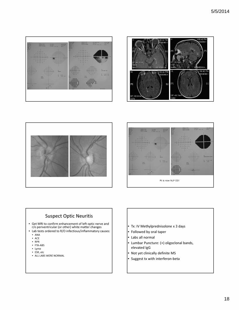
54 year-old woman
• Pain in OD x 5 days
• Gray area in OD x 2-3 days
• Colors don’t seem as bright OD
• No other neurologic symptoms
• VA OD: 20/200 OS: 20/20
• Color: 0/14 OD 14/14 OS
• PERRL (+) RAPD OD (1.8 log NDF)
• Neurologic Exam is normal
5/5/2014
18
Suspect Optic Neuritis
• Get MRI to confirm enhancement of left optic nerve and r/o periventricular (or other) white matter changes
• Lab tests ordered to R/O infectious/inflammatory causes:• ANA
• ACE
• RPR
• FTA-ABS
• Lyme
• ESR, etc
• ALL LABS WERE NORMAL
Pt is now NLP OD!
• Tx: IV Methylprednisolone x 3 days
• Followed by oral taper
• Labs all normal
• Lumbar Puncture: (+) oligoclonal bands,
elevated IgG
• Not yet clinically definite MS
• Suggest tx with interferon-beta

5/5/2014
19
2 months later
Optic Neuritis – Clinical History
• Young adult
– More likely a woman
• Unilateral visual loss
• Progresses over hours
to days
• 90% with pain,
particularly with eye
movement
Visual Prognosis
• Spontaneous visual recovery begins within 3
weeks in 80% of patients
• Improvement continues for up to 1 year
• If some improvement does not occur in 5
weeks, reconsider the diagnosis
Treatment Effect on Vision
• No difference between steroid and placebo groups at 6 months
• IV Steroids (not oral) may accelerate recovery by 2 to 3 weeks
(NEJM 326:581, 1992)
Probability of Recurrent Optic Neuritis in
Either Eye by Treatment Group41%
Prednisone
25%
Placebo
25%
Intravenous
P=0.004 Prednisone vs Placebo
P=0.003 Prednisone vs Intravenous
Oral prednisone in standard
doses is likely contraindicated
in optic neuritis!
5/5/2014
20
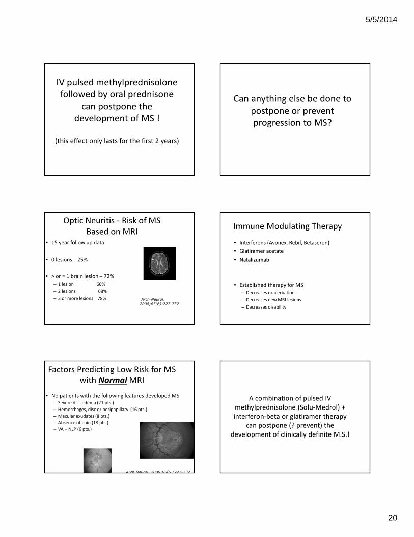
IV pulsed methylprednisolone
followed by oral prednisone
can postpone the
development of MS !
(this effect only lasts for the first 2 years)
Optic Neuritis - Risk of MS
Based on MRI
• 15 year follow up data
• 0 lesions 25%
• > or = 1 brain lesion – 72%
– 1 lesion 60%
– 2 lesions 68%
– 3 or more lesions 78% Arch Neurol.
2008;65(6):727-732
Factors Predicting Low Risk for MS
with Normal MRI
• No patients with the following features developed MS
– Severe disc edema (21 pts.)
– Hemorrhages, disc or peripapillary (16 pts.)
– Macular exudates (8 pts.)
– Absence of pain (18 pts.)
– VA – NLP (6 pts.)
Arch Neurol. 2008;65(6):727-732
Can anything else be done to
postpone or prevent
progression to MS?
Immune Modulating Therapy
• Interferons (Avonex, Rebif, Betaseron)
• Glatiramer acetate
• Natalizumab
• Established therapy for MS
– Decreases exacerbations
– Decreases new MRI lesions
– Decreases disability
A combination of pulsed IV
methylprednisolone (Solu-Medrol) +
interferon-beta or glatiramer therapy
can postpone (? prevent) the
development of clinically definite M.S.!

5/5/2014
21
Current Treatment of Optic
Neuritis: Abnormal MRI
• IV methylprednisolone (1 g/d x 3d)
• Oral prednisone taper (1 mg/kg/d x 11d, 4-d taper)
• Immunomodulating therapy
• Avoid oral prednisone alone
Balcer LJ. Optic neuritis. Current Treatment Options in Neurology 2001;3:1-10.
Söderström M. Optic neuritis and multiple sclerosis. Acta Ophthalmol Scand 2001;79:223-227.
With Normal MRI
• Consider IV methylprednisolone
• Avoid oral prednisone alone
• Clinical follow up and repeat MRI periodically
Gilenya� First FDA approved ORAL
immunomodulating agent
� Sequesters lymphocytes where there is an F1P receptor
� Higher dosages are associated with macular edema
� Not used for clinically isolated events
� Newly Approved:
� Aubagio (teriflunomide) � Once daily
Differential Diagnosis� Hereditary
� Leber’s hereditary optic neuropathy
� Infectious
� Syphilis, Cat-Scratch fever, Lyme disease
� Inflammatory
� Sarcoidosis
� Immune – Neuromyelitis optica
� Vascular
� Compressive/Infiltrative
Additional Tests if Atypical
• RPR/FTA/Lyme/Bartonella titers
• ANA, ESR
• ACE level/CXR
• Mitochondrial DNA testing for Leber’s or other
mitochondrial processes
• Neuromyelitis optica (NMO) antibody
• CSF analysis
(lumbar puncture)
CASE 10

5/5/2014
22
• 8 days ago, she awoke with an ache in her left eye
• The ache occurred when she moved her eye
• When she covered the right eye, she realized
that she couldn’t see with the left eye
• Everything looked black centrally with the left
eye.
• The vision has been fairly stable since that time.
• She still notices some pain when she moves her
eye.
50 year old woman
• hot and painful rash on her right arm 5 weeks
ago
• parasthesias of her right hand and arm
• She saw a dermatologist, who gave her a
steroid spray for an apparent allergic reaction.
– Rash resolved
• Systemic health: unremarkable
– During pregnancy was told of + syphilis test
– Her sister was told the same thing
– Neither of them had syphilis
• Ocular health: unremarkable
• Family history:
– Sister: Stiff-Person syndrome
– Daughter: joint aches with no specific diagnosis
• BCVA: OD 20/20 OS 20/40
• Color Vision: OD 14/14 OS 12/14
• 90% decreased red saturation OS
• 50% reduced brightness sense OS
• (+) RAPD OS 1.5-1.8 log
• Normal efferent testing
• Normal slit lamp exam
• Normal IOP
• Normal DFE
• Get MRI
• Get labs– CBC with platelet count
– ESR
– C-reactive protein
– Lyme titer
– RPR, FTA-ABS
– ANA with reflex titer
– anti-ds DNA
– SSA/SSB
– rheumatoid factor
– anti-GAD antibodies.

5/5/2014
23
• MRI indicates an enlarged left optic nerve with
abnormal enhancement particularly in the
region of the apex. No note was made of any
white matter lesions.
• The RPR was reactive at a1:2 titer, but the FTA-
ABS was nonreactive
• (+ )ANA screen with a 1:1280 titer in a
nucleolar pattern
• high double-stranded DNA antibody at 15
BCVA: OD 20/25 OS 20/50Color: OD 14/14 OS 8/14
• Pt Hospitalized
– IV Methylprednisolone x 5 days
– Cytoxan infusion
– Rheumatology confirmed diagnosis of Lupus
• Pt reports improvement in vision
• BCVA: OD 20/25 OS 20/20
• Color Vision: OD 14/14 OS 14/14
• 10% decreased red saturation OS
• 20% reduced brightness sense OS
• (+) RAPD OS 0.3 log
3 weeks later

5/5/2014
24
CASE 11
28 year old woman
• 2 weeks prior, noticed blurry vision OS
• Blur persists
• No pain in the left eye
• 3 days prior, noticed pain on eye movements
OD
• 1 day prior noticed decreased vision inferior OD
• She is now having difficulty functioning
• OTHER SYMPTOMS:
– Weakness and tingling left thigh
• Several episodes over past few years
• Though to be sciatica
• Thought to be related to being over-weight
– Lost 30 pounds, but symptom still recurred
• SYSTEMIC HEALTH:
– asthma
• MEDICATIONS:
– Albuterol, Advair
• OCULAR HISTORY:
– Unremarkable
• SOCIAL HISTORY:
– unremarkable
• BCVA: OD 20/25 and OS 20/40
• Color 14/14 OD and 10/14 OS
• 25% reduced red saturation OS
• 25% reduced brightness sense OS
• Pupils – pharm dilated
• Normal ocular motility exam

5/5/2014
25
• Slit Lamp Exam: normal OU
• TONOMETRY: OD-16 mm Hg, OS- 16 mm Hg
• BP: 132/92
DDX
• Optic Neuritis OD, Optic Neuropathy OS
• ? Demyelinating Disease
• WORK-UP
– Lab testing
– MRI brain and orbits with contrast
– MRI spine
– Possible lumbar puncture
• TREATMENT:
• IV steroids x 3 days
RESULTS
• MRI brain and orbits – mild enhancement of
posterior right optic nerve
• MRI spine: lesion at C3 level
• Pt diagnosed with Multiple Sclerosis at
hospital

5/5/2014
26
FOLLOW-UP
• Eye pain returns (was better on steroids)
• Severe headaches
• Leg paresthesias recur (better on steroids)
• BCVA: OD 20/25 and OS 20/20
• Color 14/14 OD and 10/14 OS
• Pupils – equivocal RAPD OS
• Normal ocular motility exam
• Because vision did not improve significantly
after steroid treatment, and there was spine
involvement but no brain involvement, need
to consider:
• NMO (neuro-myelitis optica or Devic’s disease)
• Order NMO antibody testing
• Also test for other causes of optic neuritis
• (ANA, Lyme, ACE, FTA-ABS, etc)
• NMO antibody was POSITIVE
• Positive ANA, positive Sjogren’s antibodies
• Pt started on Azathioprine
• Then put on Rituximab
– some improvement in filed of right eye

5/5/2014
27
Neuro Myelitis Optica (NMO)
Neuromyelitis optica
is, like Multiple Sclerosis an
inflammatory, demyelinating
syndrome of the central
Nervous system
Wingerchuk DM, Hogancamp WF, O’Brien PC, Weinshenker BG. The clinical course of neuromyelitis optica (Devic’s syndrome). Neurology 1999; 53: 1107–14.
preferentially affects the
-optic nerve (optic
neuritis)
-spinal cord (myelitis)
Neuromyelitis Optica (NMO)
(Devic’s Disease)Multiple Sclerosis
affects the CNS
-optic nerve and
-spinal cord
-brain
Wingerchuk DM, Hogancamp WF, O’Brien PC, Weinshenker BG. The clinical course of neuromyelitis optica (Devic’s syndrome). Neurology 1999; 53: 1107–14.
Within 5 years of
disease onset, more than 50%
of patients with relapsing
neuromyelitis optica are blind
in one or both eyes or
require ambulatory help.
Dean M Wingerchuk(et al (The Lancet Neurology, 2007, 6:805-15)

5/5/2014
28
Typical Features of Neuromyelitis
Optica:
myelitis with
-muscle weakness
-sensory dysfunction
- bladder dysfunction
optic nerve (optic neuritis)
Spinalcord
Neurogenic respiratory failure
can occur-- serious
Exceedingly rare in multiple
sclerosis
19 Pittock SJ, Weinshenker BG, Wijdicks EF. Mechanical ventilation and tracheostomy in multiple sclerosis. J Neurol Neurosurg Psychiatry 2004; 75: 1331–33.
The detection of
neuromyelitis
opticaimmunoglobulin G
(NMO-IgG),
an autoantibody, in
the serum of patients with
neuromyelitis optica,
distinguishes neuromyelitis
optica from other
demyelinating disorders
Lennon VA, Wingerchuk DM, Kryzer TJ, et al. A serum
autoantibody marker of neuromyelitis optica: distinction from
multiple sclerosis. Lancet 2004; 364: 2106–12.
NMO-IgG binds to
aquaporin 4,
which is the main channel that
regulates
Water homoeostasis in the
Central nervous system
Lennon VA, Kryzer TJ, Pittock SJ, VerkmanAS, Hinson SR. IgGmarker of optic-spinal multiple sclerosis binds to the aquaporin 4 water channel. J Exp Med 2005; 202: 473–77.
NMO-IgG
attacks proteins of the
AQP4 channels on
Astrocytes (toxic to them)
The resultant immune
reaction, causes
Demyelination
This is different than what
happens in Multiple Sclerosis
blood
brain
CASE 12

5/5/2014
29
40 year old asymptomatic
woman
• Hx of keratoconus
• Came in for CL eval, and anterior uveitis was noted
• On Pred Forte qid OS and Nevanac (NSAID prodrug)
qid OS
• Photosensitivity – stable x years
• Denies other symptoms
• Systemic Hx:
– Hypercholesterolemia – no meds
– S/p removal of axillary lymph nodes (infxn?)
– S/p 2 full term uncomplicated pregnancies
– Asthma
– Meds: Advair, Combivent, Zyrtec, Claritin
• VA: OD 20/25 OS 20/60
• Color: 13/14 OD and 0/14 OS
• PERRL (+) >1.8 log RAPD OS
• No ptosis or proptosis
• Normal ocular motility exam
• SLE: only few residual cells
• TA: OD 18 mmHg OS 21 mm Hg
• Normal neurologic exam
Labs for Optic Neuropathy
• CBC
• C-reactive protein
• ESR
• Platelet count
• Lyme titer (if + get Western blot IgG and IgM)
• ANA with reflex titer
• ACE
• RPR & FTA-ABS
• Vitamin B 12
• Folic acid
• Methylmalonic acid
• SPEP

5/5/2014
30
Work-Up
• MRI – brain & orbits w & w/o gad• Arnold Chiari I malformation• No other structural abnormalities or abnormal enhancement
• Labs• CBC **elevated eosinophils 11.6%• Platelets• ESR **H 85 mm/hr• CRP **H 1.22• Folate• B12• Lyme titer• RPR / FTA-ABS• ACE **H 254 • ANA
Chest CT
• Findings compatible with longstanding sarcoidosis• Bulky lymphadenopathy• Parenchymal changes• Scarring
• Lower lobe infiltrate ? Possible superimposed bacterial pneumonic component
• Pt referred to pulmonologist and surgeon• Lung biopsy performed (+) for sarcoid
Neurosarcoid
• Occurs in 5-15% of pts with sarcoid
• CN VII is commonly affected
• CN II and VIII also affected
• Can be a mononeuropathy, peripheral nerve
involvement, CNS involvement
• Can see leptomeningeal enhancement
• Active inflammation responds very well to steroids
(Oral steroids are fine for sarcoid)
CASE 13
• reduced VA OS X 4 days
• not feeling well for 2-3 weeks – nasal congestion, pressure / pain over left eye, sometimes when moves eyes
•PCP Rx’d Biaxin (macrolide antibiotic – interferes w/ protein synthesis) and Prelone oral steroid (Prednisolone syrup)
•Systemic Hx: asthma, many food allergies
• VA OD 20/20 OS CF at 2’ (SOSH 1/10 temporally)
• PERRLA (+) APD OS (>1.8 log)
• Decreased red saturation and brightness sense OS
•Normal efferent system
12 year old girl

5/5/2014
31
OS OD
Suspect Optic Neuritis
• Pt admitted to Children’s Hospital
• Get MRI to confirm enhancement of left optic nerve and r/o other abnormalities
• Lab tests needed, including:
– ANA
– ACE
– RPR
– FTA-ABS
– Lyme
– ESR
– C-reactive protein, etc
ONTT
• Diseases associated with optic neuritis
• (must be screened for these)
– Sarcoid
– Systemic Lupus Erythematosis
– Syphilis
– Lyme disease
– Other connective tissue disorders
MRI
• Enlargement and enhancement of left optic
nerve (intraorbital portion)
• No indication of abnormal parenchymal signal
intensity or enhancement
Lab Testing
• Remarkable for
– Positive ANA
– Titer of 1 : 1280

5/5/2014
32
Causes of a Positive ANALupus (SLE)
SclerodermaLupus Erythematosis
Polymyositis / DermatomyositisRheumatoid arthritisSjogren's syndrome
Normal finding (with aging and being female)Some drugs (procainamide, hydralazine, isoniazid,
antibiotics)Infectious mononucleosis (mono) and other
infectionsChronic liver disease
Positive ANA
• • Order additional testing
– – Anti-ds DNA (for Lupus)
– – SS-A / SS-B (for Sjogren’s)
– – p-ANCA / c-ANCA (for vasculitis / Wegner’s)
– -- RF
Lab Testing
• Remarkable for
– Positive ANA 1 : 1280
– Elevated Anti-SS-A Ab (110) normal range 0-19
– Elevated Anti-SS-B Ab (87) normal range 0-19
– - Suggestive of Sjogren’s Disease
– Subsequent lip biopsy (salivary glands)was diagnostic (cluster of inflammatory cells) of Sjogren’s Disease
– No definite indication of Lupus, Rheumatoid Arthritis at this point
– IV Methylprednisolone. Began treatment with CellCept (immunosuppressant), Prednisone taper
Sjogren’s Syndrome
• Autoimmune disorder
• Typically in 4th-5th decade, but any age possible
• Associated with the sicca complex
– Dry mouth
– Dry eye
– Lymphocytic infiltration of the exocrine glands
• (10% can get non-Hodgkin lymphoma)
– Any organ can be affected
– 2nd most common rheumatologic disorder after SLE
Sjogren’s
• Lab test
– Elevated ESR in 80%
– + RF possible
– Anemia
– Leukopenia
– Anti-SS-A
– Anti-SS-B
– Antisalivary duct antibodies (in secondary Sjogren’s)
• Biopsy (definitive diagnosis)
– Salivary gland
– Lower lip

5/5/2014
33
CASE 14
20 year-old man
• Pain OD on upgaze x few days
• Today, vision OD is “off”
• Denies diplopia, transient vision loss
• Denies headache
• A few days ago, he felt feverish, but did not
check his temperature
• Fam HX: Father dx with Lupus in 20s
• BCVA: OD 20/50 and OS 20/20
• Color 14/14 OD and 14/14 OS
• Pupils – pharm dilated
• CF: full OU
• HVF: essentially normal OU
• SLE and IOP normal OU
• BP: 104/70
• Temp: 98.8 degrees
Right EyeLeft Eye
Humphrey Visual FIeld: At initial presentation. Not e only slightlyenlarged blind spot in the right eye, and fairly p reserved central visual field.
Initial Presentation
Right Eye Left Eye
• Pt denies any rashes (only when asked)
• He does admit to a scratch by a cat (kitten)
several weeks ago (only when asked)
• A few weeks ago, his right eyelid was swollen
• Pt has several scars on his forehead, above
right eye, and on his nose

5/5/2014
34
Labs Ordered – told to have done today!
• CBC• C-reactive protein• ESR• Platelet count• Lyme titer (if + get Western Blot Lyme IgG and IgM)• ANA with reflex titer• ACE• RPR• FTA-ABS• Bartonella Quintana titer• Bartonella Henselae titer
Follow-up 5 days later
• Pt notes a spot in right vision, that is getting larger
• Reduced central vision
• Since last visit, has had chills and fever
• Has also had headache
• Decreased appetite
• Unable to work – doesn’t feel right
• Labs not done until 2 days ago – not complete
Lab results (so far)
• ANA ( + ) titer and pattern not yet known
• Lyme titer is ( + ) WB IgG (-), IgM (+)
• ACE slightly elevated at 70
• ESR: 44
• CRP: pending
• Bartonella titers: pending
• BCVA: OD 20/200 and OS 20/20
• Color 1/14 OD and 14/14 OS
• Pupils – trace RAPD OD
• CF: very large blindspot OD
• HVF: large blindspot OD – extending past
fixation and superiorly out to 10 degrees
• SLE and IOP normal OU (-) cells
Right EyeLeft Eye
Humphrey Visual Field: 6 days after initial present ation. Note significant increase in blind spot in the right eye. The left field was unreliable due to patient fatigue.
Follow-Up: 6 days after initial presentation
Right Eye Left Eye

5/5/2014
35
Follow-Up: 6 days after initial presentation
Right Eye Left Eye
• Need to r/o Lyme, sarcoid, auto-immune
disease
• Ds DNA (-)
• Repeat ACE (-), CXR (-)
• LP (-) for Lyme, sarcoid
• Bartonella titers
• Bartonella Quintana (-)
• Bartonella Hensalea (+)
• (+) IgG > 1:2560
• (+) IgM > 1:800
• DX: Cat-scratch Disease
Follow-Up: 15 days after initial presentation
Right Eye Left Eye
Neuro-retinitis
• Cat-scratch
• Sarcoid
• Syphilis
• Lyme
• Toxo
• NOT typical in MS

5/5/2014
36
• Treatment:
– Antibiotics
» Doxycycline (pt vomited every time he took this medication)
» Rifampin
» Bactrim prescribed in place of Doxycycline
» Pt then switched to Azithromycin by Infectious Disease
Cat Scratch Disease
Typically transmitted by a kitten (by a scratch or a lick)
Only a minority of the exposures to B. Henselae result in cat-scratch disease. The ability of the cat to transmit the disease is transient
Most cases occur in fall / early summer - related to kitten births and flea infestations
80% of cases occur in patients under age 21
Starts with local infection, then lymphadenopathy, and rarely progresses – eg. Neuroretinitis, etc.
Cat Scratch Disease
– Treatment / Response:
– Excellent prognosis - Most cases are
self-limiting and fully resolve, even
when involving the CNS
– Drugs of choice – Bactrim,
Gentamicin, Ciprofloxacin, Rifampin,
Azithromycin
Humphrey Visual Field: 34 days after initial presentation. Note reduction in blindspot size in the right eye corresponding with a reduction in optic disc edema. A cen tral scotoma persists in the
right eye, related to the macular star and the reduced visual acuity.
Right EyeLeft Eye
Follow-Up: 34 days after initial presentation
Right Eye Left Eye
Follow-Up: 34 days after initial presentation
Right EyeLeft Eye

5/5/2014
37
Optical Coherence Tomography (OCT) -2 different sections through the right
macular region. Note the OCT appearance
of the retinal exudates (arrows).
Follow-Up: 34 days after initial presentation
Thank You!
Questions?