u e s in b t h n iq advanced techniques in m a biology edicine giorgadze tg, khutsishvili ig,...
TRANSCRIPT
Research Article Open Access
Giorgadze et al., Adv Tech Biol Med 2017, 5:2DOI: 10.4172/2379-1764.1000215
Research Article OMICS Iinternational
Advanced Techniques in Biology & MedicineAdv
ance
d Te
chniques in Biology & M
edicine
ISSN: 2379-1764
Volume 5 • Issue 2 • 1000215Adv Tech Biol Med, an open access journalISSN: 2379-1764
Keywords: DNA; Intercalator; Resonance energy transfer; Re-radiation
IntroductionFor the last several decades, there has been great interest in a
bloodless surgery, especially for the treatment of malignant tumors and other growths has been observed. Such therapeutic treatments include: hadron, isotope, γ-radiation, microwave, chemo, fermento, photochemo, photothermo, plasma, etc.
A significant interest has appeared recently in the use of non-equilibrium atmospheric pressure cold plasma in medicine, especially in the treatment of cancer, the so-called "blood dialysis", healing wounds, and sterilization of expensive medical instruments, in which sterilization can’t be used at temperature higher than 40-50°C [1-9]. Besides, there are few papers in which non-equilibrium low-pressure plasma is used, mainly as a source of light in atomic emission analysis [10-16], in spite of the unique properties of inductively coupled low pressure plasma (1-20 Torr).
We will shortly discuss properties of non-equilibrium reduced pressure plasma for the purpose of using it as a light source for light therapy in medicine.
If electron distribution function in plasma is close to Maxwell's distribution, then, in case of weakly ionized plasma, we can consider separately electronic Te and atomic Ta temperatures [17]. This occurs in case when the frequency of collisions of electrons with each other νee is much more then frequency of energy transfer from electrons to atoms νea, i.e.
νee >> νea
Thus
Ta << Te
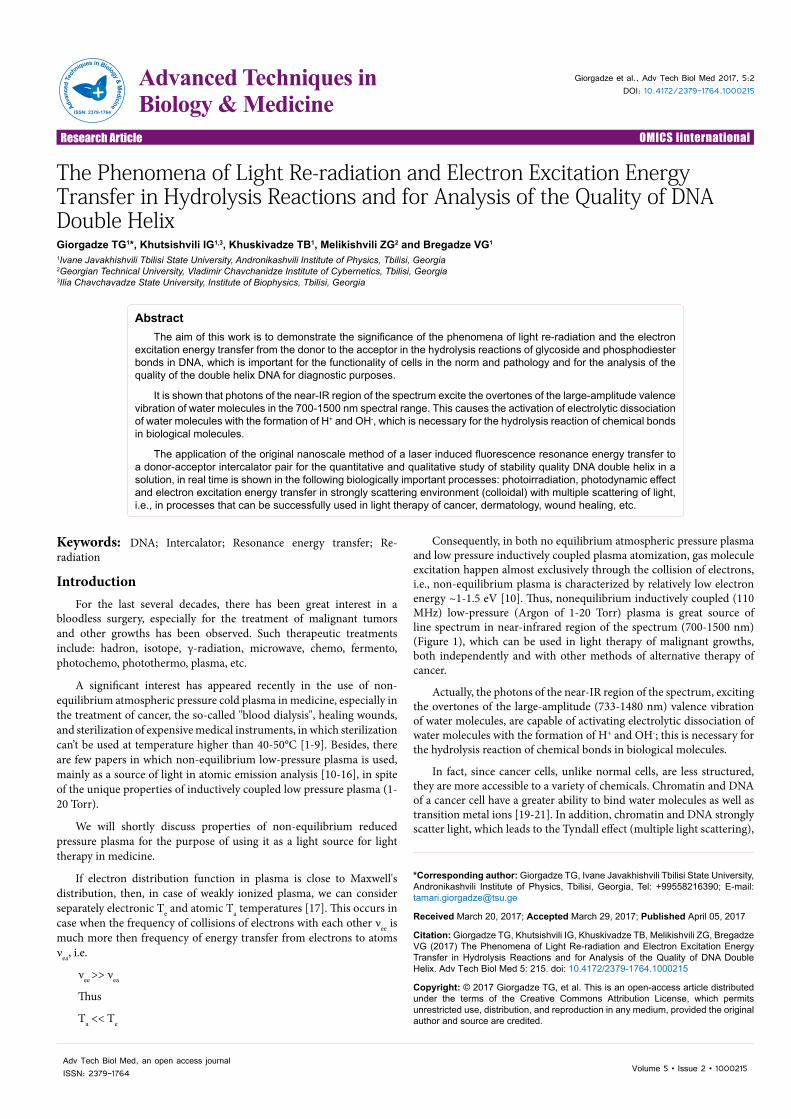
Consequently, in both no equilibrium atmospheric pressure plasma and low pressure inductively coupled plasma atomization, gas molecule excitation happen almost exclusively through the collision of electrons, i.e., non-equilibrium plasma is characterized by relatively low electron energy ~1-1.5 eV [10]. Thus, nonequilibrium inductively coupled (110 MHz) low-pressure (Argon of 1-20 Torr) plasma is great source of line spectrum in near-infrared region of the spectrum (700-1500 nm) (Figure 1), which can be used in light therapy of malignant growths, both independently and with other methods of alternative therapy of cancer.
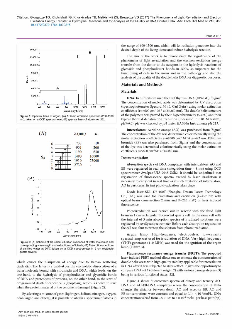
Actually, the photons of the near-IR region of the spectrum, exciting the overtones of the large-amplitude (733-1480 nm) valence vibration of water molecules, are capable of activating electrolytic dissociation of water molecules with the formation of H+ and OH-; this is necessary for the hydrolysis reaction of chemical bonds in biological molecules.
In fact, since cancer cells, unlike normal cells, are less structured, they are more accessible to a variety of chemicals. Chromatin and DNA of a cancer cell have a greater ability to bind water molecules as well as transition metal ions [19-21]. In addition, chromatin and DNA strongly scatter light, which leads to the Tyndall effect (multiple light scattering),
*Corresponding author: Giorgadze TG, Ivane Javakhishvili Tbilisi State University, Andronikashvili Institute of Physics, Tbilisi, Georgia, Tel: +99558216390; E-mail: [email protected]
Received March 20, 2017; Accepted March 29, 2017; Published April 05, 2017
Citation: Giorgadze TG, Khutsishvili IG, Khuskivadze TB, Melikishvili ZG, Bregadze VG (2017) The Phenomena of Light Re-radiation and Electron Excitation Energy Transfer in Hydrolysis Reactions and for Analysis of the Quality of DNA Double Helix. Adv Tech Biol Med 5: 215. doi: 10.4172/2379-1764.1000215
Copyright: © 2017 Giorgadze TG, et al. This is an open-access article distributed under the terms of the Creative Commons Attribution License, which permits unrestricted use, distribution, and reproduction in any medium, provided the original author and source are credited.
AbstractThe aim of this work is to demonstrate the significance of the phenomena of light re-radiation and the electron
excitation energy transfer from the donor to the acceptor in the hydrolysis reactions of glycoside and phosphodiester bonds in DNA, which is important for the functionality of cells in the norm and pathology and for the analysis of the quality of the double helix DNA for diagnostic purposes.
It is shown that photons of the near-IR region of the spectrum excite the overtones of the large-amplitude valence vibration of water molecules in the 700-1500 nm spectral range. This causes the activation of electrolytic dissociation of water molecules with the formation of H+ and OH-, which is necessary for the hydrolysis reaction of chemical bonds in biological molecules.
The application of the original nanoscale method of a laser induced fluorescence resonance energy transfer to a donor-acceptor intercalator pair for the quantitative and qualitative study of stability quality DNA double helix in a solution, in real time is shown in the following biologically important processes: photoirradiation, photodynamic effect and electron excitation energy transfer in strongly scattering environment (colloidal) with multiple scattering of light, i.e., in processes that can be successfully used in light therapy of cancer, dermatology, wound healing, etc.
The Phenomena of Light Re-radiation and Electron Excitation Energy Transfer in Hydrolysis Reactions and for Analysis of the Quality of DNA Double HelixGiorgadze TG1*, Khutsishvili IG1,3, Khuskivadze TB1, Melikishvili ZG2 and Bregadze VG1
1Ivane Javakhishvili Tbilisi State University, Andronikashvili Institute of Physics, Tbilisi, Georgia 2Georgian Technical University, Vladimir Chavchanidze Institute of Cybernetics, Tbilisi, Georgia 3Ilia Chavchavadze State University, Institute of Biophysics, Tbilisi, Georgia
Citation: Giorgadze TG, Khutsishvili IG, Khuskivadze TB, Melikishvili ZG, Bregadze VG (2017) The Phenomena of Light Re-radiation and Electron Excitation Energy Transfer in Hydrolysis Reactions and for Analysis of the Quality of DNA Double Helix. Adv Tech Biol Med 5: 215. doi: 10.4172/2379-1764.1000215
Page 2 of 7
Volume 5 • Issue 2 • 1000215Adv Tech Biol Med, an open access journalISSN: 2379-1764
which causes the dissipation of energy due to Raman scattering (inelastic). The latter is a catalyst for the electrolytic dissociation of a water molecule bound with chromatin and DNA, which leads, on the one hand, to the hydrolysis of phosphodiester and glycosidic bonds of DNA and proteolysis of proteins, on the other hand, to the start of programmed death of cancer cells (apoptosis), which is known to start when the protein material of the genome is damaged (Figure 2).
By selecting a mixture of gases (hydrogen, helium, nitrogen, oxygen, neon, argon and others), it is possible to obtain a spectrum of atoms in
the range of 600-1500 nm, which will let radiation penetrate into the desired depth of the living tissue and induce hydrolysis reaction.
The aim of the work is to demonstrate the significance of the phenomena of light re-radiation and the electron excitation energy transfer from the donor to the acceptor in the hydrolysis reactions of glycoside and phosphodiester bonds in DNA, so important for the functioning of cells in the norm and in the pathology and also the analysis of the quality of the double helix DNA for diagnostic purposes.
Materials and MethodsMaterials
DNA: In our tests we used the Calf thymus DNA (40% GC), ‘Sigma’. The concentration of nucleic acids was determined by UV absorption (spectrophotometer Specord M 40, Carl Zeiss) using molar extinction coefficients (ɛ=6600 cm-1 M-1 at λ=260 nm). The double helix structure of the polymers was proved by their hyperchromicity (>30%) and their typical thermal denaturation transition (measured in 0.01 M NaNO3, pH≅6.0). pH was checked by pH meter HANNA Instruments pH 213.
Intercalators: Acridine orange (AO) was purchased from ‘Sigma’. The concentration of the dye was determined colorimetrically using the molar extinction coefficients ε=68500 cm-1 M-1at λ=492 nm. Ethidium bromide (EB) was also purchased from ‘Sigma’ and the concentration of the dye was determined colorimetrically using the molar extinction coefficients ε=5600 cm-1M-1 at λ=480 nm.
Instrumentation
Absorption spectra of DNA complexes with intercalators AO and EB were registered in real time (integration time – 8 ms) using CCD spectrometer AvaSpec ULS 2048-USB2. It should be underlined that registration of fluorescence spectra excited by laser irradiation is necessary to carry out in real time as at such excitation of intercalators, AO in particular; its fast photo-oxidation takes place.
Diode laser SDL-475-100T (Shanghai Dream Lasers Technology Co., Ltd.) was used for irradiation and excitation (λ=457 nm with optical beam cross-section 2 mm and P=200 mW) of laser induced fluorescence.
Photoirradiation was carried out in reactor with the fixed light beam in 1 cm rectangular fluorescent quartz cell. In the same cell with the interval of 5 min absorption spectra of irradiated solutions were registered by AvaSpec spectrometer. Before each absorption registration the cell was shut to protect the solution from photo irradiation.
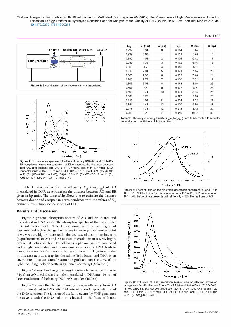
Argon lamp: High-frequency, electrodeless, low-capacity spectral lamp was used for irradiation of DNA. Very high frequency (VHF) generator (110 MHz) was used for the ignition of the argon lamp (Figure 3).
Fluorescence resonance energy transfer (FRET): The proposed laser-induced FRET method allows one to estimate the concentration of double helix areas with high quality stability applicable for intercalation in DNA after it was subjected to stress effect. It gives the opportunity to compare DNAs of 1) different origin; 2) with various damage degrees; 3) being in various functional states [22].
Figure 4 shows fluorescence spectra of binary and ternary AO-DNA and AO-EB-DNA complexes where the concentration of DNA changes the distance between donor AO and acceptor EB. AO and EB concentrations were constant and equal to 0.14 × 10⁻⁴ mol/L. DNA concentration varied from 0.5 × 10⁻⁴ to 5 × 10⁻⁴ mol/L per base pair (bp).
Figure 1: Spectral lines of Argon, (A) Ar lamp emission spectrum (200-1100 nm), taken on a CCD spectrometer; (B) spectral lines of atomic Ar [18].
Figure 2: (A) Scheme of the valent vibration overtones of water molecules and corresponding wavelength and extinction coefficients; (B) Absorption spectrum of distilled water at 20°C taken on a CCD spectrometer in 5 cm rectangle quartz cuvette.
Citation: Giorgadze TG, Khutsishvili IG, Khuskivadze TB, Melikishvili ZG, Bregadze VG (2017) The Phenomena of Light Re-radiation and Electron Excitation Energy Transfer in Hydrolysis Reactions and for Analysis of the Quality of DNA Double Helix. Adv Tech Biol Med 5: 215. doi: 10.4172/2379-1764.1000215
Page 3 of 7
Volume 5 • Issue 2 • 1000215Adv Tech Biol Med, an open access journalISSN: 2379-1764
Table 1 gives values for the efficiency EET=(1-qD/q0D) of AO intercalated in DNA depending on the distance between AO and EB given in bp units. The same table allows one to estimate the distance between donor and acceptor in correspondence with the values of EET evaluated from fluorescence spectra of FRET.
Results and DiscussionFigure 5 presents absorption spectra of AO and EB in free and
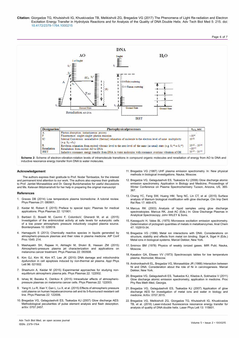
intercalated in DNA states. The absorption spectra of the dyes, under their interaction with DNA duplex, move into the red region of spectrum and highly change their intensity. From photochemical point of view, we are highly interested in the decrease of absorption intensity (hypochromism) of AO and EB at their intercalation into DNA-highly ordered structure duplex. Hypochromism phenomena are connected with it light re-radiation and, in our case re-radiation to DNA, leads to strong increase by 4-5 orders scattering cross-section. Dye-intercalator in this case acts as a trap for the falling light beam, and DNA is an environment that can strongly scatter a significant part (10-20%) of the light, including inelastic scattering (Raman scattering) (Scheme 2).
Figure 6 shows the change of energy transfer efficiency from 13 bp to 7 bp from AO to ethidium bromide intercalated in DNA after 20 min of laser irradiation of the binary DNA-AO complex (Table 2).
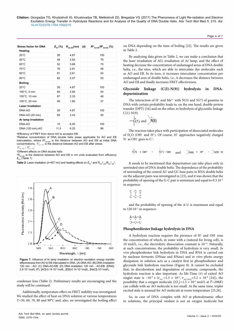
Figure 7 shows the change of energy transfer efficiency from AO to EB intercalated in DNA after 120 min of argon lamp irradiation of the DNA solution. The ignition of the lamp occurs by VHF generator; the cuvette with the DNA solution is located in the focus of double
Figure 3: Block-diagram of the reactor with the argon lamp.
Figure 4: Fluorescence spectra of double and ternary DNA-AO and DNA-AO-EB complexes where concentration of DNA changes the distance between donor AO and acceptor EB. [AO]-0.14·10-4 mol/L, [EB]-0.14·10-4 mol/L, DNA concentrations: (C0)-2.8·10-4 mol/L (P), (C1)-10∙10-4 mol/L (P), (C2)-8·10-4 mol/L (P); (C3)-6·10-4 mol/L (P); (C4)-4·10-4 mol/L (P); (C5)-2.8·10-4 mol/L (P); (C6)-1.4·10-4 mol/L (P); (C7)-10-4 mol/L (P).
Figure 5: Effect of DNA on the electronic absorption spectra of AO and EB in 10-2 mol/L, NaCl solution Dye concentration was 10-5 mol/L, DNA concentration 10-4 mol/L. Left ordinate presents optical density of EB, the right one of AO.
Figure 6: Influence of laser irradiation (λ=457 nm) on electron excitation energy transfer effectiveness from AO to EB intercalated in DNA. (A) AO-DNA; (B) AO-DNA-EB; (C) AO-DNA irradiation 20 min; (D) AO-DNA irradiation 20 min + EB. [DNA]-7 × 10⁻⁴ mol/L (P), [AO]-0.14 × 10⁻⁴ mol/L, [EB]-0.14 × 10⁻⁴ mol/L, [NaNO3]-10-2 mol/L.
EET R (nm) R (bp) EET R (nm) R (bp)0.999 0.34 0 0.184 5.44 150.999 0.68 1 0.151 5.78 160.995 1.02 2 0.124 6.12 170.983 1.36 3 0.102 6.46 180.959 1.7 4 0.085 6.8 190.919 2.04 5 0.071 7.14 200.860 2.38 6 0.059 7.48 210.783 2.72 7 0.050 7.82 220.693 3.06 8 0.043 8.16 230.597 3.4 9 0.037 8.5 240.503 3.74 10 0.031 8.84 250.500 3.75 0.027 9.18 260.416 4.08 11 0.024 9.52 270.341 4.42 12 0.020 9.86 280.278 4.76 13 0.018 10.2 290.226 5.1 14 0.016 10.54 30
Table 1: Efficiency of energy transfer EET=(1-qD/q0D) from AO donor to EB acceptor depending on the distance R between them.
Citation: Giorgadze TG, Khutsishvili IG, Khuskivadze TB, Melikishvili ZG, Bregadze VG (2017) The Phenomena of Light Re-radiation and Electron Excitation Energy Transfer in Hydrolysis Reactions and for Analysis of the Quality of DNA Double Helix. Adv Tech Biol Med 5: 215. doi: 10.4172/2379-1764.1000215
Page 4 of 7
Volume 5 • Issue 2 • 1000215Adv Tech Biol Med, an open access journalISSN: 2379-1764
condenser lens (Table 2). Preliminary results are encouraging and the study will be continued.
Additionally, temperature effect on FRET stability was investigated. We studied the effect of heat on DNA solution at various temperatures T=50, 60, 70, 80 and 90°C and, also, we investigated the boiling effect
Stress factor for DNA EET (%) RAO-EB(nm) (d) RstAO-EB/R0
AO-EB (%)Heating20°C 26 4.87 10050°C 48 3.83 7960°C 52 3.68 7570°C 76 2.81 5880°C 81 2.61 54
90°C 82 2.57 53
Boiling20°C 26 4.87 100100°C, 5 min 80 2.65 54100°C, 10 min 87 2.33 48
100°C, 20 min 95 1.80 37
Laser IrradiationDNA-AO 26 4.87 100
DNA-AO (20 min) 85 2.43 50Ar lamp IrradiationDNA-AO 10 6.50 100DNA (120 min)-AO 11.5 6.25 96
aEfficiency of FRET from donor AO to acceptor EB.bRelative concentration of DNA double helix areas applicable for AO and EB intercalation, where R0
AO-EB is the distance between AO and EB at initial DNA concentrations, st
AO EBR − is the distance between AO and EB after stress
0 /st bAO EB AO EB
R R− −
cDifferent effects on DNA double helixdRAO-EB is the distance between AO and EB in nm units evaluated from efficiency EET (Table 1)Table 2: Laser irradiation (λ=457 nm) and heating effects on EET
a and RAOst
-EB/RAO0-EB
b.
Figure 7: Influence of Ar lamp irradiation on electron excitation energy transfer effectiveness from AO to EB intercalated in DNA. (A) DNA-AO; (B) DNA-irradiation 120 min - AO; (C) DNA-AO-EB; (D) DNA irradiation 120 min - AO-EB. [DNA]-2.8∙10-4 mol/L (P), [AO]-0.14∙10-4 mol/L, [EB]-0.14∙10-4 mol/L, [NaCl]-10-2 mol/L.
on DNA depending on the time of boiling [22]. The results are given in Table 2.
By analysing data given in Table 2, we can make a conclusion that the laser irradiation of AO, irradiation of Ar lamp, and the effect of heating decrease the concentration of undamaged areas of DNA double helix, i.e., the sites, which are able to intercalate dye molecules such as AO and EB. In its turn, it increases intercalator concentration per undamaged area of double helix, i.e., it decreases the distance between AO and EB and finally increases FRET effectiveness.
Glycoside linkage (C(I)-N(9)) hydrolysis in DNA-depurinization
The interaction of H+ and Men+ with N(3) and N(7) of guanine in DNA with certain probability leads to, on the one hand, double proton transfer (DPT) [16] and on the other, to hydrolysis of glycosidic linkage C(1)-N(9).
and
The reaction takes place with participation of dissociated molecules of H2O (OH– and H+). Of course, H+ approaches negatively charged N– as OH– goes to C+:
and
It needs to be mentioned that depurination can take place only in unwinded sites of DNA double helix. The dependence of the probability of unwinding of the central AU and GC base pairs in RNA double helix on the adjacent pairs was investigated in [23], and it was shown that the probability of opening of the G-C pair is minimum and equal to 0.3·10–5 in sequence:
and the probability of opening of the A-U is maximum and equal to 120·10–5 in sequence:
Phosphordiester-linkage hydrolysis in DNA
A hydrolysis reaction requires the presence of H+ and OH- ions the concentration of which, in water with a (natural for living cell), is 10 mol/L, i.e., the electrolytic dissociation constant is 10-14. Naturally, at such concentrations, the probability of hydrolysis is very small. In vivo phosphodiester link hydrolysis in DNA and RNA is carried out by nuclease ferments (DNase and RNase) and in vitro photo energy dissipation in solution acts as a catalyst first in phosphodiester and glycoside link hydrolysis reactions (Figure 8). It cannot be excluded that, in discoloration and degradation of aromatic compounds, the hydrolysis reaction is also important. As life-Time (𝜏) of exited AO singlet state is ~10-9 s (𝜏AO =1.5 × 10⁻⁹, 𝜏AO-DNA=5.2 × 10⁻⁹ [24]), the possibility that a oxygen molecule ([O2]=1.3 × 10⁻⁶ mol/L at 𝑇=298𝐾) can collide with an AO molecule is too small. At the same time, triplet excited state is unusual for AO molecule at room temperature [25,26].
So, in case of DNA complex with AO at photodynamic effect in solutions, the principal oxidant is not an oxygen molecule but
Citation: Giorgadze TG, Khutsishvili IG, Khuskivadze TB, Melikishvili ZG, Bregadze VG (2017) The Phenomena of Light Re-radiation and Electron Excitation Energy Transfer in Hydrolysis Reactions and for Analysis of the Quality of DNA Double Helix. Adv Tech Biol Med 5: 215. doi: 10.4172/2379-1764.1000215
Page 5 of 7
Volume 5 • Issue 2 • 1000215Adv Tech Biol Med, an open access journalISSN: 2379-1764
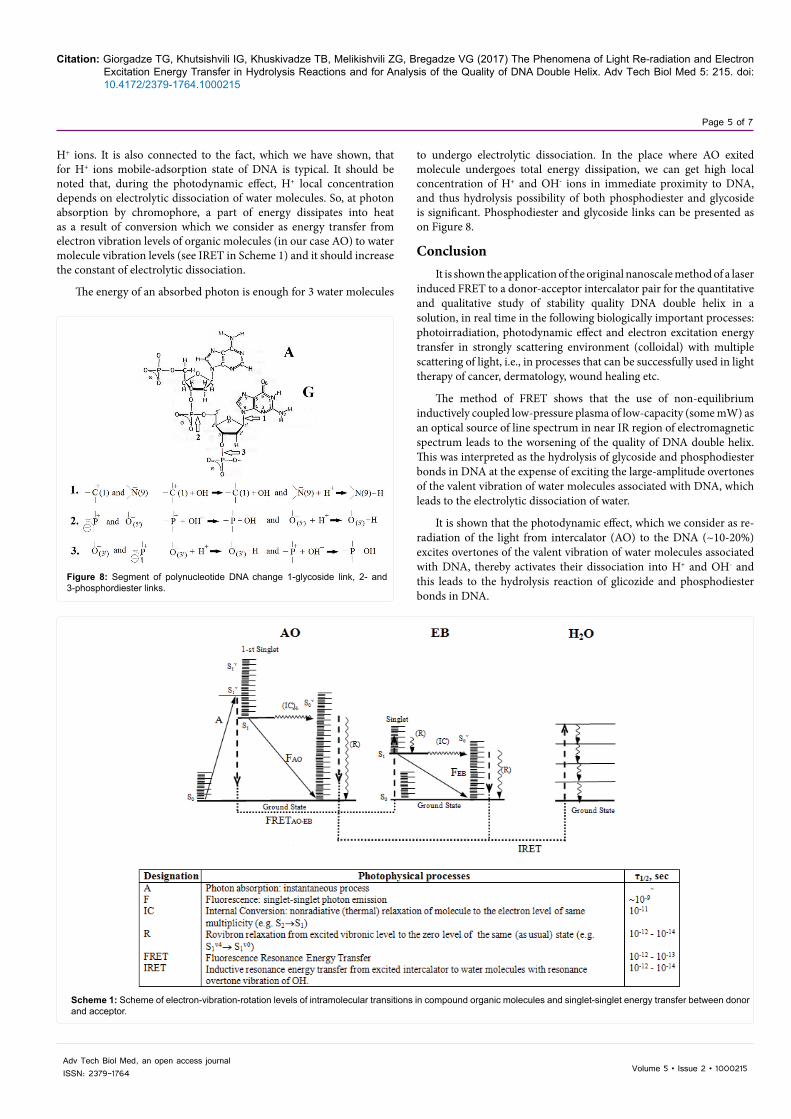
H+ ions. It is also connected to the fact, which we have shown, that for H+ ions mobile-adsorption state of DNA is typical. It should be noted that, during the photodynamic effect, H+ local concentration depends on electrolytic dissociation of water molecules. So, at photon absorption by chromophore, a part of energy dissipates into heat as a result of conversion which we consider as energy transfer from electron vibration levels of organic molecules (in our case AO) to water molecule vibration levels (see IRET in Scheme 1) and it should increase the constant of electrolytic dissociation.
The energy of an absorbed photon is enough for 3 water molecules
to undergo electrolytic dissociation. In the place where AO exited molecule undergoes total energy dissipation, we can get high local concentration of H+ and OH- ions in immediate proximity to DNA, and thus hydrolysis possibility of both phosphodiester and glycoside is significant. Phosphodiester and glycoside links can be presented as on Figure 8.
ConclusionIt is shown the application of the original nanoscale method of a laser
induced FRET to a donor-acceptor intercalator pair for the quantitative and qualitative study of stability quality DNA double helix in a solution, in real time in the following biologically important processes: photoirradiation, photodynamic effect and electron excitation energy transfer in strongly scattering environment (colloidal) with multiple scattering of light, i.e., in processes that can be successfully used in light therapy of cancer, dermatology, wound healing etc.
The method of FRET shows that the use of non-equilibrium inductively coupled low-pressure plasma of low-capacity (some mW) as an optical source of line spectrum in near IR region of electromagnetic spectrum leads to the worsening of the quality of DNA double helix. This was interpreted as the hydrolysis of glycoside and phosphodiester bonds in DNA at the expense of exciting the large-amplitude overtones of the valent vibration of water molecules associated with DNA, which leads to the electrolytic dissociation of water.
It is shown that the photodynamic effect, which we consider as re-radiation of the light from intercalator (AO) to the DNA (~10-20%) excites overtones of the valent vibration of water molecules associated with DNA, thereby activates their dissociation into H+ and OH- and this leads to the hydrolysis reaction of glicozide and phosphodiester bonds in DNA.
Figure 8: Segment of polynucleotide DNA change 1-glycoside link, 2- and 3-phosphordiester links.
Scheme 1: Scheme of electron-vibration-rotation levels of intramolecular transitions in compound organic molecules and singlet-singlet energy transfer between donor and acceptor.
Citation: Giorgadze TG, Khutsishvili IG, Khuskivadze TB, Melikishvili ZG, Bregadze VG (2017) The Phenomena of Light Re-radiation and Electron Excitation Energy Transfer in Hydrolysis Reactions and for Analysis of the Quality of DNA Double Helix. Adv Tech Biol Med 5: 215. doi: 10.4172/2379-1764.1000215
Page 6 of 7
Volume 5 • Issue 2 • 1000215Adv Tech Biol Med, an open access journalISSN: 2379-1764
Scheme 2: Scheme of electron-vibration-rotation levels of intramolecule transitions in compound organic molecules and reradiation of energy from AO to DNA and inductive resonance energy transfer from DNA to water molecules.
Acknowledgement
The authors express their gratitude to Prof. Nodar Tsintsadze, for the interest and permanent kind attention to our work. The authors also express their gratitude to Prof. Jamlet Monaselidze and Dr. Georgi Burdzhanadze for useful discussions and Ms. Ketevan Mdzinarishvili for her help in preparing the original manuscript
References
1. Graves DB (2014) Low temperature plasma biomedicine: A tutorial review. Phys Plasmas 21: 080901.
2. Keidar M, Robert E (2015) Preface to special topic: Plasmas for medical applications. Phys Plasmas 22: 121901.
3. Barbieri D, Boselli M, Cavrini F, ColomboV, Gherardi M, et al. (2015) Investigation of the antimicrobial activity at safe levels for eukaryotic cells of a low power atmospheric pressure inductively coupled plasma source. Biointerphases 10: 029519.
4. Hamaguchi S (2013) Chemically reactive species in liquids generated by atmospheric-pressure plasmas and their roles in plasma medicine. AIP Conf Proc 1545: 214.
5. Mashayekh SH, Rajaee H, Akhlaghi M, Shokri B, Hassan ZM (2015) Atmospheric-pressure plasma jet characterization and applications on melanoma cancer treatment. Phys Plasmas 22: 093508.
6. Kim GJ, Kim W, Kim KT, Lee JK (2010) DNA damage and mitochondria dysfunction in cell apoptosis induced by non-thermal air plasma. Appl Phys Lett 96: 021502.
7. Shashurin A, Keidar M (2015) Experimental approaches for studying non-equilibrium atmospheric plasma jets. Phys Plasmas 22: 122002.
8. Ishaq M, Bazaka K, Ostrikov K (2015) Intracellular effects of atmospheric-pressure plasmas on melanoma cancer cells. Phys Plasmas 22: 122003.
9. Yang H, Lu R, Xian Y, Gan L, Lu X, et al. (2015) Effects of atmospheric pressure cold plasma on human hepatocarcinoma cell and its 5-fluorouracil resistant cell line. Phys Plasmas 22: 122006.
10. Bregadze VG, Gelagutashvili ES, Tsakadze KJ (2007) Glow discharge AES: Methodological peculiarities of pulse element analysis and flash desorption. arXiv: 0707.2457.
11. Bregadze VG (1987) UHF plasma emission spectrometry. In: New physical methods in biological investigations. Nauka, Moscow.
12. Bregadze VG, Gelagutashvili ES, Tsakadze KJ (2006) Glow discharge atomic emission spectrometry. Application in Biology and Medicine, Proceedings of Winter Conference on Plasma Spectrochemistry Tucson, Arizona, US, 365-367.
13. Chang YC, Feng SW, Huang HM, Teng NC, Lin CT, et al. (2015) Surface analysis of titanium biological modification with glow discharge. Clin Imp Dent Rel Res 17: 469-475.
14. Marcus RK (2003) Analysis of liquid samples using glow discharge spectroskopies. Marcus RK, José AC (Eds.) In: Glow Discharge Plasmas in Analytical Spectroscopy. John WILEY & Sons.
15. Kawaguchi H, Valee BL (1975) Microwave excitation emission spectrometry. Determination of pictogram quantities of metals in metalloenzymes. Anal Chem 47: 102910-34.
16. Bregadze VG (1996) Metal ion interactions with DNA: Considerations on structure, stability and effects from metal ion binding. Sigel A, Sigel H (Eds.) Metal ions in biological systems. Marcel Dekker, New York.
17. Smirnov BM (1978) Physics of weakly ionized gases. MIR Publ, Nauka, Moscow.
18. Kasabov GA, Eliseev VV (1973) Spectroscopic tables for low temperature plasma. Atomizdat, Moscow.
19. Andronikashvili EL, Bregadze VG, Monaselidze JR (1988) Interaction between Ni and DNA: Consideration about the role of Ni in carcinogenesis. Marcel Dekker, New York.
20. Bregadze VG, Gelagutashvili ES, Tsakadze KJ, Kiladze A, Sokhadze V (2011) Glow discharge atomic emission spectrometry, application in medicine. Proc Phy Res Meth Med, Georgia.
21. Bregadze VG, Gelagutashvili ES, Tsakadze KJ (2007) Application of glow discharge AES for investigation of metal ions and water in biology and medicine. ArXiv: 0707.3015.
22. Bregadze VG, Melikishvili ZG, Giorgadze TG, Khutsishvili IG, Khuskivadze TB, et al. (2016) Laser-induced fluorescence resonance energy transfer for analysis of quality of DNA double helix. Laser Phys Lett 13: 115601.
Citation: Giorgadze TG, Khutsishvili IG, Khuskivadze TB, Melikishvili ZG, Bregadze VG (2017) The Phenomena of Light Re-radiation and Electron Excitation Energy Transfer in Hydrolysis Reactions and for Analysis of the Quality of DNA Double Helix. Adv Tech Biol Med 5: 215. doi: 10.4172/2379-1764.1000215
Page 7 of 7
Volume 5 • Issue 2 • 1000215Adv Tech Biol Med, an open access journalISSN: 2379-1764
23. Gralla J, Crothers DM (1973) Free energy of imperfect nucleic acid helices: III. Small internal loops resulting from mismatches. J Mol Biol 78: 301-319.
24. Booij ML, Maund I, (1976) Fluorescence of acridine orange bound to DNA. Histochemie 21: 203-207.
25. Zakharov SD, Ivanov AV, Wolf EB, Danilov VP, Murina TM, et al. (2003) Structural rearrangements in the aqueous phase of cell suspensions and protein solutions induced by a light-oxygen effect. Quantum Electron 33: 149-162.
26. Yusupov AS, Zakharov SD (2012) The laser-induced light-oxygen effect in oncological practice. Creat Surg Oncol 2: 24-32.
Citation: Giorgadze TG, Khutsishvili IG, Khuskivadze TB, Melikishvili ZG, Bregadze VG (2017) The Phenomena of Light Re-radiation and Electron Excitation Energy Transfer in Hydrolysis Reactions and for Analysis of the Quality of DNA Double Helix. Adv Tech Biol Med 5: 215. doi: 10.4172/2379-1764.1000215
OMICS International: Open Access Publication Benefits & Features Unique features:
• Increased global visibility of articles through worldwide distribution and indexing• Showcasing recent research output in a timely and updated manner• Special issues on the current trends of scientific research
Special features:
• 700+ Open Access Journals• 50,000+ editorial team• Rapid review process• Quality and quick editorial, review and publication processing• Indexing at major indexing services• Sharing Option: Social Networking Enabled• Authors, Reviewers and Editors rewarded with online Scientific Credits• Better discount for your subsequent articles
Submit your manuscript at: http://www.omicsonline.org/submission/