pdz domains — common players in the cell signaling · pdz domains — common players in the cell...
TRANSCRIPT
Review
PDZ domains — common players in the cell signaling
Filip Jeleń, Arkadiusz Oleksy, Katarzyna Śmietana and Jacek Otlewski��
Institute of Biochemistry and Molecular Biology, University of Wrocław, Wrocław, Poland
Received: 14 June, 2003; revised: 06 October, 2003; accepted: 09 October, 2003
Key words: PDZ domain, signaling, protein–protein interaction, module
PDZ domains are ubiquitous protein interaction modules that play a key role in cel-lular signaling. Their binding specificity involves recognition of the carboxyl-termi-nus of various proteins, often belonging to receptor and ion channel families. PDZdomains also mediate more complicated molecular networks through PDZ–PDZ in-teractions, recognition of internal protein sequences or phosphatidylinositol moi-eties. The domains often form a tandem of multiple copies, but equally often suchtandems or single PDZ domain occur in combination with other signaling domains(for example SH3, DH/PH, GUK, LIM, CaMK). Common occurrence of PDZ domainsin Metazoans strongly suggests that their evolutionary appearance results from thecomplication of signaling mechanisms in multicellular organisms. Here, we focus ontheir structure, specificity and role in signaling pathways.
Vol. 50 No. 4/2003
985–1017
QUARTERLY
�Jacek Otlewski was supported by a scholarship from the Foundation for Polish Science.�Correspondence: Jacek Otlewski, Institute of Biochemistry and Molecular Biology, University ofWrocław, Tamka 2, 50-137 Wrocław, Poland; tel.: (48 71) 375 2824; fax.: (48 71) 375 2608; e-mail:[email protected]
Abbreviations: �2AR, �2-adrenergic receptor; NPRAP, �-catenin/neural plakophilin-related armadillorepeat protein; AF-6, ALL-1 fusion partner from chromosome 6; AIPC, activated in prostate cancer;AMPA, �-amino-3-hydroxy-5-methyl-4-isoxazolepropionic acid; APC, adenomatous polyposis coli;aPKC, atypical protein kinase C; ASICs, acid-sensing ion channels; BP75, bromodomain-containingprotein; CAMGUK, calcium/calmodulin-dependent serine protein kinase membrane-associatedguanylate kinase; CaMK, calcium/calmodulin-dependent protein kinase domain; CASK,calcium/calmodulin-dependent serine protein kinase; Cdc42, cell division control protein 42; CFTR,cystic fibrosis transmembrane conductance regulator; CIPP, channel-interacting PDZ domain protein;Clik1, CLP-36 interacting kinase; CLP-36, C-terminal LIM domain protein 1; CFTR, cystic fibrosistransmembrane conductance regulator; DAX, domain present in Dishevelled and axin; DEP,Dishevelled, Egl-10, and pleckstrin; DH/PH, Dbl homology/pleckstrin homology; Dlg, Disc-large; Dlt,Discs Lost; DAT, dopamine transporter; E3KARP, NHE3 kinase A regulatory protein or NHERF-2;EBP50, ezrin-radixin-moesin binding phosphoprotein-50; ERM, ezrin-radixin-moesin; FAP-1,Fas-associated phosphatase-1; FERM, 4.1, ezrin, radixin, moesin; FH, forming homology domains;
Continued overleaf
PDZ domains are the most common proteininteraction modules representing 0.2 to 0.5%of open reading frames in three currently se-quenced metazoan genomes (Schultz et al.,1998b; 2000). Originally PDZ domains wererecognized in the postsynaptic density pro-tein PSD-95/SAP90 (Tsunoda et al., 1998),Drosophila septate junction proteinDiscs-large and the epithelial tight junctionprotein ZO-1 (Kennedy, 1995), hence the acro-nym PDZ. PDZ domains are also known asthe Discs-large homology regions (DHRs) orGLGF repeats (after the highly conservedfour-residue motif within the domain).PDZ domains are built of 80–100
amino-acid residues, specialized for bindingof C-termini in partner proteins, most oftentransmembrane receptors and channel pro-teins, and/or other PDZ domains. Such inter-actions localize membrane proteins to spe-cific subcellular domains, thus enabling as-sembly of supramolecular complexes. This issupported by the fact that overwhelming ma-jority of the PDZ-containing proteins is asso-ciated with the plasma membrane (Fanning &
Anderson, 1999). The role of PDZ domains inclustering and localization of proteins at theplasma membrane has important biologicalimplications, e.g., in signaling, mediating theadhesive properties of particular cells, iontransport, and formation of the paracellularbarriers also known as tight junctions.PDZ domains often occur in multiple copies
within a single polypeptide chain, for exam-ple, MUPP1 (multi-PDZ domain protein 1) isa tandem of 13 PDZ domains. The multiplic-ity of PDZ domains suggests their role as“glue” combining many different proteins in aform of supramolecular complexes (Schultz etal., 1998b; 2000).
OCCURRENCE OF PDZ DOMAINS
All the putative biological functions of PDZdomain containing proteins — signaling, ad-hesion, transport, etc. — are of crucial signifi-cance to multicellular organisms. It is possi-ble that PDZ domains coevolved with multi-cellularity and development of intercellular
986 F. Jeleń and others 2003
protein; GluR2, glutamate receptor; GRASP-1, GRIP1-associated scaffold protein; GRIP, glutamatereceptor-interacting protein; GRK-5, G-protein-coupled receptor kinase 5; GUK, guanylate kinasehomology domain; htrA, high temperature requirement A; IKEPP, intestinal and kidney enriched PDZprotein; ILR5�� IL5 receptor alpha; INAD, inactivation no afterpotential D; IRS, insulin receptorsubstrates; JAMs, junctional adhesion molecules; KIF17, kinesin family member 17; LAP, leucine-richrepeats and PDZ; LARG, leukemia-associated Rho guanine-nucleotide exchange factor; LRRs, leucinerepeats; LIM, Zinc-binding domain present in Lin-11, Isl-1, Mec-3; MAGUIN-1, membrane-associatedguanylate kinase-interacting protein 1; MAGUK, membrane-associated guanylate kinase; mGluRs,metabotropic glutamate receptors; Mint1-1, Msx2 interacting nuclear target; MRE, Magukrecruitment; MUPP1, multi-PDZ domain protein 1; NHE3, type 3 Na+/H exchanger; NHERF-1, Na+/H+
exchanger regulatory factor; nNOS, neuronal nitric oxide synthase; NorpA, no receptor potential A;Par, partition-defective protein; PATJ, Pals-1 associated tight junction protein; PDGFR, platelet-derivedgrowth factor receptor; PDZK1, PDZ domain containing-protein; PICK1, protein interacting withC-kinase; PIP2, phosphatidylinositol 4,5-bisphosphate; PMCA, plasma membrane Ca2+-ATPase; PSD,postsynaptic density; PRK2, protein kinase C-related kinase 2; PTP-BL, protein tyrosine phosphataseBL; RIL, reversion-induced LIM protein; CRIB, Cdc42 and Rac interactive binding motif; SH, Srchomology domain; Shank, SH3 and multiple ankyrin repeat domains protein; Shc, Src homology 2 do-main-containing protein; SOC, store-operated calcium channels; SSTR2, somatostatin receptor type 2;SAM, sterile alpha motif domain; TAZ, transcriptional co-activator with PDZ-binding motif; Tiam-1,T-lymphoma invasion and metastasis inducing protein 1; TRP, transient receptor potential channel;TRIP-6, thyroid receptor interacting protein 6; Tsp, tail-specific protease; YAP, Yes-associated protein;ZO, zonula occludens.
signaling. This structural motif is widespreadamong metazoans, but rare in single cellularorganisms — SMART (a Simple Modular Ar-chitecture Research Tool) database lists 1163PDZ domains in 484 human proteins, 259 do-mains in 153 proteins of Drosophila melano-gaster and 130 PDZ domains in 95 Caenor-habditis elegans proteins, 26 in 23 proteins ofArabidopsis thaliana while only 3 inSaccharomyces cerevisiae and 5 in Escherichiacoli (Schultz et al., 1998b; 2000). Estimatedoccurence values vary significantly depend-ing on the tools used for the calculations, nev-ertheless PDZ domains are always abundantin animals, yet scarce in yeast and bacteria(Ponting, 1997). Interestingly, as indicatedabove, PDZ domains are also rare in plants.Since the plant cell wall is a barrier in thecell–to–cell communication, plants may havedeveloped other signaling mechanism(Venter et al., 2001). Using database search-ing tools Ponting found 19 bacterial proteinsegments of significant similarity to previ-ously described metazoan PDZ domains(Ponting, 1997). Each of them was homolo-gous to either of the two Escherichia coliperiplasmic proteases: high temperaturerequirement A (htrA or protease Do)(Lipinska et al., 1989) and Tsp (tail-specific)protease (Silber et al., 1992). HtrA and Tsphomologues were previously shown to occurin humans and in higher plants (Oelmuller etal., 1996), respectively. Further searches re-vealed three additional ‘PDZ-like’ families:the yeast htrA-like hypothetical protein(N1897), Escherichia coli Yael proteins, andthe Bacillus subtilis stage IV sporulation pro-tein B (spoIVB) (Ponting, 1997). A PDZ-likedomain was also found in the photosystem IID1 C-terminal protease (Liao et al., 2000). Astrong similarity between bacterial and mam-malian PDZ domains suggests a horizontalmode of transmission, since primordial PDZsarose probably relatively late in theeukaryotic evolution (Ponting, 1997).
STRUCTURAL BASIS OF LIGANDRECOGNITION
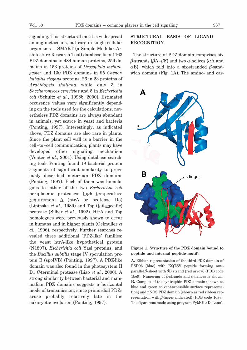
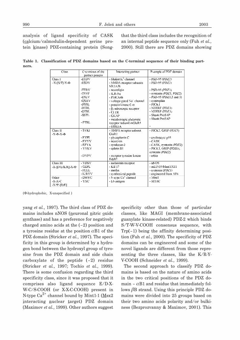
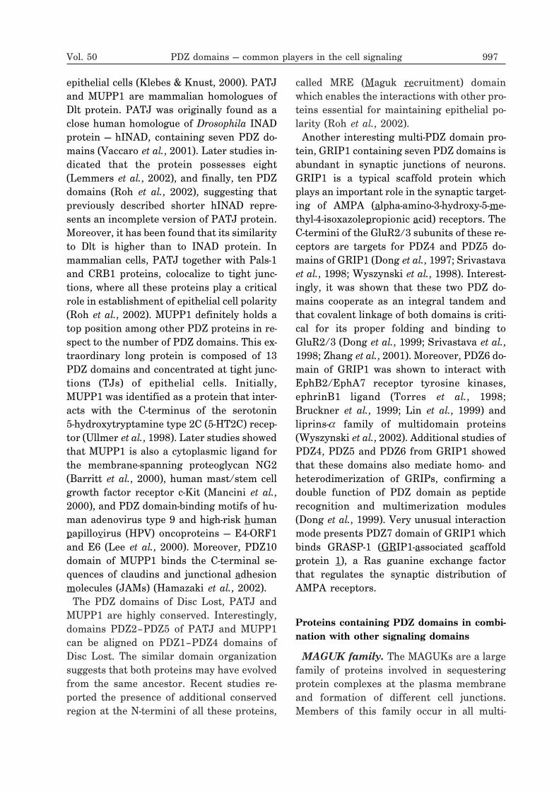
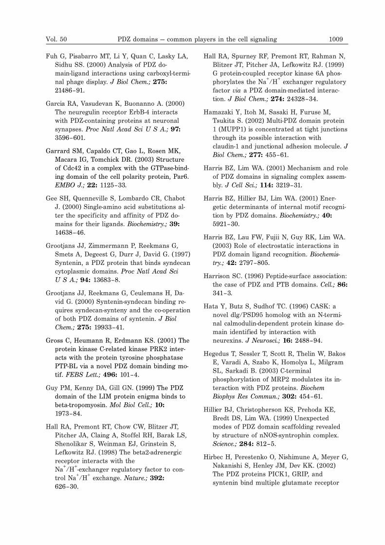
The structure of PDZ domain comprises six�-strands (�A–�F) and two �-helices (�A and�B), which fold into a six-stranded �-sand-wich domain (Fig. 1A). The amino- and car-
Vol. 50 PDZ domains — common players in the cell signaling 987
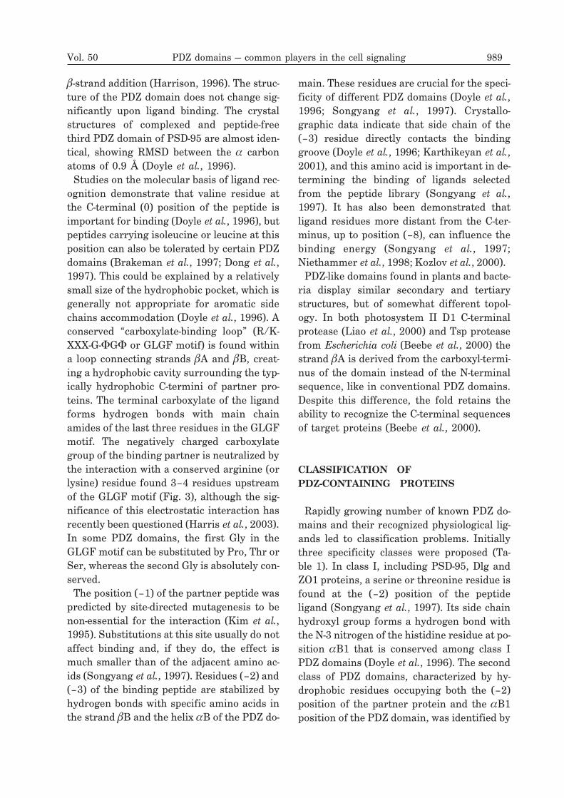
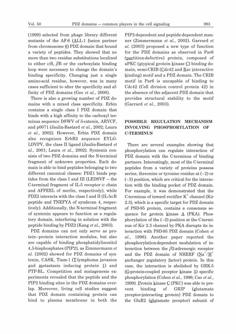
Figure 1. Structure of the PDZ domain bound topeptide and internal peptide motif.
A. Ribbon representation of the third PDZ domain ofPSD95 (blue) with KQTSV peptide forming anti-parallel �-sheet with �B strand (red arrow) (PDB code1be9). Numering of �-strands and �-helices is shown.B. Complex of the syntrophin PDZ domain (shown asblue and green solvent-accessible surface representa-tion) and nNOS PDZ domain (shown as red ribbon rep-resentation with �-finger indicated) (PDB code 1qav).The figure was made using program PyMOL (DeLano).
boxyl-termini of PDZ domains are close to-gether, facilitating incorporation of the do-main into different multi-domain proteins(Harris & Lim, 2001).PDZ domains specifically recognize short
(typically about five residues long) carboxyl-terminal peptide motifs. These sequences are
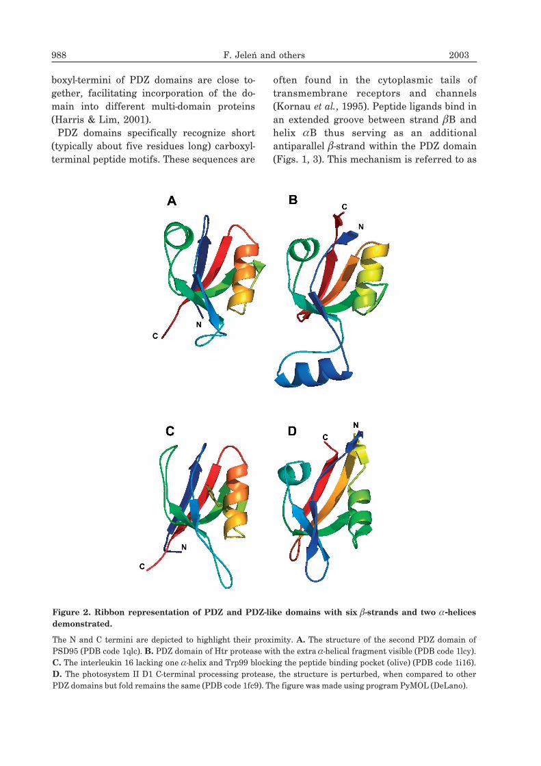
often found in the cytoplasmic tails oftransmembrane receptors and channels(Kornau et al., 1995). Peptide ligands bind inan extended groove between strand �B andhelix �B thus serving as an additionalantiparallel �-strand within the PDZ domain(Figs. 1, 3). This mechanism is referred to as
988 F. Jeleń and others 2003
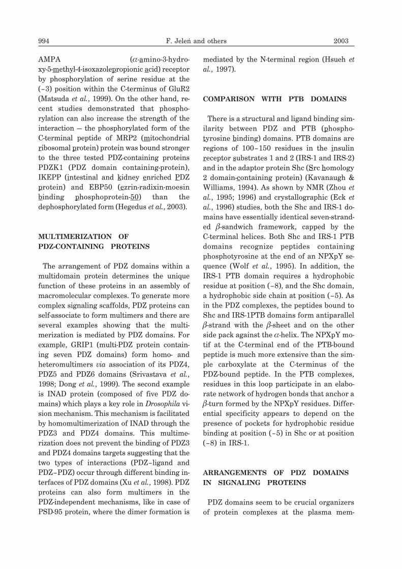
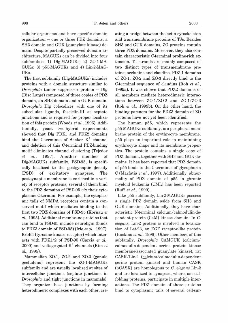
Figure 2. Ribbon representation of PDZ and PDZ-like domains with six �-strands and two �-helicesdemonstrated.
The N and C termini are depicted to highlight their proximity. A. The structure of the second PDZ domain ofPSD95 (PDB code 1qlc). B. PDZ domain of Htr protease with the extra �-helical fragment visible (PDB code 1lcy).C. The interleukin 16 lacking one �-helix and Trp99 blocking the peptide binding pocket (olive) (PDB code 1i16).D. The photosystem II D1 C-terminal processing protease, the structure is perturbed, when compared to otherPDZ domains but fold remains the same (PDB code 1fc9). The figure was made using program PyMOL (DeLano).
�-strand addition (Harrison, 1996). The struc-ture of the PDZ domain does not change sig-nificantly upon ligand binding. The crystalstructures of complexed and peptide-freethird PDZ domain of PSD-95 are almost iden-tical, showing RMSD between the � carbonatoms of 0.9 � (Doyle et al., 1996).Studies on the molecular basis of ligand rec-
ognition demonstrate that valine residue atthe C-terminal (0) position of the peptide isimportant for binding (Doyle et al., 1996), butpeptides carrying isoleucine or leucine at thisposition can also be tolerated by certain PDZdomains (Brakeman et al., 1997; Dong et al.,1997). This could be explained by a relativelysmall size of the hydrophobic pocket, which isgenerally not appropriate for aromatic sidechains accommodation (Doyle et al., 1996). Aconserved “carboxylate-binding loop” (R/K-XXX-G-�G� or GLGF motif) is found withina loop connecting strands �A and �B, creat-ing a hydrophobic cavity surrounding the typ-ically hydrophobic C-termini of partner pro-teins. The terminal carboxylate of the ligandforms hydrogen bonds with main chainamides of the last three residues in the GLGFmotif. The negatively charged carboxylategroup of the binding partner is neutralized bythe interaction with a conserved arginine (orlysine) residue found 3–4 residues upstreamof the GLGF motif (Fig. 3), although the sig-nificance of this electrostatic interaction hasrecently been questioned (Harris et al., 2003).In some PDZ domains, the first Gly in theGLGF motif can be substituted by Pro, Thr orSer, whereas the second Gly is absolutely con-served.The position (–1) of the partner peptide was
predicted by site-directed mutagenesis to benon-essential for the interaction (Kim et al.,1995). Substitutions at this site usually do notaffect binding and, if they do, the effect ismuch smaller than of the adjacent amino ac-ids (Songyang et al., 1997). Residues (–2) and(–3) of the binding peptide are stabilized byhydrogen bonds with specific amino acids inthe strand �B and the helix �B of the PDZ do-
main. These residues are crucial for the speci-ficity of different PDZ domains (Doyle et al.,1996; Songyang et al., 1997). Crystallo-graphic data indicate that side chain of the(–3) residue directly contacts the bindinggroove (Doyle et al., 1996; Karthikeyan et al.,2001), and this amino acid is important in de-termining the binding of ligands selectedfrom the peptide library (Songyang et al.,1997). It has also been demonstrated thatligand residues more distant from the C-ter-minus, up to position (–8), can influence thebinding energy (Songyang et al., 1997;Niethammer et al., 1998; Kozlov et al., 2000).PDZ-like domains found in plants and bacte-
ria display similar secondary and tertiarystructures, but of somewhat different topol-ogy. In both photosystem II D1 C-terminalprotease (Liao et al., 2000) and Tsp proteasefrom Escherichia coli (Beebe et al., 2000) thestrand �A is derived from the carboxyl-termi-nus of the domain instead of the N-terminalsequence, like in conventional PDZ domains.Despite this difference, the fold retains theability to recognize the C-terminal sequencesof target proteins (Beebe et al., 2000).
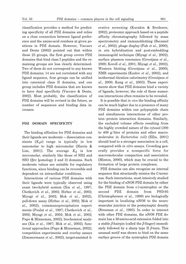
CLASSIFICATION OFPDZ-CONTAINING PROTEINS
Rapidly growing number of known PDZ do-mains and their recognized physiological lig-ands led to classification problems. Initiallythree specificity classes were proposed (Ta-ble 1). In class I, including PSD-95, Dlg andZO1 proteins, a serine or threonine residue isfound at the (–2) position of the peptideligand (Songyang et al., 1997). Its side chainhydroxyl group forms a hydrogen bond withthe N-3 nitrogen of the histidine residue at po-sition �B1 that is conserved among class IPDZ domains (Doyle et al., 1996). The secondclass of PDZ domains, characterized by hy-drophobic residues occupying both the (–2)position of the partner protein and the �B1position of the PDZ domain, was identified by
Vol. 50 PDZ domains — common players in the cell signaling 989
analysis of ligand specificity of CASK(calcium/calmodulin-dependent serine pro-tein kinase) PDZ-containing protein (Song-
yang et al., 1997). The third class of PDZ do-mains includes nNOS (neuronal nitric oxidesynthase) and has a preference for negativelycharged amino acids at the (–2) position anda tyrosine residue at the position �B1 of thePDZ domain (Stricker et al., 1997). The speci-ficity in this group is determined by a hydro-gen bond between the hydroxyl group of tyro-sine from the PDZ domain and side chaincarboxylate of the peptide (–2) residue(Stricker et al., 1997; Tochio et al., 1999).There is some confusion regarding the thirdspecificity class, since it was proposed that itcomprises also ligand sequence E/D-X-W-C/S-COOH (or X-X-C-COOH) present inN-type Ca2+ channel bound by Mint1-1 (Msx2interacting nuclear target) PDZ domain(Maximov et al., 1999). Other authors suggest
that the third class includes the recognition ofan internal peptide sequence only (Fuh et al.,2000). Still there are PDZ domains showing
specificity other than those of particularclasses, like MAGI (membrane-associatedguanylate kinase-related) PDZ-2 which bindsS/T-W-V-COOH consensus sequence, withTrp(–1) being the affinity determining posi-tion (Fuh et al., 2000). The specificity of PDZdomains can be engineered and some of thenovel ligands are different from those repre-senting the three classes, like the K/R-Y-V-COOH (Schneider et al., 1999).The second approach to classify PDZ do-
mains is based on the nature of amino acidsin the two critical positions of the PDZ do-main – �B1 and residue that immediately fol-lows �B strand. Using this principle PDZ do-mains were divided into 25 groups based ontheir two amino acids polarity and/or bulki-ness (Bezprozvanny & Maximov, 2001). This
990 F. Jeleń and others 2003
Table 1. Classification of PDZ domains based on the C-terminal sequence of their binding part-ners.
(�-hydrophobic, X-unspecified )
classification provides a method for predict-ing specificity of all PDZ domains and relieson a close connection between ligand prefer-ence and the amino-acid residues at given po-sitions in PDZ domain. However, Vaccaroand Dente (2002) pointed out that withinthese 25 groups, the first group covers PDZdomains that bind class I peptides and the re-maining groups are less clearly determined.Two of them do not correspond to any knownPDZ domains; 14 are not correlated with anyligand sequence, four groups can be unifiedinto canonical class II domains, and onegroup includes PDZ domains that are knownto have dual specificity (Vaccaro & Dente,2002). Most probably, the classification ofPDZ domains will be revised in the future, asnumber of sequences and binding data in-creases.
PDZ DOMAIN SPECIFICITY
The binding affinities for PDZ domains andtheir ligands are moderate — dissociation con-stants (Kds) range is typically in lownanomolar to high micromolar (Harris &Lim, 2001). The average Kd is lowmicromolar, similarly like those of SH2 andSH3 (Src homology 2 and 3) domains. Suchmoderate values are suitable for regulatoryfunctions, since binding can be reversible anddependent on intracellular conditions.Interactions of various PDZ domains with
their ligands were typically observed usingyeast two-hybrid system (Xia et al., 1997;Chetkovich et al., 2002; Hirbec et al., 2002;Miyagi et al., 2002; Mok et al., 2002),pull-down assay (Hirbec et al., 2002; Mok etal., 2002), coimmunoprecipitation experi-ments (Poulat et al., 1997; Chetkovich et al.,2002; Miyagi et al., 2002; Mok et al., 2002;Pupo & Minneman, 2002), biochemical analy-ses (Xia et al., 1997; Mok et al., 2002), func-tional approaches (Pupo & Minneman, 2002),competition experiments and overlay assays(Zimmermann et al., 2002), target-assisted it-
erative screening (Kurakin & Bredesen,2002), proteomic approach based on a peptideaffinity chromatography followed by massspectrometry and immunoblotting (Becamelet al., 2002), phage display (Fuh et al., 2000),in situ hybridization and post-embeddingimmunogold technique (Miyagi et al., 2002),surface plasmon resonance (Grootjans et al.,2000; Koroll et al., 2001; Miyagi et al., 2002),Western blotting (Grootjans et al., 2000),NMR experiments (Kozlov et al., 2002), andisothermal titration calorimetry (Grootjans etal., 2000; Kang et al., 2003). These experi-ments show that PDZ domains bind a varietyof ligands, however, the role of these numer-ous interactions often remains to be revealed.It is possible that in vivo the binding affinity
can be much higher due to a presence of manyPDZ domains within one polypeptide chainand simultanous interactions of other pro-tein–protein interaction domains. Similarly,the excluded volume effects resulting fromthe highly crowded nature of the cytosol (300to 400 g/liter of proteins and other macro-molecules in Escherichia coli) (Ellis, 2001)should lead to a stronger association in a cell,compared with in vitro assays. Crowding gen-erally provides a nonspecific force formacromolecular compaction and association(Minton, 2000), which may be crucial to theformation of large protein complexes.PDZ domains can also recognize an internal
sequence that structurally mimics the C-termi-nus. Such interactions, most intesively studiedfor the binding of nNOS PDZ domain by eitherthe PDZ domain from �1-syntrophin or thesecond PDZ domain from PSD-95(Christopherson et al., 1999) are biologicallyimportant in localizing nNOS to the neuro-muscular junction or the postsynaptic density(Brenman et al., 1995). In order to interactwith other PDZ domains, the nNOS PDZ do-main has a 30-amino-acid extension folded intoa stable �-hairpin (called the �-finger) immedi-ately followed by a sharp type II �-turn. Thisunusual motif was shown to bind on the samesurface groove of the syntrophin PDZ domain
Vol. 50 PDZ domains — common players in the cell signaling 991
as the C-terminal peptide ligand, with its�-turn positioned directly in place of the pep-tide’s carboxyl-terminus (Hillier et al., 1999).Closer insight into this interaction reveals thatthe �-finger of nNOS contains an internal pep-tide whose sequence and binding orientationare very similar to those of canonical C-termi-nal peptide ligands (Fig. 1B). In addition, thereis an extensive area of contacts between thecore PDZ domains of syntrophin and nNOS(Harris et al., 2001). Thus, binding of the twodifferent regions of nNOS by syntrophin is farmore specific than recognition through a shortC-terminal sequence.The multi-domain scaffolding protein INAD
(inactivation no after-potential D) containsfive PDZ domains which independently bindvarious proteins including NorpA (noreceptor potential A) and the phospholipaseC-� isoenzyme. These interactions are re-quired for the proper intracellular targetingand spatial arrangement of proteins involvedin the fly phototransduction. The structure ofthe N-terminal PDZ domain of INAD with theC-terminal heptapeptide (GKTEFCA) derivedfrom NorpA reveals an intermoleculardisulfide bond necessary for the interaction(Kimple et al., 2001). Since other proteinsalso possess similar, cysteine-containing con-sensus sequences adequate for binding to thePDZ domains, this disulfide-mediated interac-tion may be a common mode of interaction be-tween PDZ domains and their target proteins.Moreover, there are also other important dif-ferences in INAD(PDZ1)-NorpA interaction.The NorpA peptide contains an abrupt turn atPhe(–2), while all other peptides are in an ex-tended conformation (Doyle et al., 1996;Daniels et al., 1998; Schultz et al., 1998a).Furthermore, even though PDZ1 of INADpossesses a characteristic hydrophobic cleftthat normally buries the side chain of the ter-minal residue of the peptide, position (0) ofthe NorpA derived peptide is exposed to a sol-vent (Kimple et al., 2001).Erbin interacts with the receptor tyrosine
kinase ErbB2 and plays a role in its localiza-
tion at the basolateral membrane of epithelialcells (Borg et al., 2000). The protein is alsohighly concentrated at neuronal postsynapticmembranes and neuromuscular junctions.The crystal structure of the Erbin andErbB2-derived peptide reveals an interactionof the peptidic Tyr(–7) with the extended�2–�3 loop of the Erbin PDZ (Birrane et al.,2003). The second crystal structure of this do-main bound to the phosphotyrosine-contain-ing ErbB2 peptide shows that phosphory-lation of Tyr(–7) abolishes its interactionwith the �2–�3 loop. Phosphorylation of theTyr(–7) residue reduces 2.5-fold the affinityof the Erbin-ErbB2 interaction (Birrane et al.,2003).IL-16 has no significant sequence homology
to other interleukins or any other member ofthe chemokine family and is the first knownextracellular protein with the PDZ-do-main-like fold. However, the protein does notexhibit any peptide binding properties of PDZdomains (Muhlhahn et al., 1998), since itsGLGF cleft is smaller and blocked with abulky Trp side chain at its center.Recently solved NMR structure of the sec-
ond PDZ domain of PTP-BL (protein tyrosinephosphatase BL) shows a unique feature,compared to the canonical PDZ fold. An ex-tended flexible loop at the base of the bindingpocket, called L1, folds back onto the proteinbackbone and modulates the domain selectiv-ity (Walma et al., 2002).The specificity of a PDZ domain can be eas-
ily altered by substituting residues in or di-rectly adjacent to the strand �B and the helix�B. Stricker et al. (1997) changed the specific-ity of nNOS PDZ domain from D-X-V-COOHto T-X-V- COOH by introducing only two mu-tations — Tyr77His and Asp78Glu. Moreover,it was reported that PDZ domains could beengineered to specifically recognize a largenumber of proteins by combining differentbackbone templates with a computer-aidedprotein design (Reina et al., 2002). Phage dis-play approach was also used to alter thespecificities of PDZ domains. Schneider et al.
992 F. Jeleń and others 2003
(1999) selected from phage library differentmutants of the AF-6 (ALL-1 fusion partnerfrom chromosome 6) PDZ domain that bounda variety of peptides. They showed that nomore than two residue substitutions localizedto either �B, �B or the carboxylate bindingloop were necessary to change the domain’sbinding specificity. Changing just a singleamino-acid residue, however, was in manycases sufficient to alter the specificity and af-finity of PDZ domains (Gee et al., 2000).There is also a growing number of PDZ do-
mains with a mixed class specificity. Erbincontains a single class I PDZ domain thatbinds with a high affinity to the carboxyl ter-minus sequence DSWV of �-catenin, ARVCF,and p0071 (Jaulin-Bastard et al., 2002; Lauraet al., 2002). However, Erbin PDZ domainalso recognizes ErbB2 sequence EYLG-LDVPV, the class II ligand (Jaulin-Bastard etal., 2001; Laura et al., 2002). Syntenin con-sists of two PDZ domains and the N-terminalfragment of unknown properties. Each do-main is able to bind peptides belonging to twodifferent canonical classes: PDZ1 binds pep-tides from the class I and III (LEDSVF — theC-terminal fragment of IL-5 receptor � chainand AFFEEL of merlin, respectively), whilePDZ2 interacts with the class I and II (IL-5�Rpeptide and TNEFYA of syndecan 4, respec-tively). Additionally, the N-terminal fragmentof syntenin appears to function as a regula-tory domain, interfering in solution with thepeptide binding by PDZ2 (Kang et al., 2003).PDZ domains can not only serve as pro-
tein–protein interaction modules, but alsoare capable of binding phosphatidylinositol4,5-bisphosphates (PIP2), as Zimmermann etal. (2002) showed for PDZ domains of syn-tenin, CASK, Tiam-1 (T-lymphoma invasionand metastasis inducing protein 1) andPTP-BL. Competition and mutagenesis ex-periments revealed that the peptide and thePIP2 binding sites in the PDZ domains over-lap. Moreover, living cell studies suggestthat PDZ domain containing protein canbind to plasma membrane in both the
PIP2-dependent and peptide-dependent man-ner (Zimmermann et al., 2002). Garrard etal. (2003) proposed a new type of functionfor the PDZ domains as observed in Par6(partition-defective) protein, composed ofaPKC (atypical protein kinase C) binding do-main, semi-CRIB (Cdc42 and Rac interactivebinding) motif and a PDZ domain. The CRIBmotif in Par6 is uncapable of binding toCdc42 (Cell division control protein 42) inthe absence of the adjacent PDZ domain thatprovides structural stability to the motif(Garrard et al., 2003).
POSSIBLE REGULATION MECHANISMINVOLVING PHOSPHORYLATION OFC-TERMINUS
There are several examples showing thatphosphorylation can regulate interaction ofPDZ domain with the C-terminus of bindingpartners. Interestingly, most of the C-terminalpeptides from a variety of proteins possessserine, threonine or tyrosine residue at (–2) or(–3) position, which are critical for the interac-tion with the binding pocket of PDZ domain.For example, it was demonstrated that theC-terminus of inward rectifier K+ channel (Kir2.3), which is a specific target for PDZ domainof PSD-95 protein, contains a consensus se-quence for protein kinase A (PKA). Phos-phorylation of the (–2) position at the C-termi-nus of Kir 2.3 channel by PKA disrupts its in-teraction with PSD-95 PDZ domain (Cohen etal., 1996). Another paper reported thephosphorylation-dependent modulation of in-teraction between the �2-adrenergic receptorand the PDZ domain of NHERF (Na+/H+
exchanger regulatory factor) protein. In thiscase, the interaction is abolished by GRK-5(G-protein-coupled receptor kinase 5) specificphosphorylation (Cohen et al., 1996; Cao et al.,1999). Protein kinase C (PKC) was able to pre-vent binding of GRIP (glutamatereceptor-interacting protein) PDZ domain tothe GluR2 (glutamate receptor) subunit of
Vol. 50 PDZ domains — common players in the cell signaling 993
AMPA (�-amino-3-hydro-xy-5-methyl-4-isoxazolepropionic acid) receptorby phosphorylation of serine residue at the(–3) position within the C-terminus of GluR2(Matsuda et al., 1999). On the other hand, re-cent studies demonstrated that phospho-rylation can also increase the strength of theinteraction — the phosphorylated form of theC-terminal peptide of MRP2 (mitochondrialribosomal protein) protein was bound strongerto the three tested PDZ-containing proteinsPDZK1 (PDZ domain containing-protein),IKEPP (intestinal and kidney enriched PDZprotein) and EBP50 (ezrin-radixin-moesinbinding phosphoprotein-50) than thedephosphorylated form (Hegedus et al., 2003).
MULTIMERIZATION OFPDZ-CONTAINING PROTEINS
The arrangement of PDZ domains within amultidomain protein determines the uniquefunction of these proteins in an assembly ofmacromolecular complexes. To generate morecomplex signaling scaffolds, PDZ proteins canself-associate to form multimers and there areseveral examples showing that the multi-merization is mediated by PDZ domains. Forexample, GRIP1 (multi-PDZ protein contain-ing seven PDZ domains) form homo- andheteromultimers via association of its PDZ4,PDZ5 and PDZ6 domains (Srivastava et al.,1998; Dong et al., 1999). The second exampleis INAD protein (composed of five PDZ do-mains) which plays a key role in Drosophila vi-sion mechanism. This mechanism is facilitatedby homomultimerization of INAD through thePDZ3 and PDZ4 domains. This multime-rization does not prevent the binding of PDZ3and PDZ4 domains targets suggesting that thetwo types of interactions (PDZ–ligand andPDZ–PDZ) occur through different binding in-terfaces of PDZ domains (Xu et al., 1998). PDZproteins can also form multimers in thePDZ-independent mechanisms, like in case ofPSD-95 protein, where the dimer formation is
mediated by the N-terminal region (Hsueh etal., 1997).
COMPARISON WITH PTB DOMAINS
There is a structural and ligand binding sim-ilarity between PDZ and PTB (phospho-tyrosine binding) domains. PTB domains areregions of 100–150 residues in the insulinreceptor substrates 1 and 2 (IRS-1 and IRS-2)and in the adaptor protein Shc (Src homology2 domain-containing protein) (Kavanaugh &Williams, 1994). As shown by NMR (Zhou etal., 1995; 1996) and crystallographic (Eck etal., 1996) studies, both the Shc and IRS-1 do-mains have essentially identical seven-strand-ed �-sandwich framework, capped by theC-terminal helices. Both Shc and IRS-1 PTBdomains recognize peptides containingphosphotyrosine at the end of an NPXpY se-quence (Wolf et al., 1995). In addition, theIRS-1 PTB domain requires a hydrophobicresidue at position (–8), and the Shc domain,a hydrophobic side chain at position (–5). Asin the PDZ complexes, the peptides bound toShc and IRS-1PTB domains form antiparallel�-strand with the �-sheet and on the otherside pack against the �-helix. The NPXpY mo-tif at the C-terminal end of the PTB-boundpeptide is much more extensive than the sim-ple carboxylate at the C-terminus of thePDZ-bound peptide. In the PTB complexes,residues in this loop participate in an elabo-rate network of hydrogen bonds that anchor a�-turn formed by the NPXpY residues. Differ-ential specificity appears to depend on thepresence of pockets for hydrophobic residuebinding at position (–5) in Shc or at position(–8) in IRS-1.
ARRANGEMENTS OF PDZ DOMAINSIN SIGNALING PROTEINS
PDZ domains seem to be crucial organizersof protein complexes at the plasma mem-
994 F. Jeleń and others 2003
brane. They are important in transport andtargeting of different proteins to the sites ofcellular signaling thus assuring localizationand organization of both relevant receptorsand downstream effectors to proper regionsof the cell. PDZ-containing proteins createscaffolds for the assembly of supramolecularsignaling complexes, thereby coordinatingand guiding the flow of regulatory informa-tion. This is possible due to the ability ofPDZ-containing proteins to both bind an ar-ray of target proteins and oligomerize intobranched networks.According to arrangement type, all known
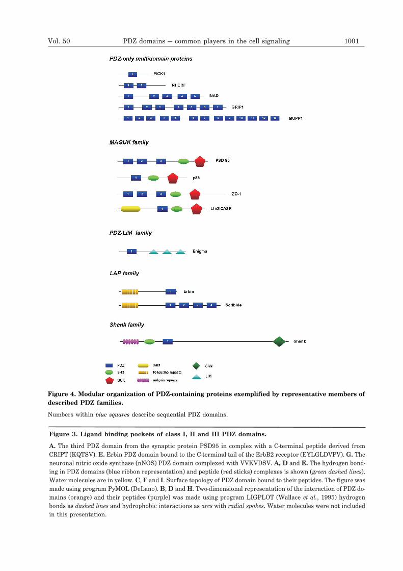
PDZ-containing proteins can be divided into twogroups. The first group comprises proteins con-taining only PDZ domains, typically several PDZdomainswith different specificities within a singlepolypeptide chain called multi-PDZ proteins. Ex-amples include single-PDZ domain proteinsPICK1 (protein interacting with C-kinase), Par6and multi-PDZ domain proteins NHERF (2 PDZdomains), CIPP (channel-interacting PDZ domainprotein, 4 PDZ domains), INAD (5 PDZ domains),GRIP (7 PDZ domains), PATJ (Pals-1 associatedtight junction protein, 10 PDZ domains) andMUPP1 (contains remarkable 13 PDZ domains).Proteins possessing single or multiple PDZ do-mains in combination with other functional do-mains form the second group. Among these, theMAGUK (membrane-associated guanylatekinase) proteins represent a very common classcontaining invariably one or three PDZ domains,a SH3 domain and a guanylate kinase homology(GUK) domain. Other PDZ-containing proteinspresent more diversified combinations with a va-riety of interaction domains such WW, LIM(zinc-binding domain present in Lin-11, Isl-1,Mec-3), CaMK (calcium/calmodulin-dependentprotein kinase domain), DH/PH (Dblhomology/pleckstrin homology), ankyrin orleucine-rich repeats.
PDZ-only proteins
Single-PDZ domain proteins. Despite thepresence of only a single PDZ domain, some
of them can effectively multimerize and inconsequence link partner proteins. This is il-lustrated by PICK1, a single-PDZ domain pro-tein expressed at synapses and originally iso-lated due to its ability to bind the C-terminusof protein kinase C (Staudinger et al., 1995;1997). It was shown that PICK1 canhomooligomerize through its coiled-coil re-gion and this self-association is essential forclustering of the synaptic metabotropicglutamate receptors (mGluR7a) (Staudingeret al., 1997). Besides this interaction,PICK1-binding partners include the GluR2subunit of AMPA receptors (Dev et al., 1999;Daw et al., 2000; Osten et al., 2000; Xia et al.,1999; 2000; Iwakura et al., 2001; Kim et al.,2001; Perez et al., 2001; Braithwaite et al.,2002), the dopamine transporter (DAT)(Torres et al., 2001), the ERBB2/HER2 recep-tor (Jaulin-Bastard et al., 2001), the mito-gen-stimulated TIS21 protein (Lin et al.,2001) and the ASICs (acid-sensing ionchannels) (Baron et al., 2002). Taken to-gether, it seems that PICK1 may serve asadaptor protein that links variety of synaptictransmembrane receptors and channels toprotein kinase C.Another single-PDZ domain containing pro-
tein, Par6, first identified in C. elegans, playsa critical role in the asymmetric cell divisionand the polarized cell growth (Hung &Kemphues, 1999). Later studies revealed afamily of mammalian Par6 proteins, similarto C. elegans forms (Joberty et al., 2000). Be-sides PDZ domain, Par6 protein contains asemi-CRIB motif, which can bind to Cdc42GTPase but only in the presence of an adja-cent PDZ domain. Moreover, it was shownthat Par6 PDZ domain effects structural sta-bility of the CRIB motif (Garrard et al., 2003).Both PDZ and semi-CRIB motif are also nec-essary for binding to Par3, another proteincontaining three copies of PDZ domain. Func-tional complex of Par6 with Cdc42-GTP, Par3and with the regulatory domains of atypicalprotein kinase C is implicated in the forma-tion of tight junctions at epithelial cell–cell
Vol. 50 PDZ domains — common players in the cell signaling 995
contacts (Joberty et al., 2000). This suggeststhat Par6 is an adaptor protein responsiblefor cross-talk between activated Cdc42 or RacGTPases and apical protein kinase signaling.Multi PDZ-domain proteins. NHERF-1
and its second isoform, NHERF-2, highly ex-pressed in epithelial cells, serve as specializedadaptors for broad range of signaling pro-teins. Both isoforms contain two highly ho-mologous PDZ domains and the C-terminalregion that associates with members of theERM (ezrin-radixin-moesin) family of mem-brane-cytoskeletal adapters. NHERF was firstidentified as a regulator of NHE3 (type 3Na+/H+ exchanger) activity (Weinman et al.,2000). However, the list of functions ofNHERF protein in epithelial cell physiologycan be extended. For example, regulation ofmembrane proteins such as �2AR��2-adrenergic receptor) (Hall et al., 1998)and CFTR (cystic fibrosis transmembraneconductance regulator) (Raghuram et al.,2001) is mediated by PDZ1 domain ofNHERF. Other proteins identified as poten-tial partners for NHERF PDZ1 domain in-clude PDGFR (platelet-derived growth factorreceptor) (Maudsley et al., 2000), GRK6A (anisoform of G-protein-coupled receptor kinase)(Hall et al., 1999), SOC (store-operatedcalcium) channels, such as Trp4 and Trp5, aswell as the phospholipases C�1 and C�2(Tang et al., 2000). On the other hand, besidesthe binding of NHE3, the PDZ2 domain ofNHERF is reported to bind two additional tar-gets: YAP-65 (Yes-associated protein) inYAP-65/c-Yes complex (Mohler et al., 1999)and phospholipase C�3 (Hwang et al., 2000).Thus, both isoforms of two PDZ-domain pro-tein NHERF are involved in regulation ofmultiple signaling pathways such as growthregulation, phosphoinositide metabolism, re-ceptor modulation and targeting non-receptorkinases (Voltz et al., 2001).The CIPP is an example of multi-PDZ do-
main protein, which was found to interact se-lectively with the C-termini of signaling recep-tors in synaptic membranes. CIPP is com-
posed of four PDZ domains possessing differ-ent specificities; PDZ2 domain binds to theC-terminus of the inward rectifier K+ (Kir)channel, Kir4.1, and neuroligin, PDZ3 inter-acts with the NR2C subunit of NMDA recep-tors and neurexin (Kurschner et al., 1998),whereas PDZ4 domain was recently reportedto bind the ASIC3 (acid-sensing ion channel3) (Anzai et al., 2002). Additionally, the C-ter-mini of NR2B subunit of NMDA and Kir4.2are specific ligands for both PDZ2 and PDZ3domains of CIPP (Kurschner et al., 1998). Incontrast, the binding partners for PDZ1 do-main have not yet been identified. Thus, theCIPP protein appears to be a typical scaffold-ing protein that links different types ofneuronal cell surface molecules to inter-cellular signaling network in neurons.INAD — Drosophila protein composed of five
PDZ modules plays a central role in organiza-tion of supramolecular signaling complex inthe phototransduction cascade. All five PDZdomains of INAD have been shown to interactwith various phototransduction proteins.PDZ1 and PDZ5 domains of INAD wereshown to bind the phospholipase C (PLC)(Tsunoda et al., 1997; van Huizen et al., 1998;Xu et al., 1998), whereas PDZ2 and PDZ4 do-mains, the C-terminus of eye-specific proteinkinase C (Huber et al., 1996b; Tsunoda et al.,1997; Adamski et al., 1998; Xu et al., 1998).Additionally, light-responsive, transientreceptor potential (TRP) channel is a targetfor PDZ3 of INAD (Huber et al., 1996a; Shieh& Zhu, 1996).Three multi-PDZ domain proteins, Dlt
(Discs Lost), PATJ (Pals-1 associated tightjunction protein) and MUPP1 (multi-PDZ do-main protein 1) are examples of proteins es-sential in organizing protein complexes cru-cial to maintaining polarity of epithelial andneuronal cells. First of them, the DrosophilaDlt contains four PDZ domains and its PDZ1domain can interact with the C-terminal fouramino acids of the dCrumbs protein — apicalpolarity determinant responsible also for po-sitioning of the zonula adherens in Drosophila
996 F. Jeleń and others 2003
epithelial cells (Klebes & Knust, 2000). PATJand MUPP1 are mammalian homologues ofDlt protein. PATJ was originally found as aclose human homologue of Drosophila INADprotein — hINAD, containing seven PDZ do-mains (Vaccaro et al., 2001). Later studies in-dicated that the protein possesses eight(Lemmers et al., 2002), and finally, ten PDZdomains (Roh et al., 2002), suggesting thatpreviously described shorter hINAD repre-sents an incomplete version of PATJ protein.Moreover, it has been found that its similarityto Dlt is higher than to INAD protein. Inmammalian cells, PATJ together with Pals-1and CRB1 proteins, colocalize to tight junc-tions, where all these proteins play a criticalrole in establishment of epithelial cell polarity(Roh et al., 2002). MUPP1 definitely holds atop position among other PDZ proteins in re-spect to the number of PDZ domains. This ex-traordinary long protein is composed of 13PDZ domains and concentrated at tight junc-tions (TJs) of epithelial cells. Initially,MUPP1 was identified as a protein that inter-acts with the C-terminus of the serotonin5-hydroxytryptamine type 2C (5-HT2C) recep-tor (Ullmer et al., 1998). Later studies showedthat MUPP1 is also a cytoplasmic ligand forthe membrane-spanning proteoglycan NG2(Barritt et al., 2000), human mast/stem cellgrowth factor receptor c-Kit (Mancini et al.,2000), and PDZ domain-binding motifs of hu-man adenovirus type 9 and high-risk humanpapillovirus (HPV) oncoproteins — E4-ORF1and E6 (Lee et al., 2000). Moreover, PDZ10domain of MUPP1 binds the C-terminal se-quences of claudins and junctional adhesionmolecules (JAMs) (Hamazaki et al., 2002).The PDZ domains of Disc Lost, PATJ and
MUPP1 are highly conserved. Interestingly,domains PDZ2–PDZ5 of PATJ and MUPP1can be aligned on PDZ1–PDZ4 domains ofDisc Lost. The similar domain organizationsuggests that both proteins may have evolvedfrom the same ancestor. Recent studies re-ported the presence of additional conservedregion at the N-termini of all these proteins,
called MRE (Maguk recruitment) domainwhich enables the interactions with other pro-teins essential for maintaining epithelial po-larity (Roh et al., 2002).Another interesting multi-PDZ domain pro-
tein, GRIP1 containing seven PDZ domains isabundant in synaptic junctions of neurons.GRIP1 is a typical scaffold protein whichplays an important role in the synaptic target-ing of AMPA (alpha-amino-3-hydroxy-5-me-thyl-4-isoxazolepropionic acid) receptors. TheC-termini of the GluR2/3 subunits of these re-ceptors are targets for PDZ4 and PDZ5 do-mains of GRIP1 (Dong et al., 1997; Srivastavaet al., 1998; Wyszynski et al., 1998). Interest-ingly, it was shown that these two PDZ do-mains cooperate as an integral tandem andthat covalent linkage of both domains is criti-cal for its proper folding and binding toGluR2/3 (Dong et al., 1999; Srivastava et al.,1998; Zhang et al., 2001). Moreover, PDZ6 do-main of GRIP1 was shown to interact withEphB2/EphA7 receptor tyrosine kinases,ephrinB1 ligand (Torres et al., 1998;Bruckner et al., 1999; Lin et al., 1999) andliprins-� family of multidomain proteins(Wyszynski et al., 2002). Additional studies ofPDZ4, PDZ5 and PDZ6 from GRIP1 showedthat these domains also mediate homo- andheterodimerization of GRIPs, confirming adouble function of PDZ domain as peptiderecognition and multimerization modules(Dong et al., 1999). Very unusual interactionmode presents PDZ7 domain of GRIP1 whichbinds GRASP-1 (GRIP1-associated scaffoldprotein 1), a Ras guanine exchange factorthat regulates the synaptic distribution ofAMPA receptors.
Proteins containing PDZ domains in combi-nation with other signaling domains
MAGUK family. The MAGUKs are a largefamily of proteins involved in sequesteringprotein complexes at the plasma membraneand formation of different cell junctions.Members of this family occur in all multi-
Vol. 50 PDZ domains — common players in the cell signaling 997
cellular organisms and have specific domainorganization — one or three PDZ domains, aSH3 domain and GUK (guanylate kinase) do-main. Despite partially preserved domain ar-chitecture, MAGUKs can be divided into foursubfamilies: 1) Dlg-MAGUKs; 2) ZO-1-MA-GUKs; 3) p55-MAGUKs and 4) Lin-2-MAG-UKs.The first subfamily (Dlg-MAGUKs) includes
proteins with a domain structure similar toDrosophila tumor suppressor protein — Dlg(Disc Large) composed of three copies of PDZdomain, an SH3 domain and a GUK domain.Drosophila Dlg colocalizes with one of itssubcellular ligands, fasciclin-III at septatejunctions and is required for proper localiza-tion of this protein (Woods et al., 1996). Addi-tionally, yeast two-hybrid experimentsshowed that Dlg PDZ1 and PDZ2 domainsbind the C-terminus of Shaker K+ channeland deletion of this C-terminal PDZ-bindingmotif eliminates channel clustering (Tejedoret al., 1997). Another member ofDlg-MAGUKs subfamily, PSD-95, is specifi-cally localized to the postsynaptic density(PSD) of excitatory synapses. Thepostsynaptic membrane is enriched in a vari-ety of receptor proteins; several of them bindto the PDZ domains of PSD-95 via their cyto-plasmic C-termini. For example, the cytoplas-mic tails of NMDA receptors contain a con-served motif which mediates binding to thefirst two PDZ domains of PSD-95 (Kornau etal., 1995). Additional membrane proteins thatcan bind to PSD-95 include neuroligin (bindsto PDZ3 domain of PSD-95) (Irie et al., 1997),ErbB4 (tyrosine kinase receptor) which inter-acts with PDZ1/2 of PSD-95 (Garcia et al.,2000) and voltage-gated K+ channels (Kim etal., 1995).Mammalian ZO-1, ZO-2 and ZO-3 (zonula
occludens) represent the ZO-1-MAGUKssubfamily and are usually localized at sites ofintercellular junctions (septate junctions inDrosophila and tight junctions in mammals).They organize these junctions by formingheterodimeric complexes with each other, cre-
ating a bridge between the actin cytoskeletonand transmembrane proteins of TJs. BesidesSH3 and GUK domains, ZO proteins containthree PDZ domains. Moreover, they also con-tain characteristic C-terminal proline-rich ex-tension. TJ strands are mainly composed oftwo distinct types of transmembrane pro-teins: occludins and claudins. PDZ-1 domainsof ZO-1, ZO-2 and ZO-3 directly bind to theC-terminal sequence of claudins (Itoh et al.,1999a). It was shown that PDZ2 domains ofall members mediate heterodimeric interac-tions between ZO-1/ZO-2 and ZO-1/ZO-3(Itoh et al., 1999b). On the other hand, thebinding partners for the PDZ3 domain of ZOproteins have not yet been identified.The human p55, which represents the
p55-MAGUKs subfamily, is a peripheral mem-brane protein of the erythrocyte membrane.p55 plays an important role in maintainingerythrocyte shape and its membrane proper-ties. The protein contains a single copy ofPDZ domain, together with SH3 and GUK do-mains. It has been reported that PDZ domainof p55 binds to the C-terminus of glycophorinC (Marfatia et al., 1997). Additionally, abnor-mality of PDZ domain of p55 in chronicmyeloid leukemia (CML) has been reported(Ruff et al., 1999).Like p55 subfamily, Lin-2-MAGUKs possess
a single PDZ domain aside from SH3 andGUK domains. Additionally, they have char-acteristic N-terminal calcium/calmodulin-de-pendent protein (CaM) kinase domain. In C.elegans, Lin-2 protein is involved in localiza-tion of Let-23, an EGF receptor-like protein(Hoskins et al., 1996). Other members of thissubfamily, Drosophila CAMGUK (calcium/calmodulin-dependent serine protein kinasemembrane-associated guanylate kinase), ratCASK/Lin-2 (calcium/calmodulin-dependentserine protein kinase) and human CASK(hCASK) are homologous to C. elegans Lin-2and are localized to synapses, where, as scaf-folding proteins, participate in multiple inter-actions. The PDZ domain of these proteinsbind to cytoplasmic tails of several cell-sur-
998 F. Jeleń and others 2003
face proteins. For example, the C-terminus ofneurexin I is a high affinity binding partnerfor PDZ domain of CASK protein (Hata et al.,1996) and the cytoplasmic tails of junctionaladhesion molecules are targets forCASK/Lin2 PDZ domain (Martinez-Estradaet al., 2001). Moreover, yeast two-hybridscreening showed an interaction between thehuman homolog hCASK and the C-terminalsequence of the membrane protein syn-decan-2 (Cohen et al., 1998). In another exam-ple, the C terminus of Parkin protein, selec-tively truncated by a Parkinson’s dis-ease-causing mutation, can selectively bind tothe PDZ domain of CASK (Fallon et al.,2002). Recent studies reported a new interac-tion partner for PDZ domain of CASK protein— plasma membrane Ca2+-ATPase (PMCA),which is major regulator of Ca2+ homeostasis(Schuh et al., 2003).PDZ-LIM family. It has been suggested
that cytoskeletal proteins belonging to thePDZ-LIM family serve as adapters for directLIM-binding proteins to the cytoskeleton(Vallenius et al., 2000). They contain a PDZdomain at the N-terminus followed by one orthree LIM domains. All six members of thefamily associate with the cytoskeleton, five ofthem via interactions with �-actinin and/or�-tropomyosin. CLP-36 (C-terminal LIM do-main protein 1) protein is expressed in epithe-lial cells and localizes to actin stress fibers.This localization is mediated via the PDZ do-main of CLP-36 that associates with thespectrin-like repeats of �-actinin. Yeasttwo-hybrid analysis indicated a highly specificassociation of CLP-36 and Clik1 (CLP-36interacting kinase) (Vallenius et al., 2000).The association is mediated by the C-terminalpart of CLP-36 containing LIM domain andleads to relocalization of the otherwise nu-clear Clik1 kinase to actin stress fibers(Vallenius & Makela, 2002).Cypher, a striated muscle-restricted protein,
has two mRNA splice variants designatedCypher1 and Cypher2. Both proteins containPDZ domain at the N-terminus. Cypher1, but
not Cypher2, contains three LIM domainsclose to the C-terminus. Cypher1 and Cypher2bind to �-actinin via their PDZ domains at theZ-lines of cardiac muscle. These data suggestthat Cypher functions as an adaptor in striatedmuscle to link protein kinase C-mediated sig-naling to the cytoskeleton (Zhou et al., 1999).In turn, PDZ domain of Enigma, another mem-ber of PDZ-LIM family, is present at the Z-linein skeletal muscle and its PDZ domain binds tothe actin-binding protein tropomyosin (skele-tal �-TM). The interaction suggests a role forEnigma as an adapter protein that directsLIM-binding proteins to actin filaments ofmuscle cells (Guy et al., 1999).LAP family. The LAP (leucine-rich repeats
and PDZ) family of PDZ proteins plays a role inestablishment of cell polarity, and mutation ofthese proteins can have oncogenic conse-quences. Sixteen leucine-rich repeats (LRRs) atthe N-terminus and single PDZ domain at theC-terminus presents a characteristic architec-ture of all members of LAP family.The LAP protein, Erbin was identified as an
adaptor protein present in the basolateralepithelia and involved in proper localizationof ERBB2/HER2 receptors to the basolateralmembrane of epithelial cells. This process ismediated by Erbin PDZ domain, which wasshown to bind the C-terminus of the receptorboth in vitro and in vivo (Borg et al., 2000). Itwas also reported that �-catenin and ARVCFserve as interaction partners for Erbin PDZdomain (Laura et al., 2002).Another LAP family member, Densin, was
identified as a transmembrane specific adhesionmolecule mediating adhesion between pre- andpostsynaptic membranes at glutamatergic syn-apses (Apperson et al., 1996). Screening of hu-man brain cDNA library resulted in identifica-tion of �-catenin/neural plakophilin-relatedarmadillo repeat protein (NPRAP) as a potentialbinding partner of PDZ domain of Densin.Colocalization of densin with �-catenin/NPRAPat synapses suggested an important role in orga-nization of the synaptic cell–cell junction (Izawaet al., 2002). Later studies, showed binding of
Vol. 50 PDZ domains — common players in the cell signaling 999
1000 F. Jeleń and others 2003
Vol. 50 PDZ domains — common players in the cell signaling 1001
Figure 4. Modular organization of PDZ-containing proteins exemplified by representative members ofdescribed PDZ families.
Numbers within blue squares describe sequential PDZ domains.
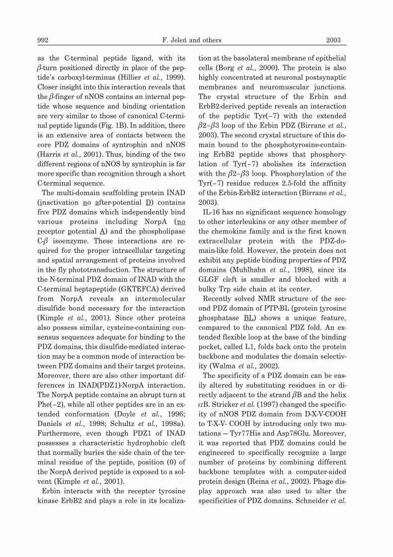
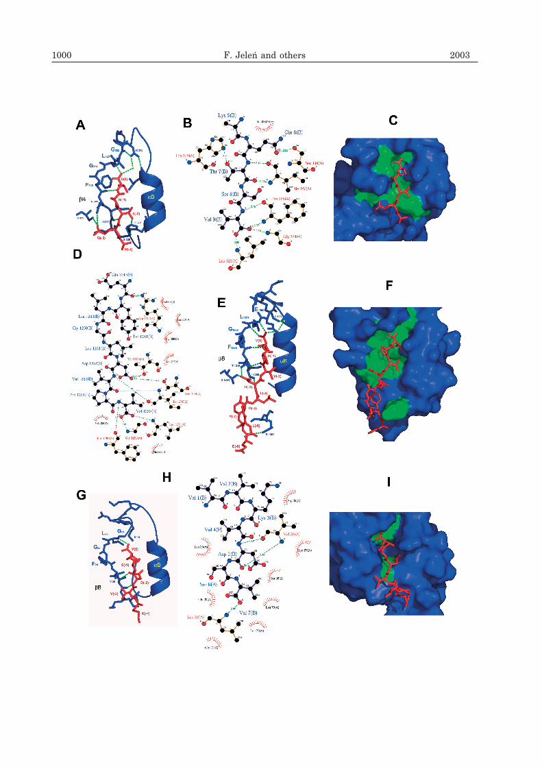
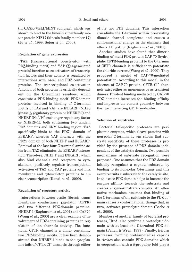
Figure 3. Ligand binding pockets of class I, II and III PDZ domains.
A. The third PDZ domain from the synaptic protein PSD95 in complex with a C-terminal peptide derived fromCRIPT (KQTSV). E. Erbin PDZ domain bound to the C-terminal tail of the ErbB2 receptor (EYLGLDVPV). G. Theneuronal nitric oxide synthase (nNOS) PDZ domain complexed with VVKVDSV. A, D and E. The hydrogen bond-ing in PDZ domains (blue ribbon representation) and peptide (red sticks) complexes is shown (green dashed lines).Water molecules are in yellow. C, F and I. Surface topology of PDZ domain bound to their peptides. The figure wasmade using program PyMOL (DeLano). B, D and H. Two-dimensional representation of the interaction of PDZ do-mains (orange) and their peptides (purple) was made using program LIGPLOT (Wallace et al., 1995) hydrogenbonds as dashed lines and hydrophobic interactions as arcs with radial spokes. Water molecules were not includedin this presentation.
Densin PDZ domain to C-terminus ofMAGUIN-1 (membrane-associated guanylatekinase-interacting protein 1) protein what is es-sential for assembly of PSD-95, MAGUIN-1,Densin ternary complex at the postsynapticmembrane of hippocampal neurons (Ohtakara etal., 2002). The Drosophila tumor suppressorScribble is a PDZ-containing protein belongingto the LAP family and required for maintainingepithelial cell polarity. At the larval neuro-muscular junction, Scribble colocalizes and indi-rectly interacts with another tumor suppressorand PDZ protein, Dlg. Scribble was identified asan essential regulator of synaptic architecture,plasticity and physiology (Roche et al., 2002).Shank family. The scaffold proteins,
Shank1, Shank2, and Shank3 (SH3 and multi-ple ankyrin repeat domains proteins) are mem-bers of the Shank family. These complex pro-teins (each about 2000 aa) possess several typesof binding modules such as (from the N to C-ter-minus) multiple ankyrin repeats, an SH3 do-main, a PDZ domain, a long proline-rich regionand a sterile alpha motif (SAM) domain. AllShank proteins are highly concentrated inpostsynaptic density of brain excitatory synap-ses (Boeckers et al., 1999; Lim et al., 1999;Naisbitt et al., 1999) where they play an impor-tant role in assembly of signaling complexes be-tween membrane and cytoplasmic proteins.The Shank PDZ domain was shown to bind theC-terminus of GKAP (guanylate kinase-asso-ciated protein) protein, that is also abundant inPSD of brain synapses (Boeckers et al., 1999;Naisbitt et al., 1999; Tu et al., 1999; Yao et al.,1999). Additionally, the C-termini of mGluRsand of SSTR2 (somatostatin receptor type 2)were reported to interact directly with theShank PDZ domain in yeast two-hybrid (Tu etal., 1999).
PDZ-containing proteins not classified tofamilies
Besides PDZ-containing proteins classifiedto the families and subfamilies, there aremany proteins possessing PDZ modules in a
unique arrangement with other signaling do-mains and which cannot be simply grouped.This situation demonstrates and confirms agigantic potential and spreading of PDZ do-mains among many types of existing pro-teins. Several interesting examples of suchproteins are briefly described below.The protein tyrosine phosphatase, PTP-BL,
localized to the submembranous region of epi-thelial cells is characterized by having theN-terminal FERM (4.1, ezrin, radixin,moesin) domain, five PDZ domains and theC-terminal catalytic phosphatase domain. ItsPDZ domains are involved in interactionswith several partners; in particular, PDZ2and PDZ4 interact with two LIM domain con-taining proteins, RIL (reversion-induced LIMprotein) and TRIP-6 (thyroid receptorinteracting protein 6) (Cuppen et al., 2000),which are found in actin-rich structures of thecell. In addition, PDZ1 can interact with BP75(bromodomain-containing protein) (Cuppenet al., 1999), PDZ2 with the tumor supressorprotein APC (adenomatous polyposis coli)(Erdmann et al., 2000) and PDZ3 with theRho effector kinase PRK2 (protein kinaseC-related kinase 2) (Gross et al., 2001). An-other interesting PDZ-containing protein,Delphilin, is the first reported protein thatcontains a single PDZ domain in combinationwith two forming homology (FH) domains.This unique protein has been reported to in-teracts with the GluR�2 C-terminus via itsPDZ domain (Miyagi et al., 2002). PDZ-Rho-GEF and LARG (leukemia-associated Rhoguanine-nucleotide exchange factor) proteins,essential for activation of biochemical path-ways specific to Rho-like GTPases also pos-sess a single N-terminal PDZ domain in theirmultidomain architecture. Recent studieshave shown the interaction between PDZ do-main of PDZ-RhoGEF and LARG and theC-terminus of B-plexins, suggesting B-plexin-mediated activation of Rho signaling (Swierczet al., 2002). Another signaling pathway, ex-tremely important for vertebrate and non-ver-tebrate embryogenesis, called the canonical
1002 F. Jeleń and others 2003
Wnt signal transduction cascade, also em-ploys the PDZ-containing protein, Dishev-elled, with a conserved arrangement of threedomains: DAX (domain present in Dishev-elled and axin), PDZ and DEP (Dishevelled,Egl-10, and pleckstrin). The PDZ domain ofDishevelled is necessary for its ability to in-duce nuclear accumulation of �-catenin(Kishida et al., 1999).
UNCONVENTIONAL FUNCTIONS OFPDZ-CONTAINING PROTEINS IN ALIVING CELL
Emphasizing a primary and critical role ofPDZ proteins in the organization of large sig-naling complexes at the plasma membrane,several additional functions of PDZ-contain-ing proteins were reported.
Protein targeting
Lin2, Lin7 and Lin10 are C. elegans proteinsrequired for the normal basolateral localiza-
tion of LET-23 receptor in vulval epithelialcells (Simske et al., 1996; Kim, 1997; Bredt,1998; Whitfield et al., 1999). On the otherhand, mammalian homologues of these pro-teins (mLin-2/CASK/PALS, mLin-7/VELI/MALS and mLin-10/MINT/X11) are mainlylocalized to neuronal cells where are responsi-ble for trafficking of the NMDA receptors tocell membrane. According to the proposedmechanism of this targeting, Lin-2/CASK,Lin-7/VELI and Lin-10/MINT have ability toform a ternary complex (Borg et al., 1998;Butz et al., 1998; Kaech et al., 1998). In caseof C. elegans homologue in epithelial cells,both Lin-7 and Lin-10 bind to the Lin-2 pro-tein (MAGUK protein) through thenon-PDZ-mediated fashion and the PDZ do-main of Lin-7 binds directly to the C-terminusof the LET-23 (lethal) receptor. In mamma-lian neurons, PDZ domain of VELI proteinfrom the CASK/VELI/MINT complex, bindsto the C-terminus of NMDA receptor subunitNR2B. Targeting of NMDA receptor toplasma membrane along microtubules is de-pendent on the PDZ domain of MINT protein
Vol. 50 PDZ domains — common players in the cell signaling 1003
Table 2. PDZ containing proteins and their interaction partners.
In case of proteins containing additional domains, only interactions involving PDZ domains are included. Dig-its in the brackets indicate the number of PDZ domains.
(in CASK/VELI/MINT complex), which wasshown to bind to the kinesin superfamily mo-tor protein KIF17 (kinesin family member 17)(Jo et al., 1999; Setou et al., 2000).
Regulation of gene expression
TAZ (transcriptional co-activator withPDZ-binding motif) and YAP (Yes-associatedprotein) function as co-activators of transcrip-tion factors and their activity is regulated byinteractions with 14-3-3 and PDZ containingproteins. The transcriptional co-activationfunction of both proteins is critically depend-ent on the C-terminal residues, whichconsitute a PDZ binding motif. PDZ-domainproteins involved in binding of C-terminalmotifs of TAZ and YAP are E3KARP (NHE3kinase A regulatory protein or NHERF-2) andNHERF (Na+/H+ exchanger regulatory factoror NHERF-1), both containing two tandemPDZ domains and ERM binding region. TAZspecifically binds to the PDZ1 domain ofE3KARP, whereas YAP interacts with thePDZ2 domain of both NHERF and E3KARP.Removal of the last four C-terminal amino ac-ids from TAZ eliminates the E3KARP interac-tion. Therefore, NHERF and E3KARP, whichalso bind channels and receptors to cyto-skeleton, positively regulate transcriptionalactivation of TAZ and YAP proteins and linkmembrane and cytoskeleton proteins to nu-clear transcription (Kanai et al., 2000).
Regulation of receptors activity
Interactions between cystic fibrosis trans-membrane conductance regulator (CFTR)and two different PDZ-domain proteins:NHERF-1 (Raghuram et al., 2001) and CAP70(Wang et al., 2000) are a clear example of in-volvement of PDZ-containing proteins in reg-ulation of ion channels activity. The func-tional CFTR channel is a dimer containingtwo PDZ-binding motifs. It has been demon-strated that NHERF-1 binds to the cytoplas-mic tails of CFTR Cl– channels through either
of its two PDZ domains. This interactioncross-links the C-termini within pre-existingdimeric channel complexes and causes aconformational change in the channels thataffects Cl– gating (Raghuram et al., 2001).Another studies have found that dimeric
binding of multi-PDZ protein CAP-70 (hydro-philic CFTR-binding protein) to the C-terminiof CFTR channels is sufficient to potentiatethe chloride current (Wang et al., 2000). Theyproposed a model of CAP-70-mediatedpotentiation. According to this model, in theabsence of CAP-70 protein, CFTR Cl– chan-nels exist either as monomers or as transientdimers. Bivalent binding mediated by CAP-70PDZ domains increases the binding affinityand improves the contact geometry betweenthe two interacting CFTR molecules.
Selection of substrates
Bacterial tail-specific proteases are peri-plasmic enzymes, which cleave proteins withnon-polar C-termini. It was shown that sub-strate specificity of these proteases is pro-vided by the presence of PDZ domain inde-pendent of the catalytic domain. Two possiblemechanisms of substrate recognition wereproposed. One assumes that the PDZ domaininitially recognizes a cognate substrate bybinding to its non-polar C-terminus and thisevent recruits a substrate to the catalytic site.In this case PDZ domain helps to increase theenzyme affinity towards the substrate andcreates enzyme-substrate complex. An alter-native mechanism assumes that binding ofthe C-terminus of the substrate to the PDZ do-main causes a conformational change that, inturn, activates proteolytic domain (Beebe etal., 2000).Members of another family of bacterial pro-
teases, HtrA, also combine a proteolytic do-main with at least one C-terminal PDZ do-main (Pallen & Wren, 1997). Finally, tricornproteases forming proteasome-like capsidsin Archea also contain PDZ domains whichin cooperation with a �-propeller fold play a
1004 F. Jeleń and others 2003
role in substrate selection (Pallen et al.,2001).
EFFECTS OF PDZ DOMAINMALFUNCTIONS
Malfunction of many PDZ domain-contain-ing proteins is implicated in a variety ofpathophysiological phenomena, includingcancer. Analysis of p55 MAGUK proteinmRNA from patients with acute mega-caryoblastic CML revealed a 69 base pair de-letion in the PDZ domain. This observation isthe first abnormality of a PDZ domain linkedto a human disease. Mutations in a gene en-coding harmonin cause Usher syndrome type1C, an autosomal recessive disorder charac-terized by congenital sensorineural deafness,vestibular dysfunction and blindness(Bitner-Glindzicz et al., 2000; Montell, 2000;Verpy et al., 2000). PDZ1 and PDZ2 domainsof harmonin interact with two complemen-tary binding surfaces of the Cadherin 23(CDH23) cytoplasmic domain. Interaction ofPDZ1 with CDH23 is perturbed by the inser-tion of 35 amino acids within CDH23 (Sie-mens et al., 2002). Mutations in Periaxin genecause Dejerine-Sottas neuropathy, a severedemyelinating form of peripheral neuropathy(Boerkoel et al., 2001; Sherman et al., 2001).In flies, mutations in the gene encoding
INAD, a protein composed solely of PDZ do-mains, disrupt the photoinduction cascade re-sulting in the light-dependent retinal degener-ation (Shieh & Zhu, 1996). Mutations in PDZdomain-containing protein result in sub-cellular mislocalization of the LET-23 proteinand the lack of vulval differentiation. TheLAP proteins are recently described family ofscaffold proteins that are involved in the for-mation of membrane complexes and themaintenance of epithelial and neuronal cellshape and polarity (Bryant & Huwe, 2000).For example, in Drosophila mutation of theScribble LAP protein (16 leucine rich repeats
and four PDZ domains) results in loss of epi-thelial cell polarity and morphology as well asuncontrolled, tumor-like growth (Bilder &Perrimon, 2000). Moreover, disruption ofScribble gene (Scrb1) causes severe neuraltube defects (termed craniorachischisis) inthe circletail mouse. In this disorder, almostthe entire brain and spinal cord are affected,owing to a failure to initiate neural tube clo-sure. It was found, that the Scrb1 gene mu-tated in circletail (Crc) contains a single baseinsertion that creates a frame shift and leadsto a premature termination of the Scrb1 pro-tein. Scrb1 may control the subcellular local-ization of the Vangl2 protein alternativelyScrb1 and Vangl2 may form a part of a pro-tein complex, perhaps through a direct inter-action of the C-terminal PDZ-binding motif ofVangl2 with the PDZ domains of Scrb1(Murdoch et al., 2003).Syntenin was originally discovered as a pro-
tein containing a tandem of PDZ domains andinteracting with transmembrane proteo-glycans called syndecans (Grootjans et al.,1997). Syntenin was subsequently shown tobind class B ephrins, proTGF-�, neurofascin,schwannomin (also known as merlin), IL5 re-ceptor � (ILR5�) and various glutamate re-ceptor subtypes. Very recently, it was discov-ered that syntenin is overexpressed and pro-motes cell migration in metastatic humanbreast and gastric cancer cell lines (Koo et al.,2002). Expression analysis shows that level ofsyntenin correlated well with invasive andmetastatic potential in these cell lines. Fur-thermore, syntenin-trasfected cells migratedmore actively, and showed numerous cell sur-face extensions, suggesting that syntenin isactive upstream of pathways affecting actincytoskeleton (Koo et al., 2002). There is someexperimental evidence that PDZ domains con-stitute for good drug targets. Fas (APO-1/CD95), a member of the tumor necrosis factorreceptor superfamily and a cell surface recep-tor, which induces apoptosis, interacts withthe PDZ domain of the Fas-associated phos-phatase-1 (FAP-1). Direct cytoplasmic micro-
Vol. 50 PDZ domains — common players in the cell signaling 1005
injection of a tripeptide (Ac-SLV) correspond-ing to the C-terminal fragment of Fas, re-sulted in apoptosis in a colon cancer cell linethat expresses both Fas and FAP-1 (Yana-gisawa et al., 1997). It is therefore possiblethat other PDZ-mediated pathways may beequally sensitive to selective inhibitors.PDZ domains are involved in tumorigenesis,
cell migration and metastasis. Among highlyexpressed proteins in the human primaryprostate tumors is AIPC (activated inprostate cancer), a protein containing sixPDZ domains (Chaib et al., 2001). It is possi-ble, that disrupting the pathways mediated bythese domains might inhibit early promotionof prostate tumorigenesis. In colon, breast,liver, lung, pancreas, stomach, and prostatetumors, a protein containing PDZ and LIMdomains, denoted PCD1, was significantlyoverexpressed, in contrast to normal tissues(Kang et al., 2000). It has been suggested thatit participates in cytoskeletal reorganizationin cancer, and that it could be a target fordrug design.
CONCLUDING REMARKS
PDZ domains are ubiquitous element of cy-toplasmic proteins in organisms from bacte-ria to mammals. Due to a common multiplecopy occurrence within a single protein theymediate formation of extensive protein–pro-tein networks. Diversity and size of such pro-tein complexes is further enhanced by combi-nation of PDZ domains with other protein in-teraction modules (SH3, PTB, LIM, WW, andankiryn repeats). Among major cellular tar-gets of PDZ domains are proteins associateddirectly with the plasma membrane like ionchannels, receptors and cytoskeleton proteinsThe structural basis of their specificity tobind four to six C-terminal residues of theseproteins appears relatively simple and sug-gests redundancy of recognized target se-quences. However, since PDZ domains can
also bind other PDZ domains in a head-to-tailfashion, recognize internal structural motifsin their target proteins or bindphosphatidylinositol derivatives, it is likelythat diversity of their cellular interactions ismuch broader. Significant problems can beexpected in deciphering cellular function andregulation of PDZ containing proteins sincecurrently the technology to study in vivotransient multidomain protein complexes isnot developed.
R E F E R E N C E S
Adamski FM, Zhu MY, Bahiraei F, Shieh BH.(1998) Interaction of eye protein kinase Cand INAD in Drosophila. Localization ofbinding domains and electrophysiologicalcharacterization of a loss of association intransgenic flies. J Biol Chem.; 273: 17713–9.
Anzai N, Deval E, Schaefer L, Friend V,Lazdunski M, Lingueglia E. (2002) Themultivalent PDZ domain-containing proteinCIPP is a partner of acid-sensing ion channel3 in sensory neurons. J Biol Chem.; 277:16655–61.
Apperson ML, Moon IS, Kennedy MB. (1996)Characterization of densin-180, a newbrain-specific synaptic protein of theO-sialoglycoprotein family. J Neurosci.; 16:6839–52.
Baron A, Deval E, Salinas M, Lingueglia E,Voilley N, Lazdunski M. (2002) Proteinkinase C stimulates the acid-sensing ionchannel ASIC2a via the PDZ domain-contain-ing protein PICK1. J Biol Chem.; 277:50463–8.
Barritt DS, Pearn MT, Zisch AH, Lee SS, JavierRT, Pasquale EB, Stallcup WB. (2000) Themulti-PDZ domain protein MUPP1 is a cyto-plasmic ligand for the membrane-spanningproteoglycan NG2. J Cell Biochem.; 79:213–24.
Becamel C, Alonso G, Galeotti N, Demey E,Jouin P, Ullmer C, Dumuis A, Bockaert J,
1006 F. Jeleń and others 2003
Marin P. (2002) Synaptic multiprotein com-plexes associated with 5-HT(2C) receptors: aproteomic approach. EMBO J.; 21: 2332–42.
Beebe KD, Shin J, Peng J, Chaudhury C, KheraJ, Pei D. (2000) Substrate recognitionthrough a PDZ domain in tail-specific prote-ase. Biochemistry.; 39: 3149–55.
Bezprozvanny I, Maximov A. (2001) Classifica-tion of PDZ domains. FEBS Lett.; 509:457–62.
Bilder D, Perrimon N. (2000) Localization ofapical epithelial determinants by thebasolateral PDZ protein Scribble. Nature.;403: 676–80.
Birrane G, Chung J, Ladias JA. (2003) Novelmode of ligand recognition by the Erbin PDZdomain. J Biol Chem.; 278: 1399–402.
Bitner-Glindzicz M, Lindley KJ, Rutland P,Blaydon D, Smith VV, Milla PJ, Hussain K,Furth-Lavi J, Cosgrove KE, Shepherd RM,Barnes PD, O’Brien RE, Farndon PA,Sowden J, Liu XZ, Scanlan MJ, Malcolm S,Dunne MJ, Aynsley-Green A, Glaser B.(2000) A recessive contiguous gene deletioncausing infantile hyperinsulinism,enteropathy and deafness identifies theUsher type 1C gene. Nat Genet.; 26: 56–60.
Boeckers TM, Kreutz MR, Winter C,Zuschratter W, Smalla KH, Sanmarti-Vila L,Wex H, Langnaese K, Bockmann J, GarnerCC, Gundelfinger ED. (1999) Proline-richsynapse-associated protein-1/cortactin bind-ing protein 1 (ProSAP1/CortBP1) is aPDZ-domain protein highly enriched in thepostsynaptic density. J Neurosci.; 19:6506–18.
Boerkoel CF, Takashima H, Stankiewicz P, Gar-cia CA, Leber SM, Rhee-Morris L, LupskiJR. (2001) Periaxin mutations cause reces-sive Dejerine-Sottas neuropathy. Am J HumGenet.; 68: 325–33.
Borg JP, Straight SW, Kaech SM, deTaddeo-Borg M, Kroon DE, Karnak D,Turner RS, Kim SK, Margolis B. (1998)Identification of an evolutionarily conservedheterotrimeric protein complex involved inprotein targeting. J Biol Chem.; 273:31633–6.
Borg JP, Marchetto S, Le Bivic A, Ollendorff V,Jaulin-Bastard F, Saito H, Fournier E,Adelaide J, Margolis B, Birnbaum D. (2000)ERBIN: a basolateral PDZ protein that inter-acts with the mammalian ERBB2/HER2 re-ceptor. Nat Cell Biol.; 2: 407–14.
Braithwaite SP, Xia H, Malenka RC. (2002) Dif-ferential roles for NSF and GRIP/ABP inAMPA receptor cycling. Proc Natl Acad SciU S A.; 99: 7096–101.
Brakeman PR, Lanahan AA, O’Brien R, RocheK, Barnes CA, Huganir RL, Worley PF.(1997) Homer: a protein that selectivelybinds metabotropic glutamate receptors. Na-ture.; 386: 284–8.
Bredt DS. (1998) Sorting out genes that regu-late epithelial and neuronal polarity. Cell.;94: 691–4.
Brenman JE, Chao DS, Xia H, Aldape K, BredtDS. (1995) Nitric oxide synthase complexedwith dystrophin and absent from skeletalmuscle sarcolemma in Duchenne musculardystrophy. Cell.; 82: 743–52.
Bruckner K, Pablo Labrador J, Scheiffele P,Herb A, Seeburg PH, Klein R. (1999)EphrinB ligands recruit GRIP family PDZadaptor proteins into raft membranemicrodomains. Neuron.; 22: 511–24.
Bryant PJ, Huwe A. (2000) LAP proteins:what’s up with epithelia? Nat Cell Biol.; 2:E141–3.
Butz S, Okamoto M, Sudhof TC. (1998) A tri-partite protein complex with the potential tocouple synaptic vesicle exocytosis to cell ad-hesion in brain. Cell.; 94: 773–82.
Cao TT, Deacon HW, Reczek D, Bretscher A,von Zastrow M. (1999) A kinase-regulatedPDZ-domain interaction controls endocyticsorting of the beta2-adrenergic receptor. Na-ture.; 401: 286–90.
Chaib H, Rubin MA, Mucci NR, Li L, TaylorJMG, Day ML, Rhim JS, Macoska JA. (2001)Activated in prostate cancer: a PDZ do-main-containing protein highly expressed inhuman primary prostate tumors. CancerRes.; 61: 2390–4.
Vol. 50 PDZ domains — common players in the cell signaling 1007
Chetkovich DM, Bunn RC, Kuo SH, KawasakiY, Kohwi M, Bredt DS. (2002) Postsynaptictargeting of alternative postsynaptic den-sity-95 isoforms by distinct mechanisms. JNeurosci.; 22: 6415–25.
Christopherson KS, Hillier BJ, Lim WA, BredtDS. (1999) PSD-95 assembles a ternary com-plex with the N-methyl-D-aspartic acid recep-tor and a bivalent neuronal NO synthasePDZ domain. J Biol Chem.; 274: 27467–73.
Cohen NA, Brenman JE, Snyder SH, Bredt DS.(1996) Binding of the inward rectifier K+
channel Kir 2.3 to PSD-95 is regulated byprotein kinase A phosphorylation. Neuron.;17: 759–67.
Cohen AR, Woods DF, Marfatia SM, Walther Z,Chishti AH, Anderson JM, Wood DF. (1998)Human CASK/LIN-2 binds syndecan-2 andprotein 4.1 and localizes to the basolateralmembrane of epithelial cells. J Cell Biol.;142: 129–38.
Cuppen E, van Ham M, Pepers B, Wieringa B,Hendriks W. (1999) Identification and molec-ular characterization of BP75, a novelbromodomain-containing protein. FEBS Lett.;459: 291–8.
Cuppen E, van Ham M, Wansink DG, de LeeuwA, Wieringa B, Hendriks W. (2000) Thezyxin-related protein TRIP6 interacts withPDZ motifs in the adaptor protein RIL andthe protein tyrosine phosphatase PTP-BL.Eur J Cell Biol.; 79: 283–93.
Daniels DL, Cohen AR, Anderson JM, BrungerAT. (1998) Crystal structure of the hCASKPDZ domain reveals the structural basis ofclass II PDZ domain target recognition. NatStruct Biol.; 5: 317–25.
Daw MI, Chittajallu R, Bortolotto ZA, Dev KK,Duprat F, Henley JM, Collingridge GL, IsaacJT. (2000) PDZ proteins interacting withC-terminal GluR2/3 are involved in a PKC-dependent regulation of AMPA receptors athippocampal synapses. Neuron.; 28: 873–86.
DeLano WL. “The PyMOL Molecular GraphicsSystem.” DeLano Scientific LLC, San Carlos,CA, U S A. http://www.pymol.org
Dev KK, Nishimune A, Henley JM, Nakanishi S.(1999) The protein kinase C alpha bindingprotein PICK1 interacts with short but notlong form alternative splice variants ofAMPA receptor subunits.Neuropharmacology.; 38: 635–44.
Dong H, O’Brien RJ, Fung ET, Lanahan AA,Worley PF, Huganir RL. (1997) GRIP: a syn-aptic PDZ domain-containing protein that in-teracts with AMPA receptors. Nature.; 386:279–84.
Dong H, Zhang P, Song I, Petralia RS, Liao D,Huganir RL. (1999) Characterization of theglutamate receptor-interacting proteinsGRIP1 and GRIP2. J Neurosci.; 19:6930–41.
Doyle DA, Lee A, Lewis J, Kim E, Sheng M,MacKinnon R. (1996) Crystal structures of acomplexed and peptide-free membrane pro-tein-binding domain: molecular basis of pep-tide recognition by PDZ. Cell.; 85: 1067–76.
Eck MJ, Dhe-Paganon S, Trub T, Nolte RT,Shoelson SE. (1996) Structure of the IRS-1PTB domain bound to the juxtamembraneregion of the insulin receptor. Cell.; 85:695–705.
Ellis RJ. (2001) Macromolecular crowding: animportant but neglected aspect of theintracellular environment. Curr Opin StructBiol.; 11: 114–9.
Erdmann KS, Kuhlmann J, Lessmann V,Herrmann L, Eulenburg V, Muller O,Heumann R. (2000) The adenomatouspolyposis coli-protein (APC) interacts withthe protein tyrosine phosphatase PTP-BL viaan alternatively spliced PDZ domain. Onco-gene.; 19: 3894–901.
Fallon L, Moreau F, Croft BG, Labib N, Gu WJ,Fon EA. (2002) Parkin and CASK/LIN-2 as-sociate via a PDZ-mediated interaction andare co-localized in lipid rafts andpostsynaptic densities in brain. J Biol Chem.;277: 486–91.
Fanning AS, Anderson JM. (1999) PDZ do-mains: fundamental building blocks in theorganization of protein complexes at theplasma membrane. J Clin Invest.; 103:767–72.
1008 F. Jeleń and others 2003
Fuh G, Pisabarro MT, Li Y, Quan C, Lasky LA,Sidhu SS. (2000) Analysis of PDZ do-main-ligand interactions using carboxyl-termi-nal phage display. J Biol Chem.; 275:21486–91.
Garcia RA, Vasudevan K, Buonanno A. (2000)The neuregulin receptor ErbB-4 interactswith PDZ-containing proteins at neuronalsynapses. Proc Natl Acad Sci U S A.; 97:3596–601.
Garrard SM, Capaldo CT, Gao L, Rosen MK,Macara IG, Tomchick DR. (2003) Structureof Cdc42 in a complex with the GTPase-bind-ing domain of the cell polarity protein, Par6.EMBO J.; 22: 1125–33.
Gee SH, Quenneville S, Lombardo CR, ChabotJ. (2000) Single-amino acid substitutions al-ter the specificity and affinity of PDZ do-mains for their ligands. Biochemistry.; 39:14638–46.
Grootjans JJ, Zimmermann P, Reekmans G,Smets A, Degeest G, Durr J, David G. (1997)Syntenin, a PDZ protein that binds syndecancytoplasmic domains. Proc Natl Acad SciU S A.; 94: 13683–8.
Grootjans JJ, Reekmans G, Ceulemans H, Da-vid G. (2000) Syntenin-syndecan binding re-quires syndecan-synteny and the co-operationof both PDZ domains of syntenin. J BiolChem.; 275: 19933–41.
Gross C, Heumann R, Erdmann KS. (2001) Theprotein kinase C-related kinase PRK2 inter-acts with the protein tyrosine phosphatasePTP-BL via a novel PDZ domain binding mo-tif. FEBS Lett.; 496: 101–4.
Guy PM, Kenny DA, Gill GN. (1999) The PDZdomain of the LIM protein enigma binds tobeta-tropomyosin. Mol Biol Cell.; 10:1973–84.
Hall RA, Premont RT, Chow CW, Blitzer JT,Pitcher JA, Claing A, Stoffel RH, Barak LS,Shenolikar S, Weinman EJ, Grinstein S,Lefkowitz RJ. (1998) The beta2-adrenergicreceptor interacts with theNa+/H+-exchanger regulatory factor to con-trol Na+/H+ exchange. Nature.; 392:626–30.
Hall RA, Spurney RF, Premont RT, Rahman N,Blitzer JT, Pitcher JA, Lefkowitz RJ. (1999)G protein-coupled receptor kinase 6A phos-phorylates the Na+/H+ exchanger regulatoryfactor via a PDZ domain-mediated interac-tion. J Biol Chem.; 274: 24328–34.
Hamazaki Y, Itoh M, Sasaki H, Furuse M,Tsukita S. (2002) Multi-PDZ domain protein1 (MUPP1) is concentrated at tight junctionsthrough its possible interaction withclaudin-1 and junctional adhesion molecule. JBiol Chem.; 277: 455–61.
Harris BZ, Lim WA. (2001) Mechanism and roleof PDZ domains in signaling complex assem-bly. J Cell Sci.; 114: 3219–31.
Harris BZ, Hillier BJ, Lim WA. (2001) Ener-getic determinants of internal motif recogni-tion by PDZ domains. Biochemistry.; 40:5921–30.
Harris BZ, Lau FW, Fujii N, Guy RK, Lim WA.(2003) Role of electrostatic interactions inPDZ domain ligand recognition. Biochemis-try.; 42: 2797–805.
Harrison SC. (1996) Peptide-surface association:the case of PDZ and PTB domains. Cell.; 86:341–3.
Hata Y, Butz S, Sudhof TC. (1996) CASK: anovel dlg/PSD95 homolog with an N-termi-nal calmodulin-dependent protein kinase do-main identified by interaction withneurexins. J Neurosci.; 16: 2488–94.
Hegedus T, Sessler T, Scott R, Thelin W, BakosE, Varadi A, Szabo K, Homolya L, MilgramSL, Sarkadi B. (2003) C-terminalphosphorylation of MRP2 modulates its in-teraction with PDZ proteins. BiochemBiophys Res Commun.; 302: 454–61.
Hillier BJ, Christopherson KS, Prehoda KE,Bredt DS, Lim WA. (1999) Unexpectedmodes of PDZ domain scaffolding revealedby structure of nNOS-syntrophin complex.Science.; 284: 812–5.
Hirbec H, Perestenko O, Nishimune A, Meyer G,Nakanishi S, Henley JM, Dev KK. (2002)The PDZ proteins PICK1, GRIP, andsyntenin bind multiple glutamate receptor
Vol. 50 PDZ domains — common players in the cell signaling 1009
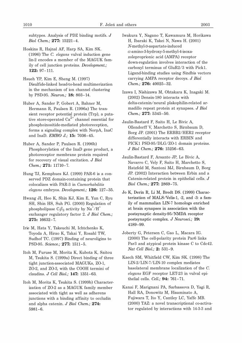
subtypes. Analysis of PDZ binding motifs. JBiol Chem.; 277: 15221–4.
Hoskins R, Hajnal AF, Harp SA, Kim SK.(1996) The C. elegans vulval induction genelin-2 encodes a member of the MAGUK fam-ily of cell junction proteins. Development.;122: 97–111.
Hsueh YP, Kim E, Sheng M. (1997)Disulfide-linked head-to-head multimerizationin the mechanism of ion channel clusteringby PSD-95. Neuron.; 18: 803–14.
Huber A, Sander P, Gobert A, Bahner M,Hermann R, Paulsen R. (1996a) The tran-sient receptor potential protein (Trp), a puta-tive store-operated Ca2+ channel essential forphosphoinositide-mediated photoreception,forms a signaling complex with NorpA, InaCand InaD. EMBO J.; 15: 7036–45.
Huber A, Sander P, Paulsen R. (1996b)Phosphorylation of the InaD gene product, aphotoreceptor membrane protein requiredfor recovery of visual excitation. J BiolChem.; 271: 11710–7.
Hung TJ, Kemphues KJ. (1999) PAR-6 is a con-served PDZ domain-containing protein thatcolocalizes with PAR-3 in Caenorhabditiselegans embryos. Development.; 126: 127–35.
Hwang JI, Heo K, Shin KJ, Kim E, Yun C, RyuSH, Shin HS, Suh PG. (2000) Regulation ofphospholipase C-�3 activity by Na+/H+
exchanger regulatory factor 2. J Biol Chem.;275: 16632–7.
Irie M, Hata Y, Takeuchi M, Ichtchenko K,Toyoda A, Hirao K, Takai Y, Rosahl TW,Sudhof TC. (1997) Binding of neuroligins toPSD-95. Science.; 277: 1511–5.
Itoh M, Furuse M, Morita K, Kubota K, SaitouM, Tsukita S. (1999a) Direct binding of threetight junction-associated MAGUKs, ZO-1,ZO-2, and ZO-3, with the COOH termini ofclaudins. J Cell Biol.; 147: 1351–63.
Itoh M, Morita K, Tsukita S. (1999b) Character-ization of ZO-2 as a MAGUK family memberassociated with tight as well as adherensjunctions with a binding affinity to occludinand alpha catenin. J Biol Chem.; 274:5981–6.
Iwakura Y, Nagano T, Kawamura M, HorikawaH, Ibaraki K, Takei N, Nawa H. (2001)N-methyl-D-aspartate-induced�-amino-3-hydroxy-5-methyl-4-isoxa-zoleproprionic acid (AMPA) receptordown-regulation involves interaction of thecarboxyl terminus of GluR2/3 with Pick1.Ligand-binding studies using Sindbis vectorscarrying AMPA receptor decoys. J BiolChem.; 276: 40025–32.
Izawa I, Nishizawa M, Ohtakara K, Inagaki M.(2002) Densin-180 interacts withdelta-catenin/neural plakophilin-related ar-madillo repeat protein at synapses. J BiolChem.; 277: 5345–50.
Jaulin-Bastard F, Saito H, Le Bivic A,Ollendorff V, Marchetto S, Birnbaum D,Borg JP. (2001) The ERBB2/HER2 receptordifferentially interacts with ERBIN andPICK1 PSD-95/DLG/ZO-1 domain proteins.J Biol Chem.; 276: 15256–63.
Jaulin-Bastard F, Arsanto JP, Le Bivic A,Navarro C, Vely F, Saito H, Marchetto S,Hatzfeld M, Santoni MJ, Birnbaum D, BorgJP. (2002) Interaction between Erbin and aCatenin-related protein in epithelial cells. JBiol Chem.; 277: 2869–75.
Jo K, Derin R, Li M, Bredt DS. (1999) Charac-terization of MALS/Velis-1, -2, and -3: a fam-ily of mammalian LIN-7 homologs enrichedat brain synapses in association with thepostsynaptic density-95/NMDA receptorpostsynaptic complex. J Neurosci.; 19:4189–99.
Joberty G, Petersen C, Gao L, Macara IG.(2000) The cell-polarity protein Par6 linksPar3 and atypical protein kinase C to Cdc42.Nat Cell Biol.; 2: 531–9.
Kaech SM, Whitfield CW, Kim SK. (1998) TheLIN-2/LIN-7/LIN-10 complex mediatesbasolateral membrane localization of the C.elegans EGF receptor LET-23 in vulval epi-thelial cells. Cell.; 94: 761–71.
Kanai F, Marignani PA, Sarbassova D, Yagi R,Hall RA, Donowitz M, Hisaminato A,Fujiwara T, Ito Y, Cantley LC, Yaffe MB.(2000) TAZ: a novel transcriptional co-activa-tor regulated by interactions with 14-3-3 and
1010 F. Jeleń and others 2003
PDZ domain proteins. EMBO J.; 19:6778–91.
Kang S, Xu H, Duan X, Liu JJ, He Z, Yu F,Zhou S, Meng XQ, Cao M, Kennedy GC.(2000) PCD1, a novel gene containing PDZand LIM domains, is overexpressed in sev-eral human cancers. Cancer Res.; 60:5296–302.
Kang BS, Cooper DR, Jelen F, Devedjiev Y,Derewenda U, Dauter Z, Otlewski J,Derewenda ZS. (2003) PDZ tandem of hu-man syntenin. Crystal structure and func-tional properties. Structure (Camb).; 11:459–68.
Karthikeyan S, Leung T, Ladias JA. (2001)Structural basis of the Na+/H+ exchangerregulatory factor PDZ1 interaction with thecarboxyl-terminal region of the cystic fibrosistransmembrane conductance regulator. JBiol Chem.; 276: 19683–6.
Kavanaugh WM, Williams LT. (1994) An alter-native to SH2 domains for binding tyro-sine-phosphorylated proteins. Science.; 266:1862–5.
Kennedy MB. (1995) Origin of PDZ (DHR,GLGF) domains. Trends Biochem Sci.; 20:350.
Kim SK. (1997) Polarized signaling: basolateralreceptor localization in epithelial cells byPDZ-containing proteins. Curr Opin CellBiol.; 9: 853–9.
Kim CH, Chung HJ, Lee HK, Huganir RL.(2001) Interaction of the AMPA receptor sub-unit GluR2/3 with PDZ domains regulateshippocampal long-term depression. Proc NatlAcad Sci U S A.; 98: 11725–30.
Kim E, Niethammer M, Rothschild A, Jan YN,Sheng M. (1995) Clustering of Shaker-typeK+ channels by interaction with a family ofmembrane-associated guanylate kinases. Na-ture.; 378: 85–8.
Kimple ME, Siderovski DP, Sondek J. (2001)Functional relevance of the disulfide-linkedcomplex of the N-terminal PDZ domain ofInaD with NorpA. EMBO J.; 20: 4414–22.
Kishida S, Yamamoto H, Hino S, Ikeda S,Kishida M, Kikuchi A. (1999) DIX domains
of Dvl and axin are necessary for protein in-teractions and their ability to regulatebeta-catenin stability. Mol Cell Biol.; 19:4414–22.
Klebes A, Knust E. (2000) A conserved motif inCrumbs is required for E-cadherin localisa-tion and zonula adherens formation inDrosophila. Curr Biol.; 10: 76–85.
Koo TH, Lee JJ, Kim EM, Kim KW, Kim HD,Lee JH. (2002) Syntenin is overexpressedand promotes cell migration in metastatichuman breast and gastric cancer cell lines.Oncogene.; 21: 4080–8.
Kornau HC, Schenker LT, Kennedy MB,Seeburg PH. (1995) Domain interaction be-tween NMDA receptor subunits and thepostsynaptic density protein PSD-95. Sci-ence.; 269: 1737–40.
Koroll M, Rathjen FG, Volkmer H. (2001) Theneural cell recognition molecule neurofascininteracts with syntenin-1 but not withsyntenin-2, both of which reveal self-associat-ing activity. J Biol Chem.; 276: 10646–54.
Kozlov G, Gehring K, Ekiel I. (2000) Solutionstructure of the PDZ2 domain from humanphosphatase hPTP1E and its interactionswith C-terminal peptides from the Fas recep-tor. Biochemistry.; 39: 2572–80.
Kozlov G, Banville D, Gehring K, Ekiel I. (2002)Solution structure of the PDZ2 domain fromcytosolic human phosphatase hPTP1Ecomplexed with a peptide reveals contribu-tion of the beta2-beta3 loop to PDZ do-main-ligand interactions. J Mol Biol.; 320:813–20.
Kurakin A, Bredesen D. (2002) Target-assistediterative screening reveals novel interactorsfor PSD95, Nedd4, Src, Abl and Crk pro-teins. J Biomol Struct Dyn.; 19: 1015–29.
Kurschner C, Mermelstein PG, Holden WT,Surmeier DJ. (1998) CIPP, a novelmultivalent PDZ domain protein, selectivelyinteracts with Kir4.0 family members,NMDA receptor subunits, neurexins, andneuroligins. Mol Cell Neurosci.; 11: 161–72.
Laura RP, Witt AS, Held HA, Gerstner R,Deshayes K, Koehler MF, Kosik KS, Sidhu
Vol. 50 PDZ domains — common players in the cell signaling 1011
SS, Lasky LA. (2002) The Erbin PDZ domainbinds with high affinity and specificity tothe carboxyl termini of delta-catenin andARVCF. J Biol Chem.; 277: 12906–14.
Lee SS, Glaunsinger B, Mantovani F, Banks L,Javier RT. (2000) Multi-PDZ domain proteinMUPP1 is a cellular target for bothadenovirus E4-ORF1 and high-riskpapillomavirus type 18 E6 oncoproteins. JVirol.; 74: 9680–93.
Lemmers C, Medina E, Delgrossi MH, MichelD, Arsanto JP, Le Bivic A. (2002)hINADl/PATJ, a homolog of discs lost, inter-acts with crumbs and localizes to tight junc-tions in human epithelial cells. J Biol Chem.;277: 25408–15.
Liao DI, Qian J, Chisholm DA, Jordan DB,Diner BA. (2000) Crystal structures of thephotosystem II D1 C-terminal processingprotease. Nat Struct Biol.; 7: 749–53.
Lim S, Naisbitt S, Yoon J, Hwang JI, Suh PG,Sheng M, Kim E. (1999) Characterization ofthe Shank family of synaptic proteins. Multi-ple genes, alternative splicing, and differen-tial expression in brain and development. JBiol Chem.; 274: 29510–8.
Lin D, Gish GD, Songyang Z, Pawson T. (1999)The carboxyl terminus of B class ephrinsconstitutes a PDZ domain binding motif. JBiol Chem.; 274: 3726–33.
Lin WJ, Chang YF, Wang WL, Huang CY.(2001) Mitogen-stimulated TIS21 protein in-teracts with a protein-kinase-C�-binding pro-tein rPICK1. Biochem J.; 354: 635–43.
Lipinska B, Fayet O, Baird L, Georgopoulos C.(1989) Identification, characterization, andmapping of the Escherichia coli htrA gene,whose product is essential for bacterialgrowth only at elevated temperatures. JBacteriol.; 171: 1574–84.
Mancini A, Koch A, Stefan M, Niemann H,Tamura T. (2000) The direct association ofthe multiple PDZ domain containing proteins(MUPP-1) with the human c-Kit C-terminusis regulated by tyrosine kinase activity.FEBS Lett.; 482: 54–8.
Marfatia SM, Morais-Cabral JH, Kim AC, ByronO, Chishti AH. (1997) The PDZ domain ofhuman erythrocyte p55 mediates its bindingto the cytoplasmic carboxyl terminus ofglycophorin C. Analysis of the binding inter-face by in vitro mutagenesis. J Biol Chem.;272: 24191–7.
Martinez-Estrada OM, Villa A, Breviario F,Orsenigo F, Dejana E, Bazzoni G. (2001) As-sociation of junctional adhesion moleculewith calcium/calmodulin-dependent serineprotein kinase (CASK/LIN-2) in human epi-thelial caco-2 cells. J Biol Chem.; 276:9291–6.
Matsuda S, Mikawa S, Hirai H. (1999)Phosphorylation of serine-880 in GluR2 byprotein kinase C prevents its C terminusfrom binding with glutamate receptor-inter-acting protein. J Neurochem.; 73: 1765–8.
Maudsley S, Zamah AM, Rahman N, Blitzer JT,Luttrell LM, Lefkowitz RJ, Hall RA. (2000)Platelet-derived growth factor receptor asso-ciation with Na(+)/H(+) exchanger regulatoryfactor potentiates receptor activity. Mol CellBiol.; 20: 8352–63.
Maximov A, Sudhof TC, Bezprozvanny I. (1999)Association of neuronal calcium channelswith modular adaptor proteins. J BiolChem.; 274: 24453–6.
Minton AP. (2000) Implications ofmacromolecular crowding for protein assem-bly. Curr Opin Struct Biol.; 10: 34–9.
Miyagi Y, Yamashita T, Fukaya M, Sonoda T,Okuno T, Yamada K, Watanabe M,Nagashima Y, Aoki I, Okuda K, Mishina M,Kawamoto S. (2002) Delphilin: a novel PDZand formin homology domain-containing pro-tein that synaptically colocalizes and inter-acts with glutamate receptor delta 2 subunit.J Neurosci.; 22: 803–14.
Mohler PJ, Kreda SM, Boucher RC, Sudol M,Stutts MJ, Milgram SL. (1999) Yes-associ-ated protein 65 localizes p62(c–Yes) to theapical compartment of airway epithelia byassociation with EBP50. J Cell Biol.; 147:879–90.
1012 F. Jeleń and others 2003
Mok H, Shin H, Kim S, Lee JR, Yoon J, Kim E.(2002) Association of the kinesin superfamilymotor protein KIF1Balpha with postsynapticdensity-95 (PSD-95), synapse-associated pro-tein-97, and synaptic scaffolding moleculePSD-95/discs large/zona occludens-1 pro-teins. J Neurosci.; 22: 5253–8.
Montell C. (2000) A PDZ protein ushers in newlinks. Nat Genet.; 26: 6–7.
Muhlhahn P, Zweckstetter M, Georgescu J,Ciosto C, Renner C, Lanzendorfer M, LangK, Ambrosius D, Baier M, Kurth R, HolakTA. (1998) Structure of interleukin 16 resem-bles a PDZ domain with an occluded peptidebinding site. Nat Struct Biol.; 5: 682–6.
Murdoch JN, Henderson DJ, Doudney K,Gaston-Massuet C, Phillips HM, PaternotteC, Arkell R, Stanier P, Copp AJ. (2003) Dis-ruption of scribble (Scrb1) causes severeneural tube defects in the circletail mouse.Hum Mol Genet.; 12: 87–98.
Naisbitt S, Kim E, Tu JC, Xiao B, Sala C,Valtschanoff J, Weinberg RJ, Worley PF,Sheng M. (1999) Shank, a novel family ofpostsynaptic density proteins that binds tothe NMDA receptor/PSD-95/GKAP complexand cortactin. Neuron.; 23: 569–82.
Niethammer M, Valtschanoff JG, Kapoor TM,Allison DW, Weinberg TM, Craig AM, ShengM. (1998) CRIPT, a novel postsynaptic pro-tein that binds to the third PDZ domain ofPSD-95/SAP90. Neuron.; 20: 693–707.
Oelmuller R, Herrmann RG, Pakrasi HB. (1996)Molecular studies of CtpA, the carboxyl-ter-minal processing protease for the D1 proteinof the photosystem II reaction center inhigher plants. J Biol Chem.; 271: 21848–52.
Ohtakara K, Nishizawa M, Izawa I, Hata Y,Matsushima S, Taki W, Inada H, Takai Y,Inagaki M. (2002) Densin-180, a synapticprotein, links to PSD-95 through its directinteraction with MAGUIN-1. Genes Cells.; 7:1149–60.
Osten P, Khatri L, Perez JL, Kohr G, Giese G,Daly C, Schulz TW, Wensky A, Lee LM, ZiffEB. (2000) Mutagenesis reveals a role forABP/GRIP binding to GluR2 in synaptic sur-
face accumulation of the AMPA receptor.Neuron.; 27: 313–25.
Pallen MJ, Wren BW. (1997) The HtrA familyof serine proteases. Mol Microbiol.; 26:209–21.
Pallen MJ, Lam AC, Loman N. (2001)Tricorn-like proteases in bacteria. TrendsMicrobiol.; 9: 518–21.
Perez JL, Khatri L, Chang C, Srivastava S,Osten P, Ziff EB. (2001) PICK1 targets acti-vated protein kinase C� to AMPA receptorclusters in spines of hippocampal neuronsand reduces surface levels of the AMPA-type glutamate receptor subunit 2. JNeurosci.; 21: 5417–28.
Ponting CP. (1997) Evidence for PDZ domainsin bacteria, yeast, and plants. Protein Sci.; 6:464–8.
Poulat F, Barbara PS, Desclozeaux M, SoullierS, Moniot B, Bonneaud N, Boizet B, Berta P.(1997) The human testis determining factorSRY binds a nuclear factor containing PDZprotein interaction domains. J Biol Chem.;272: 7167–72.
Pupo AS, Minneman KP. (2002) Interaction ofneuronal nitric oxide synthase with�1-adrenergic receptor subtypes intransfected HEK-293 cells. BMC Pharmacol.;2: 17.
Raghuram V, Mak DD, Foskett JK. (2001) Regu-lation of cystic fibrosis transmembrane con-ductance regulator single-channel gating bybivalent PDZ-domain-mediated interaction.Proc Natl Acad Sci U S A.; 98: 1300–5.
Reina J, Lacroix E, Hobson SD,Fernandez-Ballester G, Rybin V, Schwab MS,Serrano L, Gonzalez C. (2002) Com-puter-aided design of a PDZ domain to rec-ognize new target sequences. Nat StructBiol.; 9: 621–7.
Roche JP, Packard MC, Moeckel-Cole S, BudnikV. (2002) Regulation of synaptic plasticityand synaptic vesicle dynamics by the PDZprotein Scribble. J Neurosci.; 22: 6471–9.
Roh MH, Makarova O, Liu CJ, Shin K, Lee S,Laurinec S, Goyal M, Wiggins R, Margolis B.(2002) The Maguk protein, Pals1, functions
Vol. 50 PDZ domains — common players in the cell signaling 1013
as an adapter, linking mammalian homo-logues of Crumbs and Discs Lost. J CellBiol.; 157: 161–72.
Ruff P, Chishti AH, Grimm E, Bischoff D, KimAC. (1999) Exon skipping truncates the PDZdomain of human erythroid p55 in a patientwith chronic myeloid leukemia in acutemegakaryoblastic blast crisis. Leuk Res.; 23:247–50.
Schneider S, Buchert M, Georgiev O, Catimel B,Halford M, Stacker SA, Baechi T, MoellingK, Hovens CM. (1999) Mutagenesis and se-lection of PDZ domains that bind new pro-tein targets. Nat Biotechnol.; 17: 170–5.
Schuh K, Uldrijan S, Gambaryan S, RoethleinN, Neyses L. (2003) Interaction of theplasma membrane Ca2+ pump 4b/CI withthe Ca2+/calmodulin-dependent mem-brane-associated kinase CASK. J Biol Chem.;278: 9778–83.
Schultz J, Hoffmuller U, Krause G, Ashurst J,Macias MJ, Schmieder P, Schnei-der-Mergener J, Oschkinat H. (1998a) Spe-cific interactions between the syntrophinPDZ domain and voltage-gated sodium chan-nels. Nat Struct Biol.; 5: 19–24.
Schultz J, Milpetz F, Bork P, Ponting CP.(1998b) SMART, a simple modular architec-ture research tool: identification of signalingdomains. Proc Natl Acad Sci U S A.; 95:5857–64.
Schultz J, Copley RR, Doerks T, Ponting CP,Bork P. (2000) SMART: a web-based tool forthe study of genetically mobile domains. Nu-cleic Acids Res.; 28: 231–4.
Setou M, Nakagawa T, Seog DH, Hirokawa N.(2000) Kinesin superfamily motor proteinKIF17 and mLin-10 in NMDA receptor- con-taining vesicle transport. Science.; 288:1796–802.
Sherman DL, Fabrizi C, Gillespie CS, BrophyPJ. (2001) Specific disruption of a schwanncell dystrophin-related protein complex in ademyelinating neuropathy. Neuron.; 30:677–87.
Shieh BH, Zhu MY. (1996) Regulation of theTRP Ca2+ channel by INAD in Drosophilaphotoreceptors. Neuron.; 16: 991–8.
Siemens J, Kazmierczak P, Reynolds A, StickerM, Littlewood-Evans A, Muller U. (2002) TheUsher syndrome proteins cadherin 23 andharmonin form a complex by means ofPDZ-domain interactions. Proc Natl Acad SciU S A.; 99: 14946–51.
Silber KR, Keiler KC, Sauer RT. (1992) Tsp: atail-specific protease that selectively de-grades proteins with nonpolar C termini.Proc Natl Acad Sci U S A.; 89: 295–9.
Simske JS, Kaech SM, Harp SA, Kim SK.(1996) LET-23 receptor localization by thecell junction protein LIN-7 during C. elegansvulval induction. Cell.; 85: 195–204.
Songyang Z, Fanning AS, Fu C, Xu J, MarfatiaSM, Chishti AH, Crompton A, Chan AC, An-derson JM, Cantley LC. (1997) Recognitionof unique carboxyl-terminal motifs by distinctPDZ domains. Science.; 275: 73–7.
Srivastava S, Osten P, Vilim FS, Khatri L,Inman G, States B, Daly C, DeSouza S,Abagyan R, Valtschanoff JG, Weinberg RJ,Ziff EB. (1998) Novel anchorage of GluR2/3to the postsynaptic density by the AMPA re-ceptor-binding protein ABP. Neuron.; 21:581–91.
Staudinger J, Zhou J, Burgess R, Elledge SJ,Olson EN. (1995) PICK1: a perinuclear bind-ing protein and substrate for protein kinaseC isolated by the yeast two-hybrid system. JCell Biol.; 128: 263–71.
Staudinger J, Lu J, Olson EN. (1997) Specificinteraction of the PDZ domain proteinPICK1 with the COOH terminus of proteinkinase C-alpha. J Biol Chem.; 272:32019–24.
Stricker NL, Christopherson KS, Yi BA, SchatzPJ, Raab RW, Dawes G, Bassett DE Jr,Bredt DS, Li M. (1997) PDZ domain ofneuronal nitric oxide synthase recognizesnovel C-terminal peptide sequences. NatBiotechnol.; 15: 336–42.
1014 F. Jeleń and others 2003
Swiercz JM, Kuner R, Behrens J, OffermannsS. (2002) Plexin-B1 directly interacts withPDZ-RhoGEF/LARG to regulate RhoA andgrowth cone morphology. Neuron.; 35:51–63.
Tang Y, Tang J, Chen Z, Trost C, Flockerzi V,Li M, Ramesh V, Zhu MX. (2000) Associationof mammalian Trp4 and phospholipase Cisozymes with a PDZ domain-containing pro-tein, NHERF. J Biol Chem.; 275: 37559–64.
Tejedor FJ, Bokhari A, Rogero O, Gorczyca M,Zhang J, Kim E, Sheng M, Budnik V. (1997)Essential role for dlg in synaptic clusteringof Shaker K+ channels in vivo. J Neurosci.;17: 152–9.
Tochio H, Zhang Q, Mandal P, Li M, Zhang M.(1999) Solution structure of the extendedneuronal nitric oxide synthase PDZ domaincomplexed with an associated peptide. NatStruct Biol.; 6: 417–21.
Torres R, Firestein BL, Dong H, Staudinger J,Olson EN, Huganir RL, Bredt DS, Gale NW,Yancopoulos GD. (1998) PDZ proteins bind,cluster, and synaptically colocalize with Ephreceptors and their ephrin ligands. Neuron.;21: 1453–63.
Torres GE, Yao WD, Mohn AR, Quan H, KimKM, Levey AI, Staudinger J, Caron MG.(2001) Functional interaction betweenmonoamine plasma membrane transportersand the synaptic PDZ domain-containing pro-tein PICK1. Neuron.; 30: 121–34.
Tsunoda S, Sierralta J, Sun Y, Bodner R,Suzuki E, Becker A, Socolich M, Zuker CS.(1997) A multivalent PDZ-domain protein as-sembles signalling complexes in a G-pro-tein-coupled cascade. Nature.; 388: 243–9.
Tsunoda S, Sierralta J, Zuker CS. (1998) Speci-ficity in signaling pathways: assembly intomultimolecular signaling complexes. CurrOpin Genet Dev.; 8: 419–22.
Tu JC, Xiao B, Naisbitt S, Yuan JP, Petralia RS,Brakeman P, Doan A, Aakalu VK, LanahanAA, Sheng M, Worley PF. (1999) Coupling ofmGluR/Homer and PSD-95 complexes by theShank family of postsynaptic density pro-teins. Neuron.; 23: 583–92.
Ullmer C, Schmuck K, Figge A, Lubbert H.(1998) Cloning and characterization ofMUPP1, a novel PDZ domain protein. FEBSLett.; 424: 63–8.
Vaccaro P, Dente L. (2002) PDZ domains: trou-bles in classification. FEBS Lett.; 512:345–9.
Vaccaro P, Brannetti B, Montecchi-Palazzi L,Philipp S, Helmer Citterich M, Cesareni G,Dente L. (2001) Distinct binding specificityof the multiple PDZ domains of INADL, ahuman protein with homology to INAD fromDrosophila melanogaster. J Biol Chem.; 276:42122–30.
Vallenius T, Luukko K, Makela TP. (2000)CLP-36 PDZ-LIM protein associates withnonmuscle alpha-actinin-1 and al-pha-actinin-4. J Biol Chem.; 275: 11100–5.
Vallenius T, Makela TP. (2002) Clik1: a novelkinase targeted to actin stress fibers by theCLP-36 PDZ- LIM protein. J Cell Sci.; 115:2067–73.
van Huizen R, Miller K, Chen DM, Li Y, LaiZC, Raab RW, Stark WS, Shortridge RD, LiM. (1998) Two distantly positioned PDZ do-mains mediate multivalent INAD-phospholipase C interactions essential for Gprotein-coupled signaling. EMBO J.; 17:2285–97.
Venter JC, et al. (2001) The sequence of the hu-man genome. Science.; 291: 1304–51.
Verpy E, Leibovici M, Zwaenepoel I, Liu XZ,Gal A, Salem N, Mansour A, Blanchard S,Kobayashi I, Keats BJ, Slim R, Petit C.(2000) A defect in harmonin, a PDZ do-main-containing protein expressed in the in-ner ear sensory hair cells, underlies Ushersyndrome type 1C. Nat Genet.; 26: 51–5.
Voltz JW, Weinman EJ, Shenolikar S. (2001)Expanding the role of NHERF, a PDZ-do-main containing protein adapter, to growthregulation. Oncogene.; 20: 6309–14.
Wallace AC, Laskowski RA, Thornton JM.(1995) LIGPLOT: a program to generateschematic diagrams of protein-ligand interac-tions. Protein Eng.; 8: 127–34.
Vol. 50 PDZ domains — common players in the cell signaling 1015
Walma T, Spronk CA, Tessari M, Aelen J,Schepens J, Hendriks W, Vuister GW. (2002)Structure, dynamics and binding characteris-tics of the second PDZ domain of PTP-BL. JMol Biol.; 316: 1101–10.
Wang S, Yue H, Derin RB, Guggino WB, Li M.(2000) Accessory protein facilitatedCFTR-CFTR interaction, a molecular mecha-nism to potentiate the chloride channel ac-tivity. Cell.; 103: 169–79.
Weinman EJ, Steplock D, Donowitz M,Shenolikar S. (2000) NHERF associationswith sodium-hydrogen exchanger isoform 3(NHE3) and ezrin are essential for cAMP-me-diated phosphorylation and inhibition ofNHE3. Biochemistry.; 39: 6123–9.
Whitfield CW, Benard C, Barnes T, Hekimi S,Kim SK. (1999) Basolateral localization ofthe Caenorhabditis elegans epidermal growthfactor receptor in epithelial cells by the PDZprotein LIN-10. Mol Biol Cell.; 10: 2087–100.
Wolf G, Trub T, Ottinger E, Groninga L, LynchA, White MF, Miyazaki M, Lee J, ShoelsonSE. (1995) PTB domains of IRS-1 and Shchave distinct but overlapping bindingspecificities. J Biol Chem.; 270: 27407–10.
Woods DF, Hough C, Peel D, Callaini G, BryantPJ. (1996) Dlg protein is required for junc-tion structure, cell polarity, and proliferationcontrol in Drosophila epithelia. J Cell Biol.;134: 1469–82.
Wyszynski M, Kim E, Yang FC, Sheng M.(1998) Biochemical and immunocytochemicalcharacterization of GRIP, a putative AMPAreceptor anchoring protein, in rat brain.Neuropharmacology.; 37: 1335–44.
Wyszynski M, Kim E, Dunah AW, Passafaro M,Valtschanoff JG, Serra-Pages C, Streuli M,Weinberg RJ, Sheng M. (2002) Interactionbetween GRIP and liprin-alpha/SYD2 is re-quired for AMPA receptor targeting. Neuron.;34: 39–52.
Xia H, Winokur ST, Kuo WL, Altherr MR,Bredt DS. (1997) Actinin-associated LIM pro-tein: identification of a domain interactionbetween PDZ and spectrin-like repeat motifs.J Cell Biol.; 139: 507–15.
Xia J, Zhang X, Staudinger J, Huganir RL.(1999) Clustering of AMPA receptors by thesynaptic PDZ domain-containing proteinPICK1. Neuron.; 22: 179–87.
Xia J, Chung HJ, Wihler C, Huganir RL, Lin-den DJ. (2000) Cerebellar long-term depres-sion requires PKC-regulated interactions be-tween GluR2/3 and PDZ domain-containingproteins. Neuron.; 28: 499–510.
Xu XZ, Choudhury A, Li X, Montell C. (1998)Coordination of an array of signaling pro-teins through homo- and heteromeric inter-actions between PDZ domains and targetproteins. J Cell Biol.; 142: 545–55.
Yanagisawa J, Takahashi M, Kanki H,Yano-Yanagisawa H, Tazunoki T, Sawa E,Nishitoba T, Kamishohara M, Kobayashi E,Kataoka S, Sato T. (1997) The molecular in-teraction of Fas and FAP-1. A tripeptideblocker of human Fas interaction with FAP-1promotes Fas-induced apoptosis. J BiolChem.; 272: 8539–45.
Yao I, Hata Y, Ide N, Hirao K, Deguchi M,Nishioka H, Mizoguchi A, Takai Y. (1999)MAGUIN, a novel neuronal membrane-asso-ciated guanylate kinase-interacting protein. JBiol Chem.; 274: 11889–96.
Zhang Q, Fan JS, Zhang M. (2001) Interdomainchaperoning between PSD-95, Dlg, and Zo-1(PDZ) domains of glutamate receptor-inter-acting proteins. J Biol Chem.; 276:43216–20.
Zhou MM, Ravichandran KS, Olejniczak EF,Petros AM, Meadows RP, Sattler M, HarlanJE, Wade WS, Burakoff SJ, Fesik SW.(1995) Structure and ligand recognition ofthe phosphotyrosine binding domain of Shc.Nature.; 378: 584–92.
Zhou MM, Huang B, Olejniczak ET, MeadowsRP, Shuker SB, Miyazaki M, Trub T,Shoelson SE, Fesik SW. (1996) Structuralbasis for IL-4 receptor phosphopeptide recog-nition by the IRS-1 PTB domain. Nat StructBiol.; 3: 388–93.
Zhou Q, Ruiz-Lozano P, Martone ME, Chen J.(1999) Cypher, a striated muscle-restrictedPDZ and LIM domain-containing protein,
1016 F. Jeleń and others 2003
binds to alpha-actinin-2 and protein kinase C.J Biol Chem.; 274: 19807–13.
Zimmermann P, Meerschaert K, Reekmans G,Leenaerts I, Small JV, Vandekerckhove J,
David G, Gettemans J. (2002) PIP(2)-PDZdomain binding controls the association ofsyntenin with the plasma membrane. MolCell.; 9: 1215–25.
Vol. 50 PDZ domains — common players in the cell signaling 1017