peripheral intravenous cannulation …...5 | p a g e introduction this learning package provides the...
TRANSCRIPT

PERIPHERAL INTRAVENOUS
CANNULATION
LEARNING PACKAGE

2 | P a g e
Created: 2002
Reviewed and updated: 2006, 2008, 2012, 2013, 2014, 2017, 2018
Authors: Continuing Education and Development Unit Educators
Approved by: Education Service Manager
Next Review: 2019
File Pathway: M:\Programs & Services\Continuing Professional Development\IV Cannulation Accreditation\IV Cannulation Learning Package\IV Cannulation Learning Package Revised Aug 2018
3 | P a g e
Contents
Introduction ................................................................................................................ 5
Objectives .................................................................................................................. 5
Accreditation Requirements ....................................................................................... 6
Venous Anatomy ........................................................................................................ 7
Indications .................................................................................................................. 9
Contraindications ....................................................................................................... 9
Selecting a Vein ....................................................................................................... 10
Selecting a Cannula ................................................................................................. 11
Patient Identification ................................................................................................. 11
Consent .................................................................................................................... 12
Technique for Insertion ............................................................................................. 12
Potential Complications ............................................................................................ 13
References ............................................................................................................... 16

4 | P a g e
This page has been intentionally left blank
5 | P a g e
Introduction
This learning package provides the theoretical knowledge related to Peripheral
Intravenous (IV) cannulation following the Peninsula Health clinical practice
guidelines.
Peripheral IV cannulation is the establishment of an access into the bloodstream
using a cannula that is inserted into a peripheral vein1.
Objectives
On completion of this learning package, the reader will be expected to:
State the accreditation requirements for insertion of a peripheral IV cannula
Explain the venous anatomy and physiology
Provide rationale behind the selection of the appropriate site and cannula
size for peripheral IV cannulation
Outline the hospital policy requirements for peripheral IV cannulation
State the potential complications of peripheral IV cannulation and prevention
of these

6 | P a g e
Accreditation Requirements
Registered Nurses/Midwives and Enrolled Nurses who are intravenous medication
endorsed may become accredited to perform peripheral IV cannulation at Peninsula
Health. This accreditation is for IV insertion in adult patients only.
To become accredited the nurse must:
Attend the ‘Peripheral IV Cannulation Workshop
Complete the ‘Record of Supervised Practice’ form within 2 months from
completion of the workshop. This includes:
o Observation of two (2) peripheral IV cannulations, then
o Perform five (5) supervised cannulations
These cannulations must be observed by a medical officer or a nursing
staff member who has been cannulating for more than 12 months
The final cannulation must be assessed by a Nurse Educator or an
accredited IV cannulation supervisor using the IV Cannulation Assessment
form2.
Click on the link below to access the IV Cannulation Assessment form under
Standard 3.
http://moodle.phcn.vic.gov.au/mod/resource/view.php?id=3881
On completion of 2 observed cannulations and 5 supervised cannulations the
Record of Supervised Practice form must be sent to the Continuing Education and
Development Unit (CEDU) 2.
A record of peripheral IV cannulation accreditation is kept by CEDU and placed on
the online reporting system.
Staff that hold current IV cannulation accreditation prior to commencement at
Peninsula Health may contact Nurse Education on 7732 or [email protected] to
discuss their accreditation requirements.
..
7 | P a g e
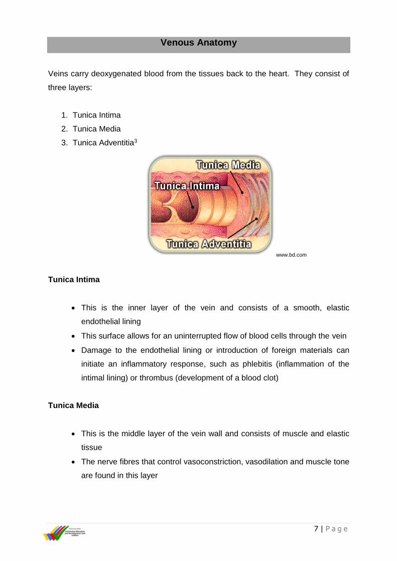
Venous Anatomy
Veins carry deoxygenated blood from the tissues back to the heart. They consist of
three layers:
1. Tunica Intima
2. Tunica Media
3. Tunica Adventitia3
www.bd.com
Tunica Intima
This is the inner layer of the vein and consists of a smooth, elastic
endothelial lining
This surface allows for an uninterrupted flow of blood cells through the vein
Damage to the endothelial lining or introduction of foreign materials can
initiate an inflammatory response, such as phlebitis (inflammation of the
intimal lining) or thrombus (development of a blood clot)
Tunica Media
This is the middle layer of the vein wall and consists of muscle and elastic
tissue
The nerve fibres that control vasoconstriction, vasodilation and muscle tone
are found in this layer
8 | P a g e
Tunica Adventitia
This is the outer layer of the vein and consists of connective tissue
The tunica adventitia provides support and protection for the vein
This layer contains the vessels that supply nutrients to the vein
Valves
Valves are structures within the lumen of the veins that are formed by the
endothelial lining of the tunica intima
They are a system of half-moon shaped flaps that are arranged in pairs
Valves are predominately found in the large veins of the extremities
Valves act like ‘trap-doors’ to keep the blood flowing towards the heart
Valves present as bumps along the course of the vein and also occur at
bifurcations (an area where two veins join)
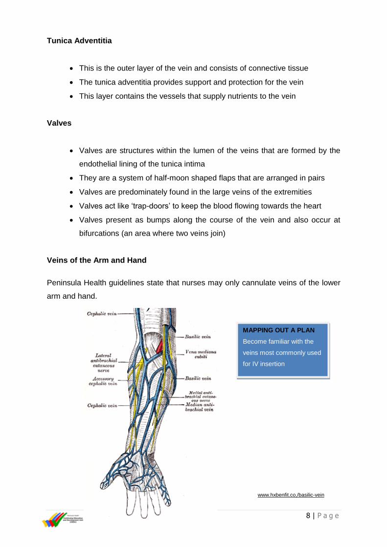
Veins of the Arm and Hand
Peninsula Health guidelines state that nurses may only cannulate veins of the lower
arm and hand.
www.hxbenfit.co,/basilic-vein
MAPPING OUT A PLAN
Become familiar with the
veins most commonly used
for IV insertion
9 | P a g e
Indications
A peripheral IV cannula may be required for the following reasons:
Administration of IV fluids/blood products
Administration of IV medications
Maintenance of intravenous access
Contraindications
The only absolute contraindication for peripheral IV cannulation is when appropriate
therapy can be provided by a less invasive route (e.g. orally) 4, 5.
Relative contraindications to use of a particular extremity include:
A history of mastectomy or lymph node dissection
Presence of an arteriovenous shunt or fistula
Oedema or deep vein thrombosis
Procedure requirement or injury on extremity
If peripheral IV cannulation is deemed necessary in one of the above conditions it
must be discussed with a medical officer.
Sites to Be Avoided
Veins below a previous IV infiltration
Areas of phlebitis or infection
Sclerosed or thrombosed veins
Areas of skin inflammation, disease, bruising or breakdown
Points of flexion over joints
Dominant hand or arm
10 | P a g e
Inner aspect of wrist or within a 5 cm radius. This will reduce the risk of
damage to the radial, median or ulnar nerves 4, 6
Selecting a Vein
When choosing an appropriate vein, you should consider the following:
The patient’s medical history
The patient’s age, size and general condition
The location and condition of the vein
The purpose of the cannula
The type of fluid and medication required
Hypertonic or irritant solutions must be infused through a vein with ample
blood flow.
The expected duration of IV therapy
Your cannulation skill 3, 4, 6
The vein that is suitable for cannulation should feel round, firm, elastic and
engorged, not hardened, bumpy or flat.
The site of catheter insertion influences the risk of infection and phlebitis. Lower
extremity insertions sites are linked to a higher risk of infection than upper extremity
sites. Veins on the hands have a lower risk for phlebitis than veins on the wrist.
The general rule is to start at the most distal site available (the hand) and move up
as necessary. Starting with the patient’s hand (preferably non-dominant) leaves
more proximal sites for subsequent cannulations.
A disposable tourniquet is used to enhance venous dilation by impeding venous
return. It is placed 5 – 10cm proximal to the anticipated cannulation site with a
pressure that should be greater than venous pressure, but less than arterial
pressure. Avoid excessive pressure and prolonged application of the tourniquet to
reduce risk of vein injury and haematoma formation.
11 | P a g e
Additional methods that may be used to enhance venous dilation if required include:
Placing the anticipated cannulation site below the level of the heart to
reduce venous return
Applying a warm compress to the intended cannulation site
Gently stroking the vein along its length in a proximal to distal direction
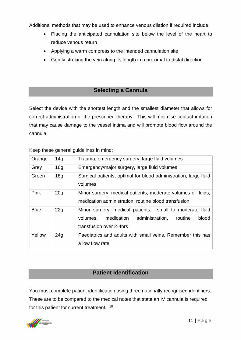
Selecting a Cannula
Select the device with the shortest length and the smallest diameter that allows for
correct administration of the prescribed therapy. This will minimise contact irritation
that may cause damage to the vessel intima and will promote blood flow around the
cannula.
Keep these general guidelines in mind:
Orange 14g Trauma, emergency surgery, large fluid volumes
Grey 16g Emergency/major surgery, large fluid volumes
Green 18g Surgical patients, optimal for blood administration, large fluid
volumes
Pink 20g Minor surgery, medical patients, moderate volumes of fluids,
medication administration, routine blood transfusion
Blue 22g Minor surgery, medical patients, small to moderate fluid
volumes, medication administration, routine blood
transfusion over 2-4hrs
Yellow 24g Paediatrics and adults with small veins. Remember this has
a low flow rate
Patient Identification
You must complete patient identification using three nationally recognised identifiers.
These are to be compared to the medical notes that state an IV cannula is required
for this patient for current treatment. 10

12 | P a g e
Consent
A valid and informed consent must be obtained and documented prior to undertaking
any non - emergency treatment and/or procedure. Consent for peripheral IV
cannulation may be both ‘verbal’ as the procedure is discussed with the
patient/client, and ‘implied’ as the patient/client offers their arm for IV cannulation 7.
You must complete patient identification using three nationally recognised identifiers.
11
Technique for Insertion
Aseptic non touch technique is required for peripheral IV cannulation with the goal of
minimising contamination of the insertion site and the key parts. An aseptic
(procedural) hand wash is required prior to the procedure. The five moments of
hand hygiene must be followed throughout the procedure 9.
A clean trolley and an IV insertion pack must be used to prepare the equipment for
peripheral IV cannulation. The insertion site is cleaned with 2% chlorhexidine and
75% alcohol to help prevent catheter related infection. The solution must be allowed
to dry for 15-30 seconds prior to cannulation. A non- touch technique must be used
and the insertion site should not be re-palpated once it has been cleaned.
A transparent semipermeable occlusive dressing is placed over the insertion site on
completion and the date must be recorded on the dressing using the documentation
label.
Protective eyewear and sterile gloves must be worn when performing peripheral IV
cannulation.
No more than 2 attempts should be made by any one nurse to insert a peripheral IV
cannula. This prevents unnecessary trauma to the patient and the potential to limit
future vascular access 2.
13 | P a g e
Click below to read the Peninsula Health ‘Peripheral Intravenous Cannulation’
Clinical Practice Guideline:
http://prompt.phcn.vic.gov.au/Search/download.aspx?filename=17837197\17837615
\22590217.pdf
Potential Complications
1. Infection
Strict adherence to hand hygiene and the use of aseptic technique in insertion and
maintenance is the most important measure to prevent infection.
Other preventative measures include:
Choosing the appropriate site and cannula size
Use of 2% chlorhexidine and 70% alcohol to prepare the site and allowing the
solution to dry
Use of sterile gloves for IV cannulation
Use of a transparent semipermeable dressing over the insertion site
Review of IV site each shift for patency, inflammation, infection or bruising
Perform hand hygiene before accessing intravenous site
Always clean the end cap with an alcohol swab allowing time to dry before
accessing. The cap must be replaced each time it is accessed
Changing the cannula within 72 hours or earlier if signs of phlebitis are
present
Changing the cannula within 24 hours if it placed in ED or in an emergency
situation where aseptic technique may be compromised
Ensuring removal of the cannula when it is no longer required.
14 | P a g e
2. Superficial Venous Thrombosis
Intravenous catheters cause endothelial trauma and inflammation which can lead to
venous thrombosis. Signs and symptoms include inflammation, pain and tenderness
along the vein and/or oedema.
Factors that can be related to formation of a venous thrombosis include:
Diameter of catheter relative to size of the vein
It is recommended to use the smallest diameter catheter appropriate for
the patient to allow blood flow around catheter and prevent stagnation of
blood
Catheter related infection
Refer to preventative measure under 1. Infection
Infusion of irritant solutions
Ensure all medications are diluted appropriately and the cannula is flushed
with normal saline at the completion of medication administration
Phlebitis refers to the inflammatory reaction within the vein, usually due to
thrombus. Clinical findings include pain, tenderness and inflammation along the
vein.
If phlebitis is noted the intravenous cannula must be removed and resited.
Superficial phlebitis is generally benign and resolves when the catheter is removed.
Elevation of the limb and warm or cool compress may also assist once the catheter
has been removed to reduce inflammation.
3. Haematoma
Haematoma is the collection of blood that can be formed following a leakage of
blood from the vein into the surrounding tissues at the insertion site. It can occur as
a result of failure to puncture the vein properly during cannula insertion or following
the removal of the cannula.
If a haematoma forms during insertion of a peripheral IV cannula it must be removed
immediately and an alternative site accessed with a new cannula.
15 | P a g e
4. Infiltration
Infiltration occurs when the fluid infused through an IV cannula enters the
subcutaneous tissue rather than the vein. Leakage into tissues of certain solutions,
such as those used for chemotherapy can cause tissue necrosis.
Once a cannula has been inserted it must be flushed with 5-10mls of normal saline
to ensure it is patent and correctly situated in the vein.
5. Air Embolism
The IV cannula provides a potential port of entry for air into the venous system.
Ensure all connections are secure.
6. Pain
Pain may occur at the time of peripheral intravenous cannulation. Topical local
anaesthetic lotions are not routinely used in adults but may be applied if considered
advantageous. Topical local anaesthetics require a medical order 3, 6, 8 .
16 | P a g e
References
1. Frank R. Peripheral venous access in adults. UptoDate, [Internet]. 2013 [cited
2018 April 30]; Available from: https://www.uptodate.com/contents/peripheral-
venous-access-in-adults
2. Peninsula Health Clinical Practice Guidelines, Peripheral Intravenous
Cannulation [Online] available,
http://prompt.phcn.vic.gov.au/Search/download.aspx?filename=17837197\178
37615\22590217.pdf, Accessed May 2018.
3. Band J, Gaynes R. Prevention of intravascular catheter-related infections
UptoDate, [Internet] 2013 Available from:
http://www.uptodate.com./prevention-of-intravascular-catheter-related-
infection .
4. The Joanna Briggs Institute. Management of peripheral intravascular devices.
Best Practice: evidence-based information sheets for health professionals.
2008:12(5).
5. Ackley B, Ladwig Swan B, Tucker S. Evidence-Based Nursing Care
Guidelines Medical-Surgical Interventions. Canada: Mosby Incorporated;
2008.
6. Berube C, Aehnder J. Catheter-Induced Upper Extremity Venous
Thrombosis, UptoDate [Internet] 2013 [cited 2018 May 1]; Available from:
http://www.uptodate.com/contents/catheter-induced-upper-extremity-venous-
thrombosis,
7. Webster J, Osborne S, Rickard C, Hall J, Clinically-Indicated Replacement
Versus Routine Replacement of Peripheral Venous Catheters. The Cochrane
Library, [Internet] 2010 Available from:
http://onlinelibrary.wiley.com/doi/10.1002/14651858.CD007798.pub/abstract,

17 | P a g e
8. Peninsula Health Policy, Infection Prevention and Control [Online] available,
http://intranet.phcn.vic.gov.au/search?SearchableText=infection+prevention+a
nd+control&path=%2FPlone, Accessed March 2017.
9. Australasian College for Infection Prevention and Control [Online]. Standard
AT Clinician Competency. Aseptic Technique Resources – ACIPC, [cited
2018 May 3] Available from: Aseptic Technique Resources – ACIPC
10. Peninsula Health Policy, Patient Identification and Procedure Matching
[Online] available,
2017.http://prompt.phcn.vic.gov.au/Search/download.aspx?filename=1783559
6\17835614\22312099.pdf, Accessed 7th May 2018
11. Peninsula Health Policy, Medical Treatment Decisions and Consent [Online]
available,
http://prompt.phcn.vic.gov.au/Search/download.aspx?filename=17835655\178
35660\34843806.pdf, Accessed 7th May 2018