petra stoerig- the neuroanatomy of phenomenal vision: a psychological perspective
TRANSCRIPT

8/3/2019 Petra Stoerig- The Neuroanatomy of Phenomenal Vision: A Psychological Perspective
http://slidepdf.com/reader/full/petra-stoerig-the-neuroanatomy-of-phenomenal-vision-a-psychological-perspective 1/19
176
The Neuroanatomy of Phenomenal Vision:
A Psychological Perspective
PETRA STOERIG
Institute of Experimental Psychology II, Heinrich-Heine-University,
D-40225 Düsseldorf, Germany
ABSTRACT: Somewhere in the visual system, phenomenal vision—the seeing of colors, brightness, depths, shades, and motion—is generated not only from thedistribution of light on the retina, but also when the eyes are closed, in dreams,hallucinations, phosphenes, and (possibly) imagery. Whether these differentforms of phenomenal vision share a common substrate although their originsare different (optical, mechanical, electrical, endogenous) is discussed in thelight of evidence from neuropsychological and functional imaging studies.Whereas extrastriate visual cortical areas appear to be involved in all types of phenomenal vision that have been studied, the necessity of a contribution fromprimary visual cortex is demonstrated by the loss of conscious vision that followsits destruction. If both extrastriate and primary cortical activation are needed,the latter may not just provide an indispensable input, but may also need toreceive the output of the extrastriate processing via reentrant connections.
KEYWORDS: Veridical vision; Blindsight; Nonveridical vision; Phosphenes;Dreams; Hallucinations; Imagery; Afterimages; V1; extrastriate cortex; Vision
INTRODUCTION
Where in the visual system does the neuronal processing of visual information
become conscious? In the past ten years, several suggestions have been put forward:they focus on the extrastriate cortical areas, either in isolation1,2 or conjointly withthe primary visual cortex,3,4 or on those extrastriate visual areas that form the ven-tral, occipitotemporal stream of visual cortical processing only,5 or they include ex-tra-visual areas in the frontal lobes,6–8 or in the reticular formation.9 In addition tothis already wide range of structures, within the broader discussion of the neuronalbasis of consciousness, thalamocorticothalamic loops linking the visual cortical ar-eas with specific and unspecific thalamic nuclei are discussed.10–12
Focusing specifically on visual consciousness instead of on the general questionof conscious representation restricts the problem to the best-studied of the sensorysystems: Whatever mechanisms mediate the mysterious transformation of neural in-formation into sensory awareness in the visual system would, presumably, play asimilar role in other sensory systems. Unfortunately, however restricted consciousvision appears when compared to the entirety of conscious experience, it is still verycomplex. It includes awareness of brightness and darkness, of colors and motion, of
Address for correspondence: Institute of Experimental Psychology II, Heinrich-Heine-Univer-sity, D-40225 Düsseldorf, Germany. Voice: +49.211.81-12265; fax: 49.211.81-14522.
8/3/2019 Petra Stoerig- The Neuroanatomy of Phenomenal Vision: A Psychological Perspective
http://slidepdf.com/reader/full/petra-stoerig-the-neuroanatomy-of-phenomenal-vision-a-psychological-perspective 2/19
177STOERIG: NEUROANATOMY OF PHENOMENAL VISION
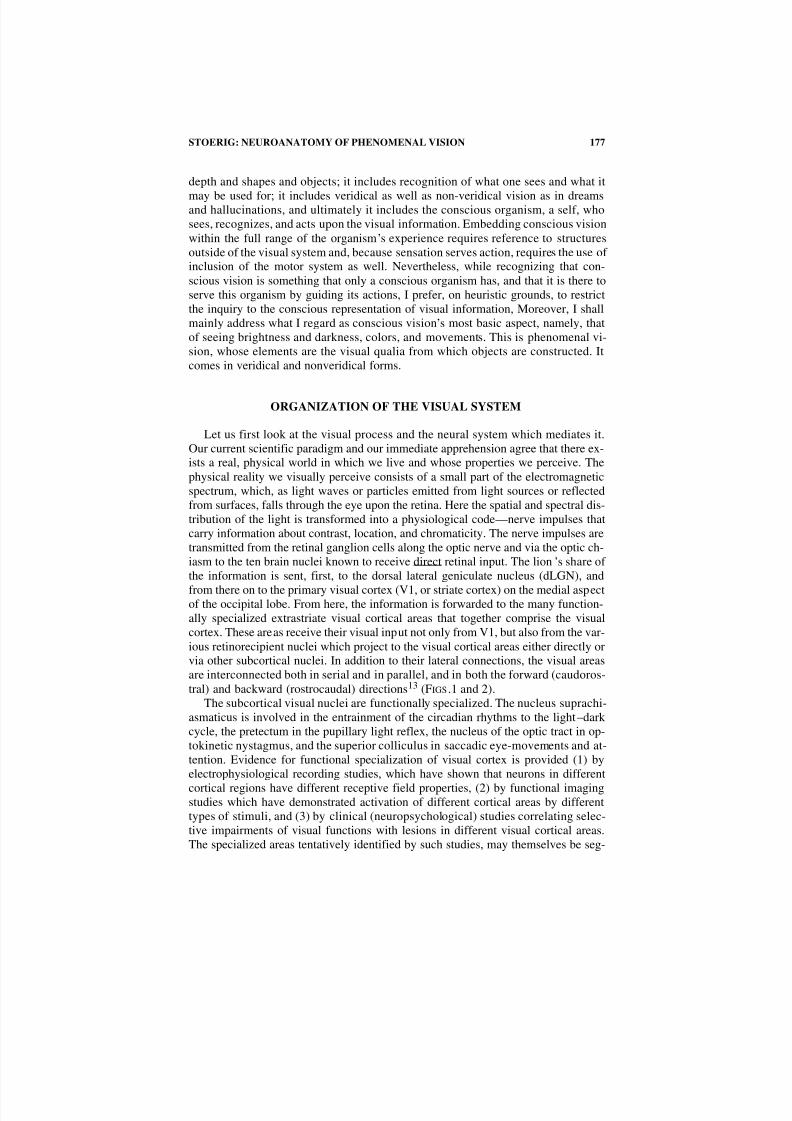
depth and shapes and objects; it includes recognition of what one sees and what it
may be used for; it includes veridical as well as non-veridical vision as in dreamsand hallucinations, and ultimately it includes the conscious organism, a self, who
sees, recognizes, and acts upon the visual information. Embedding conscious vision
within the full range of the organism’s experience requires reference to structures
outside of the visual system and, because sensation serves action, requires the use of
inclusion of the motor system as well. Nevertheless, while recognizing that con-
scious vision is something that only a conscious organism has, and that it is there to
serve this organism by guiding its actions, I prefer, on heuristic grounds, to restrict
the inquiry to the conscious representation of visual information, Moreover, I shall
mainly address what I regard as conscious vision’s most basic aspect, namely, that
of seeing brightness and darkness, colors, and movements. This is phenomenal vi-
sion, whose elements are the visual qualia from which objects are constructed. It
comes in veridical and nonveridical forms.
ORGANIZATION OF THE VISUAL SYSTEM
Let us first look at the visual process and the neural system which mediates it.
Our current scientific paradigm and our immediate apprehension agree that there ex-
ists a real, physical world in which we live and whose properties we perceive. The
physical reality we visually perceive consists of a small part of the electromagnetic
spectrum, which, as light waves or particles emitted from light sources or reflected
from surfaces, falls through the eye upon the retina. Here the spatial and spectral dis-
tribution of the light is transformed into a physiological code—nerve impulses that
carry information about contrast, location, and chromaticity. The nerve impulses are
transmitted from the retinal ganglion cells along the optic nerve and via the optic ch-
iasm to the ten brain nuclei known to receive direct retinal input. The lion ’s share of
the information is sent, first, to the dorsal lateral geniculate nucleus (dLGN), and
from there on to the primary visual cortex (V1, or striate cortex) on the medial aspectof the occipital lobe. From here, the information is forwarded to the many function-
ally specialized extrastriate visual cortical areas that together comprise the visual
cortex. These areas receive their visual input not only from V1, but also from the var-
ious retinorecipient nuclei which project to the visual cortical areas either directly or
via other subcortical nuclei. In addition to their lateral connections, the visual areas
are interconnected both in serial and in parallel, and in both the forward (caudoros-
tral) and backward (rostrocaudal) directions13 (FIGS .1 and 2).
The subcortical visual nuclei are functionally specialized. The nucleus suprachi-
asmaticus is involved in the entrainment of the circadian rhythms to the light–dark
cycle, the pretectum in the pupillary light reflex, the nucleus of the optic tract in op-
tokinetic nystagmus, and the superior colliculus in saccadic eye-movements and at-
tention. Evidence for functional specialization of visual cortex is provided (1) by
electrophysiological recording studies, which have shown that neurons in different
cortical regions have different receptive field properties, (2) by functional imagingstudies which have demonstrated activation of different cortical areas by different
types of stimuli, and (3) by clinical (neuropsychological) studies correlating selec-
tive impairments of visual functions with lesions in different visual cortical areas.
The specialized areas tentatively identified by such studies, may themselves be seg-

8/3/2019 Petra Stoerig- The Neuroanatomy of Phenomenal Vision: A Psychological Perspective
http://slidepdf.com/reader/full/petra-stoerig-the-neuroanatomy-of-phenomenal-vision-a-psychological-perspective 3/19
178 ANNALS NEW YORK ACADEMY OF SCIENCES
regated into still smaller functional compartments, increasing the difficulty of deter-mining how and where vision becomes conscious. Were the visual system organized
in a quasi-Cartesian fashion so that all retinal input eventually converged onto a sin-
gle structure whose destruction abolished all conscious vision, we should happily ac-
cept that structure as “the mind’s eye.” Instead, we find a network of heavily
interconnected, functionally specialized structures at both cortical and subcortical
levels. Visual signals originating at the same retinal locus will be conveyed along
different routes, via differing numbers of relay nuclei, by axons with different con-
duction velocities, arriving at their destination at different times.
How can perceptual unity arise from such a distributed network? Where in this
dense mesh of interconnected visual neurons does the visual neural code get trans-
formed into the phenomenal visual world? After all, qualia do not exist in the phys-
ical world, nor are they properties of neuronal processes. Instead, they represent a
mental level of reality; their perception by an animal defines its vision as conscious.
The construction of qualia may depend uniquely upon a single neuronal processingfeature, or a combination of such features, from microtubules, synapses, and neu-
rotransmitters, to neuronal morphology and connectivity, velocity of signal trans-
mission, and the synchronous activity of cell assemblies distributed in cortical and
subcortical structures. Several of these possibilities are dealt with in other chapters
FIGURE 1. A schematic representation of functionally specialized visual cortical areas.Almost all known connections are bidirectional. (Data from Felleman and Van Essen.13)
8/3/2019 Petra Stoerig- The Neuroanatomy of Phenomenal Vision: A Psychological Perspective
http://slidepdf.com/reader/full/petra-stoerig-the-neuroanatomy-of-phenomenal-vision-a-psychological-perspective 4/19
179STOERIG: NEUROANATOMY OF PHENOMENAL VISION
of this volume. In this chapter I shall focus on the macroscopic level, that is, the level
of structures and networks of structures, and ask: where are visual qualia made?
VERIDICAL PHENOMENAL VISION
Absolute blindness is an absence of all visual qualia. It may result from destruc-
tion of one or more of the following visual structures: the eye, the retina, the optic
nerve, the optic tract, the dorsal lateral geniculate nucleus (dLGN), the optic radia-tion, and the primary (V1) and secondary visual cortex (V2). A unilateral lesion will
affect the visual field of one eye if it is prechiasmatic, and the visual field of both
eyes if it is behind the chiasma (F IG. 2). The more distant it is from the retina, the
more fibers projecting to extrageniculate visual nuclei will be spared, and the more
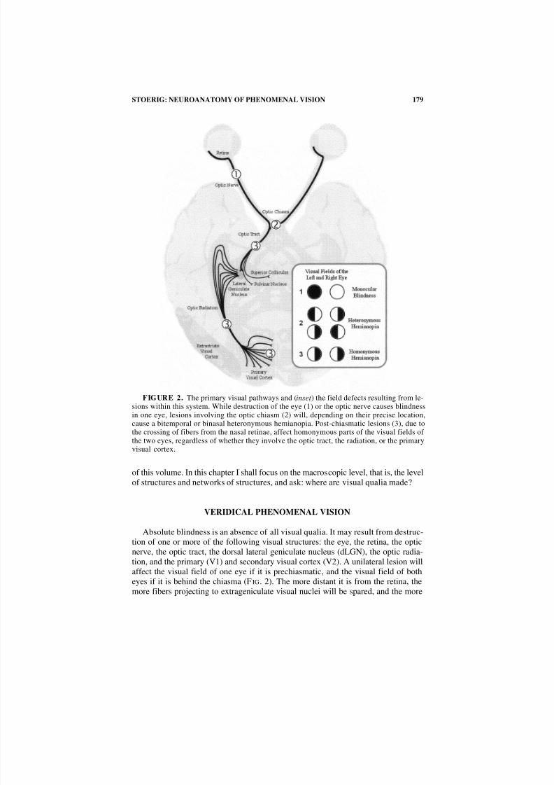
FIGURE 2. The primary visual pathways and (inset ) the field defects resulting from le-sions within this system. While destruction of the eye (1) or the optic nerve causes blindness
in one eye, lesions involving the optic chiasm (2) will, depending on their precise location,cause a bitemporal or binasal heteronymous hemianopia. Post-chiasmatic lesions (3), due tothe crossing of fibers from the nasal retinae, affect homonymous parts of the visual fields of the two eyes, regardless of whether they involve the optic tract, the radiation, or the primaryvisual cortex.
8/3/2019 Petra Stoerig- The Neuroanatomy of Phenomenal Vision: A Psychological Perspective
http://slidepdf.com/reader/full/petra-stoerig-the-neuroanatomy-of-phenomenal-vision-a-psychological-perspective 5/19
180 ANNALS NEW YORK ACADEMY OF SCIENCES
visual functions will remain intact. While a retrochiasmatic lesion may spare only
the projection to the nucleus suprachiasmaticus, a retrogeniculate lesion will leaveintact all retinofugal fibers projecting to extrageniculate nuclei. Visual reflexes such
as the pupillary light reflex can then be elicited from the blind visual field, and so
can nonreflexive visual functions, provided procedures such as forced-choice guess-
ing are used to circumvent the blindness that the patients experience. The visual
functions that have thus far been demonstrated include localization, detection, and
discrimination of visual flux, and of the size, orientation, motion, motion direction,
wavelength, and shape of stimuli presented in the blind field. These types of visual
function, which are demonstrable in a subject’s blind field, have been termed blind-
sight (see Weiskrantz et al.,14 Weiskrantz,15 and Stoerig and Cowey16 for reviews).
This phenomenon demonstrates that nonreflexive visual processing is possible in the
absence of visual awareness of the processed information. Since the extrageniculo-
striate cortical visual system is undamaged in these patients, we must infer that the
extrageniculo-striate system is, by itself, insufficient to mediate visual awareness.
Like destruction of the dLGN, destruction of the primary visual cortex causes acomplete loss of phenomenal vision, but only the cortical lesion will permit the var-
ious nonreflexive visual functions of blindsight. Destruction of the secondary visual
cortical area which surrounds V1 on all sides appears to have similar effects to those
of a lesion in V1. Destruction of lower V2 causes a quadrantanopia in the upper con-
tralateral hemifield, and destruction of upper V2 is followed by an anopia of the low-
er quadrant.17 Disconnection of the primary visual cortex both from its geniculate
input and from the higher visual cortical areas thus causes a loss of visual qualia.
However, if the lesion selectively destroys extrastriate visual cortical areas without
disconnecting V1 from the remaining cortical regions, visual qualia remain. Thus,
when the occipitotemporal areas V4/V8 (the so-called “color complex”) is selective-
ly affected, color vision is lost, but movement and brightness remain. Conversely,
when area MT, which is part of the human motion complex (hMT+), is affected, con-
scious motion vision is compromised, but color and brightness vision remain. It fol-
lows that phenomenal vision depends on the functional integrity of the early visualcortical areas surrounding and including V1/V2, with different areas supporting dif-
ferent qualia.
THE ROLE OF V1
The role that V1 plays in the concerted action mediating visual awareness is un-
clear. One hypothesis suggests that V1 functions like a cortical retina,18 providing an
input into the array of extrastriate areas without which they are incapacitated and
cannot mediate phenomenal vision.6 Alternatively, V1 could participate in visual
awareness directly, supporting a quale of its own; indeed, the mediation of brightness
has been suggested as its primary contribution to conscious vision.19,20 Finally, its
role could be to receive the results of extrastriate cortical visual processing via its ex-
tensive recurrent fibers, this feedback providing the crucial information necessary togenerate qualia.4,21 The first of these hypotheses affords V1 only a comparatively
trivial role, but, like the third, is consistent with the loss of phenomenal vision pro-
duced by destruction of V1. The second is based largely upon a body of data suggest-
ing the gradual development of some forms of conscious vision in a hemianopic
8/3/2019 Petra Stoerig- The Neuroanatomy of Phenomenal Vision: A Psychological Perspective
http://slidepdf.com/reader/full/petra-stoerig-the-neuroanatomy-of-phenomenal-vision-a-psychological-perspective 6/19
181STOERIG: NEUROANATOMY OF PHENOMENAL VISION
patient, GY, who suffered unilateral damage to his occipital lobe at age 8. This pa-
tient, now in his forties, has participated in a large number of studies of his residualvisual functions, which have suggested that he is now aware of visual stimuli provid-
ed they have some salient feature, such as movement.22–25 This visual awareness is
phenomenal, or at least that is the conclusion drawn by the authors from GY’s ability
to find a perceptually satisfactory match in the intact field for a stimulus presented in
the impaired field.26 We have observed a similar slow change from absolute to rela-
tive blindness in another patient (FS) whose lesion occurred later in life (age 42), but
who, like GY, has extensively used his hemianopic field in numerous blindsight stud-
ies.27–30 In neither patient was any evidence for ipsilesional V1 activation found in
functional imaging studies.1,8,30–32 Within the limits of current technology, this dem-
onstrates that some conscious vision may become possible without ipsilesional V1.
VERIDICAL VISION WITHOUT V1
What evidence suggests that it is specifically brightness that is missing in this
kind of V1-less, low-level vision? Morland et al.,19 using a forced-choice procedure,
asked GY to try and match colors, motion velocity, and brightness between his nor-
mal and impaired hemifield. This involved his manually adjusting the visual proper-
ties of the matching stimuli so as to make a stimulus presented in one hemifield
resemble another presented in the other field. [As in all forced-choice procedures,
guessing was an available option]. While the patient’s color and velocity matches
were quite successful, his brightness matches bore little resemblance to those of a
normal observer, leading the authors to suggest that brightness, rather than color and
motion, depended on V1. These results, taken alone, are insufficient to support the
hypothesis. Not only may a forced-choice match in a blindsight subject be based on
phenomenal properties quite different from those that mediate a match in unimpaired
subjects, but also it may reflect isomorphic processes underlying quite different per-ceptual representations such as conscious and unconscious ones. More importantly,
GY strongly denies “seeing” colors, and even his extensively studied motion pro-
cessing performance1,22,23 may reflect his inferences about positional information
rather than motion perception per se.23,27,32 It is therefore premature to conclude,
even in GY’s case, that brightness is the only quale dependent upon V1’s integrity.
Independently of whether V1 plays a special role in brightness perception, one
wonders what the source is of residual conscious vision in the absence of ipsilesional
V1? Given the observation that destruction of V1 produces cortical blindness, how
can its absence be compensated? One possibility is that qualia may be mediated by
extrastriate cortex alone; another that qualia require the joint activation of both ex-
trastriate and extravisual systems. The first alternative is contradicted by the fact that
stimulation of the impaired hemifield, although it activates extastriate cortical areas
in patients with absolute cortical blindness, does not produce even rudimentary
awareness of the stimuli.33 Extrastriate cortical activation alone is therefore insuffi-cient, even if it involves not just dorsal but also those ventral extrastriate cortical
areas30 that have been implicated in the mediation of conscious vision.5 But perhaps
ipsilesional extrastriate cortical activation, though initially insufficient, could with
practice recover to the point of supporting low-level phenomenal vision. Increasing
8/3/2019 Petra Stoerig- The Neuroanatomy of Phenomenal Vision: A Psychological Perspective
http://slidepdf.com/reader/full/petra-stoerig-the-neuroanatomy-of-phenomenal-vision-a-psychological-perspective 7/19
182 ANNALS NEW YORK ACADEMY OF SCIENCES
use of the hemianopic field might be the most likely process mediating such recov-
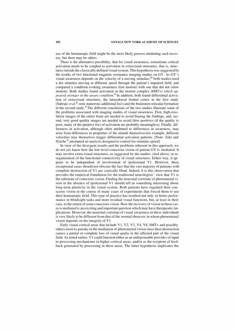
ery, but there may be others.There is the alternative possibility, that for visual awareness, extrastriate cortical
activation needs to be coupled to activation in extravisual structures, that is, struc-
tures outside the classically defined visual system. This hypothesis was suggested by
the results of two functional magnetic resonance imaging studies on GY. As GY ’svisual awareness depends on the velocity of a moving stimulus,24 both studies used
a dot stimulus moving at different speed through the patient’s impaired field, and
compared a condition evoking awareness (fast motion) with one that did not (slow
motion). Both studies found activation in the motion complex (hMT+) which ap-
peared stronger in the aware condition.9 In addition, both found differential activa-
tion of extravisual structures, the laterodorsal frontal cortex in the first study
(Sahraie et al.8; note numerous additional foci) and the brainstem reticular formation
in the second study.9 The different conclusions of the two studies illustrate some of
the problems associated with imaging studies of visual awareness. First, high-reso-
lution images of the entire brain are needed to avoid biasing the findings, and, sec-ond, very good quality images are needed to avoid false positives (if the quality is
poor, many of the putative foci of activation are probably meaningless). Finally, dif-
ferences in activation, although often attributed to differences in awareness, may
arise from differences in properties of the stimuli themselves;for example, different
velocities may themselves trigger differential activation patterns. [Note: Zeki and
ffytche 9 attempted an analysis designed to control for stimulus speed].
In view of the divergent results and the problems inherent in this approach, we
do not yet know how the low-level conscious vision of patient GY is mediated. It
may involve extra-visual structures, as suggested by the studies cited above, or re-
organization of the functional connectivity of visual structures. Either way, it ap-
pears to be independent of involvement of ipsilesional V1. However, these
exceptional cases should not obscure the fact that the vast majority of patients with
complete destruction of V1 are cortically blind. Indeed, it is this observation that
provides the empirical foundation for the traditional neurologists’ view that V1 isthe substrate of conscious vision. Finding the neuronal correlate of phenomenal vi-
sion in the absence of ipsilesional V1 should tell us something interesting about
long-term plasticity in the visual system. Both patients have regained their con-
scious vision in the course of many years of experiments that forced them to use
their hemianopic field. This type of practice has resulted not only in better perfor-
mance in blindsight tasks and more residual visual functions, but, at least in their
case, to the return of some conscious vision. How the recovery of vision in these cas-
es is mediated is an exciting and important question which may have therapeutic im-
plications. However, the neuronal correlate of visual awareness in these individuals
is very likely to be different from that of the normal observer, in whom phenomenal
vision depends on the integrity of V1.
Early visual cortical areas that include V1, V2, V3, V4, V8, hMT+ and possibly
others seem to partake in the mediation of phenomenal vision since their destruction
causes a partial or complete loss of visual qualia in the affected part of the visualfield. As noted earlier, V1 could function either as an indispensable provider of input
to processing mechanisms in higher cortical areas, and/or as the recipient of feed-
back generated by processing in these areas. The latter hypothesis implicates the
8/3/2019 Petra Stoerig- The Neuroanatomy of Phenomenal Vision: A Psychological Perspective
http://slidepdf.com/reader/full/petra-stoerig-the-neuroanatomy-of-phenomenal-vision-a-psychological-perspective 8/19
183STOERIG: NEUROANATOMY OF PHENOMENAL VISION
very extensive feedback connections among the visual cortical areas whose inactiva-
tion, via inactivation of an up-stream visual area, markedly alters the functional tun-ing of V1 neurons (e.g., Hupé et al.34). If the results of processing by higher-order
areas were fed back to V1, this could explain the unambiguous positioning of objects
in the visual field on the basis of the high spatial resolution of V1, which is in con-
trast to the much lower resolution in increasingly higher areas. This hypothesis gains
support from a number of recent neurophysiological studies that used very different
approaches. They have shown that the late response components (80–100 ms) of V1
neurons differ from the early ones in orientation tuning,35 in preserving figure-
ground segregation,36 and most importantly, in the perceptual interpretation of stim-
uli.37 Independent confirmation of this result comes, first, from experiments in bin-
ocular rivalry showing that a small percentage of V1 neurons respond according to
the monkey’s present percept and independent of its visual input,38 and, second from
a study showing that the responses of V1 neurons in cats reflected brightness rather
than physical contrast.39 Additional support for the hypothesis comes from studies
of the effects of experimental interventions, such as masks and transcranial magneticpulses, upon the conscious perception of a stimulus. These psychophysical studies
have identified two different time windows for effective disruption of visual aware-
ness. In addition to an early time window (ca. 20–30 ms), which coincides with the
arrival of the retinal information in V1, a much later one (ca. 100–120 ms) was par-
ticularly effective at suppressing the conscious perception of a visual stimulus. The
earlier window coincides with the initial processing of visual inputs, and will prevent
those inputs from being forwarded to higher areas. But at 100–120 ms, when the in-
formation has long since reached extrastriate cortex, presentation of a masking stim-
ulus still interferes with the after-discharges of V1 neurons,40 and a magnetic pulse
over the occipital pole still suppresses stimulus perception.41 If visual awareness
were indeed dependent upon the reception by V1 of feedback from extrastriate visual
processing, V1 would play a more interesting role than that of a visual relay. The
“feedback ” hypothesis is certainly consistent with the observation that the late-re-
sponse components of V1 neurons reflect the perceptual rather than the physicalproperties of a visual stimulus, since these late components are most likely to be af-
fected by the results of feedback from extrastriate areas. Interestingly, lesions in
higher visual cortical areas do not abolish or diminish the patient’s repertoire of vi-
sual qualia, but cause higher-order perceptual deficits.
NONVERIDICAL PHENOMENAL VISION
The previous section dealt with veridical vision—the situation in which light fall-
ing onto the retina is transduced into nerve impulses, and eventually transformed
into visual qualia. But there are other means to evoke phenomenal vision. Afterim-
ages are seen although the stimulus that induced them has disappeared. Phosphenes
are phenomenal events, caused by mechanical, electrical, or magnetic stimulation of
the retina or the visual cortex. Visual dreams are phenomenal, and result from invol-untary intrinsic neuronal activation, as do hallucinations, while visual imagery may
also be phenomenal, and is caused by voluntary intrinsic activation. Do all these
kinds of phenomenal vision share a common mechanism? Do they all depend on V1
8/3/2019 Petra Stoerig- The Neuroanatomy of Phenomenal Vision: A Psychological Perspective
http://slidepdf.com/reader/full/petra-stoerig-the-neuroanatomy-of-phenomenal-vision-a-psychological-perspective 9/19
184 ANNALS NEW YORK ACADEMY OF SCIENCES
and its extrastriate partners? Or is the neuronal correlate of phenomenal vision so
varied that quite different structures may mediate it under different conditions, sothat it is dependent not on a particular structure or set of structures but upon such
features as the strength or the temporal patterning of nerve impulses?
Let us begin with the phenomenon of phosphenes. Rubbing one’s eyes causes vi-
sual experiences that can be complexly patterned and colorful, while a bump on the
head causes one to “see” the “stars” with which we are familiar from cartoons. Elec-
trical stimulation of the eyes or brain also causes phosphenes, as does the much new-
er and painless transcranial magnetic stimulation (TMS). Both kinds, electro- and
magnetophosphenes, can be evoked in people who have lost their eyesight because
of damage to the eye or optic nerve: A retinal input is not needed. But what about
cortical areas? In the majority of studies using TMS, the technique has been used to
suppress rather than evoke vision42 (see Walsh and Cowey43 for recent review).
These studies have found that the timing of the magnetic pulse in relation to the pre-
sentation of the visual stimulus is critical (see above). However, several studies have
also described magnetophosphenes evoked from stimulation over the occipitallobe,44–46 suggesting that phosphenes were mediated by activity in early visual cor-
tical areas or even the optic radiation. An obvious problem with attempts to infer the
site of phosphene production from the location of visual structure most directly stim-
ulated is that the strong TMS pulse may activate a relatively wide network of struc-
tures. Nevertheless, the kind of phosphenes—simple arrangements of dots or lines
or stars—provide a cue as to the involvement of early visual cortex, because it is di-
rect electrical stimulation of these structures that evokes this type of phosphene. 47 If
V1 is necessary for the seeing of phosphenes, its destruction should prevent their ap-
pearance. As yet, there is no published study, but in our own tests of three patients
with homonymous visual field defects we failed to elicit phosphenes in the blind
field. Because we used stimulation parameters optimized for normal observers,
these data are preliminary; however, they do suggest a critical role for the early vi-
sual cortex in the production of magnetophosphenes.
Afterimages are another instance of exogenously induced phenomenal vision. Arecent functional magnetic resonance imaging study in normal observers has re-
vealed that both the presentation of a saturated color stimulus, and the long-lasting
colored after-image induced by prolonged viewing of the stimulus are accompanied
by activation in V1 and in the color complex (V4/V8). In addition to these areas, the
motion complex hMT+ was activated during the afterimage phase only, which may
account for the observation that the subjective dynamic component was seen only
during the after-image.48 Thus activation in the cortical motion complex area pro-
duces a phenomenal motion effect in normal observers even though the stimulus it-
self is not moving (see Tootell et al.49 for MT’s role in the motion aftereffect). The
role of V1 in perception of the afterimage was first explored by Bender and Kahn50
in patients with V1 lesions using colored figures that they presented entirely or partly
in their patient’s field defect. Their results showed that afterimages were not reported
when the stimulus fell entirely into the blind field, but that it was subject to some
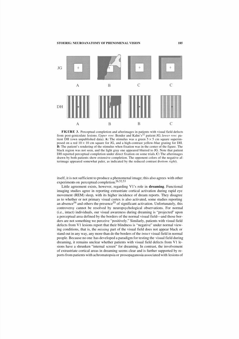
perceptual completion upon central fixation, that is, when only part of the figure wasinvisible to their patient (FIG. 3, top). These findings agree with our own data (FIG. 3,
bottom) as well as those of Marcel,51 demonstrating that information from the cor-
tically blind field may complete or otherwise influence the percept, but that, by
8/3/2019 Petra Stoerig- The Neuroanatomy of Phenomenal Vision: A Psychological Perspective
http://slidepdf.com/reader/full/petra-stoerig-the-neuroanatomy-of-phenomenal-vision-a-psychological-perspective 10/19
185STOERIG: NEUROANATOMY OF PHENOMENAL VISION
itself, it is not sufficient to produce a phenomenal image; this also agrees with otherexperiments on perceptual completion.26,52,53
Little agreement exists, however, regarding V1’s role in dreaming. Functional
imaging studies agree in reporting extrastriate cortical activation during rapid eye
movement (REM) sleep, with its higher incidence of dream reports. They disagree
as to whether or not primary visual cortex is also activated, some studies reporting
an absence54 and others the presence55 of significant activation. Unfortunately, this
controversy cannot be resolved by neuropsychological observations. For normal
(i.e., intact) individuals, our visual awareness during dreaming is “projected” upon
a perceptual area defined by the borders of the normal visual field—and those bor-
ders are not something we perceive “positively.” Similarly, patients with visual field
defects from V1 lesions report that their blindness is “negative” under normal view-
ing conditions, that is, the missing part of the visual field does not appear black or
stand out in any way, any more than do the borders of the intact visual field in normal
people. Because no one has developed a paradigm for testing the visual field duringdreaming, it remains unclear whether patients with visual field defects from V1 le-
sions have a shrunken “internal screen” for dreaming. In contrast, the involvement
of extrastriate cortical areas in dreaming seems clear and is further supported by re-
ports from patients with achromatopsia or prosopaganosia associated with lesions of
FIGURE 3. Perceptual completion and afterimages in patients with visual field defectsfrom post-geniculate lesions. Upper row: Bender and Kahn’s52 patient JG; lower row: pa-tient DH (own unpublished data). A: The stimulus was a green 5 × 5 cm square superim-posed on a red 10 × 10 cm square for JG, and a high-contrast yellow-blue grating for DH.B: The patient’s rendering of the stimulus when fixation was in the center of the figure. Theblack region was not seen, and the light gray one appeared blurred to JG. Note that patientDH reported perceptual completion under direct fixation on some trials. C: The afterimagesdrawn by both patients show extensive completion. The opponent colors of the negative af-terimage appeared somewhat paler, as indicated by the reduced contrast (bottom right ).

8/3/2019 Petra Stoerig- The Neuroanatomy of Phenomenal Vision: A Psychological Perspective
http://slidepdf.com/reader/full/petra-stoerig-the-neuroanatomy-of-phenomenal-vision-a-psychological-perspective 11/19
186 ANNALS NEW YORK ACADEMY OF SCIENCES
extrastriate visual cortex who experience an absence of color or recognizable faces
not only in their waking life, but in their dreams as well.Studies on the role of V1 in visual imagery are as controversial in their results as
they are plentiful in number. Whether the system that mediates veridical vision also
mediates visual imagery is the core question of the still unresolved “imagery de-
bate.” The available psychophysical, functional imaging, and neuropsychological
data are in agreement with respect to the involvement of higher visual cortical areas.
This was first shown convincingly by Roland et al.56 in a positron emission tomog-
raphy (PET) study on normal observers, and confirmed in numerous subsequent
studies. What remains at issue is the involvement of early visual cortex. Several stud-
ies have demonstrated activation only in higher visual cortical areas,56–58 while oth-
ers have found it to extend all the way down to areas V1/V259–61 or even the lateral
geniculate nucleus.62 Kosslyn’s psychological investigations of the properties of vi-
sual imagery led him to conclude that imagery must be supported by the “normal”visual system including early, topographically organized visual cortex (see
Kosslyn65 for review). He has argued that the failure to find activation in early visualcortex was caused by the lack of experimental control during the subject’s “resting”condition, which serves as the control against which activation in the “imagery” con-
dition is compared. If the subjects do not “wipe their screen clean” during the resting
condition, but instead imagine something of their own accord, they will generate suf-
ficient activation during the resting condition to minimize the contrast between the
resting and imagery conditions. As a result, the study will fail to implicate V1/V2 as
part of the visual network activated during visual imagery.
Another complicating factor in imagery studies is the variation in the imagining
tasks used. These may range from imagining simple geometric pattern and letters to
visualizing complex scenes, or even to imagining walking from home to a familiar
place. It is generally accepted that simple visual patterns are processed largely in the
early cortical areas, while complex spatial and object recognition tasks involve high-
er extrastriate cortical processing. In the complex tasks, less activation would be ex-
pected in the early cortical areas even if the results of the higher processing were tobe fed back to earlier areas, because such feedback should require much less “capac-
ity” than the processing task. Lower activation levels in the early areas would then
be more likely to be missed. Finally, the task ’s demands on spatial resolution may
influence the extent of early visual cortical involvement, with higher-resolution tasks
requiring more processing by the early areas with their superior spatial maps of the
visual field. This hypothesis is consistent with the report that imagining the same ob-
ject in different sizes and at different positions within the visual field leads to early
visual cortical activation.60–62
If the extent of top-down activation of the visual cortical areas is task-dependent
then the deficits of patients with lesions in different visual cortical areas should re-
flect the presumed processing capacities of the specific areas. The results of several
studies of patients with early visual cortical damage are consistent with this assump-
tion. These patients did not show a deficit in tasks requiring answers to “high imag-
ery content” questions such as “Does a bear have round or pointed ears?” or “Is agrapefruit larger or smaller than an orange?,” which despite their referring to con-
crete objects can be answered on the basis of stored visual information. 64,65 Failure
to correctly respond to such questions is more common in patients with visual agno-

8/3/2019 Petra Stoerig- The Neuroanatomy of Phenomenal Vision: A Psychological Perspective
http://slidepdf.com/reader/full/petra-stoerig-the-neuroanatomy-of-phenomenal-vision-a-psychological-perspective 12/19
187STOERIG: NEUROANATOMY OF PHENOMENAL VISION
sia, suggesting that interference with this type of visual information retrieval reflects
a disturbance of higher visual function rather than of cortical blindness. In contrast,
in a patient available for testing both pre-and postoperatively, unilateral removal of
the occipital lobe did reveal a striking change associated with the lesion and the sub-
sequent hemianopia.66 The patient was asked to imagine herself walking towards
various objects of well-defined size, to stop when the object filled her internal screen
to the point of overflow, and then to estimate the imagined distance between herself
and the object. On the preoperative test, the patient said, for example, 33 cm for a
kitten, and 188 cm for a car. During the postoperative test she more than doubled the
distance, as if her internal screen had shrunk to the same extent as the visual field
(FIG. 4). Furthermore, by repeating the tests with a ruler that had to be imagined in
a horizontal or vertical orientation, this shrinkage was found to affect only the hori-
zontal, but not the vertical extent of the image, as it should if the cortical blindnesswas responsible. Possibly, this task is more visuo-perceptual in nature, although it
too draws on visual memory and could theoretically be solved without phenomenal
imagery. An impairment of imagery requiring relatively high spatial resolution was
also found in Butter et al.’s67 study of hemianopic patients. When patients were
asked to indicate whether an arrow was pointing at one of the dots in a pattern they
had been shown in free vision but that was no longer visible, they were found to
make more errors with arrows on the side of their hemianopia. Taken together, stud-
ies of imagery have usually reported involvement of higher extrastriate visual corti-
cal areas, while the evidence for participation of early visual areas is much less
consistent. The outcomes of these studies will be influenced both by task demands
and individual problem-solving strategies, and, in the case of functional imaging
studies, the design of the protocol and the analysis will be critical.
The last form of nonveridical vision to be addressed here are hallucinations.
They may appear to patients with blindness due to (post-) retinal pathology—-Charles-Bonnet syndrome68—in whom the visual cortex remains excitable, as well
as to patients with cortically blind visual fields caused by post-geniculate le-
sions.69–72 They can be simple or highly complex, ranging from phosphenes to
FIGURE 4. Farah et al. 68reported that their patient required approximately twice thedistance to imagine an object of a definite size after unilateral occipital lobectomy, indicat-ing a shrinkage of the “field of visual imagery” that reflects the hemianopia induced by thesurgery.
8/3/2019 Petra Stoerig- The Neuroanatomy of Phenomenal Vision: A Psychological Perspective
http://slidepdf.com/reader/full/petra-stoerig-the-neuroanatomy-of-phenomenal-vision-a-psychological-perspective 13/19
188 ANNALS NEW YORK ACADEMY OF SCIENCES
complex geometric patterns to objects and people that move about. The compara-
tively simple forms—lines, dots, clouds, stars, triangles—are attributed to irritationwithin the primary visual pathway up to V169; they closely resemble the magneto-
phosphenes elicited by TMS over the occipital pole. The complex ones, in contrast,
are more likely to originate in temporal cortices, where images of scenes and people
can also be evoked by electrical stimulation applied during neurosurgery.73 Wheth-
er the precise content of the hallucinated images reflects the major focus of hyper-
excitation, as argued by ffytche et al.2 for patients with Charles-Bonnet syndrome
who underwent fMRI during hallucination, is still uncertain.
The simpler hallucinations that are attributed to the early visual structures are re-
garded as indicative of some visual recovery,68,74 while the complex ones may re-
flect a hyperexcitatory response to the lesion that caused the field defect, and usually
disappear within a relatively short time.71 That they are perceived in the cortically
blind field demonstrates that strong endogenously generated activity may produce
phenomenal images even in the absence of V1/V2. How this pathological activation
differs from that caused by TMS is presently unclear. That it at least temporarilycauses phenomenal visual images is uncontroversial (or almost so; see Pollen20) so
that hallucinations are the only instance of fully conscious phenomenal vision with-
out early visual cortex that is present immediately after the blindness-producing in-
cident. The type of veridical phenomenal vision that may develop over long periods
of training in blindsight subjects like GY is very crude and low-level when compared
to such complex hallucinations as a series of identical gray–green men strolling
through the cortically blind hemifield.69
SUMMARY AND OUTLOOK: COMPLEXITY AND UNITY
Phenomenal visual images can be caused by a variety of processes, ranging from
exogenous (optical, mechanical, electrical, and magnetic events in the eyes or the vi-
sual cortex) to endogenous processes (which may be involuntary, as in visual dreamsand hallucinations, or voluntary as in visual imagery). Studies using functional im-
aging techniques have consistently found that visual images, whether veridical or
nonveridical, are associated with activation of extrastriate visual cortical areas. In
contrast, activation of early visual cortical areas V1/V2 was reported in some, but
not all such studies (see TABLE 1).
Despite their remarkable contribution to the study of functional neuroanatomy,
imaging techniques are of limited use in demonstrating causation (that is, in estab-
lishing whether or not a particular structure is necessary for a particular function).
While imaging studies can identify brain regions activated during performance of a
particular task, they do not differentiate between activation necessary for the task
and that merely associated with its performance. Moreover, interpretation of the re-
sults of imaging studies is always linked to the design of the experimental protocol,
which may not always do justice to the physiological reality, as well as to the nature
of the data analysis. Methodological problems associated with the former include thedefinition of the resting state in protocols used in studies of visual imagery, the se-
lection of the specific temporal offset used between stimulus and response, or the
control of the subject’s psychophysiological state during the measurement. Exam-

8/3/2019 Petra Stoerig- The Neuroanatomy of Phenomenal Vision: A Psychological Perspective
http://slidepdf.com/reader/full/petra-stoerig-the-neuroanatomy-of-phenomenal-vision-a-psychological-perspective 14/19
189STOERIG: NEUROANATOMY OF PHENOMENAL VISION
ples of the latter type include the use of data smoothing vs. averaging, or the choice
of statistical criteria. Such factors may yield contradictory results from comparable
data sets (see Sibersweig et al.75and Dierks et al.76 for an example). While function-
al imaging studies are undoubtedly exciting, such interpretive considerations make
them less than conclusive when considered in isolation. Their results need to be com-
plemented by neuropsychological studies of patients with lesions of circumscribed
brain regions, which can provide evidence of the functional significance of the dam-
aged regions.
VISUAL CONSCIOUSNESS AND THE ROLE OF V1/V2
The available data (summarized in TABLE 1) show that striate and extrastriate vi-
sual cortical areas need to be activated in all normal, nonpathological forms of ver-
idical or nonveridical phenomenal vision. Patients who have suffered destruction of
primary visual cortex are blind. They do not see stimuli presented in the blind field,
they do not see afterimages of stimuli that were presented to the blind field, they
have not been reported to see phosphenes from magnetic stimulation of the lesioned
hemisphere, their imagery appears unaffected when their visual memory is tapped
but not when a visuo-perceptual task requiring good spatial resolution is given.
Whether these patients have visual dreams involving the cortically blind field is un-
known. Only in hallucinations and in the rare cases of low-level vision re-establishedin former blindsight subjects such as GY and FS is phenomenal vision without ip-
silesional V1 known to occur.
Neuropsychology thus strengthens the case for V1, but does not prove its neces-
sity without exception—the hallmark of a good rule. The exceptions, both patholog-
TABLE 1. Summary of evidence from functional imaging and neuropsychology
regarding the participation of primary and higher visual cortical areas in themediation of veridical and nonveridical forms of vision
Structures Extrastriate Areas Primary Area
Evidence from Imaging Neuropsychology Imaging Neuropsychology
Function
Veridical
Normal vision yes yes yes yes
Residual vision yes yes no no
Blindsight yes yes no no
Nonveridical
Phosphenes ? ? ?/yes yes
Afterimages yes ? yes yes
Dreams yes yes yes/no ?
Imagery yes yes yes/no yes/noHallucinations yes ? no no
NOTE: All results, including those from studies on Blindsight where conscious vision is absent,agree on extrastriate visual cortical activation.
Yes/no reflects controversial results; ? indicates the answer is unknown.
8/3/2019 Petra Stoerig- The Neuroanatomy of Phenomenal Vision: A Psychological Perspective
http://slidepdf.com/reader/full/petra-stoerig-the-neuroanatomy-of-phenomenal-vision-a-psychological-perspective 15/19
190 ANNALS NEW YORK ACADEMY OF SCIENCES
ical, may indicate that V1’s absence can be compensated for under certain
conditions. In the case of hallucinations, the spontaneous extrastriate cortical activa-tion is quite strong, and may therefore spread to other structures, subcortical and cor-
tical, in the ipsi- and contralesional hemisphere. The complexity of this activation
pattern is in contrast to the very focal activation observed from visual stimulation of
fields of absolute and relative cortical blindness. Stimulation of the normal hemifield
results in activation of the normal primary and extrastriate visual cortex, and in ip-
silesional extrastriate activation as well, just as in normal observers. Stimulation of
the blind field activates ipsilesional extrastriate cortex, but the activation appears
quite isolated and focal, involving little if any activation of surrounding ipsilesional
or contralesional cortex.30 These findings make it tempting to speculate that the
blind field is blind because the neuronal activation it elicits lacks the capacity to in-
tiate a sufficiently complex pattern of visual cortical activation.77 Destruction of V1
may prevent the development of such complex patterns, not only by destroying a
large part of the input to the extrastriate cortex, but also by interfering with the ex-
trastriate feedback to V1.78 Unusually strong extrastriate activation, such as presentin hallucinations, may allow a phenomenal representation by causing a complex
widespread pattern of activation not normally evoked without V1, and extensive
training of blindsight may induce processes that to some extent can compensate for
the loss of this structure.
If conscious vision is always mediated by widespread striate–extrastriate cortical
activation, could the presence of such activation in an organism’s brain prove that it
is consciously seeing something? Although this is likely to be the case in organisms
who are in a conscious state, and not comatose or anesthetized, it is not true in un-
conscious organisms. Provided the animals were effectively anesthetized, the fact is
clearly demonstrated by recent fMRI experiments in monkeys who showed ample
cortical activation in response to visual stimulation despite being under general an-
esthesia.79 As only conscious organisms can have conscious perception, evidence
for strong activation of striate and extrastriate visual cortical areas cannot prove con-
scious vision and thus cannot prove consciousness in animals whose possible con-sciousness we cannot yet assess unequivocally. This caveat demonstrates the need to
eventually account for the substrate of conscious vision, isolated here for simplici-
ty’s sake, within the larger context of the neural substrates of conscious (as opposed
to unconscious) states in general. Such an account would attempt to explain how the
presence of a general state of consciousness—presumably mediated by unspecific
systems and temporarily abolished by anesthesia (see Lamme et al.80 for effects of
anesthesia on neuronal activity in V1)—transforms the visuo-cortical activation into
phenomenal awareness.
Finally, let us consider that while visual processing is modular, its result is a uni-
fied percept. Whether the primary visual cortex provides a neural substrate for that
perceptual unity remains an open question. The fact that it receives perceptually rel-
evant cortico-cortical feedback, and that its destruction causes blindness despite the
availability of extrageniculo-striate input, makes V1 a prime contender for that role.
Having a structure at the bottom end of a presumed cortical processing hierarchyturns out to be responsible for the visuo-perceptual unity produced by that process-
ing would add a nice twist, which the great Cajal, who cherished whodunits and who
wrote one himself (“A secreto agravio, secreta venganza”), would have appreciated.

8/3/2019 Petra Stoerig- The Neuroanatomy of Phenomenal Vision: A Psychological Perspective
http://slidepdf.com/reader/full/petra-stoerig-the-neuroanatomy-of-phenomenal-vision-a-psychological-perspective 16/19
191STOERIG: NEUROANATOMY OF PHENOMENAL VISION
REFERENCES
1. BARBUR, J.L., J.D.G. WATSON, R.S.J. FRACKOWIAK & S. ZEKI . 1993. Conscious visualperception without V1. Brain 116: 1293–1302.
2. FFYTCHE, D.H., R.J. HOWARD, M.J. BRAMMER, A. DAVID, P. WOODRUFF & S. WILLIAMS.1998. The anatomy of conscious vision: an fMRI study of visual hallucinations.Nature Neurosci. 1: 738–742.
3. STOERIG, P. 1996. Varieties of vision: from blind processing to conscious recognition.Trends Neurosci. 19: 401–406.
4. LAMME, V. 2000. Blindsight: the role of feedforward and feedback corticocortical con-nections. Acta Psych. In press.
5. MILNER, A.D. & M. GOODALE. 1995. The Visual Brain in Action. Oxford UniversityPress. New York.
6. CRICK, F. & C. KOCH. 1995. Are we aware of neural activity in primary visual cortex?Nature 375: 121–123
7. CRICK, F. & C. KOCH. 1998. Consciousness and neuroscience. Cerebral Cortex 8: 97–107.8. SAHRAIE, A., L. WEISKRANTZ, J.L. BARBUR, A. SIMMONS, S.C. WILLIAMS & M.J.
BRAMMER. 1997. Pattern of neuronal activity associated with conscious and uncon-
scious processing of visual signals. Proc. Natl. Acad. Sci. USA 94: 9406–9411.9. ZEKI , S. & D.H. FFYTCHE. 1998. The Riddoch syndrome: insights into the neurobiol-ogy of conscious vision. Brain 121: 25–45.
10. LLÍNAS, R.R. & D. PARÉ. 1991. Of dreaming and wakefulness. Neuroscience 44:521–535.
11. JONES, E.G. 1998. A new view of specific and nonspecific thalamocortical connec-tions. In Consciousness. at the Frontiers of Neuroscience. H.H. Jasper et al., Eds.Advan. Neurol. 77: 49–71.
12. LLÍNAS, R.R. & U. RIBARY. 1998. Temporal conjunction in thalamocortical transac-tions. In Consciousness. at the Frontiers of Neuroscience. H.H. Jasper et al., Eds.Advan. Neurol. 77: 95–102.
13. FELLEMANN, D..J. & D.C. VAN ESSEN. 1991. Distributed hierarchical processing in theprimate cerebral cortex. Cerebral Cortex 1: 1–47.
14. WEISKRANTZ, L., E.K. WARRINGTON, M.D. SANDERS & J. MARSHALL. 1974. Visualcapacity in the hemianopic field following a restricted cortical ablation. Brain 97:709–728.
15. WEISKRANTZ, L. 1986. Blindsight: A Case Study and Implications. Oxford University
Press. New York.16. STOERIG, P. & A. COWEY. 1997. Blindsight in man and monkey. Brain 120: 535–559.17. HORTON, J.C.& W.F. HOYT . 1991. Quadrantic visual field defects: a hallmark of
lesions in extrastriate (V2/V3) cortex. Brain 114: 1703–1718.18. HENSCHEN, S.E. 1910. Zentrale Sehstörungen. In Handbuch der Neurologie 2. M.
Lewandowski, Ed. : 89–98. Springer. Berlin.19. MORLAND, A.B., S.R. JONES, A.L. FINLAY, D. DEYZAC, S. LE & S. KEMP. 1999.
Visual perception of motion, luminance and colour in a human hemianope. Brain122: 1183–1198.
20. POLLEN, D.A. 1999. On the neural correlates of visual perception. Cerebral Cortex 9: 4–19.21. STOERIG, P. & A. COWEY. 1995. Visual perception and phenomenal consciousness.
Behav. Brain Res. 71: 147–156.22. BARBUR, J.L., K.H. RUDDOCK & V.A. WATERFIELD. 1980. Human visual responses in
the absence of the geniculo-striate projection. Brain 102: 905–928.23. BLYTHE, I.M., J.M. BROMLEY, C. KENNARD & K.H. RUDDOCK. 1986. Visual discrimi-
nation of target displacement remains after damage to the striate cortex in humans.Nature 320: 619–621.
24. WEISKRANTZ, L., J.L. BARBUR & A. SAHRAIE. 1995. Parameters affecting consciousversus unconscious visual discrimination in a patient with damage to the visual cor-tex (V1). Proc. Natl. Acad. Sci. USA 92: 6122–6126.
25. STOERIG, P. & E. BARTH. Phenomenal vision in the absence of V1. Submitted forpublication.
8/3/2019 Petra Stoerig- The Neuroanatomy of Phenomenal Vision: A Psychological Perspective
http://slidepdf.com/reader/full/petra-stoerig-the-neuroanatomy-of-phenomenal-vision-a-psychological-perspective 17/19
192 ANNALS NEW YORK ACADEMY OF SCIENCES
26. PÖPPEL, E. 1986. Long-range colour-generating interactions across the retina. Nature
320: 523–52527. PÖPPEL, E. 1985. Bridging a neuronal gap. Naturwissenschaften 72: 599.28. STOERIG, P. 1987. Chromaticity and achromaticity: evidence for a functional differen-
tiation in visual field defects. Brain 110: 869–886.29. STOERIG, P., A. KLEINSCHMIDT& J. FRAHM. 1998. No visual responses in denervated
V1: high-resolution functional magnetic resonance imaging of a blindsight patient.NeuroRep. 9: 21–25.
30. GOEBEL, R., L. MUCKLI, F.E. ZANELLA, W. SINGER & P. STOERIG. Sustained extrastri-ate cortical activation without visual awareness revealed by fMRI studies of hemian-opic patients. Submitted for publication.
31. KLEISER, R., M. NIEDEGGEN, J. WITTSACK, R. GOEBEL & P. STOERIG. Is V1 necessaryfor conscious vision in areas of relative cortical blindness? NeuroImage. In press.
32. AZZOPARDI, P. & A. COWEY. 2001. Motion discrimination in cortically blind patients.Brain 124: 30–46.
33. STOERIG, P., R. GOEBEL, L. MUCKLI, H. HACKER & W. SINGER. 1997. The functionalneuroanatomy of blindsight. Soc. Neurosci. Abs. 23: 845.
34. HUPÉ, J.M., A.C. JAMES, B.R. PAYNE, S.G. LOMBER, P. GIRARD& J. BULLIER. 1998.
Cortical feedback improves discrimination between figure and background by V1,V2 and V3 neurons. Nature 394: 784–787.35. RINGACH, D.L., M.J. HAWKEN & R. SHAPLEY. 1997. Dynamics of orientation tuning in
macaque primary visual cortex. Nature 387: 281–284.36. LAMME, V.A.F., V. RODRIGUEZ-RODRIGUEZ & H. SPEKREIJSE. 1999. Separate process-
ing dynamics for texture elements, boundaries and surfaces in primary visual cortexof the macaque monkey. Cerebral Cortex 9: 406–413.
37. ZIPSER, K., V.A.F. LAMME & P.H. SCHILLER. 1996. Contextual modulation in primaryvisual cortex. J.Neurosci. 16: 7376–7389.
38. LOGOTHETIS, N.K. 1998. Single units and conscious vision. Phil. Trans. R. Soc. Lon-don B 353: 1801–1818.
39. ROSSI, A.F., C.D. RITTENHOUSE & M.A. PARADISO. 1996. The representation of brightness in the primary visual cortex. Science 273: 1104–1107.
40. MACKNICK, S.L. & M.S. LIVINGSTONE. 1998. Neuronal correlates of visibility andinvisiblity in the primate visual system. Nature Neurosci. 1: 144–149.
41. CORTHOUT, E., B. UTTL , V. WALSH, M. HALLETT & A. COWEY. 1999. Timing of activ-ity in early visual cortex as revealed by transcranial magnetic stimulation. Neu-
roRep. 10: 2631–2534.42. AMASSIAN, V.E., R.Q. CRACCO, P.J. MACCABE, J.B. CRACCO, A. RUDELL & L.EBERLE. 1989. Suppression of visual perception by magnetic coil stimulation of human occipital cortex. Electroenceph. Clin. Neurophysiol. 74: 458–462.
43. WALSH, V. & A. COWEY. 1998. Magnetic stimulation studies of visual cognition.Trends Cogn. Sci. 2: 103–10.
44. MARG , E. & D. RUDIAK. 1994. Phosphenes induced by magnetic stimulation over theoccipital brain: description and probable sites of stimulation. Optom. Vis. Sci. 71:301–311.
45. KAMMER, T. 1998. Phosphenes and transient scotomas induced by magnetic stimula-tion of the occipital lobe: their topographical relationship. Neuropsychologia 37:191–198.
46. KASTNER, S., I. DEMMER & U. ZIEMANN. 1998. Transient visual field defects inducedby transcranial magnetic stimulation over the occipital pole. Exp. Brain Res. 118:199–226.
47. FOERSTER, O. 1937. Motorische Felder und Bahnen. In Handbuch der Neurologie 6. O.Bumke & O.Foerster, Eds. Springer. Berlin.
48. KONEN, C., R. KLEISER & P. STOERIG. 2000. Afterimages: an fMRI-study of subjectiveexperience. Soc.. Neurosci. Abs. In press.49. TOOTELL, R.B.H., J.B. REPPAS, A.M. DALE , R.B. LOOK , T.J. BRADY & B.R. ROSEN.
1995. Visual motion aftereffect in human cortical area MT revealed by functionalmagnetic resonance imaging. Nature 375: 139–141.

8/3/2019 Petra Stoerig- The Neuroanatomy of Phenomenal Vision: A Psychological Perspective
http://slidepdf.com/reader/full/petra-stoerig-the-neuroanatomy-of-phenomenal-vision-a-psychological-perspective 18/19
193STOERIG: NEUROANATOMY OF PHENOMENAL VISION
50. BENDER, M.B. & R.L. KAHN . 1949. After-imagery in defective fields of vision. J. Neu-
rol. Neurosurg. Psychiat. 12: 196–204.51. MARCEL, A.J. 1998 Blindsight and shape perception: deficit of visual consciousness orof visual function? Brain 121: 1565–1588.
52. WARRINGTON, E.K. 1962. The completion of visual forms across hemianopic fielddefects. J. Neurol. Neurosurg. Psychiatry 25: 208–217.
53. TORJUSSEN, T. 1976. Residual function in cortically blind hemifields. Scand. J. Psy-chol. 17: 320–322.
54. BRAUN, A.R., T.J. BALKI N, N.J. WESENSTEN, F. GWADRY, R.E. CARSON, M. VARGA
P. BALDWIN, G. BELENKY& P. HERSCOVITCH. 1998. Dissociated pattern of activity invisual cortices and their projections during human rapid eye movement sleep. Sci-ence 279: 91–95.
55. LÖVBLAD, K.-O., R. THOMAS, P.M. JAKONB, T. SCAMMELL, C. BASSETTI, M. GRIS-WOLD, J. IVES, J. MATHESON, R.R. EDELMAN & S. WARACH. 1999. Silent functionalmagnetic resonance imaging demonstrates focal activation in rapid eye movementsleep. Neurology 53: 2193–2195.
56. ROLAND, P.E., L ERIKSSON, S. STONE-ELANDER & L. WILDEN. 1987. Does mentalactivity change the oxidative metabolism of the brain? J. Neurosci. 7: 2373–2389.
57. ROLAND, P.E. & B. GULYAS. 1994. Visual imagery and visual representation. TrendsNeurosci. 17: 291–287.58. D’ESPOSITO, M., J.A. DETRE, G.K. AGUIRRE, M. STALLCUP, D.C. ALSOP, L.J. TIPPET
& M.J. FARAH. 1997. A functional MRI study of mental image generation. Neurop-sychologia 35: 725–730.
59. LEBIHAN, D., R. RURNER, T.A. ZEFFIRO, C.A. CUENOD, P. JEZZARD & V. BONNEROT.1993. Activation of human primary visual cortex during visual recall: a magneticresonance imaging study. Proc. Natl. Acad. Sci. USA 90: 11802–11805.
60. KOSSLYN, S.M., A.M. ALPERT, W.L. THOMPSON, V. MALJKOVIC, S.B. WEISE, C.F.CHABRIS, S.E. HAMILTON, S.L. RAUCH & F.S. BUONANNO. 1993. Visual mentalimagery activates topographically organized visual cortex: PET investigations. J.Cogn. Neurosci. 5:263–287.
61. KOSSLYN, S.M,. W.L. THOMPSON, I.J. KIM & N.M. ALPERT. 1995. Topographical rep-resentations of mental images in primary visual cortex. Nature 378: 496–498.
62. CHEN , W., T. KATO , X.-H. ZHU, S. OGAWA, D.W. TANK & K. UGURBIL. 1998. Humanprimary visual cortex and lateral geniculate nucleus activation during visual imag-ery. NeuroRep. 9: 3669–3674.
63. KOSSLYN, S.M. 1994. Image and Brain. MIT Press. Cambridge, MA.64. GOLDENBERG, G. & C. ARTNER. 1991. Visual imagery and knowledge about the visualappearance of objects in patients with posterior cerebral artery lesions. Brain Cogn.15: 160–186.
65. CHATTERJEE, A. & M.H. SOUTHWOOD. 1995. Cortical blindness and visual imagery.Neurology 45: 2189–2195.
66. FARAH, M., M.J. SOSO & R.M. DASHEIFF. 1992. Visual angle of the mind ’s eye beforeand after unilateral occipital lobectomy. J. Exp. Psychol. Hum. Percept. Perform. 18:241–246.
67. BUTTER, C.M., S. KOSSLYN, D. MIJOVIC-PRELEC & A. RIFFLE. 1997. Field-specificdeficits in visual imagery following hemianopia due to unilateral occipital infarcts.Brain 120: 217–228.
68. BONNET, C. 1769. Essai analytique sur les facultes de l’ame. 2. Aufl, Bd.2, Kopen-hagen, Genf: Philibert.
69. GLONING, I., K. GLONING & H. HOFF . 1967. Über optische Halluzinationen. Wien. Z.Nervenheil. 25: 1–19.
70. KÖLMEL, H.W. 1985. Complex visual hallucinations in the hemianopic field. J. Neu-
rol. Neurosurg. Psychiat. 48: 29–3871. KÖLMEL, H.W. 1988. The Homonymen Hemianopsien. Springer. Berlin72. LEPORE, F.E. 1990. Spontaneous visual phenomena with visual loss: 104 patients with
lesions of retinal and neural afferent pathways. Neurology 40: 444–447.73. PENFIELD, W. & P. PEROT. 1963) The brains’s record of auditory and visual experi-
ence. Brain 86: 595–696.

8/3/2019 Petra Stoerig- The Neuroanatomy of Phenomenal Vision: A Psychological Perspective
http://slidepdf.com/reader/full/petra-stoerig-the-neuroanatomy-of-phenomenal-vision-a-psychological-perspective 19/19
194 ANNALS NEW YORK ACADEMY OF SCIENCES
74. WUNDERLICH, G., B. SUCHAN, J. VOLKMANN, H. HERZOG, V. HŠMBERG & R.J. SEITZ.
2000. Visual hallucinations in recovery from cortical blindness. Arch. Neurol. 57:561–565.75. SILBERSWEIG, D.A., E. STERN, C. FRITH, C. CAHILL, A. HOLMES, S. GROOTOONK, J.
SEAWARD. P. MCKENNA, S.E. CHUA & L. SCHNORR. 1995. A functional neuroanat-omy of hallucinations in schizophrenia. Nature 378: 176–179.
76. DIERKS, T., D.E.J. LINDEN, M. JANDL, E. FORMISANO, R. GOEBEL, H. LANFERMANN &W. SINGER. 1999. Activation of Heschl’s gyrus during auditory hallucinations. Neu-ron 22: 615–621.
77. TONONI, G. & G.M. EDELMAN. 1998. Consciousness and complexity. Science 282:1846–1851.
78. BULLIER, J., P. GIRARD & P.-A. SALIN. 1994. The role of area 17 in the transfer of information to extrastriate visual cortex. In Primary Visual Cortex in Primates. A.Peters & K.S. Rockland, Eds. :301–330. Plenum. New York.
79. LOGOTHETIS, N.K., H. GUGGENBERGER, S. PELED & J. PAULS. 1999. Functional imag-ing of the monkey brain. Nature Neurosci. 2: 555–562.
80. LAMME, V.A.F., K. ZIPSER & H. SPEKREIJSE. 1998. Figure-ground activity in primaryvisual cortex is suppressed by anesthesia. Proc. Natl. Acad. Sci. USA 95: 3263–3268.