phage-directed synthesis of copper sulfide: … synthesis of copper ... this vector differed from...
TRANSCRIPT

IOP PUBLISHING NANOTECHNOLOGY
Nanotechnology 24 (2013) 325602 (8pp) doi:10.1088/0957-4484/24/32/325602
Phage-directed synthesis of coppersulfide: structural and opticalcharacterizationMohammed Shahriar Zaman1, Chung Hee Moon2,Krassimir N Bozhilov2,3 and Elaine D Haberer1,2
1 Department of Electrical Engineering, University of California, Riverside, CA 92521, USA2 Materials Science and Engineering Program, University of California, Riverside, CA 92521, USA3 Central Facility for Advanced Microscopy and Microanalysis, University of California, Riverside,CA 92521, USA
E-mail: [email protected]
Received 20 April 2013, in final form 21 June 2013Published 17 July 2013Online at stacks.iop.org/Nano/24/325602
AbstractThe growth of crystalline copper sulfide using a viral template was investigated usingsequential incubation in CuCl2 and Na2S precursors. Non-specific electrostatic attractionbetween a genetically-modified M13 bacteriophage and copper cations in the CuCl2 precursorcaused phage agglomeration and bundle formation. Following the addition of Na2S,polydisperse nanocrystals 2–7 nm in size were found along the length of the viral scaffold.The structure of the copper sulfide material was identified as cubic anti-fluorite type Cu1.8S,space group Fm3m. Strong interband absorption was observed within the ultraviolet to visiblerange with an onset near 800 nm. Furthermore, free carrier absorption, associated with thelocalized surface plasmon resonance of the copper sulfide nanocrystals, was seen in the nearinfrared with absorbance maxima at 1060 nm and 3000 nm, respectively.
S Online supplementary data available from stacks.iop.org/Nano/24/325602/mmedia
(Some figures may appear in colour only in the online journal)
1. Introduction
Copper sulfide (CuxS) is a semiconductor which exists inseveral polymorphic forms ranging in composition fromCu2S to CuS. Among the most well-characterized phasesare high chalcocite (Cu2S, hexagonal), low chalcocite (Cu2S,orthorhombic), djurleite (Cu1.95S), digenite (Cu1.8S), andcovellite (CuS) [1]. The Cu-deficient phases of this p-typematerial exhibit sizable hole concentrations which increasewith decreasing copper content and cause the band gap toincrease due to the Moss–Burstein effect [2, 3]. Althoughthe band gaps of many non-stoichiometric phases arenot well-defined, a range of 1.2–2.58 eV (Cu2S–CuS) isgenerally accepted for bulk materials [4–6]. The large holeconcentrations in these materials also cause significant freecarrier absorption within the infrared spectral region. Thespectral range of the observed interband transitions and the
tunability of the free carrier absorption of copper sulfide arehighly desirable for photovoltaic and plasmonic applications,respectively. Nonetheless, the optical and electrical propertiesare, of course, dependent on phase and composition, whichhave proven challenging to control.
Often, in Nature, molecules such as peptides andproteins closely control mineral composition and/or structure,optimizing them for specific functions. For example,magnetotactic bacteria form nearly stoichiometric magnetite(Fe3O4) for navigation; abalones control growth of calciumcarbonate polymorphs, aragonite and calcite, to createrobust shells. Because the phase diagram of copper sulfideis complex with many polymorphs, some of which arenon-stoichiometric, it is a particularly intriguing materialsystem in which to study bio-assisted synthesis. Biomoleculesof a variety of sizes, shapes, and hierarchical structureshave been used to induce growth of different copper sulfide
10957-4484/13/325602+08$33.00 c© 2013 IOP Publishing Ltd Printed in the UK & the USA

Nanotechnology 24 (2013) 325602 M S Zaman et al
compounds in a laboratory environment. Simple aminoacids such as alanine and cysteine have served as cappingligands for CuS formation [7, 8]. Colloidal synthesis usingalanine at pH 10 created polydisperse, mixed morphology(i.e. spheres, triangles, and rods) CuS particles tens ofnanometers in size [7]. The hydroxyl group of alaninereadily attached to CuS at high pH. A hydrothermal reactionusing cysteine as both a stabilizing agent and a sulfursource resulted in hierarchical CuS microstructures includingsnowflakes, flowers, and porous shells [8]. Furthermore,small proteins such as bovine serum albumin (BSA) havealso been used as capping molecules for copper sulfidesynthesis [9]. Cu2S and CuS nanoparticles were bothsuccessfully formed in colloidal solution using BSA. Thesulfhydryl or imidazole groups within BSA likely contributeto protein–metal cation interactions making particle formationpossible. Larger biomolecules with hierarchical structureshave also demonstrated an ability to mineralize coppersulfide, improving control over particle size distribution andlong range particle geometry. For example, apoferritin, acage protein which is composed of 24 subunits arrangedin a spherical geometry, has been used to producemonodisperse CuS nanoparticles [10]. The pore diameterof this multi-component protein constrained material growthresulting in tight control of nanoparticle size. Additionally,nanocrystalline tubes of Cu2S have been mineralized usingbionanotube scaffolds approximately 100 nm in diameter andseveral microns in length [11]. HG12, a histidine-rich peptide,was displayed along the length of each bionanotube with a6.4 nm spacing [11–13]. The imidazole in histidine served totemplate monodisperse Cu2S nanoparticles along the lengthof each tube. Furthermore, the pH of the precursor solutionwas used to control the aggregation of the peptides alongthe bionanotubes and vary the average nanoparticle diameterfrom 12 nm (pH 5) to 22 nm (pH 8). The well-organized,hierarchical organization of the high affinity biomoleculesenabled both the particle size and the larger scale geometryof the mineralized material to be engineered.
In this work, we used an M13 bacteriophage as atemplate to grow nanostructured copper sulfide which wascharacterized both structurally and optically. The wild-typeM13 phage is approximately 880 nm in length and 6.5 nmin diameter. This filamentous virus consists of five structuralproteins: the pIII and pVI minor coat proteins at the proximalend, the pVII and pIX minor coat proteins at the distalend, and the pVIII major coat proteins covering the lengthof the phage [14]. The viral capsid includes about 2700identical copies of the major coat protein, each serving asa potential binding site for inorganic materials. As such,like the aforementioned bionanotubes, the M13 virus is ahigh-aspect-ratio template with a high density (∼0.15 nm−2)of well-ordered binding sites on its surface [15]. Assuminga uniform distribution, the areal density of binding sites onthe M13 surface is estimated to be six times greater thanthat of the bionanotubes. Furthermore, the M13 virus isapproximately 15 times smaller in diameter. This biologicalscaffold has been used to synthesize other metal sulfides,but not yet copper sulfides [16, 17]. The study of copper-containing compounds through phage display had been
neglected, likely due to reports that Cu ions are detrimentalto phage activity, inhibiting the use of combinatorial phagedisplay techniques to identify peptides with a specificaffinity for copper sulfide [18]. Here, we explore coppersulfide growth driven by non-specific electrostatic interactionsusing a genetically-modified, carboxyl-rich M13 template.The closely-spaced, hierarchically-ordered binding sitesenabled the formation of high quality, optically-active Cu1.8Snanocrystalline material in which both band-to-band and freecarrier absorption were observed. These studies represent firststeps toward viral control of copper sulfide synthesis.
2. Experimental details
2.1. Genetic modification and amplification of M13 virus
The viral template used for synthesis of copper sulfide wasa genetically-modified M13 phage. A peptide composed ofthree glutamic acid residues (E3) was fused to the N-terminusof each of 2700 copies of pVIII protein using a previouslyreported approach [19, 20]. Specifically, the N-terminus ofthe wild-type M13 pVIII major coat protein, AEGD-, was re-placed with the more negatively charged, carboxyl-rich aminoacid sequence, AEEE-, using the M13SK vector [21, 22].This vector differed from the wild-type M13 in that PstI andBamHI restriction sites were introduced via a mutation (Tto A) at position 1372 and a mutation (C to G) at position1381, respectively, and a PstI restriction site was removed bya mutation (T to A) at position 6250 [19]. To construct the E3phage, the oligonucleotide 5′-CT ACT ACA AGG ATC CTCCTC CTC TGC AGC GAA AGA CAG CA-3′, encoding thethree glutamic acids, was hybridized and polymerase chainreaction (PCR) amplified. The circular M13SK vector andthe oligo duplex were digested with PstI and BamHI, ligatedtogether, and transformed into XL-1 Blue electrocompetentcells with an electroporator (BioRad, MicroPulser). Thetransformed cells were titered, and the bacteriophage DNAwas sequenced in the region encoding for the pVIII coatprotein to confirm the peptide fusion. To further verify theE3 peptide insert, matrix-assisted laser desorption/ionizationtime-of-flight mass spectrometry (MALDI-TOF MS, QSTARXL oMALDI MS/MS) was performed on the modified pVIIIprotein. More detailed information regarding MALDI-TOFMS sample preparation is available in the supplementary data(available at stacks.iop.org/Nano/24/325602/mmedia).
A phage stock solution with a concentration of1010 pfu µl−1 in tris-buffered saline (TBS, 50 mM Tris-HCl,150 mM NaCl, pH 7.5) was prepared using standardamplification and polyethylene glycol (PEG) purificationtechniques for the M13 phage [23, 24]. Briefly, 500 ml of luriabroth (LB) medium combined with 5 ml of ER2738 (NewEngland Biolabs) overnight culture was inoculated with asingle plaque and incubated at 37 ◦C for 8 h with shaking. TheE. coli cells were then pelleted twice at 4 ◦C and 10 000 rpmfor 20 min, transferring the supernatant to a clean tubeafter each centrifugation. Subsequently, 1/6 volume of 20%PEG/2.5 M NaCl (PEG/NaCl) was added to the supernatantand the phage was precipitated overnight at 4 ◦C. The phage
2

Nanotechnology 24 (2013) 325602 M S Zaman et al
Figure 1. Schematic of the viral-templated crystallization process.(a) E3 phage template shown with the modified pVIII protein (greenovals); (b) copper ions (blue circles) bound to the pVIII proteinduring incubation with CuCl2; (c) Cu1.8S nanocrystals (orangecircles) formed along the length of the phage after addition of Na2S.
were then pelleted at 4 ◦C and 10 000 rpm for 10 min andthe supernatant discarded. The phage pellet was dissolvedin 1 ml of TBS, transferred to a micro-centrifuge tube, andprecipitated again with 1/6 volume PEG/NaCl on ice for60 min. Finally, the phage were pelleted at 10 000 rpm for10 min, the supernatant discarded, and the phage pellet wasdissolved in 100 µl of TBS. The concentration was measuredspectrophotometrically (Evolution 60, UV/vis) and dilutedas necessary with TBS to a phage stock concentration of1010 pfu µl−1.
2.2. Synthesis of phage-templated material
Nanocrystalline copper sulfide was formed by sequentialincubation of the E3 phage template in copper chloride(CuCl2) and sodium sulfide (Na2S) chemical precursors asshown in figure 1. Stock solutions of 1 M CuCl2 (AcrosOrganics) and 1 M Na2S (MP Biomedicals) were preparedin deionized water and then diluted to concentrations of200 mM and 1 mM, respectively, for use as precursors.The E3 stock solution was diluted 1:10 with deionizedwater. Crystallization was performed by first combining equalvolumes of 200 mM CuCl2 and the diluted E3 phage solutionfor a total volume of 200 µl. The solution was vortexedto ensure mixing and allowed to incubate undisturbed for2 h. Centrifugation was used to pellet the phage templates.The excess CuCl2 precursor solution was removed leavingthe pellet intact, and 100 µl of 1 mM Na2S was added.The phage templates were incubated in the Na2S precursorsolution for 5 min with sonication. For comparison, sampleswith wild-type M13 phage and without a phage templatewere made in a similar manner. In addition, the E3 phagetemplate was added to a solution of pre-formed nanoparticlessynthesized without a template.
2.3. Zeta potential measurements and synthesis ofphage-templated material
The electrostatic interactions between the viral templateand copper cations were investigated using zeta potential
measurements and transmission electron microscopy (TEM,FEI Tecnai12 and CM300) of phage agglomerates formedduring incubation with CuCl2. The zeta potential of the E3template in dilute TBS with and without the addition of CuCl2was measured at room temperature using a zeta potentialanalyzer (Malvern Zetasizer Nano ZS). A phage concentrationof 2.5 × 10⁸ pfu µl−1 and total volume of 600 µl were usedfor all measurements. The E3 stock solution was diluted withTBS to half the original concentration. Samples were preparedby diluting 30 µl of this phage solution (5 × 10⁹ pfu µl−1)in deionized water or 0.5 mM CuCl2. Low concentrations ofCuCl2 and E3 phage template were used for zeta potentialmeasurements to avoid significant agglomeration. For eachsolvent, three samples were measured ten times and averaged.Wild-type M13 phage samples were similarly prepared andmeasured for comparison. In addition, TEM was used tostudy the phage agglomeration behavior associated with thepresence of CuCl2 in solution. For these samples, the E3stock solution was diluted 1:10 with deionized water and anequal volume of 2 mM CuCl2 was added for a total volumeof 200 µl. The solution was vortexed to ensure mixing, then5 µl volume of solution was immediately drop cast onto acarbon-coated nickel grid, incubated for 5 min, rinsed twicewith deionized water, and stained with 2% uranyl acetate.The CuCl2 concentration used here was lower than thatused for copper sulfide synthesis. Lower CuCl2 concentrationavoided the formation of extremely large agglomerates whichmight be easily removed from the TEM grid during rinsingsteps, preventing accurate analysis of agglomerate size. TEMsamples were similarly prepared with wild-type M13 phagefor comparison.
2.4. Structural characterization
The morphology of the synthesized material, as well asthe nanocrystal geometry and size were characterized byTEM. Electron diffraction and energy dispersive x-ray (EDX)spectroscopy were used to determine the crystal structure andelemental composition, respectively. To prepare samples forTEM, a 5 µl volume of suspended synthesis products wasdrop cast onto a carbon-coated nickel grid, incubated for5 min, and then rinsed twice with deionized water. Bright-fieldTEM was used to determine the morphology of the synthesisproducts, as well as to quantify the widths of individual phageencrusted by copper sulfide nanoparticles and the widthsof phage bundles coated with nanoparticles. Phage bundleswere defined as several phage aligned side-by-side forminga structure greater than one phage in width and length.Twenty-two bundles were each measured at several locationsalong their length to obtain an average width. The averagewidth of an individual phage overgrown with copper sulfidenanoparticles was measured in a similar fashion. Individualphage widths were only measured where nanocrystallinecopper sulfide material was clearly visible.
2.5. Absorption measurements
The room-temperature optical absorption of thin films ofthe phage and copper sulfide aggregates was measured
3
Nanotechnology 24 (2013) 325602 M S Zaman et al
from 360 to 3300 nm with a spectrophotometer (Cary 500UV/vis/NIR), neglecting reflection and scattering effects. Toprepare samples for absorption measurements, the phagecoated with copper sulfide were rinsed with deionized water toremove residual precursor solution and untemplated material,and then concentrated. To rinse the material, the mineralizedphage were loosely pelleted through low speed centrifugation,the supernatant was removed, and the pellet was resuspendedin deionized water. Concentration was achieved through lowspeed centrifugation and partial removal of the deionizedwater supernatant. The remaining concentrated synthesisproducts were deposited on a glass slide and dried in a vacuumdesiccator for approximately 10 min. The optical absorptionof the film was then measured.
3. Results and discussion
3.1. E3 phage template characterization
The DNA sequence of the E3 bacteriophage confirmed thefusion of the AEEE-peptide at the N-terminus of the pVIIIprotein. Furthermore, MALDI-TOF MS performed on thegenetically-modified and wild-type M13 pVIII proteins wasconsistent with the replacement of the wild-type N-terminusamino acid sequence (AEGD-) with the E3 sequence(AEEE-). The MALDI-TOF MS spectra and a more detailedanalysis of these data are available in the supplementarydata (available at stacks.iop.org/Nano/24/325602/mmedia).As expected, the addition of an acidic amino acid to the majorcoat protein increased the net negative charge of the E3 phagecompared to the wild-type M13 phage [20–22]. The averagemeasured zeta potentials of the E3 and wild-type phage indilute TBS were −33.9 ± 1.6 mV and −29.9 ± 2.2 mV,respectively.
3.2. Macroscopic fiber formation
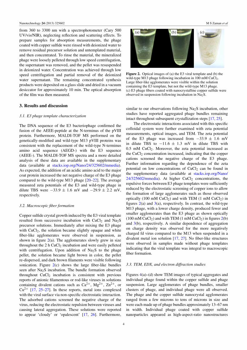
Copper sulfide crystal growth induced by the E3 viral templateresulted from successive incubation with CuCl2 and Na2Sprecursor solutions. Immediately after mixing the E3 phagewith CuCl2, the solution became slightly opaque and whitefiber-like agglomerates were observed in suspension, asshown in figure 2(a). The agglomerates slowly grew in sizethroughout the 2 h CuCl2 incubation and were easily pelletedwith centrifugation. Upon addition of Na2S to the phagepellet, the solution became light brown in color, the pelletre-dispersed, and dark brown filaments were visible followingsonication. Figure 2(c) shows the large fiber-like bundlesseen after Na2S incubation. The bundle formation observedthroughout CuCl2 incubation is consistent with previousreports of anionic filamentous or rod-like viruses in solutionscontaining divalent cations such as Ca2+, Mg2+, Zn2+, orCu2+ [17, 25–27]. In these reports, metal ions complexedwith the viral surface via non-specific electrostatic interaction.The adsorbed cations screened the negative charge of thevirus, reducing the electrostatic repulsion between viruses andcausing lateral aggregation. These solutions were reportedto appear ‘cloudy’ or ‘opalescent’ [17, 26]. Furthermore,
Figure 2. Optical images of (a) the E3 viral template and (b) thewild-type M13 phage following incubation in 100 mM CuCl2.Large fiber-like agglomerates were visible within the solutioncontaining the E3 template, but not the wild-type M13 phage.(c) E3 phage fibers coated with nanocrystalline copper sulfide wereobserved in suspension following incubation in Na2S.
similar to our observations following Na2S incubation, otherstudies have reported aggregated phage bundles remainingintact throughout subsequent crystallization steps [17, 25].
The electrostatic interactions associated with this specificcolloidal system were further examined with zeta potentialmeasurements, optical images, and TEM. The zeta potentialof the E3 phage was increased from −33.9 ± 1.6 mVin dilute TBS to −11.6 ± 1.3 mV in dilute TBS with0.5 mM CuCl2. Moreover, the zeta potential increased asthe CuCl2 concentration increased, indicating that the coppercations screened the negative charge of the E3 phage.Further information regarding the dependence of the zetapotential on low concentrations of CuCl2 can be found inthe supplementary data (available at stacks.iop.org/Nano/24/325602/mmedia). At higher CuCl2 concentrations, therepulsive forces between E3 phage templates were sufficientlyreduced by the electrostatic screening of copper ions to allowthe formation of large agglomerates such as those observedoptically (100 mM CuCl2) and with TEM (1 mM CuCl2) infigures 2(a) and 3(a), respectively. In contrast, the wild-typeM13 phage, with a lower charge density, produced fewer andsmaller agglomerates than the E3 phage as shown optically(100 mM CuCl2) and with TEM (1 mM CuCl2) in figures 2(b)and 3(b), respectively. A similar dependence of aggregationon charge density was observed for the more negativelycharged fd virus compared to the M13 when suspended in adivalent metal ion solution [17, 27]. No fiber-like structureswere observed in samples made without phage templatesindicating that the viral template was integral to macroscopicfiber formation.
3.3. TEM, EDX, and electron diffraction studies
Figures 4(a)–(d) show TEM images of typical aggregates andindividual phage found within the copper sulfide and phagesuspension. Large agglomerates of phage bundles, smallerclusters of phage, and individual phage were all observed.The phage and the copper sulfide nanocrystal agglomeratesranged from a few microns to tens of microns in size andwere each made up of phage bundles approximately 13–67 nmin width. Individual phage coated with copper sulfidenanoparticles appeared as high-aspect-ratio nanostructures
4

Nanotechnology 24 (2013) 325602 M S Zaman et al
Figure 3. The effect of the viral template surface charge density onagglomerate structure and agglomerate size in 1 mM CuCl2. (a) E3phage templates produced large, dense agglomerates. Inset: highermagnification image showing that the agglomerates were composedof very closely-packed phage bundles. (b) Wild-type M13 phageresulted in small, loose agglomerates. Inset: higher magnificationimage showing that the agglomerates were composed of fewer,loosely-packed phage.
with an average length of 946 ± 31 nm and an averagewidth of 11 ± 2.6 nm. Regardless of the configuration ofthe phage templates (bundled or individual), nanocrystallinematerial was found along the length. The density of thenanocrystalline material on the template varied greatly. Sometemplates were found to have closely-packed, continuouscoverage while others displayed sparse coverage with only afew nanoparticles. Furthermore, the nanocrystalline materialtypically appeared thicker on the exterior of phage bundles
and on individual phage templates than on phage locatedwithin a bundle. This could be due to reduced diffusion ratesof one or both of the precursors within the well-alignedbundles, despite the use of sonication during the synthesis.Alternatively, it could be caused by differences in electrostaticscreening. Further studies are necessary to fully understandthe observed difference in nanocrystalline material thickness.The sequential incubation in precursor solutions was criticalto template crystallization. E3 phage templates did not displaya strong affinity for pre-formed copper sulfide nanoparticles.Furthermore, synthesis in which the wild-type M13 phage wasused as a template produced very little to no copper sulfidealong the phage surface, as shown in the supplementary data(available at stacks.iop.org/Nano/24/325602/mmedia).
The crystalline structure and composition of the coppersulfide nanocrystalline material were studied further usingEDX and electron diffraction. As shown in figure 5, theEDX spectrum of the crystalline nanoparticles confirmed thatthey contained predominantly Cu and S. The additional peaksobserved in the spectrum are identified as C Kα, O Kα, andNi Kα lines, which are generated by spurious scattering fromthe TEM Ni grid and the amorphous carbon support film,as well as the phage. The O Kα peak was an indicationof possible partial oxidation of copper sulfide. A selectedarea electron diffraction pattern from the agglomerated coppersulfide and phage bundles is shown in the inset of figure 6.The observed ring pattern can be indexed with the digenitestructure, a slightly copper-deficient cubic polymorph ofcopper sulfide with the anti-fluorite structure and Cu1.8Sstoichiometry [28]. The measured d-spacings, 3.2, 1.95 and1.67 A, were indexed as the (111), (220) and (311) interplanardistances of digenite. High resolution TEM (HRTEM) imagesof the dense aggregates of copper sulfide and phage are alsoshown in figure 6. The material was composed of small,tightly-packed, polydisperse nanocrystals ranging in size from2 to 7 nm. Smaller nanocrystals were nearly spherical,whereas larger nanocrystals appeared slightly elongated.Figure 6 shows the 3.2 A lattice fringes of the individualnanocrystals which are consistent with the digenite (111)interplanar distances.
Nanoparticle size is difficult to predict and can typicallybe attributed to a combination of crystal growth conditions.Nonetheless, it is interesting to note that the nanocrystalsformed by the carboxyl-rich E3 template were smaller andmore polydisperse than those templated by bionanotubesfunctionalized with the imidazole-rich 12-mer peptide,HG12 [11]. The comparatively diminished binding sitespacing of the E3 may not allow for the formation of largerdiameter nanoparticles such as those observed by Banerjeeet al [11]. Further studies are necessary to determine the effectof peptide spacing on crystal growth.
3.4. Absorption studies
The absorption spectrum of a film prepared from thesynthesized material is shown in figure 7 from 360 to3300 nm. In the ultraviolet to visible wavelength region,a strong increase in absorbance was observed as thewavelength decreased below 800 nm. This optical behavior
5

Nanotechnology 24 (2013) 325602 M S Zaman et al
Figure 4. TEM images of (a) large phage bundles coated with copper sulfide, (b) higher magnification image showing phage bundlesformed from several individual phage, (c) copper sulfide nanocrystals coating a small cluster of a few phage and (d) a single phage coatedwith copper sulfide nanocrystals. Inset: high magnification image of copper sulfide nanocrystals on a single phage.
Figure 5. EDX spectrum for the copper sulfide nanocrystallinematerial showing elemental copper and sulfur peaks.
was attributed to indirect and direct band-to-band transitionsof the semiconductor material. Bandgaps near 1.55–1.6 eVhave been reported for bulk and polycrystalline digenite[4, 6]. In the near infrared (NIR) wavelength region,a broad peak near 1060 nm and a narrower set ofpeaks near 3000 nm were observed. These featureswere attributed to localized surface plasmon resonance
Figure 6. HRTEM image of nanocrystalline material showing the(111) lattice fringes of cubic Cu1.8S with digenite structure. Inset:indexed selected area electron diffraction pattern of the crystallinecopper sulfide material.
(LSPR) due to the presence of free carriers withinCu1.8S. NIR LSPR is common in non-stoichiometriccopper sulfides in which copper vacancies produce nearmetallic concentrations of free carriers, specifically holes
6

Nanotechnology 24 (2013) 325602 M S Zaman et al
Figure 7. The absorbance spectrum of a film of bio-inducedsynthesis products on a glass slide.
[6, 29–32]. The free carrier absorption peak is dependenton the copper sulfide composition, as well as nanocrystalsize and shape. The LSPR increases in strength and blueshifts with increasing hole concentration or copper vacancies.Additionally, as observed by Luther et al [30] and Kriegel et al[32] for copper sulfide nanoparticles with dimensions smallerthan the mean free path, the LSPR wavelength increaseswith decreasing particle size due to surface scattering of freecarriers. Our NIR absorbance peak near 1060 nm was broadlikely due to the polydispersity, 2–7 nm diameter particles,of the nanocrystalline material. The additional peaks near3000 nm are most likely caused by asymmetric or elongatednanocrystal geometries within the synthesis products orby plasmonic coupling between tightly-packed nanocrystalsalong the length of the viral template. The presence ofmultiple resonance peaks due to shape anisotropy was alsoobserved by Hsu et al in CuxS nanodisks [33]. The small dipnear 2250 nm is due to a spectrophotometer discontinuity.
4. Conclusion
We have examined the use of a high-aspect-ratio viral scaffoldfor bio-induced copper sulfide crystal growth. The dense,well-organized binding sites of the highly negatively charged,genetically-modified M13 template produced cubic Cu1.8S.Both interband and free carrier absorption were exhibited bythe polydisperse, nanocrystalline copper sulfide material. Dueto the complexity of the Cu–S system phase diagram witha multitude of polymorphic phases and non-stoichiometriccompounds, copper sulfide is a compelling system inwhich to study bio-assisted crystal growth. In many livingorganisms, biomolecules have demonstrated control of bothstoichiometry and crystal structure of inorganic materials.These bio-induced synthesis studies are an initial excursioninto the capacity of highly-organized, hierarchical viraltemplates to control semiconductor materials with complexphase diagrams such as copper sulfide. The ability to engineerbiomolecules to synthesize specific copper sulfide phaseswould represent significant progress in designing materialswith potentially high impact on future photovoltaic andplasmonic devices.
Acknowledgments
The authors would like to thank E L Hu (Harvard University)and A M Belcher (MIT) for the gift of the M13SK vector, aswell as our former lab member Xiaoyin Ma for completingthe genetic modification of the M13 major coat proteinusing this vector. In addition, we are grateful for earlycontributions to templated synthesis and process developmentmade by UCR undergraduate researchers Joon-Bok Lee,Steven Garcia and Hector Lopez-Hernandez. We would alsolike to thank Baharak Bahmani from Bahman Anvari’s (UCR)group for kindly training us to use the Zetasizer instrument.These studies made use of the UCR Analytical ChemistryFacilities (ACIF) which are partially supported by theNational Science Foundation under Grant No. CHE-9974924,as well as the UCR Institute for Integrative GenomeBiology (IIGB), the Bioengineering Center at UCR, and theCentral Facility for Advanced Microscopy and Microanalysis(CFAMM). This work was supported by the Regents’ FacultyFellowship and National Science Foundation under GrantNo. ECCS-1032466.
References
[1] Chakrabarti D J and Laughlin D E 1983 Bull. Alloy PhaseDiagr. 4 254–71
[2] Burstein E 1954 Phys. Rev. 93 632–3[3] Moss T S 1954 Proc. Phys. Soc. Lond. B 67 775–82[4] Mulder B J 1972 Phys. Status Solidi a 13 79–88[5] Grozdanov I and Najdoski M 1995 J. Solid State Chem.
114 469–75[6] Nair M T S, Guerrero L and Nair P K 1998 Semicond. Sci.
Technol. 13 1164–9[7] Nelwamondo S M M, Moloto M J, Krause R W M and
Moloto N 2012 Mater. Lett. 75 161–4[8] Li B X, Xie Y and Xue Y 2007 J. Phys. Chem. C 111 12181–7[9] Brelle M C, Torres-Martinez C L, McNulty J C, Mehra R K
and Zhang J Z 2000 Pure Appl. Chem. 72 101–17[10] Iwahori K, Takagi R, Kishimoto N and Yamashita I 2011
Mater. Lett. 65 3245–7[11] Banerjee I A, Muniz G, Lee S and Matsui H 2007 J. Nanosci.
Nanotechnol. 7 2287–92[12] Djalali R, Chen Y F and Matsui H 2003 J. Am. Chem. Soc.
125 5873–9[13] Banerjee I A, Yu L T and Matsui H 2003 Proc. Natl Acad. Sci.
USA 100 14678–82[14] Sidhu S S 2001 Biomol. Eng. 18 57–63[15] Merzlyak A, Indrakanti S and Lee S-W 2009 Nano Lett.
9 846–52[16] Flynn C E, Mao C, Hayhurst A, Williams J L, Georgiou G,
Iverson B and Belcher A M 2003 J. Mater. Chem.13 2414–21
[17] Wang F K, Cao B R and Mao C B 2010 Chem. Mater.22 3630–6
[18] Solis D 2006 Biological scaffolds for the peptide-directedassembly of nanoscale materials and devices DoctoralDissertation Massachusetts Institute of Technology
[19] Lee S K, Yun D S and Belcher A M 2006 Biomacromolecules7 14–7
[20] Nam K T, Lee Y J, Krauland E M, Kottmann S T andBelcher A M 2008 ACS Nano 2 1480–6
[21] Nam K T, Kim D W, Yoo P J, Chiang C Y, Meethong N,Hammond P T, Chiang Y M and Belcher A M 2006Science 312 885–8
7

Nanotechnology 24 (2013) 325602 M S Zaman et al
[22] Nuraje N, Dang X N, Qi J F, Allen M A, Lei Y andBelcher A M 2012 Adv. Mater. 24 2885–9
[23] New England Biolabs, Inc. 2009 PhD Phage DisplayLibraries Instruction Manual
[24] Barbas C, Burton D, Scott J and Silverman G 2001 PhageDisplay A Laboratory Manual (Cold Spring Harbor, NY:Cold Spring Harbor Laboratory Press)
[25] He T, Abbineni G, Cao B R and Mao C B 2010 Small6 2230–5
[26] Lee S Y, Culver J N and Harris M T 2006 J. Colloid InterfaceSci. 297 554–60
[27] Tang J X, Janmey P A, Lyubartsev A and Nordenskiold L2002 Biophys. J. 83 566–81
[28] Yamamoto K and Kashida S 1991 J. Solid State Chem.93 202–11
[29] Zhao Y X, Pan H C, Lou Y B, Qiu X F, Zhu J J and Burda C2009 J. Am. Chem. Soc. 131 4253–61
[30] Luther J M, Jain P K, Ewers T and Alivisatos A P 2011 NatureMater. 10 361–6
[31] Grozdanov I 1994 Semicond. Sci. Technol. 9 1234–41[32] Kriegel I, Jiang C, Fernandez J R, Schaller R D, Talapin D V,
Como E D and Feldmann J 2012 J. Am. Chem. Soc.134 1583–90
[33] Hsu S W, On K and Tao A R 2011 J. Am. Chem. Soc.133 19072–5
8