pharmacologyonline 2: 1025-1037 (2009) santra et...
TRANSCRIPT

Pharmacologyonline 2: 1025-1037 (2009) Santra et al.
1025
A�TIFERTILITY EFFECT OF LEAF EXTRACT OF �EEM
(AZADIRACHTA I�DICA) O� THE MALE WILD I�DIA� HOUSE
RAT (RATTUS RATTUS)
Kalyan Brata Santra
1*, Chanchal Kumar Manna
1
1Endocrinology Laboratory, Department of Zoology,
University of Kalyani, Kalyani-741235, West Bengal, India
Present Address:
* Department of Zoology, Daulatpur High School (H.S.), Daulatpur-733146, Dakshin
Dinajpur, West Bengal, India
Summary
The present study was conducted to carry out phytochemical, contraceptive efficacy on
some indicators for anti-fertility activities of crude leaf extract of neem (Azadirachta
indica). Effect of oral administration of aqueous leaf extract of neem (200 mg/kg body
weight/day, for 30 days) on the male reproductive organs of the wild Indian house rat
(Rattus rattus) was investigated and its activity was noticed at 35, 40 and 45 days of post
treated animals. The treatment had no severe effect on body weight and the reproductive
organs weight of rats. In treated rat, testes showed affected seminiferous tubules with
intraepithelial vacuolation, loosening of germinal epithelium, occurrence of giant cells,
mixing of germ cell types in stages of spermatogenesis and degenerated appearance of
germ cells. The treatment also had adverse effects on, morphology, and number of
spermatozoa in the cauda epididymidis. The histochemical distribution of different
components within the testicular tissue of 35-45days of post treated revealed that the
sudanophilic lipid granules was found with higher intensities in the seminiferous tubules.
The intensities of acid and alkaline phosphatases were decreased. But there was no
remarkable change of ∆5-3β HSDH and 17β-HSDH within the seminiferous tubules. The
concentration of some biochemical components such as the cholesterol and ascorbic acid
within the testicular tissues has been increased than the control value. The concentration of
acid phosphatase and the alkaline phosphatase were depressed. At 45 days of post
treatment, the alterations induced in the reproductive organs recovered to control levels.
The results suggested that treatment of neem (Azadirachta indica) leaf extract caused
reversible alterations in the male reproductive organs of wild Indian house rat, Rattus
rattus. It also be suggested that the aqueous extracts of neem leaf may be included as a
controlling agent in the control of fertility of the rodent pests.
Key words: Azadirachta indica , leaf extract, testis, spermatozoa, fertility, rodent pest

Pharmacologyonline 2: 1025-1037 (2009) Santra et al.
1026
Introduction
Among the vertebrate pests, rodents are the most destructive to the agricultural produce in
India. These rodents cause damage to the standing crops due to their burrowing, cutting and
hoarding activities, to food in storage, in poultry farms and to other commodities (1, 2, 3).
In addition to these damages, they are also responsible for transmitting various types of
dreaded diseases (4). Naturally on an emergency basis the rodent population should be
controlled in a judicious way. Many synthetic chemicals compounds, physical agents, plant
extracts have been used to control them. Use of aqueous leaf extract of neem (Azadirachta
indica) is one of the methods to control the rodent pest as an integrated pest management
programme.
The use of neem (Azadirachta indica) leaf and seed extracts as contraceptives is not a new
idea; these use as a spermicide has been underway since the 1960s. The injection of neem
oil into the vas deferens has been successfully tested as an alternative to surgical vasectomy
(5). Various forms of neem have been studied as potential reversible male contraceptives
(6). Male mice fed water crushed with fresh neem leaves impregnated fewer female mice
and had smaller average litter sizes (7). The antifertility effects of neem oil were observed
by Lal et al. (8). The contraceptive effect of these two forms of neem comes from a
reduction in the motility of the sperm. However, neem bark extract and neem seed oil
caused arrest of spermatogenesis within 2 months, with a decrease in the number of Leydig
cells (9). Contraceptive tablets made from neem extract are currently being used in India.
Considering all aspects and especially to control their fertility the present study has been
undertaken to pin point the site of action of aqueous leaf extract of neem (Azadirachta
indica) on the spermatogenic cells within the testis at various periods of exposure in a wild
murid rodent, the wild Indian house rat (Rattus rattus).
Material and methods
Experimental animals
In the present study adult male wild Indian house rat ( Rattus rattus ) weighing 100-120g
were taken. Before starting of the experiment, all the animals were collected from the
surrounding fields of the Kalyani University and were maintained in the usual laboratory
conditions ( light 12h : dark 12h ). Animals were fed on pellet and tap water ad libitum.
Plant collection and extract preparation
The leaves of neem (Azadirachta indica) were collected from Kalyani University
campus.The leaves were dried under shade and ground into course powder which was
macerated in distilled water for 48 hrs. and filtered with filter paper. Evaporation of the
solvent using rotary evaporator gave brownish dark sticky residue. A weighed amount of
the concentrated extract was then dissolved in distilled water to get the desired
concentration for all experiments.

Pharmacologyonline 2: 1025-1037 (2009) Santra et al.
1027
Effect of the extract on the weights of the body and other organs
A total of twenty four animals were randomly divided into four groups, namely, A, B, C
and D. Group A served as the control and the groups B, C and D as the treated groups. All
the treated groups received 200 mg/kg body weight/day, for 30 days oral administration of
aqueous leaf extract of neem (Azadirachta indica) and the other group (control) received an
equal volume of vehicle. On the 35th, 40th and 45th day of post treatment the body weights
of all rats were recorded, and the animals were sacrificed by cervical dislocation. At
necropsy, the various organs of interest ( testes, epididymes, vasdeferens, seminal vesicle,
coagulating , prostate gland and adrenal) were quickly taken out, weighed on a torsion
balance and processed for histological and spermatokinetic studies.
Tissue preparation for histological observations
For histological work, testis were fixed in Bouin's fluid and processed for routine
microtomy. 6 µm thick paraffin sections were made, stained with Haematoxylin-Erythrosin
and Fast Green sequence. From the well-stained sections of testes of various groups,
observations were made and photomicrographs taken. Well stained sections were mainly
used for the cytometric measurements.
For the quantitative estimation of the germ cell population, well stained tubular sections of
the testis of six animals were counted to give the percentage of germ cell population at the
magnification of 1000 x (oil immerssion objective 100x and ocular 10x). 200 different
seminiferous tubules were randamly selected and counted from each rat.
For the measurement of the seminiferous tubular area randomly sixty fields from each rat
were traced with the camera lucida unit and their areas were measured with the help of an
Allbritt disc planimeter with Zero setting device. To draw the areas of this component
stained sections were magnified with the help of light microscope low power (10x10)
magnification.
Tissue preparation for histochemical observations
For the cytochemical localization of lipids ( 10 ), ∆5-3β-hydroxysteroid dehydrogenase (
11, 12 ), 17β- hydroxysteroid dehydrogenase ( 13, 12 ), alkaline phosphatase ( 14 ) and
acid phosphatase ( 15 ) within the testis of control and various experimental groups,
different methods were followed.
At the end of the cytochemical procedures, sections were mounted in glycerine jelly and
observations were made under the light microscope. After thorough processing, the
reactions within the testis of the control and experimental animals were carefully studied
and their intensities were recorded properly. The necessary photomicrographs were taken
from the well-stained sections.
Tissue preparation for biochemical parameters
For the quantitative estimation of various biochemical components eg. total cholesterol
(16), ascorbic acid (17), acid and alkaline phosphatases (18) in the testis of control and
various experimental animals at least six treated rats were used from each group. At
autopsy, the testis was quickly taken out, weighed in a torsion balance, homogenised in
different required media for the extraction of cholesterol, ascorbic acid, acid and alkaline
phosphatase. After homogenisation they were centrifuged and the supernatant were taken
to estimate the mention components.

Pharmacologyonline 2: 1025-1037 (2009) Santra et al.
1028
All the measurements were made at various wavelengths with the help of a
spectrophotometer ( Spectronic 20 Genesys ). After thorough processing for each
biochemical component, the estimations were made and the data were recorded, calculated
and analysed statistically.
Assessment of sperm count
The cauda epididymis was removed within two minutes of autopsy and immediately placed
in 5ml phosphate buffered saline at 37°c. It was then finely minced with scissors and left
for 20 min. to ensure an even distribution of sperm throughout the buffer solution. A drop
of this suspension was placed in a Neubauer chamber of the haemocytometer and the
number of sperm heads were counted (19, 20).
Assessment of abnormal forms of sperm
The sperm suspension (vide above) were filtered to exclude large tissue fragments. Sperms
were prepared, fixed and stained with Giemsa stain. At least 1000 spermatozoa per animal
were assessed at 1000x magnification with bright field illumination for morphological
abnormalities (19).
Statistical analysis
All data were expressed as the mean ± S.E.M, and the level of significance was determined
by student’s t-test (21).
Results
Gravimetry
The insignificant changes in the weight of body, epididymis, seminal vesicles, coagulating
gland, vasdeferens, ventral prostate, were noticed in the 35 days groups. In the 40th and 45
th
days of post treated groups, the weight of the body, testis, epididymis, coagulating gland,
vasdeferens, seminal vesicle and ventral prostate were increased but not significant
statistically.
Histological observations
Histological sections of the control testis showed a large number of seminiferous tubules at
various stages of the seminiferous epithelial cycle ( Fig. 1 ). One or two layers of
spermatogonial cells, primary and secondary spermatocytes, cluster of spermatid and
spermatozoa were found. Spermatozoa were attached to the apical region of the Sertoli
cells.
In 35 days post treated rats
Seminiferous tubular area was decreased. The percentage of spermatogonia and spermatid
were increased, secondary spermatocyte and spermatozoa were decreased. The testis
exhibited severe necrotic damage in the 35 days of post treated group. Leydig cells
populations were decreased but there was no significant change in the Sertoli cells nuclear
diameter (Table 2).

Pharmacologyonline 2: 1025-1037 (2009) Santra et al.
1029
In 40 days post treated rats
The damaged structures noticed in the seminiferous tubules of rats (Fig. 2). They also
caused a marked decrease in the area of the seminiferous tubules of treated groups (Table
2). The percentage of different germ cells population was found to change considerably.
The percentages of spermatogonia and spermatocytes were gradually increased (Table 1).
The spermatozoa population has been decreased than the control value. No significant
change was noticed in the Sertoli cell nuclear diameter.
Fig. 1. T. s. of the testes in control rats with normal seminiferous tubules and
spermatogenesis (450X).
Fig. 2. Section of testis of 40 days leaf extract of Azadirachta indica post treated rat
showing few necrotic spermatogonia, early and late primary spermatocytes
and sperm debris (450X).
Pharmacologyonline 2: 1025-1037 (2009) Santra et al.
1030
In 45 days post treated rats
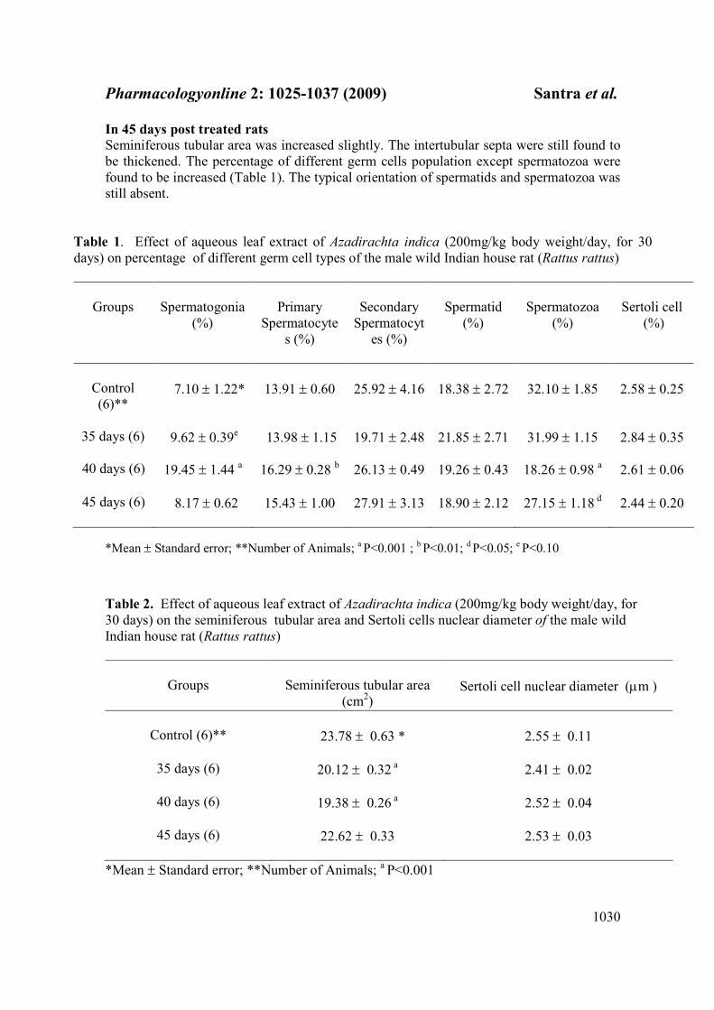
Seminiferous tubular area was increased slightly. The intertubular septa were still found to
be thickened. The percentage of different germ cells population except spermatozoa were
found to be increased (Table 1). The typical orientation of spermatids and spermatozoa was
still absent.
Table 1. Effect of aqueous leaf extract of Azadirachta indica (200mg/kg body weight/day, for 30
days) on percentage of different germ cell types of the male wild Indian house rat (Rattus rattus)
Groups
Spermatogonia
(%)
Primary
Spermatocyte
s (%)
Secondary
Spermatocyt
es (%)
Spermatid
(%)
Spermatozoa
(%)
Sertoli cell
(%)
Control
(6)**
7.10 ± 1.22*
13.91 ± 0.60
25.92 ± 4.16
18.38 ± 2.72
32.10 ± 1.85
2.58 ± 0.25
35 days (6)
9.62 ± 0.39
e 13.98 ± 1.15 19.71 ± 2.48 21.85 ± 2.71 31.99 ± 1.15 2.84 ± 0.35
40 days (6) 19.45 ± 1.44 a
16.29 ± 0.28 b 26.13 ± 0.49 19.26 ± 0.43 18.26 ± 0.98
a 2.61 ± 0.06
45 days (6) 8.17 ± 0.62 15.43 ± 1.00 27.91 ± 3.13 18.90 ± 2.12
27.15 ± 1.18 d 2.44 ± 0.20
*Mean ± Standard error; **Number of Animals; a P<0.001 ;
b P<0.01;
d P<0.05;
e P<0.10
Table 2. Effect of aqueous leaf extract of Azadirachta indica (200mg/kg body weight/day, for
30 days) on the seminiferous tubular area and Sertoli cells nuclear diameter of the male wild
Indian house rat (Rattus rattus)
Groups
Seminiferous tubular area
(cm2)
Sertoli cell nuclear diameter (µm )
Control (6)**
23.78 ± 0.63 *
2.55 ± 0.11
35 days (6) 20.12 ± 0.32 a
2.41 ± 0.02
40 days (6) 19.38 ± 0.26 a
2.52 ± 0.04
45 days (6) 22.62 ± 0.33
2.53 ± 0.03
*Mean ± Standard error; **Number of Animals; a P<0.001
Pharmacologyonline 2: 1025-1037 (2009) Santra et al.
1031
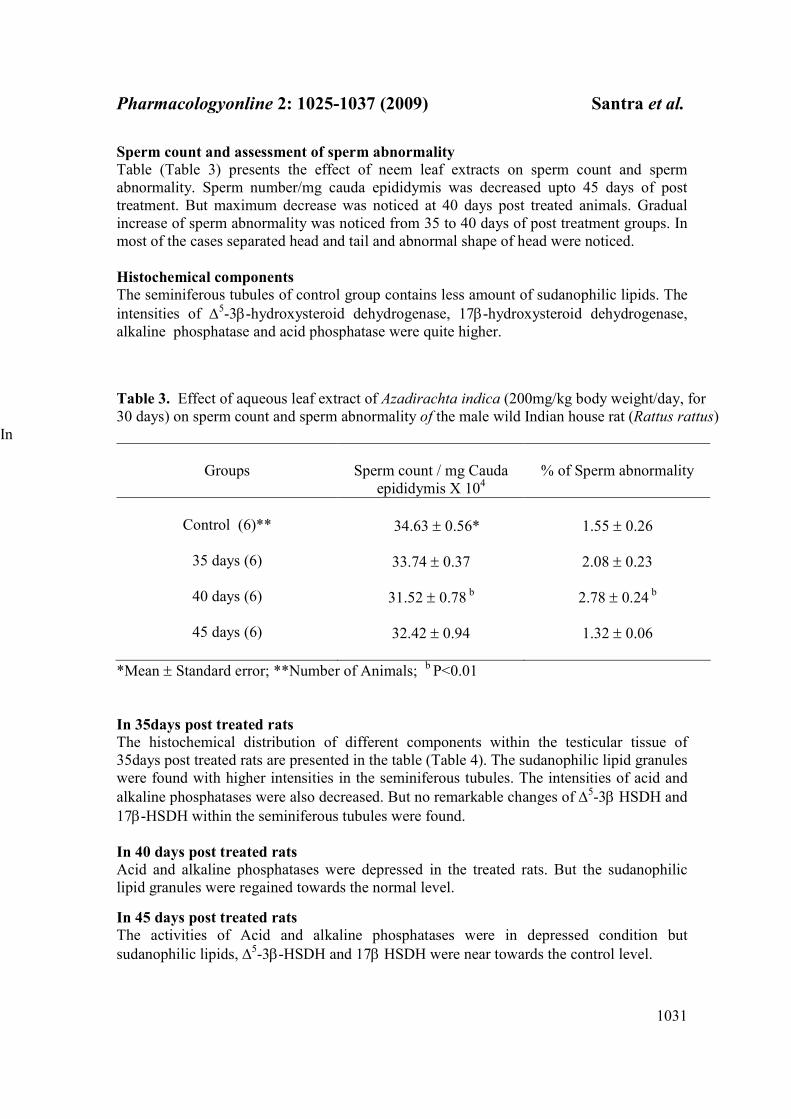
Sperm count and assessment of sperm abnormality
Table (Table 3) presents the effect of neem leaf extracts on sperm count and sperm
abnormality. Sperm number/mg cauda epididymis was decreased upto 45 days of post
treatment. But maximum decrease was noticed at 40 days post treated animals. Gradual
increase of sperm abnormality was noticed from 35 to 40 days of post treatment groups. In
most of the cases separated head and tail and abnormal shape of head were noticed.
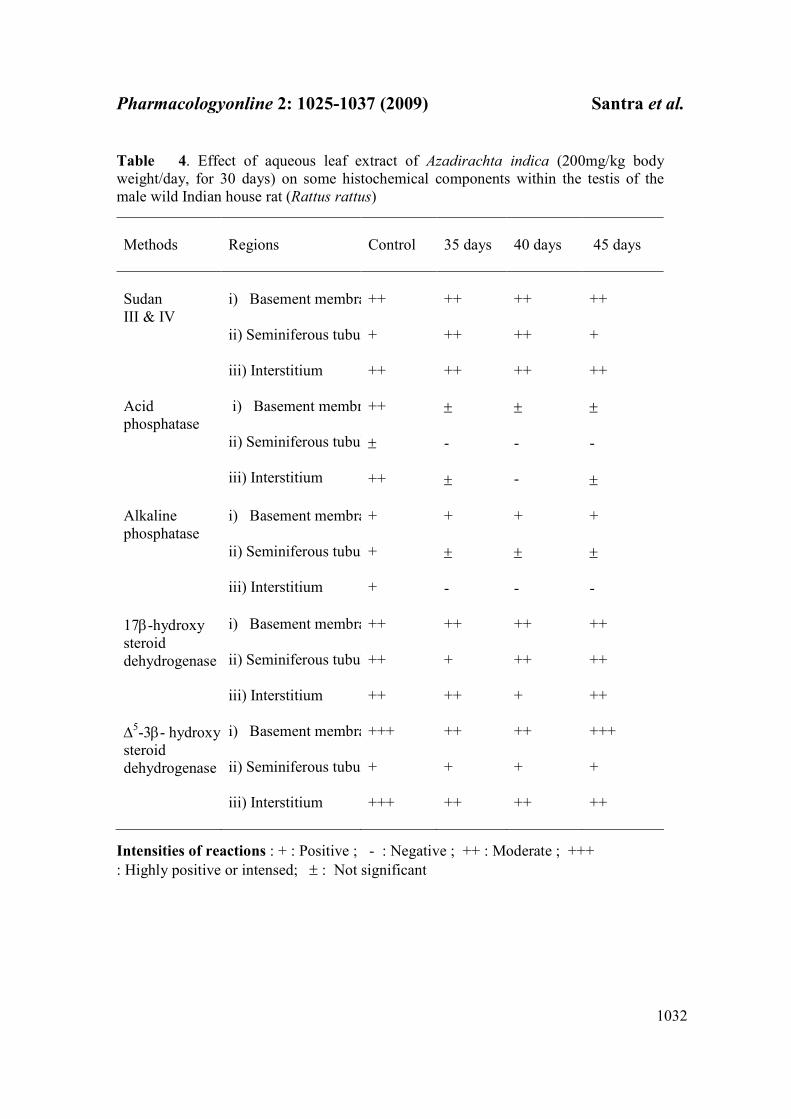
Histochemical components
The seminiferous tubules of control group contains less amount of sudanophilic lipids. The
intensities of ∆5-3β-hydroxysteroid dehydrogenase, 17β-hydroxysteroid dehydrogenase,
alkaline phosphatase and acid phosphatase were quite higher.
Table 3. Effect of aqueous leaf extract of Azadirachta indica (200mg/kg body weight/day, for
30 days) on sperm count and sperm abnormality of the male wild Indian house rat (Rattus rattus)
In
Groups
Sperm count / mg Cauda
epididymis X 104
% of Sperm abnormality
Control (6)**
34.63 ± 0.56*
1.55 ± 0.26
35 days (6)
33.74 ± 0.37 2.08 ± 0.23
40 days (6)
31.52 ± 0.78
b 2.78 ± 0.24
b
45 days (6) 32.42 ± 0.94
1.32 ± 0.06
*Mean ± Standard error; **Number of Animals; b P<0.01
In 35days post treated rats
The histochemical distribution of different components within the testicular tissue of
35days post treated rats are presented in the table (Table 4). The sudanophilic lipid granules
were found with higher intensities in the seminiferous tubules. The intensities of acid and
alkaline phosphatases were also decreased. But no remarkable changes of ∆5-3β HSDH and
17β-HSDH within the seminiferous tubules were found.
In 40 days post treated rats
Acid and alkaline phosphatases were depressed in the treated rats. But the sudanophilic
lipid granules were regained towards the normal level.
In 45 days post treated rats
The activities of Acid and alkaline phosphatases were in depressed condition but
sudanophilic lipids, ∆5-3β-HSDH and 17β HSDH were near towards the control level.
Pharmacologyonline 2: 1025-1037 (2009) Santra et al.
1032
Table 4. Effect of aqueous leaf extract of Azadirachta indica (200mg/kg body
weight/day, for 30 days) on some histochemical components within the testis of the
male wild Indian house rat (Rattus rattus)
Methods
Regions
Control
35 days
40 days
45 days
Sudan
III & IV
i) Basement membrane
ii) Seminiferous tubule
iii) Interstitium
++
+
++
++
++
++
++
++
++
++
+
++
Acid
phosphatase
i) Basement membrane
ii) Seminiferous tubule
iii) Interstitium
++
±
++
±
-
±
±
-
-
±
-
±
Alkaline
phosphatase
i) Basement membrane
ii) Seminiferous tubule
iii) Interstitium
+
+
+
+
±
-
+
±
-
+
±
-
17β-hydroxy
steroid
dehydrogenase
i) Basement membrane
ii) Seminiferous tubule
iii) Interstitium
++
++
++
++
+
++
++
++
+
++
++
++
∆5-3β- hydroxy
steroid
dehydrogenase
i) Basement membrane
ii) Seminiferous tubule
iii) Interstitium
+++
+
+++
++
+
++
++
+
++
+++
+
++
Intensities of reactions : + : Positive ; - : Negative ; ++ : Moderate ; +++
: Highly positive or intensed; ± : Not significant

Pharmacologyonline 2: 1025-1037 (2009) Santra et al.
1033
Biochemical components
Testicular extracts of control rats contain very less concentration of ascorbic acid and
cholesterol. The concentrations of acid and alkaline phosphatases were very high (Table 5).
In 35 days post treated rats
The different biochemical components in the testicular extract of 35 days post-irradiated
group are given in the table (Table 5). The concentration of cholesterol and ascorbic acid
has been increased than the control value. Whereas the concentrations of acid and alkaline
phosphatase were quite lower than the control value.
In 40 days post treated rats
The concentration of cholesterol and ascorbic acid in the treated groups were high. But the
concentration of alkaline phosphatase and alkaline phosphatase have been decreased.
In 45 days post treated rats
The concentration of ascorbic acid was nearer to the normal value. But the concentration of
cholesterol was quite higher than the normal value. The activities of alkaline phosphatase
and acid phosphatase have been decreased in the 45 days of post treated rats in comparison
to the control animals ( Table 5).
Table 5. Effect of aqueous leaf extract of Azadirachta indica (200mg/kg body weight/day, for
30 days) on some biochemical components of the testes of the male wild Indian house rat (Rattus
rattus)
Groups
(mmol/100mg fresh testicular tissue)
(mg/100mg fresh testicular
tissue)
Acid
phosphatase
Alkaline
phosphatase
Ascorbic acid
Cholesterol
Control (6)**
35 days (6)
0.694 ± 0.054*
0.669 ± 0.014
0.312 ± 0.013
0.289 ± 0.024
0.018 ± 0.009
0.022 ±
0.001
0.529 ± 0.02
0.674 ± 0.03
b
40 days (6)
0.595 ± 0.047
0.265 ± 0.026
0.025 ± 0.002
0.649 ±
0.04 c
45 days (6)
0.608 ± 0.034
0.252 ± 0.026 0.018 ± 0.001 0.595 ± 0.05
*Mean ± Standard error; ** Number of Animals: b P<0.01; c P<0.02
Pharmacologyonline 2: 1025-1037 (2009) Santra et al.
1034
Discussion
In the present study oral administration of aqueous leaf extract of neem (Azadirachta
indica) (200 mg/kg body weight/day, for 30 days) on the male reproductive organs of the
wild Indian house rat (Rattus rattus) was investigated and its activity was noticed at 35, 40
and 45 days of post treated animals.
In this study histopathological observations showed that leaf extract of neem (Azadirachta
indica) affected the process of spermatogenesis and disrupted the production of sperm. The
seminiferous tubules showed only spermatogonia, spermatocytes, Sertoli cells, few
spermatids and very small amount of deformed spermatozoa at 35 and 40 days of post
treatment. This might have been the cause of the delay in reproduction observed in the
experimental groups. The reduction in the number of round spermatids was depressed at
40days when compared to the control ratio of spermatids. There is a close relationship
between spermatid losses and the ultimate decline in sperm numbers (22). At 45 days after
treatment, there was evidence of regeneration of spermatozoa and resting spermatocytes
(pre-spermatocytes ) were present. The damaged structures noticed in the seminiferous
tubules of rats at 35and 40 days of post treated groups. They also caused a marked decrease
in the area of the seminiferous tubules of treated groups. The No significant changes were
noticed in the Sertoli cell nuclear diameter and Leydig cells population. Seminiferous
tubular area was increased slightly at 45 days of post treatment.
Though typical orientation of spermatids and spermatozoa was still absent. Sperm
number/mg cauda epididymis was decreased upto 45 days of post treatment. But maximum
decrease was noticed at 40 days post treated animals. Gradual increase of sperm
abnormality was noticed from 35 to 40 days of post treatment groups. In most of the cases
separated head and tail and abnormal shape of head were noticed.
Mishra and Singh (23) showed that oral administration 200 mg/kg body weight/day, for 28
days of aqueous leaf extract of neem (Azadirachta indica) on the male reproductive organs
of the Parkes (P) strain mice had no effect on body weight and the reproductive organs
weight. In severe cases, the tubules were lined with Sertoli cells only, Sertoli cells and rare
germ cells, or with Sertoli cells and several germ cells but without cellular association
patterns (23). Leaf extract of neem (Azadirachta indica) had adverse effects on motility,
morphology, and number of spermatozoa in the cauda epididymidis. Sander-Cramer (24)
and Garge et al. (25) showed that purified neem extract had potent spermicidal activity.
Neem extract also reduced sperm counts and motility and increased the number of
abnormal sperm (26). He also reported reductions in weight, epithelial cell height and
nuclear diameter of the ventral prostate and seminal vesicles as indirect evidence of the
anti-androgenic action of neem extract. Neem extract reduced the sperm count and
abnormal sperm morphology.
From the histochemical study, high amount of lipid materials were observed in the treated
groups. This lipid accumulation was mainly restricted to the whole tubules, basement
membrane and the interstitial cells of the testicular tissue. High amount of lipid droplets
within the Sertoli cells of mammals seem to be due to the phagocytosed lipid materials of
the degenerating germ cells, which is also pointed out by Lacy (27).

Pharmacologyonline 2: 1025-1037 (2009) Santra et al.
1035
It is also known that the lipid inclusion in the Sertoli cell may be in the control of FSH
secretion from the pituitary (28). Serum FSH have been shown to be related to the germinal
cell component, particularly spermatogonial numbers (29). Both the lipids and steroid
dehydrogenases are usually known to show inverse relationship between them. Higher
content of lipids and lower activity levels of the dehydrogenases are characteristic of
steroidogenically inactive gonads.
Acid and alkaline phosphatases were found to be decreased within the treated groups of
rats (both biochemical and histochemical). The alkaline phosphatase is said to be a
histochemical marker for primordial germ cells of various species, including rat (30) and
mouse (31). The alkaline and acid phosphatases have been widely studied in many
organisms and tissues (32) including certain animal testis or sperm (33, 34). The role of
alkaline phosphatase in the development of spermatozoa has been well studied in a number
of mammalian species. It is known that this enzyme is required for the synthesis of
glycogen, which in turn apparently participates in the metabolic process of spermatogenesis
(35). Mann (36) reported an intense activity of acid phosphatase in the seminal plasma of
several mammalian species including the human beings. Seminal and prostatic acid
phosphatase has been associated with the nutrition of spermatozoa (37, 38) and with their
fertilizing ability (39).
It is also known from the present investigation that ascorbic acid from the testicular tissues
has been increased due to oral administration of neem leaf extract. Role of ascorbic acid in
the process of steroidogenesis is well known (40). In mammals, ascorbic acid has been
found to exert an inhibitory role on steroidogenesis (40). Again as ascorbic acid is a known
catalyst for both lipid peroxidation and alteration of unsaturated fatty acid composition
(41), so the involvement of ascorbic acid in the process of steroidogenesis in the testis of
the rats of control and treated groups may be taken into consideration. Adverse effects of
leaf extract of neem on the level of fructose in the seminal vesicle and sialic acid in the
duct was noticed in mice by Mishra and Singh (23).
It has been suggested (42) that 17 β-ol-dehydrogenase ( oxidase ) is located within both the tubules and the interstitial elements and is diminished slightly during the oral
administration of neem leaf extract. This phenomenon is directly related to the destructive
changes associated with the depopulation of the seminiferous tubules.
In this study with wild rats, it can be concluded that aqueous crude leaf extract of neem
(Azadirachta indica) has effective contraceptive activity for the control of rodent pests and
reasonable safety at anti-fertility doses used, however, the study has a limitation that it was
done on crude extract that contains many anti-fertility components responsible for observed
effects was not done. Further study on the possible mechanism as well investigation on the
fractionated isolates should be investigated.
References
1. Chopra G, Dhindsa JS. Rodent pest management in poultry farms. Poult. Adv 1987 ; 20
( 3 ) : 49-60.

Pharmacologyonline 2: 1025-1037 (2009) Santra et al.
1036
2. Chopra G, Dhindsa JS. Positivity of opinion survey of rodent control in poultry farms.
Ind. Poult. Rev.1988 ; 20 ( 1 ) : 31-39.
3. Prakash I, Ghosh PK. Rodents in Indian Agriculture. Vol. I. Scientific Publishers,
Jodhpur 1992; 1-685.
4.WHO. Ecology and control of rodents of public health importance. WHO Technical
Report. Series No. 553 1974 : 1-2.
5. Upadhyay SN, Dhawan S, Talwar GP. Antifertility effects of neem (Azadirachta indica)
oil in male rats by single intra-vas administration: an alternate approach to vasectomy.”
Journal of Andrology 1993;14(4): 275-81.
6. Jensen JT. Male contraception. Current Womens Health Reports 2002; 2(5): 338-45.
7. Deshpande VY, Mendulkar KN, Sadre NL. Male antifertility activity of Azadirachta
indica in mice. Journal of Postgraduate Medicine 1980; 26: 167-70.
8. Lal R, Sankarnarv A, Mathur VS, Sharma PL. Antifertility effect of neem oil in female
albino rats by intra vaginal and oral routes. Indian J. Med. Res. 1986; 83: 89-92.
9. Randhawa NS, Parmar BS. Neem. New Age International. New Delhi, India 1996.
10. Kay WW, Whitehead R. Sudan III and IV methods for neutral fats. Histochemistry-
Theoretical and Applied. 2nd ed. Pearse . AGE ed. 1941; 853-854.
11. Wattenberg LW. Microscopic histochemical demonstration of steroid 3β-ol-
dehydrogenase in tissue sections . J. Histochem. Cytochem. 1958 ; 6 : 225-232.
12. Bilaspuri GS, Guraya SS. Histochemical studies on steroid dehydrogenases in the testis
of the goat ( Capra hircus ). J. Endocrinol. 1984 ; 101 : 359-363.
13. Pearson B, Grose F. Histochemical demonstration of 17β-hydroxysteroid
dehydrogenase by use of a tetrazolium salt. Proc. Soc. Exptl. Biol. Med. 1959 ; 100 : 636-
638.
14. Butcher RG, Chayan J. Quantitative studies on the alkaline phosphatase reaction . J.
Roy. Micros. Soc. 1966 ; 85 : 111-117.
15. Bitensky L. Modifications of the Gomori acid phosphatase technique for controlled
temperature frozen sections. Quart. J. Micros. Sci. 1963 ; 104 : 193-196.
16. Zarrow MX, Yochim JM, Mc Carthy JT. Experimental endocrinology: A source book
of basic techniques. Academic Press, New York 1964.
17. Nino HV, Prasad AS. Ascorbic acid (Vitamin C) . In : Vitamins and trace elements.
Gradwohl's Clinical Laboratory Methods and Diagnosis. Vol.1. Sonnenwirth AC, Jarett L.
ed. The C. V. Mosby Comp., St. Louis, Toronto, London 1980 ; 370-372.
18. Walter K, Schutt C. Acid and alkaline phosphatase in serum ( Two point method ). In :
Methods of Enzymatic Analysis Vol. 2. Bergmeyer, HU ed. Academic Press, New York,
Sanfrancisco, London.1974 ; 856-860.
19. Barratt CLR, Davis AG, Bansal MR, Williams ME. The effects of lead on the male rat
reproductive system. Andrologia 1989 ; 21 ( 2 ) : 161-166.
20. Didolkar AK, Keizer-Zucker A, Sundaram K, Bardin CW, Agback H, Johansson DB.
Effect of sulfasalazinc and its analogs on fertility in male rats. Contraception 1988 ; 37 ( 5 )
: 539-548.
21. Fischer RA. Statistical methods for research workers. Oliver Boyd. London 1963 .
22. Pogany GC. Effects of X- irradiation on the kinetics of abnormal sperm production and
sperm loss in the mouse. J. Repord. Fert. 1987 ; 80 :1-12.
23. Mishra RK, Singh SK. Effect of aqueous leaf extract of Azadirachta indica on the
reproductive organs in male mice. Indian journal of experimental biology 2005; 43(11):
1093-1103.

Pharmacologyonline 2: 1025-1037 (2009) Santra et al.
1037
24. Sander FV, Cramer SD. A practical method of testing the spermicidal action of
chemical contraceptives. Human Fert. 1941; 6: 134.
25. Garge S, Doncel G, Chabara S, Upadhyay SN, Talwar GP. Synergistic spermicidal
activity of neem seed extract, reetha saponins and quinine hydrochloride. Contraception
1994;50: 185-190.
26. Shaikh PD. Studies on the antifertility effect of Azadirachta indica leaves on the testis
of albino rats. M. Phil. Dissertation, Karnataka University, India 1990.
27. Lacy D. Light and electron microscopy and its use in the study of factors influencing
spermatogenesis in the rat. J. Roy. Micros. Soc. 1960 ;79 : 209-225.
28. Kerr JB, de Kretser DM. Cyclic variations in Sertoli cell lipid content throughout the
spermatogenic cycle in the rat. J. Reprod. Fert. 1975 ; 43 : 1-8.
29. de Kretser DM, Burger HG, Hudson B. The relationship between germinal cells and
serum FSH levels in males with infertility. J. Clin. Endocrinol. Metab. 1974 ; 38 : 793-797.
30. Mc Alpine RJ. Alkaline glycerophosphatase in the developing adrenals, gonads and
reproductive tract of the white rat. Anat. Rec. 1955 ; 121 : 407-408.
31. Mintz B. Embryological development of primordial germ cells in the mouse : influence
of a new mutation . W. J. Embryol. Exp. Morphol. 1957 ; 5 : 396-403.
32. Mc Comb RB, Bowers GH Jr, Posen S. Alkaline phosphatase. Plenum Publication .
New York 1979 .
33. Terner C, Mc Laughlin J, Smith BR. Changes in lipase and phosphatase activities of rat
spermatozoa in transit from caput to cauda epididymis. J. Reprod. Fert. 1975 ; 45 : 1-8.
34. Chang CH, Angellis D, Fishman WH. Presence of the rare D-variant, heat stable ,
placental type alkaline phosphatase in normal human testes . Cancer Res. 1980 ; 40 :
1506-1510.
35. Sohval AR. The anatomy and endocrine physiology of the male reproductive system. In
: The Endocrinology of reproduction. Velardo JT ed . Oxford University Press, New York
1958 ;243-312.
36. Mann T. The biochemistry of semen and of the male reproductive tract. Methuen and
Co. Ltd. London 1964 .
37. Allison AC, Hartree EF. Lysosomal enzymes in the acrosome and their possible role in
fertilization. J. Repord . fert. 1970 ; 21 : 505-515.
38. Serrano JA, Shannon WA Jr, Sternberger NJ, Wasserkrug HL, Serrano AA, Seligman
AM. The cytochemical demonstration of prostate acid phosphatase using a new substrate,
phosphorylcholine . J. Histochem.Cytochem. 1976 ; 24 : 1046-1056.
39. Singer R, Barnet M, Allalcuf U, Schwartzman S, Sagiv M, Landau B, Segenreich E,
Servadio C. Some properties of acid and alkaline phosphatase in seminal fluid and isolated
sperm. Arch. Androl. 1980 ; 5 : 195-199.
40. Kitabchi AE. Ascorbic acid in steroidogenesis. Nature 1967 ; 215 : 1385-1386.
41. Shimizu K. Effects of ascorbic acid on the side chain cleavage of cholesterol. Biochem.
Biophys. Acta. 1970 ; 210 : 333-340.
42. Ellis LC, Berliner DL. Alterations in testicular androgen biosynthesis as related
changes in spermatogenesis induced by ionizing radiations. In : The Gonads. Mc Kerns
KW ed. Appleton, New York 1969 ; 739-783.