phase-contrast ct: fundamental theorem and fast image ... · phase-contrast ct: fundamental theorem...
TRANSCRIPT
Phase-contrast CT: Fundamental theorem and fast imagereconstruction algorithms
Andrei V. Bronnikov
Bronnikov Algorithms, PO Box 77, Arnhem 6800 AB, The Netherlands
ABSTRACT
Phase-contrast x-ray computed tomography (CT) is an emerging imaging technique that can be implementedat third generation synchrotron radiation sources or by using a microfocus x-ray tube. Promising experimentalresults have recently been obtained in material science and biological applications. At the same time, the lack ofa mathematical theory comparable to that of conventional absorption-based CT limits the progress in this field.We suggest such a theory and prove a fundamental theorem that plays the same role for phase-contrast CT asthe Fourier slice theorem does for absorption-based CT. The fundamental theorem allows us to derive fast imagereconstruction algorithms in the form of filtered backprojection (FBP).
Keywords: phase contrast, micro CT, tomographic image reconstruction
1. INTRODUCTION
Conventional x-ray computed tomography (CT) is based on the difference in radiation absorption by differenttissues. At the same time, a wide range of samples used in biology and medicine demonstrate very weak absorptioncontrast, nevertheless producing significant phase shifts in the x-ray beam. The use of phase information forimaging purposes is therefore a suitable alternative here. Utilizing phase contrast also has attractive sidesitself: first, refractive properties of the medium can be studied, rather than its absorption properties, as done inabsorption-based CT and secondly, it may help to diminish the total absorbed dose, enhancing the conditions ofthe entire imaging procedure.
In this paper we present a mathematical theory which lays down the foundations of quantitative phase-contrastCT, making accurate reconstruction of phase-contrast data as easy as in conventional CT. The suggested theoryrequires no intermediate step of phase retrieval and provides direct reconstruction of the refractive index fromthe intensity distributions measured in a single plane of the near field region. In the case of a mixed phase andamplitude object, the data in the contact print plane are required as well. The theory is based on a fundamentalrelation between the three-dimensional (3D) Radon transform of the object function and the two-dimensional(2D) Radon transform of the phase-contrast projection that is established in the form a fundamental theorem.Using this theorem, reconstruction algorithms can be derived in the form of filtered backprojection.
2. PHASE-CONTRAST IMAGING
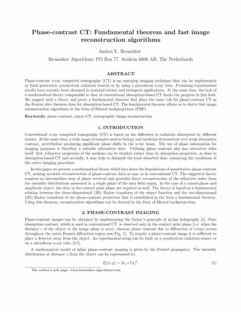
Phase-contrast images can be obtained by implementing the Gabor’s principle of in-line holography [1]. Pureabsorption contrast, which is used in conventional CT, is observed only in the contact print plane (i.e. when thedistance z of the object to the image plane is zero), whereas phase contrast due to diffraction of x-rays occursthroughout the entire Fresnel diffraction region (see Fig. 1). To acquire a phase-contrast image it is sufficient toplace a detector away from the object. An experimental setup can be built on a synchrotron radiation source oron a microfocus x-ray tube [2-5].
A mathematical model of inline phase-contrast imaging is given by the Fresnel propagator. The intensitydistribution at distance z from the object can be represented by
Izθ (x, y) = |hz ∗ Uθ|2, (1)
The author’s web page: www.bronnikov-algorithms.com
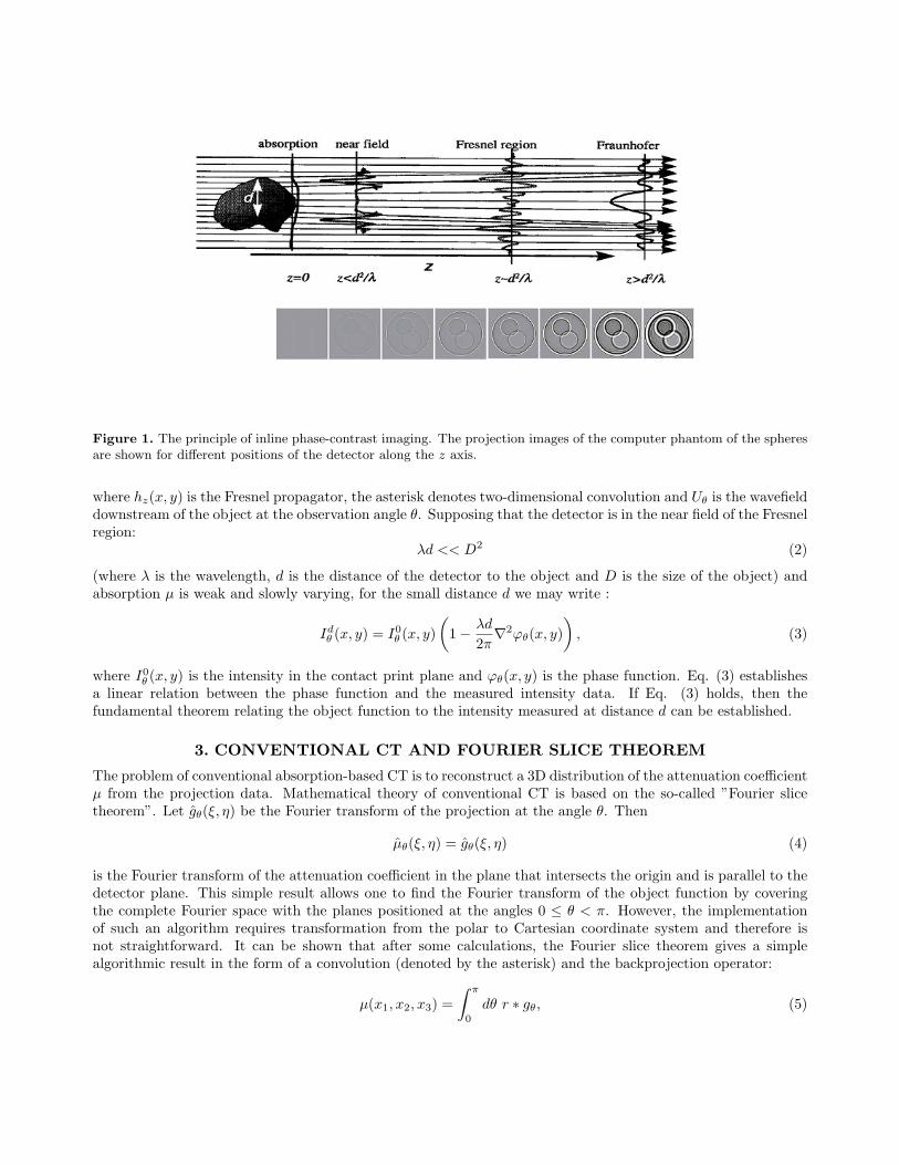
Figure 1. The principle of inline phase-contrast imaging. The projection images of the computer phantom of the spheresare shown for different positions of the detector along the z axis.
where hz(x, y) is the Fresnel propagator, the asterisk denotes two-dimensional convolution and Uθ is the wavefielddownstream of the object at the observation angle θ. Supposing that the detector is in the near field of the Fresnelregion:
λd << D2 (2)
(where λ is the wavelength, d is the distance of the detector to the object and D is the size of the object) andabsorption µ is weak and slowly varying, for the small distance d we may write :
Idθ (x, y) = I0
θ (x, y)(
1− λd
2π∇2ϕθ(x, y)
), (3)
where I0θ (x, y) is the intensity in the contact print plane and ϕθ(x, y) is the phase function. Eq. (3) establishes
a linear relation between the phase function and the measured intensity data. If Eq. (3) holds, then thefundamental theorem relating the object function to the intensity measured at distance d can be established.
3. CONVENTIONAL CT AND FOURIER SLICE THEOREM
The problem of conventional absorption-based CT is to reconstruct a 3D distribution of the attenuation coefficientµ from the projection data. Mathematical theory of conventional CT is based on the so-called ”Fourier slicetheorem”. Let gθ(ξ, η) be the Fourier transform of the projection at the angle θ. Then
µθ(ξ, η) = gθ(ξ, η) (4)
is the Fourier transform of the attenuation coefficient in the plane that intersects the origin and is parallel to thedetector plane. This simple result allows one to find the Fourier transform of the object function by coveringthe complete Fourier space with the planes positioned at the angles 0 ≤ θ < π. However, the implementationof such an algorithm requires transformation from the polar to Cartesian coordinate system and therefore isnot straightforward. It can be shown that after some calculations, the Fourier slice theorem gives a simplealgorithmic result in the form of a convolution (denoted by the asterisk) and the backprojection operator:
µ(x1, x2, x3) =∫ π
0
dθ r ∗ gθ, (5)
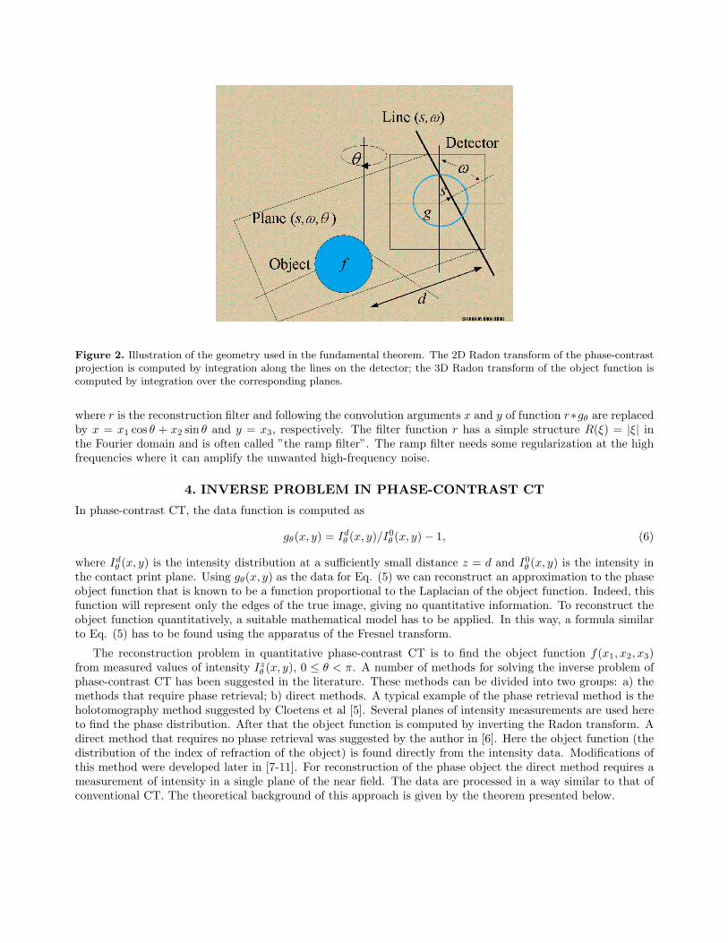
Figure 2. Illustration of the geometry used in the fundamental theorem. The 2D Radon transform of the phase-contrastprojection is computed by integration along the lines on the detector; the 3D Radon transform of the object function iscomputed by integration over the corresponding planes.
where r is the reconstruction filter and following the convolution arguments x and y of function r∗gθ are replacedby x = x1 cos θ + x2 sin θ and y = x3, respectively. The filter function r has a simple structure R(ξ) = |ξ| inthe Fourier domain and is often called ”the ramp filter”. The ramp filter needs some regularization at the highfrequencies where it can amplify the unwanted high-frequency noise.
4. INVERSE PROBLEM IN PHASE-CONTRAST CT
In phase-contrast CT, the data function is computed as
gθ(x, y) = Idθ (x, y)/I0
θ (x, y)− 1, (6)
where Idθ (x, y) is the intensity distribution at a sufficiently small distance z = d and I0
θ (x, y) is the intensity inthe contact print plane. Using gθ(x, y) as the data for Eq. (5) we can reconstruct an approximation to the phaseobject function that is known to be a function proportional to the Laplacian of the object function. Indeed, thisfunction will represent only the edges of the true image, giving no quantitative information. To reconstruct theobject function quantitatively, a suitable mathematical model has to be applied. In this way, a formula similarto Eq. (5) has to be found using the apparatus of the Fresnel transform.
The reconstruction problem in quantitative phase-contrast CT is to find the object function f(x1, x2, x3)from measured values of intensity Iz
θ (x, y), 0 ≤ θ < π. A number of methods for solving the inverse problem ofphase-contrast CT has been suggested in the literature. These methods can be divided into two groups: a) themethods that require phase retrieval; b) direct methods. A typical example of the phase retrieval method is theholotomography method suggested by Cloetens et al [5]. Several planes of intensity measurements are used hereto find the phase distribution. After that the object function is computed by inverting the Radon transform. Adirect method that requires no phase retrieval was suggested by the author in [6]. Here the object function (thedistribution of the index of refraction of the object) is found directly from the intensity data. Modifications ofthis method were developed later in [7-11]. For reconstruction of the phase object the direct method requires ameasurement of intensity in a single plane of the near field. The data are processed in a way similar to that ofconventional CT. The theoretical background of this approach is given by the theorem presented below.
Figure 3. Reconstruction of the phase object by the suggested algorithm. No phase retrieval is required; a single detectorposition in the near field of the Fresnel region is sufficient.
5. FUNDAMENTAL THEOREM OF PHASE-CONTRAST CT
In order to find an analytical solution to the inverse problem of phase-contrast CT, we make certain approxima-tions that hold in the near field of the Fresnel region (see [7] for details). Using the hat for notations of 2D and3D Radon transforms, we present the main result in the form of the following
TheoremLet the data function be given by Eq. (6) and the conditions of Eqs (2) and (3) hold, then
∂2
∂s2f(s, θ, ω) =
−1d
gθ(s, ω). (7)
The theorem shows the relationship between the measured intensity of the x-ray beam downstream of the objectand the index of refraction of the object. The theorem has a structure reminding that of the Fourier slicetheorem in conventional CT, but instead of the Fourier transform the Radon transform is applied here. TheRadon transform is an integration along lines in 2D and an integration over planes in 3D. As seen from Fig.2, the theorem says that the integrals of the data along lines in the detector plane are proportional to thesecond derivative of the corresponding plane integrals of the object. Since the formula for the inverse 3D Radontransform is known, the theorem can be directly used to find the object function.
6. RECONSTRUCTION ALGORITHMS
Using the fundamental theorem, a reconstruction algorithm can be derived. Indeed, the object function can befound from equation (7) if the 3D Radon transform is inverted. This gives us the formula
f(x1, x2, x3) =1
4π2d
∫ π
0
sinωdω
∫ π
0
dθ gθ(s′, ω). (8)
Inversion formula (8) suggests that a 2D Radon transform in the detector plane has to be computed for eachprojection gθ(x, y). The result is then back-projected into the 3D space by using the backprojection operatorof the 3D inverse Radon transform. Applying the full 2D Radon transform to each projection is a lengthycomputational task; a more practical FBP reconstruction algorithm could be obtained if Eq. (8) was simplified

Figure 4. Reconstruction of the mixed phase and amplitude object by the suggested algorithm. Two detector positions:in the contact print plane and in the near field of the Fresnel region are required.
by calculating the integral over angle ω. This result was first obtained in [6]. The algorithm is reduced to afamiliar FBP algorithm with the filter function q(x, y) = |y|/(x2 + y2):
f(x1, x2, x3) =1
4π2d
∫ π
0
dθ q ∗ ∗gθ, (9)
where following the convolution arguments x and y of function q ∗ ∗gθ are replaced by x = x1 cos θ + x2 sin θ andy = x3, respectively. Eq. (9) gives us an algorithm that is similar to the FBP algorithm of conventional CT (seeEq. (5)). The major difference is that the filtering operation in phase-contrast CT is done in two dimensions.The filter can be implemented in the Fourier domain:
Q(ξ, η) =|ξ|
ξ2 + η2, (10)
where ξ and η are the spatial frequencies.
7. MIXED PHASE AND AMPLITUDE OBJECTS
The use of the approach for mixed phase and amplitude objects is straightforward, at least theoretically. Indeed,since I0
θ (x, y) is measured in the contact print plane, it should contain information of absorption contrast onlyand therefore the absorption-contrast image is canceled by division in Eq. (6). In practical implementation thismethod may require some additional processing of the data I0
θ (x, y) that can be noisy and of insufficient contrast.Note that reconstruction of the mixed phase and amplitude objects will require measurements in two planes,which are the contact print plane and the plane at the distance d from the object. In the case of a purely phaseobject, I0
θ (x, y) = Ii, where Ii is the intensity of the incident beam, so that only a measurement in a single planein the near field is needed (compare Figs. 3 and 4).
8. STABILITY
As was pointed out already in [6,7], Q(ξ, η) is a low-pass filter so that the FBP algorithm given by Eqs. (9),(10)is stable to the high-frequency noise. This is a special property of phase-contrast reconstruction. Here we havea situation where the reconstruction algorithm does not need to be stabilized at the high frequencies, which isopposite to the situation in conventional CT. At the same time, the instability of the inverse problem appears
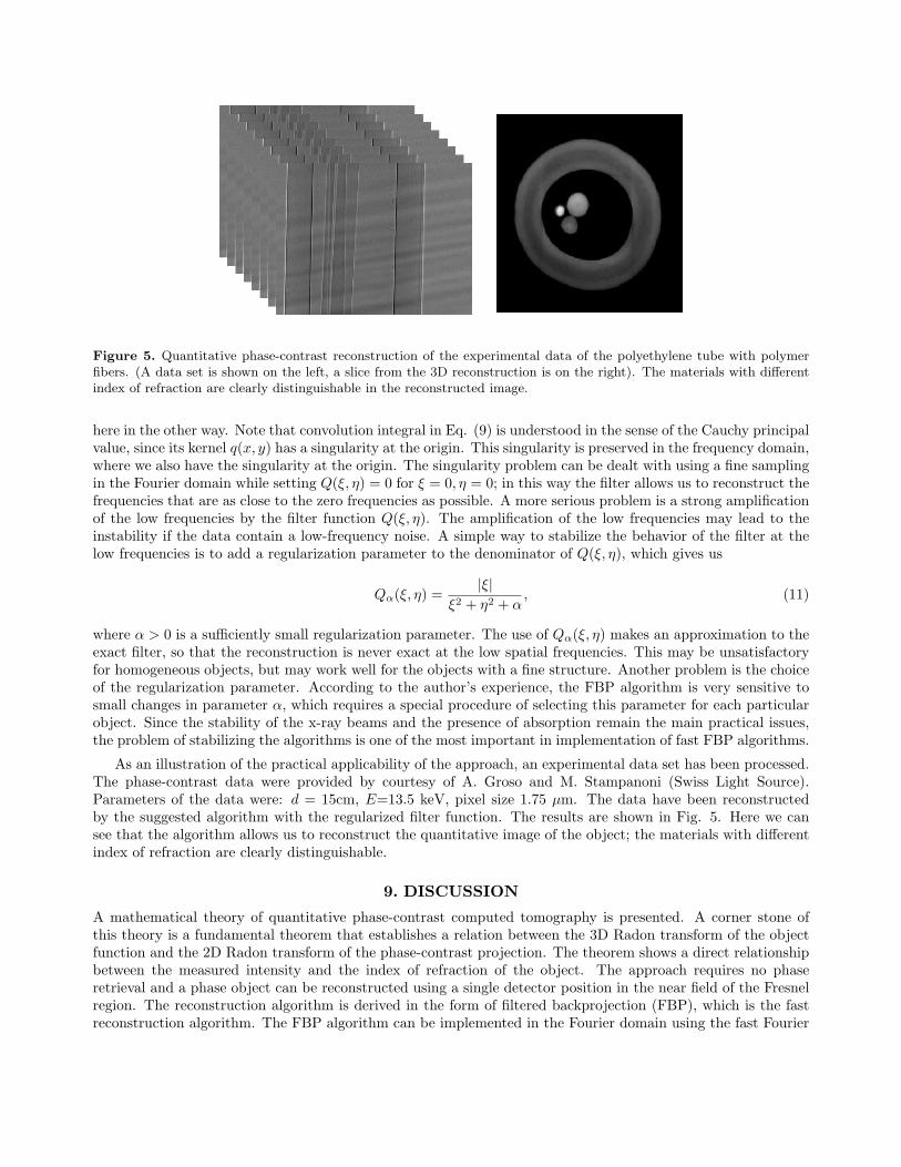
Figure 5. Quantitative phase-contrast reconstruction of the experimental data of the polyethylene tube with polymerfibers. (A data set is shown on the left, a slice from the 3D reconstruction is on the right). The materials with differentindex of refraction are clearly distinguishable in the reconstructed image.
here in the other way. Note that convolution integral in Eq. (9) is understood in the sense of the Cauchy principalvalue, since its kernel q(x, y) has a singularity at the origin. This singularity is preserved in the frequency domain,where we also have the singularity at the origin. The singularity problem can be dealt with using a fine samplingin the Fourier domain while setting Q(ξ, η) = 0 for ξ = 0, η = 0; in this way the filter allows us to reconstruct thefrequencies that are as close to the zero frequencies as possible. A more serious problem is a strong amplificationof the low frequencies by the filter function Q(ξ, η). The amplification of the low frequencies may lead to theinstability if the data contain a low-frequency noise. A simple way to stabilize the behavior of the filter at thelow frequencies is to add a regularization parameter to the denominator of Q(ξ, η), which gives us
Qα(ξ, η) =|ξ|
ξ2 + η2 + α, (11)
where α > 0 is a sufficiently small regularization parameter. The use of Qα(ξ, η) makes an approximation to theexact filter, so that the reconstruction is never exact at the low spatial frequencies. This may be unsatisfactoryfor homogeneous objects, but may work well for the objects with a fine structure. Another problem is the choiceof the regularization parameter. According to the author’s experience, the FBP algorithm is very sensitive tosmall changes in parameter α, which requires a special procedure of selecting this parameter for each particularobject. Since the stability of the x-ray beams and the presence of absorption remain the main practical issues,the problem of stabilizing the algorithms is one of the most important in implementation of fast FBP algorithms.
As an illustration of the practical applicability of the approach, an experimental data set has been processed.The phase-contrast data were provided by courtesy of A. Groso and M. Stampanoni (Swiss Light Source).Parameters of the data were: d = 15cm, E=13.5 keV, pixel size 1.75 µm. The data have been reconstructedby the suggested algorithm with the regularized filter function. The results are shown in Fig. 5. Here we cansee that the algorithm allows us to reconstruct the quantitative image of the object; the materials with differentindex of refraction are clearly distinguishable.
9. DISCUSSION
A mathematical theory of quantitative phase-contrast computed tomography is presented. A corner stone ofthis theory is a fundamental theorem that establishes a relation between the 3D Radon transform of the objectfunction and the 2D Radon transform of the phase-contrast projection. The theorem shows a direct relationshipbetween the measured intensity and the index of refraction of the object. The approach requires no phaseretrieval and a phase object can be reconstructed using a single detector position in the near field of the Fresnelregion. The reconstruction algorithm is derived in the form of filtered backprojection (FBP), which is the fastreconstruction algorithm. The FBP algorithm can be implemented in the Fourier domain using the fast Fourier
transform and is as simple as the conventional FBP algorithm. A family of related algorithms can be obtainedby modifying the filter function in order to provide the solution with the required properties.
Computer simulations showed promising results both for phase and mixed phase and amplitude objects.A thorough numerical analysis of the reconstruction problem and a series of computer experiments have beendone by the author in [7]; we refer to [7] for all necessary numerical illustration of the theory. An independentevaluation of the suggested algorithm was done by Anastasio et al [8]. Groso et al [9] have pointed out theproblem of instability of the algorithm to the low frequency noise in the data. Their conjecture was that the realsamples are never purely phase objects and therefore some absorption is always present, which can jeopardize thebehavior of the reconstruction algorithm at the low spatial frequencies. They have stabilized the reconstructionwith respect to the low frequency noise by using a regularized filter function as of Eq. (11) and provided anheuristic procedure for finding the regularization parameter [10]. Gureyev et al suggested a more theoreticalapproach for selecting the regularization parameter for the case of proportionality of the amplitude and phaseobjects [11]. A further work is needed to generalize the FBP algorithms for objects with arbitrary absorption.
REFERENCES1. D. Gabor, ”A new microscopic principle,” Nature 161 777-778 (1948).2. A. Snigirev, I. Snigireva, V. Kohn, S. Kuznetsov, and I. Schelokov, ”On the possibilities of x-ray phase contrast
microimaging by coherent high-energy synchrotron radiation,” Rev. Sci. Instrum. 66 5486-5492 (1995).3. T. Davis, D. Gao, T. Gureyev, A. Stevenson, and S. Wilkins, ”Phase contrast imaging of weakly absorbing
materials using hard x-rays,” Nature 373 595-598 (1995).4. S. W. Wilkins, T.E. Gureyev, D. Gao, A. Pogany, and A. W. Stevenson, ”Phase-contrast imaging using
polychromatic hard x-rays,” Nature 384 335-338 (1996).5. P. Cloetens, W. Ludwig, J. Baruchel, D. Van Dyke, J. Van Landuyt, J. P. Guigay, and M. Schlenker, ”Holo-
tomography: quantitative phase tomography with micrometer resolution using hard synchrotron radiation xrays,” Appl. Phys. Let. 75 2912-2914 (1999).
6. A.V. Bronnikov, ”Reconstruction formulas in phase-contrast tomography,” Optics Communications 171 239-244 (1999).
7. A.V. Bronnikov, ”Theory of quantitative phase-contrast computed tomography,” J. Opt. Soc. Am. A 19472-480 (2002).
8. M.A. Anastasio, D. Shi, F. De Carlo, and X. Pan, ”Analytic image reconstruction in local phase-contrasttomography”, Phys. Med. Biol. 49 121-144 (2004).
9. A.Groso, M. Stampanoni, R. Abela, P. Schneider, S. Linga, R. Muller, ”Phase contrast tomography: analternative approach”, Appl. Phys. Let 88 214104 (2006).
10. A.Groso, R. Abela, M. Stampanoni, ”Implementation of a fast method for high resolution phase contrasttomography”, Optics Express, to be published (2006).
11. T.E. Gureyev, D.M Paganin, G.R. Myers, Ya.I. Nesterets and S. Wilkins, ”Phase-and-amplitude computertomography,” Apll. Phys. Let 89 034102 (2006).