value of non-contrast ct examination of the urinary tract
TRANSCRIPT

Alexandria Journal of Medicine (2016) 52, 209–217
HO ST E D BYAlexandria University Faculty of Medicine
Alexandria Journal of Medicine
http://www.elsevier.com/locate/ajme
Value of non-contrast CT examination of the
urinary tract (stone protocol) in the detection of
incidental findings and its impact upon the
management
* Corresponding author at: 27 Ahmad Foad Nour Str., Camp
Caesar, Alexandria 21525, Egypt. Tel.: +20 (003)01001948320.E-mail addresses: [email protected] (M.S. Shaaban),
[email protected] (A.F. Kotb).
Peer review under responsibility of Alexandria University Faculty of
Medicine.
http://dx.doi.org/10.1016/j.ajme.2015.08.0012090-5068 � 2015 The Authors. Alexandria University Faculty of Medicine. Production and hosting by Elsevier B.V.This is an open access article under the CC BY-NC-ND license (http://creativecommons.org/licenses/by-nc-nd/4.0/).
Mohamed Samir Shaabana,*, Ahmed Fouad Kotb
b
aDiagnostic and Interventional Radiology Department, Faculty of Medicine, Alexandria University, EgyptbUrology Department, Faculty of Medicine, Alexandria University, Egypt
Received 14 April 2015; accepted 6 August 2015
Available online 4 September 2015
KEYWORDS
Stone protocol;
CT;
Urolithiasis;
Incidental;
Urinary tract stones
Abstract Background: Urolithiasis is one of the most common urinary tract diseases worldwide,
with a wide range of affected age groups. Non-contrast CT examination of the urinary tract is the
gold-standard examination for detection and characterization of urinary tract stones, with great
impact upon the choice of method of management. Aside from detection of stones, non-contrast
CT examination of the abdomen and pelvis also offers a valuable overlook upon the other abdom-
inal organs, pathologies of which may simulate a stone disease, or accompany stone disease and can
be detected incidentally, which may shift management plan dramatically.
Aim of work: To demonstrate the use of non-contrast CT examinations (stone protocol) in the
detection of abdominal pathologies other than stones, whether or not simulating the clinical picture
of urolithiasis, and its impact upon patient management.
Patients and methods: Assessment of the non-contrast examinations of the urinary tract of patients
referred for suspected stone urolithiasis recording any incidental finding and follow-up of the
impact of these incidental findings upon the management delivered to the patient.
Results: A total of 719 examinations were performed, of which 334 had urinary tract stones only,
211 had incidental finding beside urinary tract stones, 170 had an incidental finding with no urinary
tract stones, and four patients had neither stones nor incidental findings. A total number of 381
patients had incidental findings, 198 (47%) of which had an impact upon the management.
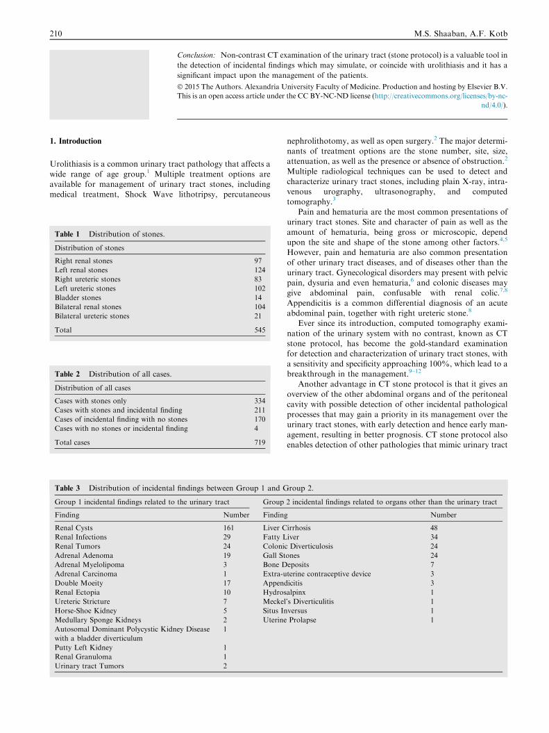
Table 1 Distribution of stones.
Distribution of stones
Right renal stones
Left renal stones
Right ureteric stones
Left ureteric stones
Bladder stones
Bilateral renal stones
Bilateral ureteric stones
Total
Table 2 Distribution of all cases.
Distribution of all cases
Cases with stones only
Cases with stones and incidental findin
Cases of incidental finding with no sto
Cases with no stones or incidental find
Total cases
Table 3 Distribution of incidental
Group 1 incidental findings related to
Finding
Renal Cysts
Renal Infections
Renal Tumors
Adrenal Adenoma
Adrenal Myelolipoma
Adrenal Carcinoma
Double Moeity
Renal Ectopia
Ureteric Stricture
Horse-Shoe Kidney
Medullary Sponge Kidneys
Autosomal Dominant Polycystic Kidn
with a bladder diverticulum
Putty Left Kidney
Renal Granuloma
Urinary tract Tumors
210 M.S. Shaaban, A.F. Kotb
Conclusion: Non-contrast CT examination of the urinary tract (stone protocol) is a valuable tool in
the detection of incidental findings which may simulate, or coincide with urolithiasis and it has a
significant impact upon the management of the patients.
� 2015 The Authors. Alexandria University Faculty of Medicine. Production and hosting by Elsevier B.V.
This is an open access article under the CC BY-NC-ND license (http://creativecommons.org/licenses/by-nc-
nd/4.0/).
1. Introduction
Urolithiasis is a common urinary tract pathology that affects a
wide range of age group.1 Multiple treatment options areavailable for management of urinary tract stones, includingmedical treatment, Shock Wave lithotripsy, percutaneous
97
124
83
102
14
104
21
545
334
g 211
nes 170
ing 4
719
findings between Group 1 and G
the urinary tract Group
Number Finding
161 Liver C
29 Fatty L
24 Coloni
19 Gall St
3 Bone D
1 Extra-u
17 Append
10 Hydros
7 Meckel
5 Situs In
2 Uterine
ey Disease 1
1
1
2
nephrolithotomy, as well as open surgery.2 The major determi-nants of treatment options are the stone number, site, size,attenuation, as well as the presence or absence of obstruction.2
Multiple radiological techniques can be used to detect andcharacterize urinary tract stones, including plain X-ray, intra-venous urography, ultrasonography, and computed
tomography.3
Pain and hematuria are the most common presentations ofurinary tract stones. Site and character of pain as well as the
amount of hematuria, being gross or microscopic, dependupon the site and shape of the stone among other factors.4,5
However, pain and hematuria are also common presentationof other urinary tract diseases, and of diseases other than the
urinary tract. Gynecological disorders may present with pelvicpain, dysuria and even hematuria,6 and colonic diseases maygive abdominal pain, confusable with renal colic.7,8
Appendicitis is a common differential diagnosis of an acuteabdominal pain, together with right ureteric stone.8
Ever since its introduction, computed tomography exami-
nation of the urinary system with no contrast, known as CTstone protocol, has become the gold-standard examinationfor detection and characterization of urinary tract stones, with
a sensitivity and specificity approaching 100%, which lead to abreakthrough in the management.9–12
Another advantage in CT stone protocol is that it gives anoverview of the other abdominal organs and of the peritoneal
cavity with possible detection of other incidental pathologicalprocesses that may gain a priority in its management over theurinary tract stones, with early detection and hence early man-
agement, resulting in better prognosis. CT stone protocol alsoenables detection of other pathologies that mimic urinary tract
roup 2.
2 incidental findings related to organs other than the urinary tract
Number
irrhosis 48
iver 34
c Diverticulosis 24
ones 24
eposits 7
terine contraceptive device 3
icitis 3
alpinx 1
’s Diverticulitis 1
versus 1
Prolapse 1

Figure 1 Fifty-two year old female patient with bilateral flank pain. CT stone protocol in axial plane at the level of gall bladder (a)
showing gall bladder stones (arrow). (b) Axial scan at the left of left renal hilum shows a left renal pelvic stone (arrow).
Figure 2 Fifty-six year old male patient with left flank pain. CT
stone protocol shows small diverticula in the sigmoid colon
(arrows). No stones were found.
Figure 3 Thirty-four year old female patient with right sided
abdominal pain and dysuria. CT stone protocol revealed an
oblong tubular structure related to the ileal loops, with stranded
adjacent fascial planes, proven at surgery to be an inflamed
Meckel’s diverticulum.
Figure 4 Thirty-four year old male patient with bilateral flank
pain. In addition to bilateral renal stones, CT stone protocol
shows a left adrenal lipid-rich adenoma (arrow).
Value of CT examination of urinary tract and its impact 211
stone in its symptoms and signs, and so redirecting the man-agement plan to its correct path.13–15
2. Methods and materials
This is a prospective study that included a total of 719 patientswho had CT stone protocol examinations performed by a 16-
detector CT Siemens Somatom Sensation, with no oral orintravenous contrast medium administration, during the per-iod between May 2012 and December 2014, with clinically sus-
pected urinary tract stone disease.No patient preparation was required, apart from assuring a
full urinary bladder.
Patients lied on CT table in supine position, with elevatedarms behind the head. Initially a topogram in antero-posterior view was extended from the lower chest down tothe upper thighs. Then, scans were obtained from the dome
of the liver to below the ischial tuberosities using 1.5 mm slicecollimation and images were reconstructed at 1 mm slice thick-ness and 0.75 intervals. Setting of the exposure factors had
been 130 KVp and 200 mAS.

Figure 5 Forty-one year old female patient with dysuria and hematuria. CT stone protocol revealed a contraceptive device that
penetrated into the urinary bladder lumen. (a) Axial cut at the urinary bladder showing the two limbs of the device (arrow). (b) Axial cut at
a lower level showing the stem of the device (arrow). Coronal (c) and sagittal (d) showing the contraceptive device (arrows).
Figure 6 Thirty-four year old female patient with left flank pain.
CT stone protocol in sagittal oblique plane through the left kidney
shows a parapelvic cyst causing mild dilatation of the upper calyx
with no stones. Cyst aspiration and sclerotherapy relieved the
pain.
212 M.S. Shaaban, A.F. Kotb
Exclusion criteria included any patient with a known dis-ease (either urinary or extra-urinary) other than the suspected
urinary tract stone.Follow-up of the cases with incidental findings was done
with documentation of the impact of detection of the inciden-
tal pathology upon management.Informed consent was obtained from all individual partici-
pants included in the study.
3. Results
A total of 719 CT stone protocol examinations were obtained.
467 of the patients were males (65%) and 252 were females(35%), age ranged from 15 years and 68 years.
334 patients (46%) had urinary tract stones only with noother associated pathologies detected by non-contrast CT,
Table 1 and 211 patients (29%) had incidental finding besideurinary tract stones, 170 patients (24%) had an incidental find-ing with no urinary tract stones, and four patients (1%) had
neither stones nor incidental findings seen in non-contrastCT study Table 2.
The most common symptom encountered in the study was
flank pain (right in n = 280, left in n= 226, bilateral inn= 129), followed by hematuria (n = 110) and finally dysuria(n = 21).
A total number of 381 patients (53% of total patients) hadincidental findings, and these incidental findings were dividedinto two groups: group 1 with incidental findings related tothe urinary system (66%), and group 2 related to organs other
than the urinary system (34%); Table 3.Considering the patients with extra-urinary incidental find-
ings (group 2), the most of the incidental findings were related
to the hepato-biliary system; 106 patients (62%) (Fig. 1),
followed by the bowel (Figs. 2 and 3); 28 patients (16%), adre-nal masses; 23 patients (14%) (Fig. 4), bone deposits; seven
patients (4%), followed by gynecological disorders; fivepatients (Fig. 5) and one patient with situs inversus.
On the other hand, renal cysts were the urinary tract related
incidental finding most commonly encountered (only one cystof about 6 cm diameter caused renal pain and hence required

Figure 7 Fifty-three year old diabetic female patient with left flank pain. CT stone protocol in axial (a) and coronal (b) planes through
the kidneys shows left renal abscess (arrows). No stones were found.
Figure 8 Sixty-one year old female patient with right flank pain. CT stone protocol through the right kidney in axial (a) and coronal (b)
planes shows dense parenchymal calcifications (arrows). No underlying masses or stones were found and stationary size was found on
follow-up, and so considered as granuloma.
Value of CT examination of urinary tract and its impact 213
intervention) (Fig. 6); 161 patients (62%), followed by renalinfections; 29 patients (11%) (Figs. 7–9), urinary tract tumors;
24 patients (9%) (Figs. 10 and 11), and double-moiety; 17patients (7%).
From the total 381 patients with incidental findings, the dis-
covered incidental findings had an impact on the managementin 198 patients (47%), either in the form of diet adjustment,medical treatment or even surgical intervention (open surgery
or endoscopy). Two patients had medullary sponge kidneys(Fig. 12).
Table 4 shows the patients with incidental findings, and themodification of management they received
4. Discussion
Flank pain and hematuria are common presentations of uri-
nary tract calculi. However, a number of other pathologies
in different abdominal organs and in the urinary tract itselfcan give a similar presentation. We aimed in our study to
assess the utility of non-contract CT examination of the uri-nary tract (stone protocol) in the detection of the pathologiesother than urinary tract stones which mimic their symptoms
and signs, and comparing our results with other similarstudies.
Ather et al.13 studied 4000 patients suspected to have uri-
nary tract stone, and found an alternate diagnosis in 398patients (9.9%), which is different than our finding of 24%stone-free patients, and it should be noted that in this study– in addition to the different sample size - the search was for
a cause for the complaint other than stone; however, in ourstudy the search was for concomitant as well as for alternatediagnosis. Ather et al. also noted a wide spectrum of significant
alternate diagnoses including urogenital (76.6%) and non-uro-genital (23.4%) conditions that could be reliably established or

Figure 9 Sixty-eight year old female patient with left flank pain. CT stone protocol through the left kidney in (a) axial, (b) coronal, and
(c) sagittal planes revealed a small calcified left kidney, consistent with putty kidney.
Figure 10 Sixty-one year old male patient with right flank pain
and hematuria. CT stone protocol shows urinary bladder mass at
the right vesico-ureteric junction (large yellow arrow) with
obstruction of the right ureter, which is filled by mass tissues
(small yellow arrow). Small lower calyceal stone was found in the
left kidney (white arrow).
214 M.S. Shaaban, A.F. Kotb
suggested on spiral CTs performed for suspected renal coliccases. However, Ather et al. included ovarian lesions in thegenitor-urinary group.13
In a study conducted by Katz et al.,14 1000 stone protocolexaminations were reviewed, ureteric stones were found on 557
examinations, findings consistent with a recently passed stonewere found on 67 examinations, and 275 CT examinationswere free. An alternative or additional diagnosis was foundor suggested on 101 examinations (10%), including 26 patients
having both urinary tract stone and an incidental pathology.Again, different sample size than that in our study may causethe different percentage of patients with incidental findings. In
Katz et al. study, there were 62 incidental findings related togenitourinary system and 39 findings not related to the geni-tourinary tract. Katz et al. included pathologies related to
the female genital system to the urinary tract group, whichwas separate in our study.
Studying incidental diseases on 233 unenhanced helicalcomputed tomography examinations performed for ureteric
colic, Ahmad et al.16 found stones-only in 148 examinations(64%), findings of recent passage of calculi in 10 examinations(4%) and no calculus in 75 examinations (32%). Overall the
incidental findings (additional or alternative diagnosis) werefound in 28 (12%) CT scans. They grouped the different inci-dental diagnoses according to the pathology into inflammatory
conditions (n = 12), tumors and masses (n = 12), and otherurological diseases (n= 4). However, by analyzing the differ-ent incidental pathologies, those related to the urinary tract
were 9 (32%), while those not related to the urinary tract were19 (68%), and the most common extra-urinary pathologieswere in adnexal masses and cysts (n= 6), followed by gallbladder diseases (n = 4) and bowel diseases (n = 4).
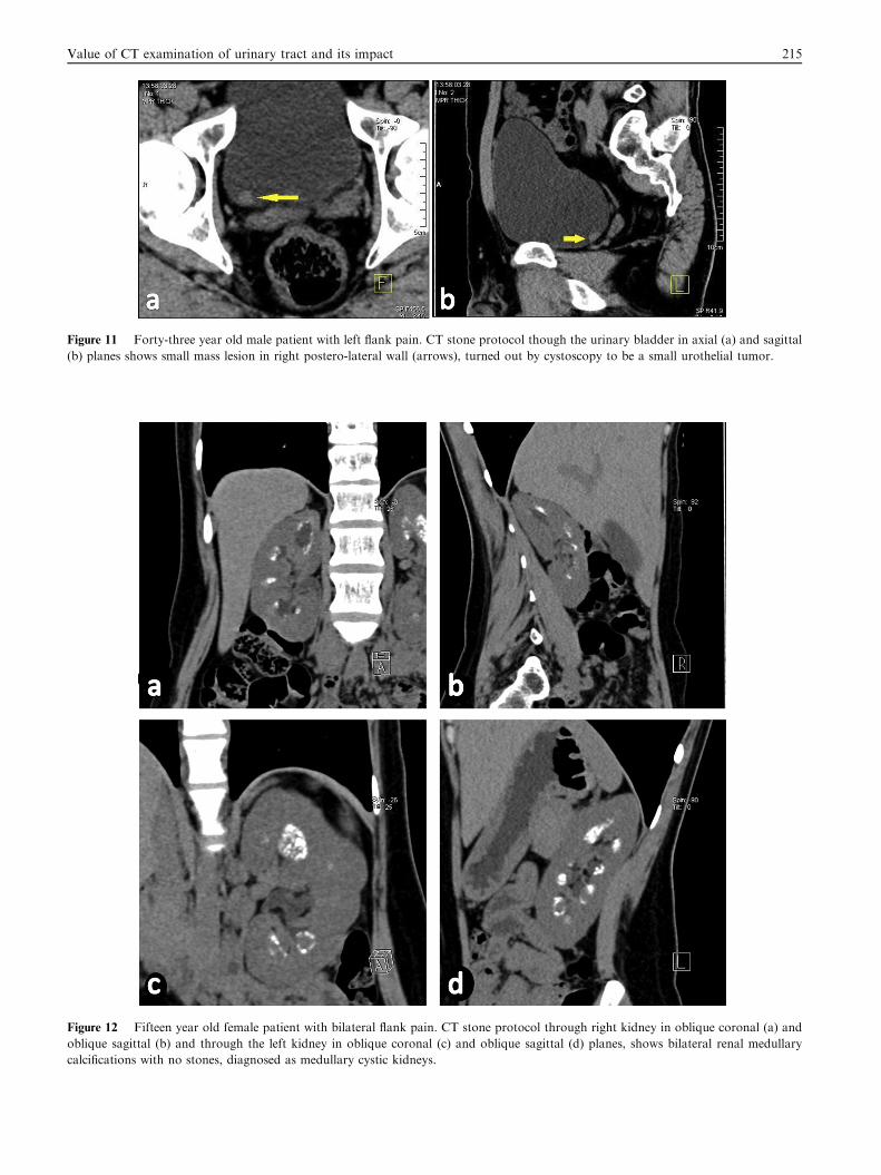
Figure 11 Forty-three year old male patient with left flank pain. CT stone protocol though the urinary bladder in axial (a) and sagittal
(b) planes shows small mass lesion in right postero-lateral wall (arrows), turned out by cystoscopy to be a small urothelial tumor.
Figure 12 Fifteen year old female patient with bilateral flank pain. CT stone protocol through right kidney in oblique coronal (a) and
oblique sagittal (b) and through the left kidney in oblique coronal (c) and oblique sagittal (d) planes, shows bilateral renal medullary
calcifications with no stones, diagnosed as medullary cystic kidneys.
Value of CT examination of urinary tract and its impact 215

Table 4 The patients with incidental findings, and the modification of management they received.
Incidental finding Total number
of patients
Number of patients with
impact on management
Change in management
Adrenocortical Carcinoma 1 1 Surgical Adrenalectomy
APKD and a urinary bladder diverticulum 1 1 Open Stone Extraction
Appendicitis 3 3 Appendicectomy
Bone Deposits 7 7 Further search For Primary
Colonic Diverticulosis 24 19 Medical Treatment
Fatty Liver 23 23 Diet modification and Medical Treatment
Fatty Liver With Gall Stones 11 11 Cholecystectomy With diet And Medical Treatment
Gall Stones 13 13 Cholecystectomy
Hydrosalpinx 2 2 Laparoscopy
Perforating contraceptive device 3 3 Endoscopic Extraction
Liver Cirrhosis 48 48 Medical Treatment
Medullary cystic Kidneys 2 2 Medical Treatment
Meckel’s Diverticulitis 1 1 Surgery
Renal Cysts 161 1 Cyst Aspiration
Renal Infections 29 29 Surgical And Medical Treatment
Renal Tumors 24 24 Nephrectomies
Stricture 7 7 Dilatation
Urinary tract Tumor 2 2 Cystoscopy
Uterine Prolapse 1 1 Surgery
Total 420 198
216 M.S. Shaaban, A.F. Kotb
In a study conducted by Hoppe et al.,17 1500 patientsunderwent unenhanced CT due to acute flank pain. 1035
(69%) had urinary tract calculi. Stones alone were found in331 of these patients (32%) and additional pathological condi-tions were noted in 704 (68%). Of all patients 1064 (71%) had
other or additional CT findings. Of all patients 207 (14%) hadnon-stone related CT findings requiring immediate or deferredtreatment, 464 (31%) had pathological conditions of little clin-
ical importance and 393 (26%) had pathological conditions ofno clinical relevance. CT was normal in 105 of all patients(7%).
The different sample sizes in our study and in the men-
tioned studies contribute to the different percentages ofpatients with stone-only, stone with incidental findings andpatients with alternate diseases. Also the fact that different
studies (including our study), different categorization of theindividual incidental/alternate findings also contributed tothe apparently different results.
Still all studies have agreed that the stone protocol exami-nation adds to the detection of pathologies other than urinarytract stones, which may mimic their presentations, or be inci-dentally found with urolithiasis.
5. Conclusion
Non-enhanced CT examination of the urinary tract offers thehighest sensitivity and specificity in the detection and charac-terization of urinary tract stones, and is also valuable in thedetection of both incidental and alternate pathologies that
may be incidentally found, with great impact on patient diag-nosis and management.
Conflict of interest
The authors declared that there is no conflict of interest.
References
1. Turney BW, Reynard JM, Noble JG, Keoghane SR. Trends in
urological stone disease. BJU Int 2012;109:1082–7.
2. Preminger GM, Tiselius HG, Assimos DG, Alken P, Buck C,
Gallucci M, et al. 2007 guideline for the management of ureteral
calculi. J Urol 2007;178:2418–34.
3. Bhargava P, Dighe MK, Lee JH, Wang C. Multimodality
imaging of ureteric disease. Radiol Clin North Am 2012;50
():271–99.
4. Kazi SN, Benz RL, Kazi SN, Benz RL. Work-up of hematuria.
Prim Care 2014;41(4):737–48.
5. Flannigan R, Choy WH, Chew B, Lange D. Renal struvite stones–
pathogenesis, microbiology, and management strategies. Nat Rev
Urol 2014;11(6):333–41.
6. Cordeiro Gonzalez P, Punal Pereira A, Blanco Gomez B, Lema
Grille J. Bladder endometriosis: report of 7 new cases and review
of the literature. Arch Esp Urol 2014;67(7):646–9.
7. Nigri G, Petrucciani N, Giannini G, Aurello P, Magistri P,
Gasparrini M, et al. Giant colonic diverticulum: clinical presen-
tation, diagnosis and treatment: systematic review of 166 cases.
World J Gastroenterol 2015;21(1):360–8.
8. Brown J. Diagnostic and treatment patterns for renal colic in US
emergency departments. Int Urol Nephrol 2006;38:87–92.
9. Tasian GE, Copelovitch L. Evaluation and medical management
of kidney stones in children. J Urol 2014;192(5):1329–36.
10. Ahmad NA, Ather MH, Rees J. Unenhanced helical computed
tomography in the evaluation of acute flank pain. Int J Urol
2003;10:287–92.
11. Larsen AS, Pedersen R, Sandbaek G. Computed tomography of
the urinary tract: optimalization of low-dose stone protocol in a
clinical setting. Acta Radiol 2005;46(7):764–8.
12. Kirpalani A, Khalili K, Lee S, Haider MA. Renal colic:
comparison of use and outcomes of unenhanced helical CT for
emergency investigation in 1998 and 2002. Radiology
2005;236:554–8.
13. Ather MH, Faizullah K, Achakzai E, et al. Alternate and
incidental diagnoses on non contrast enhanced spiral computed
tomography for acute flank pain. Urol J 2009;6:14–8.

Value of CT examination of urinary tract and its impact 217
14. Katz DS, Scheer M, Lumerman JH, Mellinger BC, Stillman CA,
Lane MJ. Alternative or additional diagnoses on unenhanced
helical computed tomography for suspected renal colic: experience
with 1000 consecutive examinations. Urology 2000;56:53–7.
15. Ather MH, Memon W, Rees J. Clinical impact of incidental
diagnosis of disease on non-contrast enhanced helical CT for acute
ureteral colic. Semin Ultrasound CT MR 2005;26:20–3.
16. Ahmad NA, Ather MH, Rees J. Incidental diagnosis of diseases
on un-enhanced helical computed tomography performed for
ureteric colic. BMC Urol 2003;3:2.
17. Hoppe H, Studer R, Kessler TM, Vock P, Studer UE, Thoeny HC.
Alternate or additional findings to stone disease on unenhanced
computerized tomography for acute flank pain can impact
management. J Urol 2006;175:1725–30.