phd thesis - ku liu.pdf · phd thesis guannan liu . studying extrachromosomal genetic elements in...
TRANSCRIPT

F A C U L T Y O F S C I E N C E
U N I V E R S I T Y O F C O P E N H A G E N
PhD thesis Guannan Liu
Studying Extrachromosomal Genetic Elements in Sulfolobus
Academic advisor: Roger A. Garrett
February 2015

Acknowledgments
This work has only been made possible because many people were supporting me
scientifically and personally.
I want to thank my supervisor Roger A. Garrett for the excellent guidance, suggestions
and critical comments that made this work progressing. I also appreciate his support and
patience for discussions and clarifications.
I am very grateful to Qunxin She who is always available for scientific suggestions,
and his encouragement during my studies.
Thanks to Xu Peng who is always the best person to ask for the experimental skills.
She provided me ideas for further studies.
Great thanks to Susanne Erdmann, who supervised me a lot during my PhD study and
also during the thesis writing even she left this lab. She taught me all the practical and
theoretical knowledge about archaeal work, especially about the virus work, inspiring me
to develop my own ideas.
Thanks to all the lab members and the people in my office for their scientific helps and
social life: Laura Martínez, Shiraz Shah, Simon Bressendorff, Carlos Leon, Ling Deng,
Kristine Uldahl, Soley Gudbergsdottir, Erica Ferrandi, Wenyuan Han, Wenfang Peng, Fei
He, Yang Guo, Marzieh Mousaei, Chandra Shekar Kenchappa, Daniel Stiefler-Jensen,
Mariah Nabi, Hien Phan, Mariana Awayez, Michael Christiaan Greeff, Eleazar
Rodriguez, Magnus Wohlfahrt Rasmussen, Signe Lolle, Milena Roux, Stephan
Thorgrimsen, Raquel Azevedo, Sabrina Stanimirovic. It has been a great pleasure
working together.
Thanks to all my friends all over the world who provided great personal supports. All
of you guys make me happy during my PhD study and beyond. I am very thankful to
Haiyan Ma, who contributed enormously to improve my English.
Last but not the least, I want to express my gratitude to my whole family. Their love
and encouragement is forever imprinted on my mind.

Table of Contents Summary ............................................................................................................................. I
Sammendrag ...................................................................................................................... II
Abbreviations .................................................................................................................. III
I. Introduction .................................................................................................................... 1 1. Archaea ................................................................................................................................... 1
1.1 Sulfolobus .......................................................................................................................... 1 2. Archaeal extrachromosomal genetic elements .................................................................... 4
2.1 Archaeal viruses ................................................................................................................. 4 2.1.1 Spindle-shaped archaeal viruses ............................................................................................... 4
2.1.1.1 Fuselloviridae ................................................................................................................... 4 2.1.1.2 Bicaudaviridae ................................................................................................................. 5 2.1.1.3 Monocaudaviruses ............................................................................................................ 6
2.1.2 Linear viruses ........................................................................................................................... 6 2.1.2.1 Rudiviridae ....................................................................................................................... 6 2.1.2.2 Lipothrixviridae ................................................................................................................ 6
2.2 Membrane vesicles .......................................................................................................... 10 2.2.1 Mechanisms of MVs biogenesis ............................................................................................. 10
2.2.1.1 Bacterial MVs ................................................................................................................. 10 2.2.1.2 Eukaryotic MVs.............................................................................................................. 11 2.2.1.3 Archaeal MVs ................................................................................................................. 11
2.3 Archaeal plasmids ............................................................................................................ 12 2.3.1 Sulfolobus plasmid ................................................................................................................. 12
2.3.1.1 Cryptic plasmids ............................................................................................................. 12 2.3.1.2 Conjugative plasmids ..................................................................................................... 13
2.4 Horizontal gene transfer .................................................................................................. 15 2.4.1 Integration .............................................................................................................................. 15 2.4.2 Transposable elements ........................................................................................................... 16
2.4.2.1 IS Elements ..................................................................................................................... 17 2.4.2.2 Non-autonomous Mobile Elements ................................................................................ 17
3. CRISPR-Cas systems ........................................................................................................... 183.1 Mechanism of CRISPR targeting .................................................................................... 19 3.2 CRISPR-Cas system of S. solfataricus P2 ....................................................................... 21 3.3 CRISPR-Cas system of S. islandicus REY15A ............................................................... 22
II. Membrane Vesicles of Sulfolobus ............................................................................ 23III. Interactions of Archaeal Virus ATV with the CRISPR Adaptive ImmuneSystem of Sulfolobus solfataricus .................................................................................... 33
IV. Conflicting Interactions between the Archaeal Conjugative Plasmid pKEF9and Different Sulfolobus Hosts ....................................................................................... 51
Perspectives ...................................................................................................................... 70
References ......................................................................................................................... 72
Summary Archaea constitute a separate domain in the universal tree of life. They exhibit
exceptional biological properties and provide important insights into the origin of cellular
life. Rapid advances in DNA sequencing and bioinformatical methods as well as the
development of versatile genetic tools have facilitated the characterization of viruses,
plasmids and membrane vesicles. Studying the interactions between Sulfolobus and
extrachromosomal genetic elements has provided many new insights into basic molecular
processes.
Secreted membrane vesicle seems to be a common characteristic for Sulfolobus. In
order to study the biochemical compositions and the genetic functions of these membrane
vesicles, production of membrane vesicles in Sulfolobus was optimized, and the
membrane vesicles were shown to contain cellular DNA. Furthermore, DNA sequencing
revealed that the DNA bound to membrane vesicles consisted of random chromosomal
fragments, including IS elements. The results suggest that membrane vesicles could serve
as vehicles for the inter-cellular transport of genetic material.
A variant of ATV, ATV2, was isolated that infected a newly isolated Sulfolobus
solfataricus P3 strain. Comparative genomics of three closely related viruses (ATV,
ATV2, ATVv) revealed a conserved genome organization, but many differences in gene
size and content. Comparison of the CRISPR loci in S. solfataricus P3 with those of three
published S. solfataricus strains showed many shared spacers, as well as different spacers,
especially those adjoining the leader region. Several spacers of the newly isolated S.
solfataricus P3 had significant sequence matches to ATV and ATV2 genomes, indicating
S. solfataricus P3 has been a host for ATV viruses previously.
Finally, interactions between pKEF9 and Sulfolobus hosts were studied to gain a better
understanding of the interactions between conjugative plasmids and hosts. The result also
demonstrated why certain archaeal conjugative plasmids are gradually lost during
continuous growth. Whereas loss of pKEF9 in S. islandicus was due to interference from
the host CRISPR-Cas system, whereas the deactivation of pKEF9 in S. solfataricus was
caused by orfB mobile elements after it had integrated into the host genome.
I

Sammendrag Arkæa udgør et separat domæne på livets træ. De er i besiddelse af exceptionelle
biologiske egenskaber og giver en vigtig indsigt i oprindelsen af cellulært liv. Fremskridt
inden for DNA sekvensering og bioinformatisk analyse samt udviklingen af alsidige
genetiske teknikker har muliggjort karakteriseringen af virus, plasmider og
membranvesikler. Studiet af interaktionerne mellem Sulfolobus og ekstrakromosale
genetiske elementer giver ny indsigt i basale molekylære processer, for eksempel
konjugation, integration og replikation.
Produktionen af membranvesikler lader til at være et fælles karakteristika for
Sulfolobus. For at studere den biokemiske sammensætning og de genetiske funktioner af
disse membranvesikler, blev produktionen af membranvesikler i Sulfolobus optimeret og
det blev vist membranvesiklerne indeholdt cellulær DNA.
Ydermere afslørede DNA sekvensering at det DNA der er bundet til
membranvesiklerne består af tilfældige kromosomale fragmenter, inklusiv insertion
sequence (IS) elementer. Resultatet antyder at membranvesikler kan fungere som vesikler
for intercellulær transport af genetisk materiale.
Der blev isoleret en variant af ATV, ATV2, der kan inficere en nyligt isoleret
Sulfolobus solfataricus P3 stamme. Sammenlignings af genomerne fra tre tæt relateret
virus (ATV, ATV2, ATVv) viste en konserveret genom organisation, men mange
forskelle i gen størrelse og indhold. Sammenligning af CRISPR loci i S. solfataricus P3
med loci i tre publicerede S. solfataricus stammer viste mange ens spacers og forskellige
spacers, specielt dem der lå op til leader regionen. Adskillige spacers fra den nyligt
isoleret S. solfataricus P3 havde signifikante sekvens matches til ATV og ATV2
genomerne, indikerende af S. solfataricus P3 tidligere har været vært for ATV virus.
Til sidst blev interaktioner mellem pKEF9 og Sulfolobus værter undersøgt for at få en
bedre forståelse af interaktionerne mellem konjugative plasmider og værter. Resultatet
demonstrerer også hvorfor visse arkæa konjugative plasmider bliver gradvist mistet ved
kontinuert vækst. Tab af pKEF9 i S. islandicus skyldtes interferens fra værts CRISPR-
Cas systemet, hvor deaktiveringen af pKEF9 i S. solfataricus var forsaget af orfB mobile
elementer efter de var integreret i værts genomet.
II

Abbreviations ABV: Acidianus bottle-shaped virus
AFV: Acidianus filamentous virus
ARV: Acidianus rod-shaped virus
ASV: Acidianus spindle-shaped virus
ATP: Adenosine triphosphate
ATV: Acidianus two-tailed virus
CRISPR: Clustered regularly interspaced short palindromic repeat
Cas: CRISPR-associated
CsCl: Caesium chloride
Cmr: CRISPR module RAMP (repeat–associated mysterious protein)
crRNA: CRISPR RNA
dsDNA: Double-strand DNA
ssDNA: Single-strand DNA
IS: Insertion Sequence
MITE: Miniature inverted-repeat transposable elements
MV: Membrane vesicle
OD: Optical density
p.c.: Post conjugation
PAM: Proto-spacer adjacent motif
qPCR: Quantitative polymerase chain reaction
rRNA: Ribosomal ribonucleic acid
SMV1: Sulfolobus monocauda virus 1
SIFV: Sulfolobus islandicus filamentous virus
SIRV: Sulfolobus islandicus rod-shaped virus
SNDV: Sulfolobus neozealandicus droplet-shaped virus
SSV: Sulfolobus spindle shaped virus
STIV: Sulfolobus turreted icosahedral virus
STSV: Sulfolobus tengchongensis spindle-shaped virus
TEM: Transmission electron microscopy
UV: Ultraviolet
III

I. Introduction 1. Archaea
Archaea contribute up to 20% of the biomass on earth (DeLong & Pace, 2001), and
they are prevalent in extreme environments, especially those with high temperature
(hyperthermophiles), high salt concentration (halophiles) and extreme pH (acidophiles
and alkalophiles) (Pikuta et al., 2007). So far, six phyla have been proposed:
Euryarchaeota, Crenarchaeota, Korarchaeota, Nanoarchaeota, Thaumarchaeota and
Aigarchaeota (Fig. 1) (Brochier-Armanet et al., 2008, Nunoura et al., 2011), the first two
of which are the most studied branches of the Archaea domain (Woese et al., 1990).
Diverse genetic elements have been discovered in archaeal cells, in particular in
Sulfolobus: viruses, plasmids, membrane vesicles and mobile elements.
1.1 Sulfolobus
Sulfolobus species were first described in 1972 (Brock et al., 1972), and belong to the
Crenarchaeota phylum. They are broadly distributed in Iceland, Italy, Russia, Japan,
China, USA and New Zealand in solfataric hot acid springs (Whitaker et al., 2003). They
optimally grow aerobically at pH around 2-3 and temperatures of 75-80°C (Zillig et al.,
1980). Sulfolobus species can grow heterotrophically utilizing organic compounds, or
chemolithotrophically via CO2 fixation as energy and carbon sources (Bernander, 2007).
Currently 17 Sulfolobus genomes have been sequenced
(http://www.ebi.ac.uk/genomes/archaea.html) (Table 1), facilitating comparative
genomics studies. The GC content of most sequenced Sulfolobus strains is around 35%,
and lower for S. tokodaii at 32.79% (Table 1). In addition, the genomic comparison of ten
Sulfolobus islandicus strains has revealed that they share an approximately 2Mb core and
long variable regions with many strain-specific genes (Jaubert et al., 2013).
The overall organization of the cell cycle in Sulfolobus is well characterized, but much
more work is needed on revealing the regulation mechanism of cell cycle and virus-host
interactions (Bernander, 2000, Bernander, 2003, Duggin & Bell, 2006). Sulfolobus
strains, including S. solfataricus and S. islandicus, are employed as hosts for propagating
diverse viruses and plasmids (Prangishvili et al., 2006, Pina et al., 2011). Several genetic
tools have been established, e.g. genetic knockout and virus-sensitive mutants
(Gudbergsdottir et al., 2011).
1

Fig.1 Unrooted Bayesian tree of the archaeal domain based on a concatenation of ribosomal proteins. The scale bar indicates the average number of substitutions per site. Numbers at branches represent posterior probabilities. (Brochier-Armanet et al., 2011).
2

Table 1. Summary of the sequenced Sulfolobus strains. GC content is calculated through (http://tubic.tju.edu.cn/GC-Profile/) (Gao & Zhang, 2006).
Sulfolobus Length (bp) GC content (%)
Genes number
Accession number
Sulfolobus. acidocaldarius
DSM639 2,225,959 36.71 2,330 CP000077
Sulfolobus acidocaldarius N8 2,176,362 36.7% 2,275 CP002817
Sulfolobus acidocaldarius
Ron12/I 2,223,983 36.7% 2,317 CP002818
Sulfolobus acidocaldarius
SUSAZ 2,061,920 36.3% 2,146 CP006977
Sulfolobus. solfataricus P2 2,992,245 35.79 3,034 AE006641
Sulfolobus. solfataricus 98/2 2,668,974 35.83 2,728 CP001800
Sulfolobus. tokodaii 2,694,756 32.79 2,875 NC003106 S. islandicus
LAL14/1 2,465,177 35.14 2,591 CP003928
Sulfolobus. islandicus HVE10/4 2,655,201 35.15 2,718 CP002426
Sulfolobus. islandicus L.D.8.5 2,722,032 35.25 3,128 CP001731
Sulfolobus. islandicus L.S.2.15 2,736,272 35.11 3,071 CP001399
Sulfolobus. islandicus M.14.25 2,608,832 35.10 2,902 CP001400
Sulfolobus. islandicus M.16.4 2,586,647 35.00 2,871 CP001402
Sulfolobus. islandicus M.16.27 2,692,402 35.01 2,958 CP001401
Sulfolobus. islandicus REY15A 2,522,992 35.31 2,819 CP002425
Sulfolobus. islandicus Y.G.57.14
2,702,058 35.39 3,083 CP001403
Sulfolobus. islandicus Y.N.15.51
2,812,165 35.29 3,271 CP001404
3

2. Archaeal extrachromosomal genetic elements Rapid developments in DNA sequencing and versatile bioinformatics approaches have
greatly facilitated the study of extrachromosomal genetic elements, e.g. viruses, plasmids
and membrane vesicles. In order to study their life cycles, host interactions and genetics,
plasmids and viruses have been used as models of choice, because of their small genomes
and relatively rapid replication (Lindas & Bernander, 2013).
2.1 Archaeal viruses
Viruses are one of the greatest reservoirs of genetic diversity on the planet, and they
play a pivotal role in horizontal gene transfer, thereby driving the evolution of their hosts
(Sorek et al., 2008). Although our understanding of archaeal viruses has advanced
significantly during the past 40 years, much remains to be explored, e.g. the detailed
mechanisms of absorption and entry, replication, assembly and release, as well as the
transcriptional regulation.
There are 65 sequenced archaeal viruses in the database
(http://www.ebi.ac.uk/genomes/archaealvirus.html). All of them contain double-stranded
DNA genomes, with the exception of HRPV1 (Halorubrum pleomorphic virus 1) which
carries a single-stranded (ss) DNA genome (Pietila et al., 2009). Based on their
morphology and genome content, archaeal viruses have been mainly classified into eight
representative viral families (Table 2), including spindle-shaped Fuselloviridae and
Bicaudaviridae, rod-shaped Rudiviridae, fiamentous Lipothrixviridae, spherical-shaped
Globuloviridae, bottle-shaped Ampullaviridae, droplet-shaped Guttaviridae and
bacilliform Clavaviridae (Fig. 2, 3) (Geslin et al., 2007, Pina et al., 2011). There are also
some unclassified pleomorphic viruses.
2.1.1 Spindle-shaped archaeal viruses
2.1.1.1 Fuselloviridae
To date, nine known species of the fuselloviruses (Table 2, Fig. 3) propagate in
Sulfolobus and/or Acidianus (Martin et al., 1984, Stedman et al., 2003, Wiedenheft et al.,
2004, Redder et al., 2009). The majority of these family members have spindle-shaped
virions, with the exceptions of SSV6 and ASV1 (Acidianus spindle-shaped virus 1)
whose virions tend to be pleomorphic (Redder et al., 2009). Fuselloviruses carry a set of
short, thin fibres at one of the pointed ends, leading to the formation of rosette-like
aggregates. All these nine fuselloviruses have circular dsDNA with a conserved integrase
4

of the tyrosine recombinase family. Owing to their integration sites within the integrase
genes, integration results in the partition of integrase genes (Muskhelishvili et al., 1993,
Letzelter et al., 2004, Clore & Stedman, 2007). In addition, the genomes of viruses have
numerous recombination sites, which can facilitate genome rearrangements to adapt to
the ever-changing environment.
Sulfolobus spindle-shaped virus (SSV)
As a typical member of the Fuselloviridae, SSV1 is one of the best studied archaeal
viruses (Fig. 2a). It was originally isolated from S. shibatae, and has been shown to be
lysogenic in Sulfolobus. SSV1 has a 15.5-kb circular dsDNA genome encoding 34
putative proteins (Table 2) (Nadal et al., 1986), most of which are not annotated in the
NCBI database. The biochemical and structural studies on the SSV1 proteome are
gradually assigned. SSV1 replication can be significantly induced by UV irradiation.
Therefore, it has been used as a pioneering model for transcription studies in Archaea
(Reiter et al., 1987, Frols et al., 2007). Moreover, SSV1 was employed to construct of the
first shuttle vectors for Sulfolobales (Jonuscheit et al., 2003).
2.1.1.2 Bicaudaviridae
Acidianus two-tailed virus (ATV)
ATV, the first characterized virus of the Bicaudaviridae family (Table 2), undergoes a
unique morphological change outside its host (Haring et al., 2005). ATV virions are
devoid of tails when released from the host, taking the form of a lemon shape particle, but
they can extend their tails at both ends extracellularly when the incubation temperature is
close to their natural infection conditions (Fig. 2b). The circular dsDNA genome of ATV
is 62.7 kb, and it encodes 72 putative genes with at least 11 structural proteins and an
integrase of the tyrosine recombinase family. The integrase allows ATV to establish two
infection modes: lysogenic (integration into the host chromosome) or lytic (interrupted by
stress factors, such as UV irradiation or mitomycin C treatment) (Prangishvili et al.,
2006). ATV2 was isolated from an enrichment culture of an environmental sample
collected from a hot spring in Pozzuoli, Italy, and maintained in a virus-sensitive strain of
S. solfataricus (See Chapter III).
Sulfolobus monocaudavirus 1 (SMV1)
Virions of SMV1 are fusiform with a single tail and a nose-like structure on the
opposite pole (Erdmann et al., 2014). The observation of plaques formed by the virus-
sensitive strain of S. solfataricus in Gelrite plates and the presence of the integrase gene
5

suggest that SMV1 could also have two life cycles. Sequence comparison shows that 14
of the SMV1 putative proteins have similarities with ATV proteins (Haring et al., 2005,
Prangishvili et al., 2006), implying they are close phylogenetically.
2.1.1.3 Monocaudaviruses
Both STSV1 and STSV2 have a spindle-shaped morphology with a single tail of
variable length protruding from one of the ends (Xiang et al., 2005, Erdmann et al.,
2014). They do not cause cell lysis. After infecting its host, STSV1 replicates rapidly and
retards the host growth. STSV2 can be stably cultured over long periods in several
laboratory strains of Sulfolobus. Both viruses may serve as good models for investigating
archaeal virus–host interactions (Erdmann et al., 2014).
2.1.2 Linear viruses
Linear viruses represent the most abundant virion morphotype in extreme
environments (Rachel et al., 2002). They are classified into two families: the stiff and
rod-like Rudiviridae, and the flexible filamentous Lipothrixviridae (Fig. 3) (Prangishvili
et al., 2006). Despite these differences, rudiviruses share at least nine genes with
lipothrixviruses, suggesting that these two families may have evolved from a common
ancestor (Peng et al., 2001, Prangishvili et al., 2013).
2.1.2.1 Rudiviridae
There are three short terminal fibres at each end of the rod-shaped, non-enveloped
Rudiviridae virions (Fig. 3d). All the known characterized rudiviruses carry linear
dsDNA genomes with long inverted terminal repeats ending in covalently closed hairpin
structures which prime DNA replication (Blum et al., 2001, Peng et al., 2001). As the
representative viruses, both SIRV1 and SIRV2 are present in carrier states in their
original hosts. However, upon infection of other host strains, SIRV2 was stable and
invariant in contrast to SIRV1 which yields many variants (Prangishvili et al., 1999).
2.1.2.2 Lipothrixviridae
Unlike rudiviruses, lipothrixvirus filaments are enveloped (Prangishvili et al., 2006).
Eight representatives of this family (Table 2, Fig.3e) can propagate in Sulfolobus. They
have different terminal structures at each end of the virion, e.g. claws (AFV1), T-bars
(AFV9), mop-like structures (SIFV), three (AFV3) or six (SFV) short filaments or tips
resembling bottle brushes (AFV2). The structure of the AFV3 virion consists of a
6

cylindrical envelope containing globular subunits in a helical formation (Vestergaard et
al., 2008).
Fig. 2 Electron micrographs of archaeal viruses with exceptional morphologies. a) SSV1 (inset) and its extrusion from the host cell, b) ATV (inset) and its extrusion from the host cell, c) ABV, d) SNDV. Bars, 100nm. Adapted from (Prangishvili et al., 2006).
Fig. 3 Morphological diversity in crenarchaeal viruses with a) Fuselloviridae, b) STIV2, c) Globuloviridae, d) Rudiviridae and e) Lipothrixiviridae. Bars, 100nm. Modified from (Krupovic et al., 2011).
7

Table 2. Morphology and taxonomical classification of archaeal viruses with the hosts from the phylum Crenarchaeota.
Virion morphology Family/genus Virus Abbreviation Host
Genome Origin Reference Lengt
h (bp) Type (C/L) Int Accession
No.
Spindle
Fuselloviridae
Sulfolobus spindle-shaped virus 1 SSV1
Sulfolobus
15,465 C + X07234 Japan (Palm et al., 1991)
Sulfolobus spindle-shaped virus 2 SSV2 14,796 C + AY370762 Italy (Stedman et al., 2003)
Sulfolobus spindle-shaped virus 4 SSV4 15,135 C + EU030938 Iceland (Peng, 2008)
Sulfolobus spindle-shaped virus 5 SSV5 15,330 C + EU030939 Iceland (Redder et al., 2009)
Sulfolobus spindle-shaped virus 6 SSV6 15,684 C + FJ870915 Iceland (Redder et al., 2009)
Sulfolobus spindle-shaped virus 7 SSV7 17,602 C + FJ870916 Iceland (Redder et al., 2009)
Sulfolobus virus Kamchatka1 SSVk1 17,385 C + AY423772 Russia (Wiedenheft et al.,
2004)
Bicaudaviridae
Acidianus two-tailed virus ATV Acidianus 62,730 C + AJ888457 Italy (Haring et al., 2005)
Acidianus two-tailed virus 2 ATV2 Acidianus 57,909 C + unpublished Italy unpublished
Sulfolobus monocaudavirus SMV1 Sulfolobus 48,775 C + HG322870 USA (Erdmann et al., 2014)
Monocauda-viruses
Sulfolobus tengchongensis
spindle-shaped virus 1 STSV1
Sulfolobus
75,294 C + AJ783769 China (Xiang et al., 2005)
Sulfolobus tengchongensis
spindle-shaped virus 2 STSV2 76,107 C + JQ287645 China (Erdmann et al., 2014)
Linear Rudiviridae
Sulfolobus islandicus rod-shaped virus 1 SIRV1
Sulfolobus 32,308 L - AJ414696 Iceland (Zillig et al., 1994)
Sulfolobus islandicus rod-shaped virus 2 SIRV2 35,450 L - AJ344259 Iceland (Peng et al., 2001)
Acidianus rod-shaped virus 1 ARV1 Acidianus 24,655 L - AJ875026 USA (Vestergaard et al.,
2005)
8

Lipothrixviridae
Acidianus filamentous virus 1 AFV1
Acidianus
20,869 L - AJ567472 Italy (Bettstetter et al., 2003)
Acidianus filamentous virus 2 AFV2 31,787 L - AJ854042 Italy (Haring et al., 2005)
Acidianus filamentous virus 3 AFV3 40,449 L - AM087120 Italy (Vestergaard et al.,
2008) Acidianus filamentous
virus 6 AFV6 39,577 L - AM087121 Italy (Vestergaard et al., 2008)
Acidianus filamentous virus 7 AFV7 36,895 L - AM087122 Italy (Vestergaard et al.,
2008) Acidianus filamentous
virus 8 AFV8 38,179 L - AM087123 Italy (Vestergaard et al., 2008)
Acidianus filamentous virus 9 AFV9 41,172 L - EU545650 Russia (Bize et al., 2008)
Sulfolobus islandicus filamentous virus SIFV Sulfolobus 40.900 L - AF440571 Iceland (Arnold et al., 2000)
Thermoproteus tenax virus 1 TTV1 Thermoproteus 13,669 L - X14855 Iceland (Janekovic et al., 1983)
Spherical Globulovirid
ae
Pyrobaculum spherical virus PSV Pyrobaculum 28,337 L - AJ635161 Italy (Haring et al., 2004)
Thermoproteus tenax spherical virus TTSV Thermoproteus 20,933 L - AY722806 Indonesia (Ahn et al., 2006)
Bottle Ampullaviridae
Acidianus bottle-shaped virus ABV Acidianus 23,814 L - EF432053 Italy (Haring et al., 2005)
Droplet Guttaviridae Sulfolobus
neozealandicus droplet-shaped virus
SNDV Sulfolobus 20,000 C - unpublished New Zealand (Arnold et al., 2000)
Bacilliform Clavaviridae Aeropyrum pernix bacilliform virus 1 APBV1 Aeropyrum 5,278 C - AB537968 Japan (Mochizuki et al., 2010)
Icosahedral Unclassified
Sulfolobus turreted icosahedral virus 1 STIV1 Sulfolobus
17,663 C - AY569307 USA (Rice et al., 2004)
Sulfolobus turreted icosahedral virus 2 STIV2 16,622 C - GU080336 Iceland (Happonen et al., 2010)
9

2.2 Membrane vesicles
Production of membrane vesicles (MVs) is a widespread feature of the microbial
world (Deatherage & Cookson, 2012). Numerous biological functions have been
attributed to these extracellular structures, including DNA and protein secretion, cell to
cell communication, formation of biofilms (Schooling & Beveridge, 2006) and stress
response (McBroom & Kuehn, 2007). As an important repository of antigens and
virulence factors, the biological impact of MVs is likely to contribute to adaptive
capabilities of microbial cells, particularly in host-pathogen interactions during infection
(Deatherage & Cookson, 2012). To some extent, if the host can regulate MV production
mediated by the environmental changes, it could influence host-pathogen interactions. In
addition to proteins, toxins, antibiotics and quorum sensing factors can be incorporated
into the MVs and be secreted (Kuehn & Kesty, 2005, Mashburn & Whiteley, 2005,
Schooling & Beveridge, 2006). For instance, the MVs secreted by Escherichia coli have
α-hemolysin inside (Balsalobre et al., 2006). Pseudomonas aeruginosa shows the
capability to produce MVs with antimicrobial activity and has been implicated in quorum
sensing (Mashburn & Whiteley, 2005). It also has been shown that MVs released by
Thermoanaerobacterium thermosulfurogenes EM1 have starch-degrading activities under
a stress condition (Specka et al., 1991, Mayer & Gottschalk, 2003).
The discovery of sulfolobicin excreted from S. islandicus unlocks an area of studying
MVs in Archaea. Sulfolobicin from several S. islandicus strains contains a protein factor
that could inhibit the growth of other Sulfolobus spp. (Prangishvili et al., 2000). Two
sulfolobicin-encoding genes with a high antimicrobial activity were identified in S.
acidocaldarius, SulA and SulB (Ellen et al., 2011). The protein and lipid compositions of
MVs from Sulfolobus show that these MVs consist of tetraether lipids and are coated with
S-layer (Ellen et al., 2009).
2.2.1 Mechanisms of MVs biogenesis
2.2.1.1 Bacterial MVs
The release of MVs, a phenomenon shared by organisms across all three branches of
life, seems to be an important physiological process that has been extensively studied in
Bacteria and Eukarya. It has been proposed that the MVs of bacteria bud from the outer
membrane (Mashburn-Warren & Whiteley, 2006), with proteins or lipopolysaccharides
involved in the process. One of the best studied examples in bacteria is the MVs from
10

Proteobacteria that are responsible for signal trafficking, delivery of virulence factors
and modulation of the host immune system (Manning & Kuehn, 2011).
2.2.1.2 Eukaryotic MVs
In Eukarya, MVs constitute at least two populations: Exosomes (40 to 100 nm in
diameter) and ectosomes (also called microparticles/shedding microvesicles, 100 to 1,000
nm in diameter). Exosomes are derived from multivesicular bodies within the cell.
Therefore they have homogenous shapes. In contrast, ectosomes bud directly from the
cell surface, resulting in heterogeneous morphologies. Therefore they may have antigens
and cytoplasmic constituents from the cell membrane (Deatherage & Cookson, 2012).
Exosomes and ectosomes are involved in many physiological processes, such as long
distance signalling, transfer of membrane and cytosolic materials (including DNA, RNA
and proteins) and modulation of the immune response. Eukaryotic cells commonly use
endosomal sorting complexes required for transport (ESCRT) to regulate the release of
MVs (Lindas & Bernander, 2013). ESCRT-III together with the ESCRT-I and ESCRT-II
proteins, is involved inthe formation of multivesicular bodies to deliver the proteins cargo
into vacuoles/lysosomes or expel it from the cell as exosomes (Wollert & Hurley, 2010).
Furthermore, ESCRT-III and the vacuolar sorting protein (Vps4) function together to
release membrane buds in an ATP-dependent way (Lata et al., 2008).
2.2.1.3 Archaeal MVs
Much evidence, from electron microscopy investigations (Nather & Rachel, 2004) to
the proteome analyse of secreted MVs of Sulfolobus (Ellen et al., 2009), especially the
presence of proteins homologous to subunits of the eukaryotic ESCRT-III and Vps4,
supports that MVs released by Archaea are also controlled by an ESCRT mechanism
(Makarova et al., 2010). However, the archaeal ESCRT-III homologous proteins remain
to be fully elucidated. Future studies should address the detailed mechanisms of vesicle
release and their functions in the cellular physiology of archaea.
Several publications have reported the presence of nucleic acids associated with MVs,
suggesting that vesicles could act as extrachromosomal genetic elements (Renelli et al.,
2004, Soler et al., 2008). For instance, MVs from Eukarya containing mRNA and
microRNA can be transferred and expressed in recipient cells (Valadi et al., 2007,
Ramachandran & Palanisamy, 2012). Bacterial MVs, which harbour endogenous
plasmids, could be delivered between cells (Dorward et al., 1989, Kolling & Matthews,
11

1999, Yaron et al., 2000, Velimirov & Hagemann, 2011). Recently, it has been shown
that MVs produced by the hyperthermophilic archaeon Thermococcus kodakaraensis can
be used as vehicles to transfer plasmid DNA from cell to cell (Gaudin et al., 2013).
Future study will focus on MVs from Sulfolobus and the functions of MVs as vehicles
(See Chapter I).
2.3 Archaeal plasmids
Although the research on archaeal plasmids is still in its infancy, much progress has
been made during the past two decades since the first crenarchaeal plasmid pRN1 from
Iceland was sequenced in 1994 (Keeling et al., 1996). The size of plasmids varies, from
the large megaplasmid pNRC100 from Haloarcula sp. NRC-1 with 191,346 bp circular
DNA (Baliga et al., 2004, Soppa, 2006), to the small plasmid pRT1 from the Pyrococcus
sp. strain JT1 with 3,373 bp circular DNA (Ward et al., 2002). Many sequenced plasmids
have facilitated our understanding of their interactions with the hosts. However, many
unknown proteins restrict a deep understanding of the details of the genetic mechanisms
of these archaeal plasmids, including conjugation, integration and replication (Lipps,
2006).
2.3.1 Sulfolobus plasmid
So far, more than 20 kinds of crenarchaeal plasmids or virus-plasmid hybrids are
available in the database (http://www.ebi.ac.uk/genomes/plasmid.html). Only pDL10 and
pAH1 were isolated from Acidianus, and all the others are from Sulfolobus (Kletzin et al.,
1999, Basta et al., 2009). Two kinds of archaeal plasmid families have been assigned for
the genus Sulfolobus, the small cryptic pRN-type plasmids and the pNOB8-type
conjugative plasmids with genomes larger than 25 kb (Lipps, 2006). The current
characterized plasmids in Sulfolobus are presented in Table 3.
2.3.1.1 Cryptic plasmids
The small cryptic pRN-type plasmids were isolated from diverse geographic locations,
with genome sizes from 5 to 14 kb (Table 3). They share three characterized genes,
CopG, PlrA and RepA, implying that they may share a common replication machinery
(Soler et al., 2010). CopG is homologous to the ribbon–helix–helix fold which functions
as a DNA-binding domain (Lipps, 2006). PlrA is a sequence-specific DNA-binding
protein (Lipps et al., 2001). Although plrA represents the plasmid regulatory gene A, the
function of this highly conserved protein still needs to be explored. The N-terminal of
12

RepA of pRN1 shows DNA primase/polymerase catalytic activities and the C-terminal
domain harbours a DNA helicase domain (Lipps et al., 2003, Lipps, 2004, Beck et al.,
2010). However, instead of pRN1 RepA, pXZ1 encodes another protein without any
similarities (Peng, 2008) and pTAU4 encodes a MCM helicase (Greve et al., 2005). In
general, these three conserved proteins function together to regulate the copy numbers of
plasmids.
In addition, two virus–plasmid hybrids have been characterized, pSSVx and pSSVi.
Both of them spread with the help of SSV1 or SSV2. The former, isolated from S.
islandicus REY15/4, is a hybrid of a pRN plasmid and a fusellovirus (Arnold et al., 1999).
The latter was isolated from an S. solfataricus P2 strain (Wang et al., 2007), and pSSVi
helps both SSV1 and SSV2 to replicate more efficiently (Ren et al., 2013). The existence
of pSSVx and pSSVi shows close evolutionary relationships between plasmids and
viruses.
2.3.1.2 Conjugative plasmids
Conjugative plasmids transfer their genomes efficiently from a donor cell to another
cell through the cellular contacts. The comparative genomics of archeal conjugative
plasmids suggests three conserved regions (Greve et al., 2004). Although none of the
proteins in the conjugation apparatus have been studied biochemically yet, two genes in
region A share low sequence similarity with the bacterial proteins TraG and TrbE which
participate in the transport of single-stranded DNA across bacterial membranes (She et al.,
1998, Stedman et al., 2000). Region B carries a putative origin of replication. Two
conserved genes in region C, the copG and the PlrA, are involved in plasmid replication
(Greve et al., 2004). Integrase is also encoded in region C, implicating horizontal gene
transfer.
Conjugative plasmids also carry numerous recombination motifs on their genomes
(Stedman et al., 2000, Greve et al., 2004). Variants of pING plasmid with deletions and
recombination were derived during propagation (She et al., 1998, Stedman et al., 2000).
The variants pING4 and pING6 are derived from pING1 by integration of genomic IS
elements. pING2, a deletion derivative of pING4 by the recombination of two motifs,
cannot mobilize without pING1 (Stedman et al., 2000). Sequencing shows that the non-
self-transmissable pING2 lacks the conjugative apparatus. pING3 has also lost the ability
to spread by conjugation. A simple explanation for the existence of these variants could
be that they are adapted best by their hosts.
13

Table 3. General properties of the plasmids propagated in Sulfolobales and their sequence accession numbers.
Plasmid Propagate in Strains Origin Accession no. Genome
Size (bp) Reference
pNOB type
pNOB8 Sulfolobus sp. NOB8 Japan AJ010405 41,299 (She et al., 1998)
pHVE14 S. solfataricus P2 Iceland AJ748324 35,422 (Greve et al., 2004)
pARN3 S. solfataricus P2 Iceland AJ748322 26,200 (Greve et al., 2004)
pARN4 S. solfataricus P2 Iceland AJ748323 26,476 (Greve et al., 2004)
pING 1 S. islandicus HEN2P2 Iceland NC004852 24,554 (Stedman et al.,
2000)
pSOG1 S. islandicus SOG2/4 Iceland DQ335583 29,000 (Erauso et al.,
2006)
pSOG2 S. islandicus SOG2/4 Iceland DQ335584 26,960 (Erauso et al.,
2006)
pYN01 S. islandicus Y.N.15.51 Iceland CP001405 42,245 (Reno et al.,
2009)
pLD8501 S. islandicus L.D.8.5 Iceland CP001732 26,615 unpublished
pKEF9 S. islandicus Iceland AJ748321 28,930 (Greve et al., 2004)
pMGB1 S. solfataricus P2 Italy NC_021914 27,795 (Erdmann et al., 2013)
pAH1 Acidianus hospitalis W1 Italy EU881703 28,649 (Basta et al.,
2009)
pTC S. tengchongensis China AY517480 20,417 (Xiang et al., 2015)
pRN type
pRN1 S. islandicus REN1H1 Iceland U36383 5,350 (Keeling et al.,
1996)
pIT3 S.solfataricus IT3 Italy AY591755 4,967 (Prato et al., 2006)
pXZ1 S. islandicus Iceland EU030940 6.970 (Peng, 2008)
pRN2 S. islandicus REN1H1 Iceland U93082 6,959 (Keeling et al.,
1998) pHEN7 S. islandicus Iceland AJ294536 7,830 (Peng, 2008)
pDL10 Acidianus ambivalens Italy AJ225333 7,598 (Kletzin et al.,
1999)
pTIK4 S. neozealandicus New Zealand NG_036063.1 13,638 (Greve et al.,
2005)
pTAU4 S. neozealandicus New Zealand NG_036062.1 7,192 (Greve et al.,
2005)
pORA1 S. neozealandicus New Zealand NC_006906.1 9,689 (Greve et al.,
2005)
pSSVx S. islandicus Iceland AJ243537.1 5,705 (Arnold et al., 1999)
pSSVi Sulfolobus solfataricus P2 Italy DQ183185 5,740 (Wang et al.,
2007)
14

2.4 Horizontal gene transfer
There is an increasing appreciation that horizontal gene transfer is a potent
evolutionary force in both Archaea and Bacteria. Many bacterial and archaeal lineages
undergo or underwent extensive horizontal gene transfer (Polz et al., 2013). So far there
are several mechanisms for horizontal gene transfer: transformation, transduction,
conjugation and integrative elements (Wozniak & Waldor, 2010). Horizontal gene
transfer results in the cells acquiring new features, e.g. antibiotic resistance (Hochhut et
al., 2001, Whittle et al., 2002, Mohd-Zain et al., 2004). Cells could also lose some
functions as a result of horizontal gene transfer. For example, the rearrangements caused
by mobile elements in pHH1 and pNRC100 result in the abortion of the gas vesicles
synthesis (Pfeifer et al., 1981, DasSarma et al., 1983, Pfeifer et al., 1989).
Transformation is a process of uptake and expression of the foreign genetic material
either naturally or under laboratories conditions. Transduction is a process where
bacterial DNA is moved from one bacterium to another by a phage virus. On the contrary,
conjugation transfers genes via specific, physical contacts between donor and recipient
cells. However, integrative elements, such as viruses, plasmids and transposable
elements, mediate the DNA movement by homologous recombination within genomes
and between genomes (Cortez et al., 2009).
2.4.1 Integration
All the currently characterized archaeal conjugative plasmids except pTC, and some
archaeal viruses, encode an integrase that: (i) belongs to the tyrosine recombinase family
where the C-terminal domain is involved in catalysis containing barely variant amino acid
residues R. . .HXXR. . .Y; (ii) catalyzes integration and excision of the genetic element;
(iii) has one highly preferred integration site in the host chromosome (attB), normally on
the tRNA; and (iv) recombines identical sequence between attB and attP . Based on
whether integrase gene was interrupted or not, the types of integration fall into two
groups: the SSV type and pNOB8 type (She et al., 2004).
SSV1 integration was the firstly shown by experiments on archaea (Fig. 4). It
integrates in the downstream half of a tRNAArg gene of S. shibatae (Muskhelishvili et al.,
1993). On insertion, the integrase gene is partitioned into two fragments where the flanks
carry perfect 44-bp direct repeats (Brugger et al., 2002). Virus genomes can also excise
from the recombination arms of the integrated chromosome, regenerating the circular
virus carrying an intact integrase gene. Some integrative pRN-type plasmids also encode
15

SSV-type integrase, which suggest they have high potential to integrate into the host
genome with the partitioned integrase genes (She et al., 2002).
Unlike the SSV-type, pNOB8-type integration occurs without disruption the integrase
gene (Fig. 4). For example, Sulfolobus conjugative plasmid pKEF9 encodes an integrase
with 56% identity to that of pNOB8 (See Chapter IV). It integrates into tRNA through a
site-specific integration.
Fig. 4 Schematic presentation of two archael integration types. A. SSV-type integrated element. B. pNOB8-type integrated element. Take the pKEF9 integration form and excision form as an example. int denotes the integrase gene, attP and attB indicate the attachment sites for integration, the tRNA overlapping the attB site is indicated, attL and attR denote the attachment sites for excision, and the target tRNA gene is restored after integration and overlaps the attL site. intN and intC denote the N-terminal and C-terminal parts of an original integrase gene. Modified from (She et al., 2004).
2.4.2 Transposable elements
The available genomes facilitate a detailed analysis of all the transposable elements of
an organism and their phylogenetic positions in the evolution trees (Redder et al., 2001).
All the known mobile elements fall into two main types, autonomous insertion sequence
(IS) elements and the non-autonomous miniature inverted transposable element (MITE)-
like elements. Both types are considered to be mobilized via transposases that are
16

encoded by the IS elements (Brugger et al., 2002). The number of mobile elements varies
between different archaeal genomes. For example, S. solfataricus P2 is considered to be
the best example to illustrate the complex interwoven as the elements constituting about
10% of the genome (She et al., 2001). On the contrary, there are none in
Methanobacterium thermoautotrophicum (Smith & Albers, 1997).
2.4.2.1 IS Elements
Many IS elements carry perfect or imperfect terminal inverted repeats which facilitate
transposases binding into the target sites, and the size of IS elements ranges from a few to
68 bp in bacteria and archaea or even longer in eukarya (Mizuuchi, 1992, Mahillon &
Chandler, 1998). IS elements insert into the genome by either a copy/paste or excise/paste
mechanism in contrast to the way in eukarya where it occurs via RNA intermediates
(Okada et al., 1997). Meanwhile, the abundant noncoding archaeal RNAs regulate the
activities of IS elements in case they accumulate too much and become detrimental for
the cells (Tang et al., 2005).
Similar classes of IS elements/transposons are observed from Bacteria to Eukarya and
Archaea. It indicates the high mobile activities may cross the domain boundaries
(Mahillon & Chandler, 1998). For example, the archaeal ISC1316 and TA1471, and
bacterial IS1136A and IS1341, belong to the same IS605 family, suggesting similar IS
elements tolerate broad hosts. In addition, there are also some examples of distantly
related IS elements from the same family within the same host, like ISC1058, ISC1212,
ISC1234 and ISC1290 from the IS5 family in S. solfataricus (Brugger et al., 2002).
Besides, the fact that pNOB8 contains two of its host mobile transposases, ISC1316 and
ISC1332 (She et al., 1998), suggests that transpoases are mobile between the
chromosomes and plasmids. Similar insertions were also observed in the megaplasmids
pNRC100 and pNRC200 (Ng et al., 2000). The hypothesis that mobile elements mediate
horizontal gene transfer is strengthened by the discovery that a 16-kb fragment flanked by
IS elements was transferred to other isolates which lack the fragment in Pyrococcus
furiosus (Diruggiero et al., 2000).
2.4.2.2 Non-autonomous Mobile Elements
Although the evolutionary history for the non-autonomous transposable elements is
still unclear, two types of miniature inverted-repeat transposable elements (MITEs) have
been characterized in Archaea (Oosumi et al., 1996). Type I MITE comes from a deletion
17

within an IS element, while type II MITE has terminal inverted repeats, including four
different kinds of repeats, SM1-4. For example, Sulfolobus solfataricus P2 contains 143
short sequence elements similar to eukaryal non-autonomous mobile elements, including
the most-conserved elements 40 SM1 (79-80 bp) and 25 SM2 (183-186 bp), and the less-
conserved elements 44 SM3 (127-139 bp) and 34 SM4 (160-168 bp) (Redder et al., 2001).
Besides, many are detected in S. islandicus, SMV1 etc.
3. CRISPR-Cas systems The clustered regularly interspaced short palindromic repeats (CRISPR) and CRISPR
associated proteins (Cas) are adaptive immune systems that are present in around 40%
Bacteria and 90% Archaea (Kunin et al., 2007, Garrett et al., 2011, Makarova et al.,
2011, Terns & Terns, 2011, Wiedenheft et al., 2012). A CRISPR-Cas system is composed
of Cas proteins and one or more arrays of 23-50 bp repeats separated by 17-84 bp spacers
(Horvath & Barrangou, 2010). A 200-400 bp sequence immediate the upstream of a
CRISPR array termed as leader contains promoter elements that drives the transcription
of the entire CRISPR array (Shah et al., 2009). The discovery of CRISPR spacers
perfectly matched to the sequences in viruses and plasmids leads to the hypothesis that
CRISPR has a regulatory effect on viruses and plasmids propagation (Bolotin et al., 2005,
Mojica et al., 2005, Pourcel et al., 2005). Consequently, this hypothesis was first
experimentally proved by the observation that DNA fragments from phages were
integrated into the CRISPR array of Streptococcus thermophiles (Barrangou et al., 2007).
Therefore, CRISPR spacer sequences provide a significant record of the invaders.
Accordingly, the CRISPR repeat-spacer units can be used to link viral genome sequences
to the bacterial or archaeal hosts present in the same environment. Therefore, the spacers
inside the CRISPR array could be used to identify new archaeal viruses or plasmids
(Andersson & Banfield, 2008). However, apart from other mechanisms which may help
viruses and plasmids avoid the host detections, e.g. abortive infection systems, high rate
of mutations facilitates the viruses and plasmids to escape from CRISPR-Cas targeting
system, at least provisionally and partially (Vestergaard et al., 2008, Garrett et al., 2010,
Garrett et al., 2011).
On the basis of CRISPR repeats and Cas proteins, CRISPR-Cas systems are classified
into three main types, Type I, II and III (Makarova et al., 2011), with a further division
into several subtypes (Vestergaard et al., 2014). The Type I, Type II and Type III-A
interference systems appear to target DNA, while Type III-B interference systems could
18

target DNA or RNA (Deng et al., 2013). Archaea only have Type I and Type III CRISPR
systems. Recently, the S. islandicus type III-B Cmr shows capability to target both DNA
and RNA (Peng et al., 2015).
3.1 Mechanism of CRISPR targeting
Overall, CRISPR-Cas systems mediate immunity to invading genetic elements in three
distinct steps (Fig. 5): (i) spacer adaptation, where Cas proteins excise the protospacer
sequence from invasive elements and integrate it into the repeat adjacent to the leader of
the host CRISPR loci; (ii) crRNA expression, where CRISPR arrays are transcribed and
are subsequently processed into mature crRNAs carrying a single spacer sequence and
portions of the adjoining repeat sequence; (iii) CRISPR interference, where crRNAs are
assembled into complexes with Cas proteins, and the complexes guide Cas proteins to
cleave the complementary nucleic acids (Marraffini & Sontheimer, 2010, Bhaya et al.,
2011, Fineran & Charpentier, 2012, Barrangou, 2013). The proteins involved in the
adaptation (especially Cas1 and Cas2) are highly conserved, while the ones in expression
and interference vary between different types and subtypes (Deveau et al., 2010, Horvath
& Barrangou, 2010, Karginov & Hannon, 2010).
Fig. 5. Scheme for the three primary processes of CRISPR system. Adapted from (Garrett et al., 2011).
Although considerable progress has been made in elaborating the structures and
targeting modes of different interference complexes, and in determining the molecular
mechanisms of interference, the molecular mechanisms involved in the adaptation
process remain to be further studied. Adaptation, as the first step for CRISPR function,
involves the selection process of protospacers from foreign invaders and integration into
CRISPR loci at the leader side of a CRISPR array. The new integrated sequence, together
19

with the duplicated repeat, composes a new repeat-spacer unit, and the unit inserts into a
CRISPR arrays adjacent to leader. During this process, the regions of the invading DNA,
termed as protospacers, are generally determined by the recognition of proto-spacer-
adjacent motifs (PAMs) (Erdmann & Garrett, 2012, Swarts et al., 2012, Yosef et al.,
2012). PAM, a 2–5 bp sequence adjacent to one end of a protospacer, varies according to
different CRISPR system (Mojica et al., 2009). This motif was initially found by
mapping the CRISPR spacers of Streptococcus strains to the protospacers of
bacteriophages (Bolotin et al., 2005). Subsequently other diverse PAMs were defined in
different types of CRISPR systems and different strains (Semenova et al., 2011).
The successful uptake of spacers in S. thermophilus accelerates the pace in studying
acquisitions (Barrangou et al., 2007, Deveau et al., 2008). In general, three proteins
(Cas1, Cas2 and in some cases Cas4), repeats and leaders, as well as PAM sequences
participate in adaptation (Garrett et al., 2011, Shah & Garrett, 2011, Vestergaard et al.,
2014). Cas1, as the most conserved protein, exhibits DNA endonuclease activity
(Wiedenheft et al., 2009, Babu et al., 2011) and its mutation in the E. coli subtype I-E
system can inhibit spacer acquisition (Yosef et al., 2012). Cas2 protein from Bacillus
halodurans exhibits dsDNA endonuclease activity (Nam et al., 2012), whereas the Cas2
from S. solfataricus and other Archaea shows a low specific ssRNA endonuclease activity
(Beloglazova et al., 2008). It has been shown that overexpression of Cas1 and Cas2 are
sufficient for E. coli to acquire new spacers (Yosef et al., 2012). Cas4 of S. solfataricus
possesses 5’ to 3’ DNA exonuclease activity that may generate recombinogenic 3’
overlaps for CRISPR spacer insertion (Zhang et al., 2012). An early experiment
demonstrated that the first 60 bp of the leader and first repeat in E. coli type I-E system
were critical for spacer acquisition (Yosef et al., 2012). Subsequently, Mojica and
colleagues shortened the essential leader region of the Type I-E system to 42 bp (Diez-
Villasenor et al., 2013). Furthermore, the assumption, CRISPR loci lose the capability to
acquire new spacers without the leader area, was reinforced in S. solfataricus (Lillestol et
al., 2006, Lillestol et al., 2009). Besides, in subtype I-A systems of the Sulfolobales,
protospacer selection specifically occurs immediately after the PAM sequence and the
diverse protospacer lengths, leading to a hypothesis that an imprecise molecular ruler
mechanism measures from the PAM (Erdmann & Garrett, 2012).
The maturation process from a long primary transcript of a CRISPR locus (pre-
crRNA) into short crRNAs is catalysed by Cas6 (van der Oost et al., 2014) or Cas5d
(Garside et al., 2012) in Type I and Type III systems, while in the bacterial Type II
20

system it is catalysed by RNase III under the guidance of a tracrRNA (Deltcheva et al.,
2011). Moreover, Sulfolobus shows bidirectional transcription (Lillestol et al., 2009).
After the crRNAs are processed, crRNAs associate with Cas proteins to form
complexes (referred to as Cascade). After annealing to the complementary protospacer
sequence, Cascade changes conformation and recruits Cas3 helicase/nuclease for
cleavage (Van der Oost et al., 2009, Garneau et al., 2010, Sontheimer & Marraffini,
2010). DNA targeting activity has been demonstrated in all three types of CRISPR
systems. Pyrococcus furiosus and S. solfataricus type IIIB have Cmr modules to mediate
RNA targeting activity (Hale et al., 2012, Zhang et al., 2012). Furthermore, in order to
distinguish self from non-self DNA, CRISPR-Cas systems of type I and type III identify
PAMs to prevents from targeting the chromosome (Tang et al., 2002, Tang et al., 2005,
Lillestol et al., 2006, Brouns et al., 2008, Lillestol et al., 2009, Hale et al., 2012). Another
strategy to recognize non-self DNA is repeat protection of chromosome-encoded CRISPR
arrays in type III systems, as was shown in the DNA interference by the Csm module of
Staphylococcus epidermidis (Marraffini & Sontheimer, 2010). It has been proposed that
crRNA seed sequence plays a role in the target recognition and location (Semenova et al.,
2011, Wiedenheft et al., 2011). Apart from seed sequence, studies with archaeal
CRISPR–Cas systems reveal a lower stringency of spacer–target complementarity
(Garrett et al., 2011, Manica et al., 2011)
3.2 CRISPR-Cas system of S. solfataricus P2
S. solfataricus P2, isolated in Pisciarelli, Italy (Zillig et al., 1980) and sequenced in
2001 (She et al., 2001), is one of the most studied organisms in the Crenarchaea. It has
been broadly used for study cell cycles and propagation plasmids and viruses. S.
solfataricus P2 contains many mobile elements, including insertion sequence (IS)
elements and miniature inverted-repeat transposable elements (MITEs) (Lillestol et al.,
2006). It underlines that caution is required in working with S. solfataricus P2 which has
a continually changing genome (Redder & Garrett, 2006).
S. solfataricus P2 carries 6 CRISPR arrays annotated from A to F, with 102, 94, 31,
95, 6, 88 spacers respectively. Based on the sequence of their repeats, their leaders, their
PAM motifs and the associated proteins, locus A and B are classified within subfamily II
and loci C to F within subfamily I (Erdmann & Garrett, 2012). The smallest locus E and
the leaderless locus F share the same repeat sequence, two base pairs different from that
of loci C and D. Infecting the Sulfolobus cells with a environmental virus mixture
21

produced hyperactive adaptation via two distinct mechanisms (Fig. 6) (Erdmann &
Garrett, 2012, Erdmann et al., 2013, Erdmann et al., 2014).
3.3 CRISPR-Cas system of S. islandicus REY15A
S. islandicus REY15A was isolated in Iceland and sequenced in 2011 (Guo et al.,
2011). Due to its minimal genome, stable genetics and easy to grow and manipulate, it
has been used for developing a versatile genetic toolbox, e.g. a D-arabinose-inducible
expression system with a lacS reporter gene (Peng et al., 2009), Sulfolobus-Escherichia
coli shuttle vectors and gene knockout strains (Deng et al., 2009). S. islandicus REY15A
is also employed as a host for many viruses to study the interactions between archaeal
virus and host (Zillig et al., 1994, Lillestol et al., 2009).
S. islandicus REY15A encodes three distinct CRISPR interference modules, including
a type IA system and two type IIIB systems: cmr-α and cmr-β (Peng et al., 2013). It has
two CRISPR arrays with 114 spacers and 92 spacers, respectively, with identical repeat
(Fig. 6). It has been shown that S. islandicus REY15A is active in interference
(Gudbergsdottir et al., 2011, Deng et al., 2013, Peng et al., 2013). In this study, it was
used to study the adaptations of the CRISPR system to conjugative plasmids (See Chapter
IV).
Fig. 6 Schematic representation of the CRISPR systems of S. islandicus REY15A and S. solfataricus P2.
22

II. Membrane Vesicles of Sulfolobus
23

Abstract
Membrane vesicles released by cells are responsible for various cellular functions of
Archaea. They can be, for example, a stress response and transport of toxic compound
from cells. Although studies have shown that membrane vesicles produced by
euryarchaeal Thermococcales could participate in DNA transfer between cells at high
temperature (Gaudin et al., 2013), the biochemical compositions and genetic functions of
membrane vesicles produced by S. solfataricus P2 remain unclear. Here, we show that the
Sulfolobus membrane vesicles contain cellular DNA. Furthermore, DNA sequencing
reveals that vesicle-bounded DNA is constituted of random genome segments, but rich in
fragments of IS elements. In addition, pyramidal structures were observed in the cells of
Solfataricus P1-pKEF9 integrated with SSV2.
Introduction
Sulfolobus, a representative organism of the crenarchaea, has been intensively studied
biochemically and genetically. These oganisms are aerobic, heterotrophic, growing at an
optimal temperature of about 80°C and pH of about 3 (Zillig et al., 1994). Although
Sulfolobus grows under these harsh conditions in natural environment, they are readily
cultivated in the laboratory in liquid cultures or on plates. Moreover, research in this area
is greatly facilitated by the sequenced genomes of several Sulfolobus species.
The release of membrane vesicles (MVs) is a universal and probably an ancient
phenomenon across all three domains of life (Soler et al., 2011). The first discovery of
MVs in Archaea arose with the characterisation of sulfolobicin extruded from S.
islandicus that inhibited the growth of other Sulfolobus spp. (Prangishvili et al., 2000). A
proteomic analysis of MVs from Sulfolobus revealed the presence of proteins
homologous to subunits of eukaryotic ESCRT and the vacuolar sorting protein (Vps4)
(Ellen et al., 2009), supporting that archaea may also use an ESCRT mechanism to
control MV release (Makarova et al., 2010). Since MV release influences cell physiology
(Deatherage et al., 2009), it has been traditionally assumed that MVs are the products of
cell stress response. Therefore, an environmental virus sample was used in this work to
stress cells. However, since CRISPR-Cas systems generate adaptive immunity against
invasive nucleic acids such as plasmids and viruses in Archaea (Barrangou & Oost,
2013), a Sulfolobus CRISPR-minus mutant was used for virus propagation.
24
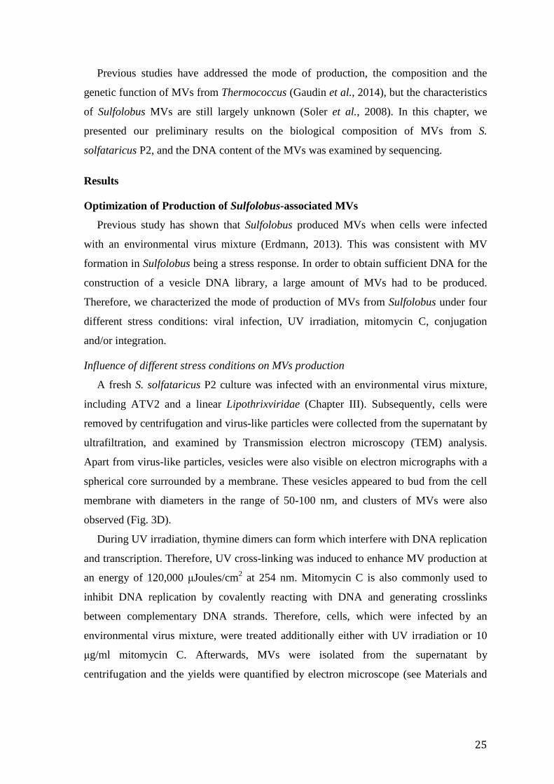
Previous studies have addressed the mode of production, the composition and the
genetic function of MVs from Thermococcus (Gaudin et al., 2014), but the characteristics
of Sulfolobus MVs are still largely unknown (Soler et al., 2008). In this chapter, we
presented our preliminary results on the biological composition of MVs from S.
solfataricus P2, and the DNA content of the MVs was examined by sequencing.
Results
Optimization of Production of Sulfolobus-associated MVs
Previous study has shown that Sulfolobus produced MVs when cells were infected
with an environmental virus mixture (Erdmann, 2013). This was consistent with MV
formation in Sulfolobus being a stress response. In order to obtain sufficient DNA for the
construction of a vesicle DNA library, a large amount of MVs had to be produced.
Therefore, we characterized the mode of production of MVs from Sulfolobus under four
different stress conditions: viral infection, UV irradiation, mitomycin C, conjugation
and/or integration.
Influence of different stress conditions on MVs production
A fresh S. solfataricus P2 culture was infected with an environmental virus mixture,
including ATV2 and a linear Lipothrixviridae (Chapter III). Subsequently, cells were
removed by centrifugation and virus-like particles were collected from the supernatant by
ultrafiltration, and examined by Transmission electron microscopy (TEM) analysis.
Apart from virus-like particles, vesicles were also visible on electron micrographs with a
spherical core surrounded by a membrane. These vesicles appeared to bud from the cell
membrane with diameters in the range of 50-100 nm, and clusters of MVs were also
observed (Fig. 3D).
During UV irradiation, thymine dimers can form which interfere with DNA replication
and transcription. Therefore, UV cross-linking was induced to enhance MV production at
an energy of 120,000 μJoules/cm2 at 254 nm. Mitomycin C is also commonly used to
inhibit DNA replication by covalently reacting with DNA and generating crosslinks
between complementary DNA strands. Therefore, cells, which were infected by an
environmental virus mixture, were treated additionally either with UV irradiation or 10
μg/ml mitomycin C. Afterwards, MVs were isolated from the supernatant by
centrifugation and the yields were quantified by electron microscope (see Materials and
25

Methods). For both UV irradiation samples and the mitomycin C-treated samples, high
yields of secreted MVs were observed (Fig.1A, B).
Fig. 1 Comparison of vesicles productions of S. solfataricus P2 under stress treatments (A. mitomycin C and B. UV irradiation). Mitomycin C and UV irradiation led to comparable vesicle production. Production of MVs by S. solfataricus P1-pKEF9 integrated with SSV2
A culture of pKEF9 conjugated S. solfataricus P1 integrated with SSV2 was grown up
and spherical MVs with diameters in the range of 50-100 nm were observed in culture
supernatant by electron microscope (Fig. 2). Although no virus-like particles were
detected in the electron micrographs, it was inferred that integrated virus produced a
stress reaction in the hosts.
Fig. 2 TEM micrograph of MVs from the supernatant of S. solfataricus P1-pKEF9 integrated with SSV2. Influence of strain type in MVs production
To determine the degree to which production of vesicles was specific to strain types,
four types of Sulfolobus strains currently used in our laboratory were examined, S.
solfataricus P2, S. islandicus REY15A and S. solfataricus P2 CRISPR-minus mutant (S.
solfataricus P2 CRISPRΔ), as well as S. islandicus REY15A CRISPR-minus mutant (S.
26

islandicus REY15A CRISPRΔ). S. solfataricus P2 CRISPR-minus mutant lacks CRISPR
loci A to D and adaptation-associated genes, and S. islandicus REY15A CRISPR mutant
lacks both CRISPR loci and associated cas genes (Gudbergsdottir et al., 2011). Since
both of the CRISPR-Cas minus strains lack of the capacity for virus interference, the
viruses are likely to propagate at high copy numbers.
Combined with UV irradiation or mitomycin treatment after viral infection, the MV
yields were determined for the four strains (Table 1, Fig. 3). Peak vesicle production was
reached, when S. solfataricus P2 was subjected to UV irradiation after infected by an
environmental virus mixture.
Table 1 Comparison of the yields of the MV obtained by different strains and methods. The symbols (-, +, ++, +++) indicate the relative yields of MV production from lowest level to the highest level.
Virus mixture Virus mixture + UV irradiation
Virus mixture + Mitomycin C
Virus Vesicles Virus Vesicles Virus Vesicles S. islandicus
REY15A CRISPRΔ
+++ - + - ++ -
S. islandicus REY15A - + - + + +
S. solfataricus P2 CRISPRΔ +++ + + ++ + +
S. solfataricus P2 - ++ - +++ - ++
27

Fig. 3 TEM micrographs of MVs produced by virus mixture infected cultures from different Sulfolobus strains. A) S. islandicus REY15A CRISPRΔ under the mitomycin C treatment with large numbers of virions but no MVs. B) S. islandicus REY15A subjected to UV irradiation with no virions but a few MVs. C) S. solfataricus P2 CRISPRΔ under the mitomycin C treatment with both virions and MVs. D) S. solfataricus P2 subjected to UV irradiation with high yield of vesicles. Structures similar to ‘‘strings of pearls’’ were observed at higher magnification.
Extracellular DNA associated with MVs from Sulfolobus
MVs were concentrated in CsCl gradients and they formed a sharp white opalescent
band. Vesicle DNA were extracted and examined on a 1 % agarose gel stained with
ethidium bromide. The results revealed that the DNA was about 10-12 kb in size. In order
to determine whether the DNA is located within these MVs or strongly bound to the
surface, MVs were separated into three equal portions, one no treatment as a control, the
second treated only with DNAase, and the third treated with both proteinase K and DNase.
The results (Fig. 4) showed that the sample treated with DNase still contained DNA,
indicating that DNA was protected by lipid or protein. The DNA concentration in DNase-
treated sample was also lower than in the control, implying that some DNA was bound
28

externally on the vesicles and was degraded. In the sample treated with both proteinase K
and DNase, a clear DNA band was still present albeit at a lower concentration than the
control. We inferred that DNA in the MVs was resistant to DNase treatment and
proteinase K, and protected within the MVs (Fig. 4).
DNA sequencing was employed to analyse DNA from MVs further. The results
showed that the DNA constituted random chromosomal fragments and in particular from
IS elements (Erdmann, 2013). The results suggest MV could function as a vector to
maintain the host genetic information under stress conditions.
Fig. 4 MVs from S. solfataricus P2 associated with chromosomal DNA fragments. (1) MV DNA without any treatment, (2) MV DNA after treatment with proteinase K and DNase, (3) MV DNA digested by DNase. All samples were loaded on a 1% agarose gel with SDS-loading buffer. M indicates size marker. Observation of Pyramidal Structures
So far, only two archaeal viruses have been shown to induce host pyramidal membrane
structures, STIV (Sulfolobus turreted icosahedral virus) in S. solfataricus P2 (Brumfield
et al., 2009) and SIRV2 (Sulfolobus islandicus rod-shaped virus 2) in S. islandicus
LAL14/1(Bize et al., 2009). These structures produce holes for virion release. During
purification MVs from S. solfataricus P1, we also observed pyramidal structures on the
cells (Fig. 5). Genome sequencing revealed that SSV2 integrated into the genome of S.
solfataricus P1-pKEF9. Therefore, we investigated possible SSV2 proteins that could
produce pyramidal structures. Since protein C92 of STIV and protein 98 of SIRV2 are the
only viral proteins known to produce pyramidal structures, we searched for homologous
of SSV2 proteins using protein BLAST searches (http://blast.ncbi.nlm.nih.gov/Blast.cgi)
which yielded pKEF9 ORF100 as a possible candidate. A Clustal Omega multiple protein
29

sequence alignment was performed to compare the three proteins
(http://www.ebi.ac.uk/Tools/msa/clustalo/) (Fig. 6). Several amino acids were conserved,
indicating their potential to structural or functional importance. The results could explain
the observation of pyramidal structures in the cells of S. solfataricus P1-pKEF9 integrated
with SSV2.
Fig. 5 (A) TEM micrograph of pyramidal structures of S. solfataricus P1-pKEF9 integrated with SSV2. (B) TEM micrograph of a cell showing the production of vesicles. Samples were negatively stained with 2% uranyl acetate. A scale bar is shown.
Fig. 6 Sequence analysis of the C92/P98-like proteins of SSV2. The multiple sequence alignment was generated using Clustal Omega. It is colour-coded according to the standard Clustal Omega colouring scheme. Sequence conservation at each position below. Protein accession numbers: SSV2 ORF100 (AAQ73268), STIV C92 (AAS89074) and SIRV2 ORF98 (CAC87324).
Discussion
Study of archaeal MVs is still in its infancy, and many questions remain to be tackled.
The production of MVs is considered a universal and important mechanism for cellular
communication. Studies of MVs from Sulfolobus reveal that they emerge from cell
membranes by a specific budding process similar to the ESCRT pathway (Ellen et al.,
2009). While this work was in progress, it was reported that MVs from Thermococcales
contain DNA and protect DNA against thermodegradation. Moreover, the fact that MVs
30

can transfer DNA indicates that they can be potentially used to fuse with recipient cells
and deliver their contents from cell to cell (Gaudin et al., 2013, Gaudin et al., 2014).
In this chapter, two experiments were aimed at improving our understanding of MVs
in Sulfolobus. We characterized factors influencing production and determined the
biochemical composition of MVs from Sulfolobus. MVs from Sulfolobus contain
intracellular DNA, and MVs protect DNA against proteinase K and DNase. Further, our
study suggests that MVs could serve as vehicles for the inter-cellular transport. DNA
isolated from MVs is rich in IS elements, suggesting a regulatory mechanism of DNA
selection and package. Remarkably, we observed the pyramidal structures of Sulfolobus
cells during purification of MVs, and these results could expand the spectrum of genetic
elements producing pyramidal structures.
Since MVs from Sulfolobus contain cellular DNA, future studies are required to
address the functions of MV in cellular physiology, especially in DNA transportation.
This should also strengthen our knowledge about their function as a vehicle for horizontal
gene transfer in natural environments. MVs can also potentially be exploited as a genetic
tool, once a MV specific genetic maker is found.
Although the experiments are still ongoing and the results presented here are
preliminary, DNA transfer mediated by MVs is expected to greatly enhance
understanding the physiological functions of MVs.
Materials and Methods
Strains and growth conditions
S. islandicus REY15A, S. solfataricus P2 and their CRISPR-deletion mutants
(Gudbergsdottir et al., 2011), S.solfataricus P1 conjugated with pKEF9 were used in this
study. Cells were cultivated in Sulfolobus medium (Zillig et al., 1994) supplemented with
0.2% tryptone, 0.1% yeast extract and 0.2% sucrose (TYS-medium), while for Sulfolobus
CRISPR mutant cells, additional 1% 2 mg/ml uracil is necessary (TYSU-medium). pH
was adjusted with 1:1 HCl to be around 3-3.5. Finally autoclaved ddH2O was added up to
the volume of 1L. Then cultures were enriched aerobically at temperatures 75°C or 78 °C.
Before infection experiments with ATV, growing cultures were always diluted two to
three times in late exponential phase to retrieve a highly active and viable culture. When
cells reached exponential stage cells, resuspended in 1ml preheat TYS medium after 6000
rpm for 10 min centrifugation. In order to stress the cells rather than completely viral
infection, 2 μl environmental virus mixture were co-incubated with the cells for over 1
31

hour. Then the cultures were transferred to 50 ml preheat TYS medium (CRISPR mutant
with addition uracil) for three days. More vesicles were enriched by adding in 550ml
preheat TYS medium (CRISPR mutant with addition uracil) for 2 days. The cultures were
grown at 75°C and upscaled adding more medium every 2-3 days.
Virus-like particles purification
Virus-like particles were prepared firstly by centrifugation of infected cultures at
6000rpm for 10 min and then the supernatants were filtered by 150 ml 0.2 μL filter. At
last, virus-like particles were collected by filtration of the culture supernatant through the
0.2 μm VIVASPIN 20 (Sartorius Stedim, Biotech) spin-filters with a molecular weight
cut off (MWCO) of 300,000 Dalton. Here virus can be stored at 4°C ready for infections
and examination by electron microscope. Virus-like particles concentrates were
concentrate by CsCl gradient ultra-centrifuge at 30,000 rpm for 55 hours. After dialysis
with 10 mM Tris-HCl (pH 8.5) and 50 mM NaCl, pure concentrates were used for the
total DNA extraction and enzyme digestion.
Transmission electron microscopy
5 μL of concentrated virus-like particles suspension was spotted onto a carbon coated
copper grid and incubated for 5 min. The grid was then rinsed with distilled water and
negatively stained with 2% uranyl acetate for 2 min. The stain was washed off of the grid
and was ready for imaging in a Jeol JEM-1010 (Japan Electron Optics Ltd, Tokyo, Japan)
TEM. The images were digitally recorded using a camera connected to a computer and
images were captured.
Enzyme digestion
One part of vesicles was treated with 5 μl 1.25 μg/ml Proteinase K (Qiagen), after
incubation in 56°C for half an hour, enzymes were inactived at 70°C for 10 min. Then 2
μl DNase I (fermentas) was added and incubated for 30 min at 37°C. The other part was
only treated with DNase for half an hour at 37°C. Samples were loaded on 1% agarose
gel with SDS-loading buffer.
32

III. Interactions of Archaeal Virus ATV with the CRISPR Adaptive Immune System of Sulfolobus solfataricus
33

Abstract
Acidianus two-tailed virus (ATV), that undergoes independent extracellular
development of two tails, was examined for its genetic diversity. ATV2 was isolated and
propagated in a S. solfataricus P3 strain isolated in 2011 from Pozzuoli, Naples. The
genome of ATV2 and the CRISPR loci of the host were sequenced. A laboratory mutant
ATVv was also available. Comparative genome analysis of the three closely related
viruses revealed a conserved genome organization, but many differences in gene size and
content. The genomes of ATV2 and ATVv were shorter as a result of deletions.
Comparison of the CRISPR loci in S. solfataricus P3 with those of three published S.
solfataricus strains showed many shared spacers, as well as different spacers, especially
those adjoining the leader region. Several spacers of the newly isolated S. solfataricus P3
had significant sequence matches to ATV and ATV2 genomes, indicating S. solfataricus
P3 has been a host for ATV viruses previously.
Introduction
As a major component of the biosphere on the planet, viruses shape the planet’s
ecosystems and co-evolve with their hosts (Krupovic et al., 2011). Rapid advances in
DNA sequencing and bioinformatics as well as versatile genetic tools have significantly
pushed forward an emerging field of isolation and characterization of new viruses,
particularly archaeal viruses (Prangishvili et al., 2006). However, archaeal viral research
is still at an early stage of development, and many processes of viral life cycles need to be
explored, including replication, integration and virus-host interactions (Ortmann et al.,
2006). So far, approximately 65 archaeal viruses have been characterized
(http://www.ebi.ac.uk/genomes/archaealvirus.html), mainly from the Euryarchaeota and
Crenarchaeota (Brochier-Armanet et al., 2011, Pina et al., 2011). To date, viruses have
been isolated and characterized for members of the archaeal genera: Sulfolobus,
Acidianus, Pyrobaculum, and Thermococcus (Schoenfeld et al., 2008).
ATV, is a member of the Bicaudaviridae, and it develops tails extracellularly and
independently of the host after extrusion from cells as spindle-shaped particles (Haring et
al., 2005). Studies of the tail development implicated p618 (ATPase) and p892 in this
process (Scheele et al., 2011). The changing morphology may represent a strategy for the
virus to communicate with the host (Ortmann et al., 2006).
34

S. solfataricus P2 has been used as a host for many archaeal viruses and conjugative
plasmids (Zillig et al., 1994, She et al., 2001). It carries complex CRISPR (clustered
regularly interspaced short palindromic repeat) systems with six CRISPR loci annotated
by A to F. Based on the sequence of CRISPR repeats, leaders, PAM motifs (protospacer-
associated motif) and associated proteins, locus A and B are classified within the Type I-
A subfamily II, while loci C to F are classified within Type I-A subfamily I (Vestergaard
et al., 2014), which employ different PAM sequence. Experiments demonstrate that the
CRISPR system of S. solfataricus P2 is active and it shows two distinct mechanisms for
spacer acquisition (Erdmann & Garrett, 2012, Erdmann et al., 2013, Erdmann et al.,
2014).
In order to counteract the deleterious effects of viruses, hosts have developed defence
systems including prevention of adsorption, blocking of injection, abortive infection,
restriction-modification, toxin-antitoxins and the CRISPR-Cas system (Samson et al.,
2013, Dy et al., 2014). The CRISPR-Cas system can target invasive DNA by inserting a
short sequence (protospacer) into a CRISPR loci after recognizing a PAM sequence
(Shah & Garrett, 2011). PAM sequences are approximately -2 to -4 bp from the end of
the protospacer which becomes leader-proximal on CRISPR insertion, and may also be
involved in avoiding self-interference of DNA targeting (Shah et al., 2013). CRISPR loci
provide a memory of previously invading genetic elements via short spacer sequences
(Mojica et al., 2005, Barrangou et al., 2007), and they are transcribed, and processed to
yield crRNAs that complement the invader nucleic acids and guide interference
complexes for cleavage (Garrett et al., 2011).
Several studies have employed CRISPR-Cas systems to analyse archaeal viral
genomes in natural environments (Andersson & Banfield, 2008, Vestergaard et al., 2008,
Anderson et al., 2011, Sencilo & Roine, 2014). Previously, our group has reported that it
is possible to determine a virus host by comparing its genome of a virus with CRISPR
spacer sequences of potential host (Garrett et al., 2010). In this study, we studied the
interactions of a specific ATV-Sulfolobus system from the same environments by
comparing the viral genome with CRISPRs of the host. We also compared the genomes
of viruses to investigate genomic diversity.
35

Results
Characterisation of virus-like particles
Virus-like particles were isolated from an environmental sample collected from a hot
spring in Pozzuoli, Italy. Probably due to the interference of the host from matching
spacers, we could not propagate the virus-like particles in S. solfataricus P2. Therefore,
The virus-like particles were enriched in the CRISPR-minus mutant strain of S.
solfataricus P2 which lacked loci A to D and the cas genes essential for adaptation and
interference (Gudbergsdottir et al., 2011). In enrichment cultures from the supernatant of
the CRISPR-minus mutant strain of S. solfataricus P2, we detected two distinct
morphologies, filamentous particles and lemon-shaped particles (Fig. 1).
A shotgun library was generated from the supernatant of the heterogenous enrichment
culture and sequenced with a five-fold genome coverage. In total, sequencing produced
10,895,598 reads with average length 90bp, and there were 124,124 reads mapped to
ATV. After clean, there were 44,408 contigs left, and 50.6% of the contigs matched to
lipothrixviral genomes AFV (Acidianus Filamentous Virus) (Bettstetter et al., 2003,
Haring et al., 2005, Keller et al., 2007, Bize et al., 2008, Vestergaard et al., 2008) and
14.5% to ARV1 (Acidianus Rod-shape Virus 1) (Genebank No. AJ875026) (Vestergaard
et al., 2005), while 29% showed significant matches to ATV. In addition, there was a low
level of contigs matching to Sulfolobus conjugative plasmids including pHVE14, pYN01,
pLD8501, pNOB8, pAH1 and pARN4 (Schleper et al., 1995, She et al., 1998, Greve et
al., 2004).
Preliminary assembly of the contigs to ATV yielded a scaffold with three gaps.
Although we tried to link them by primer-walking, there was still a significant level of
sequence heterogeneity throughout the ATV2 genome. Hence the final sequence was a
consensus, where the dominant nucleotide was taken at each position. It carried 57,909 bp
and 62 open reading frames (ORFs) and was significantly smaller than ATV with 62,730
bp, 72 ORFs (Accession no.: AJ888457), as well as ATVv (61,783 bp, 68 ORFs) isolated
from a laboratory mutant (Vestergaard, 2009).
Comparative genomics of ATV viruses
The genome sequences of ATV, ATV2 and ATVv were compared to investigate
genomic differences. The results reveal that whereas the basic genome organization is
conserved, there are numerous heterogeneities in around 50% of the annotated genes
between ATV and ATV2, while ATVv shows only about 10% difference compared with
36

ATV (Fig. 2). The variations mainly included nucleotide insertions or deletions, reading
frame shifts, and/or altered start/stop codons. The GC content of ATV2 is 40.69%, close
to that of ATVv and ATV which are 41.34% and 41.23%, respectively.
The comparison of these genomes shows that there is a large deletion in ATVv. It
lacks orf54, orf273, orf79, orf59a and orf48, but carries a new orf117 possibly with an
altered function (Fig. 2). This 1.0 kb deletion could be due to recombination between the
direct repeat sequences AAAAATAGTCGA and AAAAATGGTCGA. A similar
recombination event may also have happened in ATV2 and produced the 2.1 kb deletion
between the identical direct repeat sequences AAAAATAGTCGA. Another major
deletion in ATV2 including orf192 and orfB mobile element (orf383b), suggests that the
orfB mobile element carrying ORF192 may move to the host genome or other genetic
elements. However, sequence heterogeneities preclude the precise localisation of these
mobile elements in the genome. Two small putative ATV proteins, ORF34 and ORF45,
were not encoded in ATV2.
In contrast to ATV, there are many truncated genes in ATV2 (Table 1). In particular,
three characterized virion proteins, ORF145, ORF326a and ORF567, were truncated to
various degrees in ATV2. Moreover, the largest open reading frame in ATV, ORF1940,
is truncated to ORF1793 in ATVv. Gene diversity also occurs in a putative operon of
ATV2. Whereas ATV carried ORF59b, ORF189 and ORF60, ATV2 exhibits ORF60,
ORF87 and ORF124. Of these, ORF60 in ATV2 is homologous to S. solfataricus P2
SSO10342, a transcripitional regulator, and shows 62% amino acid residue identities to
ORF59b of ATV. ORF87 exhibits 60% identity to ORF83a of Acidianus rod-shaped virus
ARV (Vestergaard et al., 2005), and may share the same functions. Contrary to ORF60 in
ATV, ORF124 has 47% identity to PepK of pDL10 from Acidianus ambivalens (Kletzin
et al., 1999). However, ORF241, which encodes an integrase, remains intact, highlighting
the importance of viral integration. In general, proteins present in virions or involved in
virus-host interactions varied extensively, while proteins implicated in DNA replication
or integration were relatively conserved (Table. 1).
Characterization of S. solfataricus P3 CRISPR-Cas system
In order to investigate the spacer diversity of the CRISPR loci, S. solfataricus P3 was
co-isolated with ATV2 in 2011. The CRISPR spacer sequences of S. solfataricus P3 were
BLAST-matched with the NCBI sequence database. The results showed about 34% of the
spacers could be assigned to known viruses, conjugative plasmids, or the integrated
37

genetic elements of Sulfolobus genomes. We compared the total numbers of spacers in S.
solfataricus P3 with that of S. solfataricus P2, as well as the numbers of distinct spacers
which have been characterized in the database (Table 2). The numbers of significant
spacers which match to ATV and ATV2 are also given. Loci A, B and D of S.
solfataricus P3 have almost the same number of spacers against ATV/ATV2 as in S.
solfataricus P2, while loci C and E have more spacers against ATV2. Locus F was
identical in S. solfataricus P3 and S. solfataricus P2. Around 8% of the spacers in the new
isolated S. solfataricus P3 matched to ATV sequences, while a slightly lower number 7.4%
matching ATV2 (Table 2).
As shown earlier, de novo spacer acquisition generally occurs adjacent to the leader
region in loci A, B, C, D. However, locus E is exceptional in that insertions can occur
throughout the CRISPR locus. We observed two forms of locus E in different clones of S.
solfataricus P3, with spacers inserted either adjacent to the leader area or in the middle of
the locus (Table 3), reinforcing the hypothesis of two different adaptation mechanisms
(Erdmann & Garrett, 2012).
CRISPR divergence in S. solfataricus
To investigate the CRISPR locus variation, CRISPR loci of S. solfataricus P3 were
compared with S. solfataricus strains P1, P2 and 98/2 (Fig. 3) (Lillestol et al., 2006,
Lillestol et al., 2009). S. solfataricus strains P1, P2 and P3 were isolated from Naples,
Italy, while S. solfataricus 98/2 may originate from USA. The comparison showed that
leader proximal spacers were strain-specific and that the last two spacers of each locus
were conserved. It also revealed that these four sets of related CRISPR loci share
extensive genetic information despite their broad geographical distributions. The specific
spacers adjoining the leader area appear to display the environmental bias whereas the
last two conserved spacers may indicate that the strains diverge from similar ancestors
(Fig. 3).
Only three irregularities have been observed in the CRISPR loci. There are two large
insertions with no known match in the locus D of S. solfataricus P1 and S. solfataricus
P3. In addition, there is an 899 bp fragment of a pNOB8-like conjugative plasmid in locus
F of S. solfataricus P1/P2/P3 (Table 3) (Lillestol et al., 2006, Lillestol et al., 2009).
CRISPR loci provide insights into the interaction dynamics of virus and host
CRISPR loci carry the host memory of past invaders, including viruses and plasmids.
For interference to occur, the PAM sequence is recognized. CCN is the PAM motif for
38

CRISPR loci C, D and E while loci A, B require the motif TCN (Erdmann & Garrett,
2012, Shah et al., 2013). In order to understand how CRISPR-Cas systems mediate the
virus population dynamics, all the protospacers in S. solfataricus P3 matching to either
ATV or ATV2 were selected and examined for their PAM sequences (Table 4). In
general, the protospacers were prone to carry more mismatches in the ATV2 compared to
those of ATV, although there are three identical protospacers shared by ATV and ATV2
(spacer 25 in locus A, spacer 18 in locus B, spacer 38 in locus D). For spacer 4 in locus C
and spacer 1 in locus E, although they have few mismatches in the protospacers, their non
cognate PAMs inhibit interference (Table 4). Furthermore, spacer 10 in locus D has the
same mismatches in the protospacers but altered PAM, indicating the non cognate PAMs
is a way to avoid interference. For spacer 19 in locus B, spacers 25/27 in locus C and
spacers 11/33/58 in locus D, more mismatches and altered PAM sequences would likely
be ineffective in interference of ATV2. However, three sequences have perfect matches
to ATV2 and cognate PAMs, for example spacer 18 in locus B and spacers 12/65 in locus
D. This may explain why we couldn’t propagate ATV2 in S. solfataricus P2. In
conclusion, the significant spacers of S. solfataricus P3 match to ATV or ATV2 indicate
S. solfataricus P3 used to be a host for ATV viruses.
Discussion
As a result of the development of advanced DNA sequencing techniques and rapidly
expanding metagenomic datasets, as well as the availability of versatile genetic tools, our
understanding of the archaeal viruses has advanced significantly, but a comprehensive
understanding of many fundamental processes is still lacking, including the life cycle
mechanisms including, replication, integration and the virus-host interactions. Here we
examined the virus-host interactions for ATV2 and S. solfataricus P3 based on the host
CRISPR-Cas system. We isolated and characterized ATV2, and we also sequenced the
CRISPR loci of S. solfataricus P3. The comparison between the genomes of three closely
related viruses (ATV, ATV2, ATVv) reveal the genomic diversity of this kind of virus. A
comparison of the CRISPR loci in the four S. solfataricus strains shows shared spacers, as
well as strain-specific spacers especially those adjoining the leader.
ATV2 was isolated from a hot spring in Pozzuoli, Italy in 2011. The virus mixture
including ATV2 and lipothrixviruses were firstly enriched from the environmental
sample, and then propagated in S. solfataricus P2 CRISPR-minus mutant lacking of
CRISPR loci A to D and associated cas genes. Failure to infect S. solfataricus P2 and
39

successful infection of the S. solfataricus P2 CRISPR-minus mutant suggested that the
host could target ATV2 by CRISPR-Cas interference. An attempt to assemble the contigs
which have high similarities with AFV has failed. This may also indicate the high
genomic diversity of AFV. The minority ATV2 particles with numerous sequence
alterations strengthens the hypothesis that viral genomic diversity plays a key factor in
maintaining their population (Peng et al., 2004).
The genome of ATV2 shows numerous differences from that of ATV, including indels,
deletions partly from recombination, amino acid mutations and ORF size changes with
altered start or stop codons (Fig. 2). This is presumed to originate from viral adaptation to
different hosts, as is shown by the comparison of HAV1 (hyperthermophilic archaeal
virus 1) variants (Garrett et al., 2010). Together with heterogeneity in the protospacers of
ATV2, the altered PAM helps viruses to avoid CRISPR-directed interference. However,
the matching spacers may result in ATV2 being a minor viral component in the
environmental sample.
In conclusion, in this study an attempt was made to gain a broad picture of the viral
genomic diversity resulting from CRISPR-Cas interference for ATV viruses. The results
indicate that ATV2 exists as a mixture of variants, where the genome sequence is a
consensus with dominant nucleotides. It is suggested that these variants could occur in the
environment, and/or appear after propagating from a different host (in this case S.
solfataricus P2 CRISPR-minus mutant). The altered genomes may result from the rapid
adaptation of virus to new environment. Finally, our data suggest that S. solfataricus P3
could be a host for ATV previously.
Materials and methods
Viruse particles isolation, sequencing and assembly
An aqueous mud sample was taken from an acidic hot spring in Pozzuoli, Italy. One
millilitre of this sample was added to 50 ml of Sulfolobus medium supplemented with 0.2%
trypton, 0.1% yeast extract and 0.2% sucrose (TYS medium) (Zillig et al., 1994) and
incubated aerobically for 5 days at 78°C. Two liters of enrichment culture was then
established in TYS medium at 78°C. Cells were pelleted (6000 g, 10 min) and virus
particles were isolated by filtration of the supernatant through 0.2 mm pore filters
(Vivaspin®). This virus mixture was then used to infect S. solfataricus P2 CRISPR-
minus mutant cultured in the Sulfolobus medium. The concentrated virus was subjected to
CsCl density gradient ultracentrifugation and dialysed against 10 mM Tris-HCl, pH 8.
40

DNA was isolated using Dneasy® Blood&Tissue Kit (Qiagen, Hilden, Germany).
Sequencing by Illumina sequencing was performed by Beijing Genomics Institute
(Shenzhen, China). After preliminary clean, the sequences were assembled using Velvet
(Zerbino & Birney, 2008) and CLC genomic workbench (CLC Bio, Aarhus, Denmark).
Single colony of S. solfataricus P2
Cells of S. solfataricus P2 from the same mud sample were harvested from a fresh
enrichment culture by centrifuging (6000 g, 10 min) and resuspending in 1 ml of TYS
medium. After serial dilution, the cells were dispensed on Gelrite plates and incubated at
78°C for 3–6 days until single colonies were grown. If any of the products migrated
differently from the control PCR product on an agarose gel, they were sequenced. At last,
four out of 16 single colonies, which showed the majority PCR products, were chosen for
research.
Primer-walking
After a roughly assembly of the ATV2 genome, primers were designed to amplify PCR
products covering gaps between contigs. PCR products were separated on 1 % agarose
gel, excised and purified with QIAquick Gel Extraction Kit (Qiagen). Purified PCR
products were cloned using InsTAcloneTMPCR Cloning Kit (Fermentas) following the
manufacturers’ protocols. Plasmid was purified by GeneJET Plasmid Miniprep Kit
(Fermentas) and sequenced by MWG Biotech (Germany).
Genome annotation
The annotation of the ATV2 sequence was performed using Artemis software (Rutherford
et al., 2000). Each putative protein identified was compared to the NCBI database using
BLASTP with alignments created using the default settings.
Plaque assays
Viral isolates were serially diluted and mixed with preheated fresh cells followed by
mixing with 1mL preheated 0.4% gelrite. The mixture was layered over a 0.7% solid
gelrite layer. Plates were then incubated for 3-6 days at 75 °C to allow virus infection and
host growth. Plaques or of growth inhibition formed on the plates were counted.
Transmission electron microscopy
Virus particles were adsorbed onto carbon-coated copper grids for 5 min and stained with
2% uranyl acetate. Images were recorded using a Tecnai G2 transmission electron
microscope (FEI, Eindhoven, the Netherlands), with a CCD camera, at an acceleration
voltage of 120 kV.
41

DNA isolation
The DNA was prepared as follows without phenol using a modified Qiagen
Blood&Tissue Kit manual. 12ml Sulfolobus islandicus cells were collected after
centrifuged at 6000 rpm for 10mins. After resuspended the cell pellets in 40 μl medium
salt solution, added 150 μl ATL buffer and mixed well by vortexing. Then added 4 μl
RNAse, after incubated at 37C for 30 min, added 20 ul proteinase K solution and mixed
thoroughly by vortexing. Then followed the protocol’s suggestions. Primers used for
amplication of the CRISPR loci of S.solfataricus P2 were listed as below (Table S1).
42

Table 1. Changes to the characterized proteins in these three viruses. The functions of these proteins are also listed.
ATV proteins Changes in ATVv Changes in ATV2 Functions Reference
ORF145 none ORF142 (91% amino acids identities) virion protein (Prangishvili
et al., 2006)
ORF273 Deletion caused by recombination.
Deletion caused by recombination virion protein
(Prangishvili et al., 2006, Felisberto-
Rodrigues et al., 2012)
ORF326a none ORF266, C-terminal
one nucleotide deletion gave ORF shift
virion protein (Prangishvili et al., 2006)
ORF387 none ORF389 (71% amino acids identities)
Interactions p618, DNA binding
(Prangishvili et al., 2006)
ORF567 none ORF197, truncated N-terminal. virion protein (Prangishvili
et al., 2006)
ORF618 none ORF605, loss 13 amino
acids, truncated C-terminal
AAA-ATPase, role tail development
(Prangishvili et al., 2006, Scheele et al., 2011)
ORF653 none ORF652 (81% amino acids identities)
DNA-binding protein,interacts with p618,
(Prangishvili et al., 2006)
ORF529 none ORF524 (TT to AA
altered stop codon, loss 5 amino acids)
AAA-ATPase host receptor recognition,
endonuclease
(Erdmann et al., 2011,
Happonen et al., 2014)
ORF892 none ORF900 (83% amino acids identities)
VWA-containing co-chaperone for tail
develop.
(Scheele et al., 2011,
Happonen et al., 2014)
ORF241 intact intact integrase (Prangishvili et al., 2006)
ORF1940 ORF1793 (truncated C-terminal)
ORF1944 (80% amino acids identities)
Conjugative transfer protein TrbJ, ATP
synthase
(Prangishvili et al., 2006)
43

Table 2. Comparison of the total number of CRISPR spacers in both S. solfataricus P2 and S. solfataricus P3, including the distinct spacer numbers that were subjected to BLAST searches against GenBank sequences. The numbers of spacers which match to ATV and ATV2 are also given.
Locus A Locus B Locus C Locus
D Locus E Locus F Total
P2 P3 P2 P3 P2 P3 P2 P3 P2 P3 P2 P3 P2 P3 spacers
No. 102 49 95 64 31 41 96 74 6 9 10 88 418 325 326
distinct spacers
No. 36 7 30 16 12 10 34 30 3 2 3 46 161 111 112
match to ATV 4 2 2 2 0 6 8 15 0 0 1 1 15 26 27
Match to ATV2 4 2 2 2 0 5 8 14 6 0 1 1 21 24 25
conserved 10 41 4 2 6/5 88 151/150
44

Table 3. Comparison of the spacers in locus E of S. solfataricus P2 with two variants of locus E in S. solfataricus P3 (locus E1 and locus E2). S. solfataricus P2 Locus E S. solfataricus P3 Locus E1 S. solfataricus P3 Locus E2
AAGTAGATTGTTGAAACTCCTAGTTCGTGGAGTGTTTTA
TGTGAAAATCATAAAACGCCTACATTTTTATATCTTCATTG
AATGTTAGTCCCCAAGACTCTGTTTCTGATGGATTTCTCA
TGTGTATTCCCCCGTGTGGAGTGTCCACACAAAGAGTCTT
ATACTGCAAGCGAATTGGCGGAAAATTGGCAGCGACGTG ATACTGCAAGCGAATTGGCGGAAAATTGGCAGCGACGTG
TTATCTAATTTTAATAATCAAGGAAATTCATTAACTCAAATA TTATCTAATTTTAATAATCAAGGAAATTCATTAACTCAAATA
CCCATTCATCTCTTTCTTTGCAGCTTTGTTCTAACATTA CCCATTCATCTCTTTCTTTGCAGCTTTGTTCTAACATTA
ATTGAACGTTGTTGAACATTCTTGAACGTTATTGAATGTTAT TACGTATCTCTTCCATGGGGGCAAATATCCAGGTTCTT ATCCCAGAAAGCATTATCGAGTTTCGCATGGACATACG
TGGTAAATAGCTCTGTTAGGCCCAGTTATTCCATATTCTGA TGGTAAATAGCTCTGTTAGGCCCAGTTATTCCATATTCTGA TGGTAAATAGCTCTGTTAGGCCCAGTTATTCCA
TATTCTGA ATTTTCTAATATATCTAATTCACTCTGC
GTATCATTATGGATAA ATTTTCTAATATATCTAATTCACTCTGCGTATCATTATGGATAA ATTTTCTAATATATCTAATTCACTCTGCGTATCATTATGGATAA
TTAGCCCAACAATTAACTAAAGATCCTGAAGCAGTCAA TTAGCCCAACAATTAACTAAAGATCCTGAAGCAGTCAA TTAGCCCAACAATTAACTAAAGATCCTGAAGCA
GTCAA
45

Table 4. Details of spacers in newly isolated S. solfataricus P3 matching to ATV or ATV2 with numbers of mismatches indicated. PAM sequences and the corresponding protospacer positions in the viral genomes are also given.
cluster spacer match to ATV PAM position match to ATV2 PAM position A S24 100% TCT gp57 ORF653 10 mismatches TCT gp57 ORF652
S25 2 mismatches TCA gp56 ORF277 2 mismatches TCA gp56 ORF276 B S18 100% TCT gp58 ORF213 100% TCT gp58 ORF206
S19 2 mismatches CTA gp48 ORF800 4 mismatches TAG gp48 ORF800 5 mismatches TAG gp48 ORF800
C S4 5 mismatches TAT gp60 ORF710 4 mismatches TAT gp60 ORF744
S25 100% TCT gp45 ORF161 4 mismatches ACC gp45 ORF161
S27 2 mismatches TCA gp20 (ORF45) ^gp 21 (ORF240) 3 mismatches TTA gp19 (93) ^gp21 (226)
S29 2 mismatches CCA gp71 ORF1334 3 mismatches CCA gp71 ORF1334
S34 1 mismatches CCT gp30 ORF161 4 mismatches CCT gp30 ORF161
S35 4 mismatches AAA gp49 (ORF567) ^gp 50 (ORF1940)
D S7 2 mismatches TCA gp17 ORF80 11 mismatches TGA gp17 ORF80
S10 4 mismatches ATT gp16 ORF175 4 mismatches CCA gp16 ORF175
S11 4 mismatches CCT gp57 ORF653 9 mismatches TGT gp57 ORF652
S12 2 mismatches CCG gp67 ORF545 100% CCG gp67 ORF558
S32 2 mismatches CAC gp50 ORF1940 5 mismatches CCG gp50 ORF1944
S33 3 mismatches CCA gp50 ORF1940 12 mismatches TTA gp50 ORF1944
S35 6 mismatches GAC gp45 ORF161 9 mismatches CCA gp45 ORF161
S37 100% CCG gp34 ORF315 3 mismatches CCG gp34 ORF315
S38 5 mismatches CCT gp06 ORF117 5 mismatches CCT gp06 ORF63
S45 6 mismatches CCG gp63 ORF387 13 mismatches CCA gp64 ORF362
S51 12 mismatches GCG gp72 ORF241 46
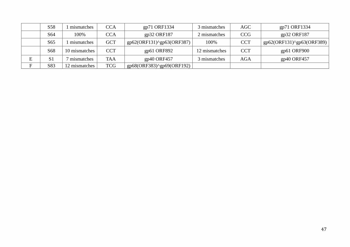
S58 1 mismatches CCA gp71 ORF1334 3 mismatches AGC gp71 ORF1334
S64 100% CCA gp32 ORF187 2 mismatches CCG gp32 ORF187
S65 1 mismatches GCT gp62(ORF131)^gp63(ORF387) 100% CCT gp62(ORF131)^gp63(ORF389)
S68 10 mismatches CCT gp61 ORF892 12 mismatches CCT gp61 ORF900
E S1 7 mismatches TAA gp40 ORF457 3 mismatches AGA gp40 ORF457 F S83 12 mismatches TCG gp68(ORF383)^gp69(ORF192)
47

Fig. 1. Electron micrographs of virus particles isolated from the supernatant of an enrichment culture. Samples were negatively stained with 1% uranyl acetate and size bars are included. The red arrows indicate the filamentous virus-like particles, while the blue ones denote ATV-like particles.
Fig. 2. Comparative genomes of the three ATV viruses. ORF organization and comparison of genome maps of the ATV viruses where predicted genes and their direction are indicated by arrows and the amino acid numbers of their products. Red, deletion; purple, truncated genes; yellow, a new gene.
48

Fig. 3. Comparison of CRISPR loci A, B, C, D, E and F from four strains of S. solfataricus. Spacers are coloured to identify the mobile genetic elements with the best sequence match. Each locus is oriented with the leader on the left. Regions with same colour or the same shape show identical sequence in each loci. Leader regions are indicated by L.
49

Table S1. Primers used for amplifying S. solfataricus P2 CRISPR loci.
Name Forward Reverse Locus A (starts from the leader area)
AF ACGCTTACGTTGCTCTCGAATTTCT As CCGGTTAAGTTCGTTTTCATGAAGTTG CTGAAAGTGGGACAACTCCTGGTTACC A1 ACAACTAAAATTGGTCGCATGAAGA AGGTGATGAGAGAAGATGAGTGATG A2 CATCACTCATCTTCTCTCATCACCT AAAATGGTTGCATCTGCGATACTGC A3 GCAGTATCGCAGATGCAACCATTTT AAGATATGAGCAAGATGGGAGTCAAC A4 GTTGACTCCCATCTTGCTCATATCTT TCTCTTCATCTAGGCATGTAGTGTC A5 GACACTACATGCCTAGATGAAGAGA AAATCTAAATCCCGTCCTATGGGCG A6 CGCCCATAGGACGGGATTTAGATTT GAGCAGAGAGGGAGGATAGTAGAATA A7 TATTCTACTATCCTCCCTCTCTGCTC CCGACCTTACCTCAGCCAATAATAT A8 ATATTATTGGCTGAGGTAAGGTCGG ATCCTACTGGATTGAGGTTATCGTG A9 CACGATAACCTCAATCCAGTAGGAT GACTACCTAATAGCGATAAGCACCA
A10 TGGTGCTTATCGCTATTAGGTAGTC CTCTTCGCACTTACTAAGAAATTGAC Locus B (starts from the leader area)
Bs CGACATTAGCCCTGGGGGTATCTAAACC
GCAATGAAAGAAAGATGAAAGGAGAGCGATAAG
B1 AAATTGGTCGCATGAAGAGTAAAGG ATATGAACAAGCTGTTGATGTGCAA B2 TTGCACATCAACAGCTTGTTCATAT GAATGGAGGGCATATAATGTTGAGC B3 GCTCAACATTATATGCCCTCCATTC CAAAAATCTCAACGACAACACCAAC B4 GTTGGTGTTGTCGTTGAGATTTTTG TAGGAGTGTAGTCATAAGGAGAGCA B5 TGCTCTCCTTATGACTACACTCCTA TTCGTTCCAACTACACCTAATGGAT B6 ATCCATTAGGTGTAGTTGGAACGAA AGATTTGAGGATGCCATCAGAAGTA B7 TACTTCTGATGGCATCCTCAAATCT CATATCTTTGGCTGAAGTTCCTTGG B8 CCAAGGAACTTCAGCCAAAGATATG TAAACAAGCTCGCACAGAAGAAAAT B9 ATTTTCTTCTGTGCGAGCTTGTTTA TAGAGAATAGAGAACAGAGAACGGC
B10 GCCGTTCTCTGTTCTCTATTCTCTA GGAAGTGTTAGAAGAGGTGTATGGT B11 ACCATACACCTCTTCTAACACTTCC AGAAACTCATTCAGAGTCTCTTCCAA B12 TTGGAAGAGACTCTGAATGAGTTTCT TGGCTTTGGAGAGGTAGAAGTAAAA
Locus C (starts from the leader area) Cs TCGCTTATCTCTCTCATGCGCCATT TGTCCCGTTTTTGTAAGTGGGGG C1 TGTCCCGTTTTTGTAAGTGGGGG TCGCTTATCTCTCTCATGCGCCATT C2 AATGGCGCATGAGAGAGATAAGCGA GAGTTCGATCGGATTAAAGAGGAGA C3 TCTCCTCTTTAATCCGATCGAACTC TTTGTGGTACTTGTAGTTGTGATGC C4 GCATCACAACTACAAGTACCACAAA AGAACACTCCCGTACCAATTTCTTA
Locus D (starts from the leader region) DF TCTGCGACCTCACAATATAATAGC CGACTCTTTTTCTCCCTCTCTCCAAC D1 AGTTCCACCCCCGAAGCTCCT AGCCGGGACAAGTTTCACAAATTGA D2 TCAATTTGTGAAACTTGTCCCGGCT GTTTATGTTTCACGGGCATTTGGCT D3 AGCCAAATGCCCGTGAAACATAAAC AGTCTTCTTGGGCGAGGTGAGTTAT D4 ATAACTCACCTCGCCCAAGAAGACT ATTCAACAGAGGAAGCTGGGAGTTG D5 CAACTCCCAGCTTCCTCTGTTGAAT GTTGGGCTAGTAAGTATGATGGCGT D6 ACGCCATCATACTTACTAGCCCAAC CTATTTCGCCTGCTATTGTTTTCGC D7 GCGAAAACAATAGCAGGCGAAATAG ACCGTTAGACCATAGCGGACTTTTG D8 CAAAAGTCCGCTATGGTCTAACGGT CAGCAATGCCGAAATTCGGTACAAT D9 ATTGTACCGAATTTCGGCATTGCTG CTATTTTTACCTTTGTGGCTTCGGG
D10 CCCGAAGCCACAAAGGTAAAAATAG CGACTCTTTTTCTCCCTCTCTCCAAC Locus E (starts from the leader area)
E ATAGGGAAAGAGTTCCCCCG TGACTCTAGTGCAATCTTCGA Locus F (starts from the leader area)
F CGGCGTTATAATGGGTATCGGAATCGG GCTCACTATCTCACCCCTATCAAT
50

IV. Conflicting Interactions between the Archaeal Conjugative Plasmid pKEF9 and Different Sulfolobus Hosts
51

Abstract
Sulfolobus conjugative plasmids tend to be gradually lost after conjugating between
cells in continuous culture. To understand this mechanism, we have investigated genome
changes in the conjugative plasmid pKEF9 and the hosts, Sulfolobus solfataricus and
Sulfolobus islandicus. Initial efficient replication of pKEF9 in conjugated strains resulted
in a dramatic retardation of cell growth. Moreover, pKEF9 integrated both strains at
tRNA[Glu] genes. Loss of pKEF9 in S. islandicus appeared to be due to spacer acquisition
followed by the immune response of the host CRISPR-Cas system, whereas the loss in S.
solfataricus coincident with integration into the host. In addition, mobile elements
probably regulated by one non-coding RNA restricted pKEF9 function after integration.
It is concluded that the deactivation and loss of pKEF9 in S. islandicus and S. solfataricus
is caused by CRISPR immune response and mobile elements regulation, respectively.
Introduction
Owing to their relative smaller genome sizes and autonomous replication, plasmids
and viruses have been widely investigated to unveil mechanisms of conjugation,
replication and integration (Norman et al., 2009). So far, two general archaeal plasmid
families have been assigned for the genus Sulfolobus, cryptic plasmids and conjugative
plasmids (Lipps, 2006).
The conjugative plasmids pKEF9 has three conserved and functionally distinct genetic
sections A, B and C. Section A encodes proteins for conjugation, of which two proteins
are distantly related to the bacterial proteins, TraG and TrbE. Section B carries a putative
replication origin, and section C contains six to nine proteins are involved in initiation
and regulation of plasmid replication and integration (Greve et al., 2004). The integrase
of pKEF9 has 56% amino acids identities to that of pNOB8, suggesting that pKEF9 may
also integrate into the hosts by the same mechanism (She et al., 1998). Two mechanisms
for integration were proposed: SSV-type and pNOB8-type (She et al., 2004). The
integrase of SSV1, as the representative example of the former type, has been
characterized (She et al., 2004). SSV1 can integrate into a tRNA gene of the host genome,
but after integration, the SSV1 integrase gene is partitioned into a smaller N-terminal part
(70 amino acids) and a larger C-terminal part (270 amino acids) bordering the provirus
(Schleper et al., 1992). However, for pNOB-type integration, the putative integrase of
pNOB8 keeps an intact integrase gene during integration (She et al., 1998). In addition,
52

the presence of a CRISPR array in pKEF9 and the transcript of pKEF9 CRISPR in S.
acidocaldaricus (Lillestol et al., 2009) provided a basis for exploring interactions
between host and plasmid.
Numerous integrative elements, such as viruses, plasmids and transposable elements,
mediate the movement of DNA within or between genomes via homologous
recombination, and play important roles in the emergence of new features. S. solfataricus
P2 was estimated to contain more than 300 mobile elements, and appears to be always
undergoing rearrangements partially as a result of transpositions (She et al., 2001). The
transpositions of mobile elements are regulated by ncRNAs (Tang et al., 2005). In
eukaryotes, ncRNAs guide complexes to bind to the 3-untranslated region (UTR) of
mRNAs, which causes translational repression and/or mRNA decay (Filipowicz et al.,
2008). In Bacteria, ncRNAs predominantly target the 5-UTR of mRNAs by non-
contiguous base-pairing (Waters & Storz, 2009). However, Sulfolobus employs a
different mode of ncRNA regulation, because two thirds of mRNAs in S. solfataricus are
devoid of 5-UTR (Wurtzel et al., 2010). Archaea often regulate gene expression by
antisense-based mechanisms with either full or partial complementarity between target
and ncRNA (Wurtzel et al., 2010). So far a few groups have identified ncRNAs in
archaea, and they have shown that at lease 8 ncRNAs are complementary to transposases
mRNAs (Dennis & Omer, 2005, Tang et al., 2005, Wurtzel et al., 2010, Martens et al.,
2013). These antisense RNAs are inferred to regulate transposition by annealing to
transposase mRNAs.
In this work, interactions between the archaeal conjugative plasmid pKEF9 and its
Sulfolobus host were examined after conjugation. Extensive genome changes occurred in
pKEF9 and its hosts during conjugative transfer and replication. Evidence is presented for
integration of pKEF9 into the hosts, and for uptake of spacers from pKEF9 into CRISPR
loci. Finally, one novel ncRNA candidate is identified which may regulate transposition
of mobile elements.
Results
pKEF9 conjugates in Sulfolobus and causes retardation of cell growth.
pKEF9 conjugates in S. islandicus REY15A.
pKEF9 was originally detected in an S. islandicus strain but was propagated in S.
solfataricus P1 in a higher copy numbers than the natural host (Greve et al., 2004).
Subsequently S. solfataricus P1 was employed as a host for pKEF9. In order to explore
53

the genetic variations of both pKEF9 and its hosts during conjugation, S. solfataricus P1
carrying pKEF9 was added to an S. islandicus REY15A culture at a donor:recipient ratio
of 1:10,000. Cultures were successively diluted to OD600 = 0.05 once the stationary
growth had been reached. Growth retardation occurred at about 50 hours p.c. (post
conjugation) (Fig. 1), indicating that pKEF9 propagated efficiently in liquid cultures of S.
islandicus REY15A. qPCR analysis showed that pKEF9 replicated at high copy numbers
in the mating strain S. islandicus REY15A (maximum 150 copies/cell at about 38 hours
p.c.), and greatly reduced cell growth. Plasmids were extracted from S. islandicus
REY15A-pKEF9 and S. solfataricus P1-pKEF9 and subjected to endonuclease EcoRI
digestion to compare the difference of pKEF9 from the donor and recipient.
Fig. 1 Growth curves for unconjugated and conjugated wildtype S. islandicus REY15A. Similar growth curves were observed in triplicate experiments.
EcoRI restriction fragment patterns of pKEF9 in agarose gels revealed similar
fragment patterns for pKEF9 from S. solfataricus P1 stock and from S. islandicus
REY15A (Fig. 2), indicating that pKEF9 was stable in the recipients after conjugation.
Fig. 2 EcoRI digestion map of pKEF9 from (1) the recipient S. islandicus REY15A 80 hours p.c. (2) the donor S. solfataricus P1-pKEF9. M indicates size marker.
0
0.4
0.8
1.2
1.6
0 50 100 150 200 250 300 350
OD
600
Time (h)
S.isl REY15A S.isl REY15A+pKEF9
54

pKEF9 conjugates in S. solfataricus P2
Similar experiments were conducted in S. solfataricus P2 and growth retardation was
also observed at around 50 hours p.c. (Fig. 3). However, pKEF9-conjugated S.
solfataricus P2 started growing rapidly again about 100 hours p.c. and finally yielded
growth curves which were similar to the wild-type. Plasmid extractions demonstrated that
pKEF9 propagated in the conjugated cultures throughout the 100 hours p.c. However,
since no plasmids were observed at 190 hours p.c., it was infered that S. solfataricus P2
was cured of pKEF9. In order to understand the curing mechanism, the host was checked
for integration of pKEF9.
Fig. 3 Growth curves of S. solfataricus P2 wild-type without and with pKEF9.
pKEF9 integrates into the host chromosome
pKEF9 and other conjugative plasmids of this family encode an integrase of the non-
partitioning pNOB8-type carring the motif R..HxxR..Y that is important for the catalysis
of DNA strand cleavage and exchange (She et al., 1998). We tested for pKEF9
investigation into the Sulfolobus host genome. By alignment of the pKEF9 attP sequences
(Erauso et al., 2006) with those of the corresponding tRNA genes in S. solfatatricus P2,
pKEF9 can integrate into two tRNAGlu candidate genes via plasmid attP site and
chromosomal attB sites (Table 1), one with a perfect match (CTC) and the other carrying
a single mismatch (CTT) (Fig. 4). These two attachment sites in both S. islandicus
REY15A and S. solfataricus P2 can be assayed by PCR amplification of attB sites within
tRNAGlu genes.
0
0.5
1
1.5
2
0 100 200 300 400
OD
600
Time (h)
ssoP2 ssoP2+pKEF9
55

Fig. 4 Diagram showing the integration of pKEF9 into the S. islandicus REY15A genome. The positions and orientations of the reverse primers (IntR) are indicated by blue arrows on the circular map of pKEF9, whereas those forward primers for both sites (perfect match IntF1 and one mismatch InF2) are shown by blue arrows on the linear genome of S. islandicus REY15A. The probe used for Southern hybridization is indicated in green.
Table. 1. Sequence alignment of attP sites of pKEF9 with the attB sites of tRNAGlu genes in S. islandicus REY15A (SiRe) and S. solfataricus P2 (SSO). The integration sites and tRNAGlu anticodons (three nucleotides) are shown in red.
Sequence SiRe_t0010 TGCGGGCCTCTCGAGCCCGTGACCCGGGTTCAAATC SiRe_t0022 TGCGGGCCTTTCGAGCCCGTGACCCGGGTTCAAATC pKEF9 TTCTAGCCTCTCGAGCCCGTGACCCGGGTTCAAATC SSOt25 TGCGGGCCTTTCGAGCCCGTGACCCGGGTTCAAATC SSOt36 TGCGGGCCTCTCGAGCCCGTGACCCGGGTTCAAATC
pKEF9 integrates into S. islandicus REY15A chromosome
pKEF9 integration at tRNAGlu sites of S. islandicus REY15A was tested by PCR
amplification and Southern blotting at 24, 38 and 85 hours p.c. The results showed that
56

pKEF9 has only integrated at the mismatch (CTC) site within 24 hours p.c., while both
host tRNAGlu sites were occupied after 38 hours p.c. (Fig. 5).
Fig. 5 Identification the integration of pKEF9 in Sulfolobus. A. Southern blotting results of the pKEF9 conjugated culture at 24 hours p.c. and 38 hours p.c., respectively. After EcoRI digestion, the size of free pKEF9 is 3.5kb, while the size for integration at the perfect site is 1.2kb in contrast to the one at one mismatch site 2.5kb. B. PCR amplification to check for integration at both tRNAGlu sites of S. islandicus REY15A at 85 hours p.c. at (1) the perfect match site (CTC) and (2) the site with one mismatch (CTC). M indicates size marker. pKEF9 integrates into the S. solfataricus P2 chromosome
PCR amplification of the two tRNAGlu integration sites of S. solfataricus P2 were
conducted and the products were sequenced. The results showed that plasmid had
integrated at both sites at 102 hours p.c. (Fig. 6), which provide an explanation for why
cells started to grow quickly again at about 100 hours p.c. (Fig. 3). It indicated that the
host had restricted pKEF9 activity after integration by an unknown mechanism. In
contrast, genome sequencing of S. solfataricus P1-pKEF9 tRNA genes showed that
integration had occurred only at the perfect match site. Possibly this difference of
integration in S. solfataricus P2 and S. solfataricus P1 could be caused by restriction
modification system.
57

Fig. 6 PCR amplification to check for integration at both tRNAGlu sites of S. solfataricus P2 at 102 hours p.c. at (1) the perfect match site (CTC) and (2) the site with one mismatch (CTC). M indicates size marker.
pKEF9 conjugation induces host chromosomal DNA degradation
The profiles of DNA contents obtained by flow cytometry showed that pKEF9-
conjugated S. islandicus cells lost genomic integrity (Fig. 7). Moreover, since the
intracellular DNA started to degrade 17 hours p.c., it suggested that conjugation occurred
fairly rapidly throughout the culture. As we can see from figure 7, the genome of cells
appeared to be normal within 10 hours p.c., while it started lost genomic integrity at 17
hours p.c.. Furthermore, considering the generation time of S. islandicus REY15A is
around 6-8 hours, the time for pair formation, transfer, and gene expression required for a
secondary transfer was estimated at about 7 hours.
Fig. 7 Flow cytometry time-course analysis of S. islandicus REY15A cells conjugated by pKEF9. DNA content distributions from unconjugated and conjugated culture. The peak shift observed in the conjugated sample indicated that host DNA was degraded after conjugation.
58

The adaptation response of the S. islandicus CRISPR immune system is activated
We tested for de novo spacer acquisition at the leader proximal end of the two
CRISPR loci of S. islandicus REY15A by PCR amplification over a 13-day period post
conjugation. After 13 days of continuous growth, PCR results of the leader proximal
regions showed slower moving bands, indicating that spacer acquisition had occurred
(Fig. 8). DNA from these bands was isolated, cloned and sequenced. In total 107 clones
were examined, including 53 sequences from locus 1 yielding 73 de novo spacers and 54
sequences from locus 2 producing 81 de novo spacers. No strong protospacer bias was
observed to either DNA strand of pKEF9. Moreover, there was no bias to genes; 12.3%
of the protospacer matches fell within intergenic regions and 12.9% of the genome is non-
protein-coding. Some genes carried a few protospacers whereas others had none (Fig. 9).
These results are consistent with pKEF9 being cured from S. islandicus REY15A due to
the CRISPR-Cas immune response. However, no de novo spacers insertions were
observed in S. solfataricus P2 (Fig. 10), suggesting that this organism uses another
mechanism to control pKEF9 replication.
In addition, eight de novo spacers matched to SSV fusellovirus DNA and there were
11 unidentified spacers. Genome sequencing of S. solfataricus P1-pKEF9 revealed these
additional spacers derived from SSV2 which excised from the genome of the SSV2
integrated S. solfataricus P1.
Fig. 8 PCR products from CRISPR locus 1 of S. islandicus REY15A (1) Non conjugated and (2) conjugated with pKEF9. PCR products of CRISPR locus 2 of S. islandicus REY15A (3) non conjugated and (4) conjugated with pKEF9. de novo spacer insertions are indicated with arrows. M indicates size marker.
59

Fig. 9 Circular genome map of pKEF9. Locations of matching de novo spacers (black) on each of the DNA strands are indicated on inner and outer concentric circles. ORFs are filled in turquoise. Integrase gene is indicated in red. Three conserved and functionally distinct sections are labelled in orange. CRISPR array is shown in blue.
Fig. 10 PCR products amplified from leader proximal regions of CRISPR loci A to F of S. solfataricus P2 wild-type strain and the pKEF9-conjugated culture. No insertions were detected. PCR products of loci (1)B, (2)C, (3)D, (4)E, (9)A leader region of S. solfataricus P2 and the corresponding PCR products of loci (5)B, (6)C, (7)D, (8)E, (10)A leader region of S. solfataricus P2 conjugated with pKEF9. M indicates size marker. Properties of the pKEF9 CRISPR locus
The CRISPR locus of pKEF9 carries seven repeats and six spacers (Table 2). It lacks a
leader region and the first repeat is corrupted, which indicate that the locus is unlikely to
undergo de novo spacer acquisition. Nevertheless, it can be transcribed and processed,
and potentially cause interference with the help of host-encoded Cas proteins (Lillestol et
al., 2009). This inference is further supported by the two spacers showing matches to the
Sulfolobus viruses SIRV and SSV (Lillestol et al., 2006, Shah & Garrett, 2011).
Moreover, a third spacer shows a match to one spacer in CRISPR locus F of S.
solfataricus P1 and P2, which could contribute to the instability of pKEF9 CRISPR locus
in S. solfataricus P2.
60

The stability of the pKEF9 CRISPR locus in conjugated S. islandicus REY15A
cultures was examined by PCR at different time points. PCR products indicated a time-
dependent gradual loss of spacer-repeat units from the CRISPR locus of pKEF9 (Fig. 11).
Sequencing revealed a mixture with different spacer-repeat units, consistent with the PCR
results. As expected, no de novo spacer acquisition was found.
Table 2. CRISPR spacer sequences (one to six) of pKEF9. Spacer Match to Mismatches GATGTTGCTGAGCGCCAGAGACTGGTATAAAAACTTTCT ACAGACGATAGATCGACTTGAACCATTGTGTTGATTATGGTA AATTTTAACGCGGAGGGAAATTCAATTCAACAAATTTCTAC SIRV1/2 4 TTGAGGATGTAGACGCCGACACCAGATACAATAGAGACTGTTA TTACCATCTCTTCGACTTCCATTAGACCTTTCTTGCTCA SSV4/5 13 TTGTACATTCCTAGGAGGGCATAAGAGCCGTTTGAGAGT S. solf P1/P2 10
Fig. 11 Identities of PCR products from the pKEF9 CRISPR locus following transcript directions. M indicates size marker. PCR products from (1) pKEF9-conjugated S. solfataricus P1 stock; (2) pKEF9-conjugated S. islandicus REY15A 200 hours p.c.; (3) pKEF9-conjugated S. islandicus REY15A 280 hours p.c.; (4) pKEF9-conjugated S. islandicus REY15A 300 hours p.c. S. solfataricus P1-pKEF9 genome
In order to determine the basis of the pKEF9 CRISPR locus heterogeneity and to
investigate when heterogeneity happened during the conjugation, the genome of pKEF9-
conjugated S. solfataricus P1 stock was sequenced. Sequencing initially yielded 40,046
reads with the mean length of 4,890 bp, and after a pre-clean, there were 31,129 reads
with the mean length 5,010 bp. These were mapped to host genome. The genome of S.
solfataricus P1, which showed high similarities to S. solfataricus P2, was assembled. No
de novo spacer acquisitions and deletions of the host CRISPR loci were found, consistent
with the PCR results which showed no additional PCR products of the CRISPR were
observed in pKEF9-conjugated S. solfataricus P2 (Fig. 10). The result reinforced that S.
61

solfataricus P2 uses a different mechanism from S. islandicus REY15A to regulate
pKEF9 activity. In addition, two forms of pKEF9 CRISPR locus were found in S.
solfataricus P1; either as same as sequenced earlier (Greve et al., 2004) or with only the
corrupted repeat-spacer 1-repeat present and no de novo spacer acquisition was observed.
Integrated pKEF9 is targeted by orfB elements in S. solfataricus
A few contigs that did not assemble into the S. solfataricus P1 or pKEF9 genomes
contained fragmented pKEF9 regions interspaced with transposable orfB (ORFB-IS605)
elements. This transposon is present in a single copy in the S. solfataricus P2 genome. It
encodes the ORFB protein in the downstream region and the upstream region carries
putative ncRNA genes. In all the non assembled contigs, orfB elements were located at
five specific pKEF9 nucleotide positions: 14098, 17040, 22423, 26494 and 28711
(Accession no. NC_006422).
In order to investigate whether the orfB element was inserted into the free plasmid or
the integrated form, plasmids were extracted from a conjugated S. solfataricus P1-pKEF9
culture and the five insertion sites were amplified by PCR. The main PCR product was
homogeneous in size and derived exclusively from free plasmid (Fig. 12), and it was
inferred, therefore, that free plasmids were not subjected to orfB transposition. Moreover,
larger PCR were obtained that could have resulted from orfB insertions (Fig. 12). We
inferred from these results that the integrated form of pKEF9 is specifically targeted by
orfB elements.
Fig. 12 PCR products amplified from the five pKEF9 sites of insertion of orfB elements. 1 to 3 –position 14098; 4 to 6 - 17040; 7 to 9 - 22423; 10 to 12 - 26494; 13 to 15 - 28711. Repetitive DNA templates were repeated every three samples: 1, 4, 7, 10, 13 isolated pKEF9 with full CRISPR locus, 2, 5, 8, 11, 14 pKEF9 isolated from S. solfataricus P1-pKEF9, and 3, 6, 9, 12, 15 S. solfataricus P1-pKEF9 genome. M indicate DNA size markers.
62

One cis-encoded antisense ncRNA potentially regulates orfB element
Transposon-associated ncRNAs are common in the S. solfataricus P2 transcriptome.
The majority of ncRNAs corresponded to cis-encoded antisense RNA transcripts which
are complementary with their targets (Tang et al., 2005, Wurtzel et al., 2010). One could
infer that the majority of the cis-encoded antisense RNAs complementary to transposase
transcripts are involved in the silencing of transposons (Tang et al., 2005).
By BLAST searches of the NCBI database with the sequence upstream from the orfB
element (1927131..1928641), one cis-encoded antisense ncRNA candidates was
identified that could regulate orfB transposition at position 1576535..1576761. It is
located in the intergenic regions between SSO1739 and SSO8813 of the S. solfataricus
P2 genome, termed ncRNA-227. It could be used as a possible regulator of the host orfB
transposon by base-paring to the upstream region of orfB transposon at position
1927131..1927408. A BLAST search revealed that ncRNA-227 had three potential
homologs complementary with the S. solfataricus P1 ncRNA: Sso-109 (AJ786211), Sso-
17 (AJ786208) and sR107 (AY722659). Moreover, the transcriptome of S. solfataricus
P2 showed that both the orfB element (1927131..1928641) and ncRNA-227 were
transcribed. Our results exemplify that S. solfataricus use cis-encoded antisense ncRNAs
complementary with transposase gene to control the mobility of the mobile elements at
the transcriptional level.
Insertion motif of orfB elements
The five specific orfB insertion positions were located on the pKEF9, including two in
intergenic areas and three in genes of unknown proteins. These five positions carried the
sequence GGC on one DNA strand (Table 3), and the last three nucleotides of the orfB
transposon (1927631..1928641) are also GGC. This implies that orfB inserts through a
GGC recognition site between plasmid and orfB element. Therefore, we assume that
cleavage occurs at the motif site and orfB element undergoes copy/paste insertion into the
new location. In addition, recombination events result in rearrangements in the pKEF9-
integrated S. solfataricus P1 genome. For example, one unitig contains seven physically
unlinked segments of S. solfataricus P1, as well as part of pKEF9, interspaced by orfB
elements (Fig. 13).
63

Table 3. Sequence properties of the five orfB insertion sites on pKEF9.
Position Sequence Location 14098 TTAGATTAGGCTATAAG intergenic 22423 AAGGGAGAGGCTATTTT ORF102 17040 CTATAGTAGGCTTTAAA ORF355 26494 GAGCGACGGGCTGACCC intergenic 28711 GAGTCATAGGCATTGCA ORF99
Fig. 13 Illustration of unitig 9. This shows the unlinked S. solfataricus P1 (in different colors) with integrated pKEF9 (yellow) is interspaced by orfB elements. The orfB element contains a putative transposase gene (light green) and ncRNA (turquoise) that is complementary to ncRNA-227. Discussion
Several archaeal plasmids have been isolated and sequenced, but relatively little is
known on their mechanisms of conjugation, integration and replication. For the
conjugative plasmids, little was known about factors determining their stability in
Sulfolobus strains, nor did was the function of their CRISPR understood. In this study, we
have developed a natural system to investigate how Sulfolobus hosts become cured of the
conjugative plasmid pKEF9. Two distinct mechanisms operate in two different hosts.
They involve pKEF9 resistance to the CRISPR-based immune system in S. islandicus,
and orfB mobile elements in S. solfataricus. Further, we examined significance of pKEF9
integration into Sulfolobus genomes.
Initially, pKEF9 propagated in S. solfataricus P2 and retarded the cell growth (Fig. 3).
After 100 hours p.c., pKEF9 was gradually cured and S. solfataricus P2-pKEF9 regained
wild-type growth rate, and no free plasmids were detected in the culture. During this
process, pKEF9 was integrated into both tRNAGlu of S. solfataricus P2 (Fig. 6). S.
solfataricus P2-pKEF9 carries numerous rearrangements caused by orfB elements, which
was confirmed by genome sequencing of S. solfataricus P1-pKEF9.
BLAST searches of the ORFB proteins encoded in STSV2, ATV, SMV1 and pMGB1
against proteins encoded by S. solfataricus P2 yield three orfB element candidates in S.
solfataricus P2 (sso1521, sso8288 and sso7710). The reason that S. solfataricus P2
employs mobile elements, rather than its CRISPR-Cas system to restrict activities of
pKEF9 remains unknown. Possibly, pKEF9 CRISPR spacer that matches a spacer in S.
64

solfataricus P2 cluster F has an inhibiting effect (Table 2). More work needs to be done
before making solid conclusions.
Although the same growth retardation and pKEF9 integration was observed in both
hosts, the conflicting interactions of pKEF9 with S. islandicus REY15A are based on the
CRISPR-Cas system. It has been reported that adaptation of the type I-B CRISPR-Cas
system of Haloarcula hispanica to a purified virus strictly requires a priming process by
partially matching spacers in the host CRISPR loci (Li et al., 2014). However, no
evidence for priming of spacer acquisition was found for pMGB1 conjugated S.
solftaricus P2 (Erdmann et al., 2013) or STSV1-infected S. islandicus REY (Erdmann et
al., 2014). In the present experiments, two CRISPR spacers (SislRE_115_114 and
SislRE_93_44) in S. islandicus REY15A show imperfect sequence matches to pKEF9.
Both protospacers in pKEF9 exhibit cognate CCN PAM motifs (Table S1). The presence
of spacer SislRE_93_44 in S. islandicus REY15A was confirmed by PCR amplification
and sequencing (Fig. S1). Therefore, if the priming theory is prevalent in Crenarchaea,
the spacer 44 of S. islandicus REY 15A may initiate the priming process. However, no
strand bias was observed between DNA sequences upstream and downstream of the
priming spacer, indicating there is no priming process of spacer acquisition in pKEF9-S.
islandicus REY15A.
Due to its homologous to other identified ncRNAs, we infer that ncRNA-227 might
function as a set of the three ncRNAs (Sso-19, Sso-17 and sR107), or could be the full
sequence of them. ncRNA-227, together with the orfB transposase, to regulate the orfB
mobility by complementary to the upstream of orfB elements.
In conclusion, these studies yield seminal insights into the interactions of a conjugative
plasmid with a Sulfolobus host. Two different mechanisms are proposed for the
inactivation of conjugative plasmids, one via the CRISPR-Cas immune system and a
second mediated by orfB mobile elements. Further studies are required to interpret the
complex mechanistic details of these different mechanisms.
65

Supplementary Materials
Fig. S1 Confirming the presence of pKEF9-matching spacer 44. M indicates size marker. (1) PCR product of spacer SislRE_93_44 of S. islandicus REY15A. Table S1. CRISPR spacers of S. islandicus REY15A and S. solfataricus P2 showing significant sequence matches to pKEF9. PAM sequence were determined in both strains.
Spacer PAM Protospacer mismatches
pKEF9 coordinates Locus pKEF9
SislRE_93_44 CCA 1 17869..17831 ORF245 SislRE_115_114 CCA 7 6248..6207 ORF147
Ssol_32_5 TTT 17 806..832 TrbE Ssol_89_73 AAT 10 19004..19038 CRISPR spacer 6 Ssol_103_59 CAG 7 442..479 TrbE
66

Table S2. All the primers used in this work.
Primers Sequences Locus C1F GTCCATAGGAGGACCAGCTTTC Locus C1R CCAACCCCTTAGTTCCTCCTCTATAG Locus C2F GTTCCTTCCACTATGGGACTAGGAAC Locus C2R CGTCACTGACACCATATTTATAC
SsoAF CCGGTTAAGTTCGTTTTCATGAAGTTG SsoAR CTGAAAGTGGGACAACTCCTGGTTACC SsoBF CGACATTAGCCCTGGGGGTATCTAAACC SsoBR GCAATGAAAGAAAGATGAAAGGAGAGCGATAAG SsoCF TCGCTTATCTCTCTCATGCGCCATT SsoCR TGTCCCGTTTTTGTAAGTGGGGG SsoDF TCTGCGACCTCACAATATAATAGC SsoDR CGACTCTTTTTCTCCCTCTCTCCAAC SsoEF ATAGGGAAAGAGTTCCCCCG SsoER TGACTCTAGTGCAATCTTCGA SsoFF CGGCGTTATAATGGGTATCGGAATCGG SsoFR GCTCACTATCTCACCCCTATCAAT SSV2F GGCTGAAGGATGGAGGAGTTA SSV2R CAGGTAGCTAACGAAACCAGTG IntF1 CGTCAAGTGAGTTAGCAAGGGA IntF2 GAGGTGTTTAAGGGTTTAACGTC IntR GAGCCAGCATTTCTGTAGCTT
probeF GGATCCGGGATCTATTAGCT probeR CGAATGAATTCTTCTCTATATGG 14098F CGCCTGGCTTTCTGCTTTTC 14098R TGCAGCAGCAATAGGACCTT 22423F AGGGAAGCCACACAATCG 22423R TCATTCCTCCTCAGCCTCC 17040F CCCTTCTCATGCTGTTCTCC 17040R ACAAGAGAAAGCTGGCTCAG 26494F ACCTCCTATATGCAGCCAGC 26494R GCTTATCTTGGGTTCCACGC 28711F AGGAAAATCACTGCGGGGAC 28711R GGACGTCTACGCTACACCAG
P2-1-ORF31 F GCTCTAAGCCCAAGGGAGTC P2-ORF31 R GAGCCAGCATTTCTGTAGCTT
P2-2-ORF31 F ACCTAATTCAGGTCGCACTTT S.isl44F ATTCGTAGAGCTTCTTATTCCTGCT S.isl44R GCAGGATGCAATTGATTTCGTAAAC
67

Materials and methods
Conjugation and growth curves
S. islandicus REY15A was grown in Sulfolobus medium supplemented with 0.1%
vitamin, 0.1% CAA, 0.2% sucrose and 0.1% uracil (SCVU medium) for two times (Zillig
et al., 1994). Then diluted the last fresh cells to OD600 about 0.05 and grew until the
OD600 around 0.2-0.3. At this point, mixed S. islandicus KEF9 with S. islandicus
REY15A at the donor/recipient cells number ratio of 1:10000. Mixtures were made in
fresh medium to give a cell concentration of 107/ml and were subsequently incubated
under moderate shaking at 75°C, as is standard. Samples were removed at the times
indicated below and were either plated or used for preparations of total DNA and for
determination of the optical density. Growth curves were made based on the measurement
of OD600 two times per day. 10 ml of cells were harvested by centrifuging (6000g, 10
min), and then we used either alkaline lysis extraction method or OMEGA BAC/PAC
DNA Kit to extract the plasmids.
PCR amplification of CRISPR loci
Samples were harvested by centrifugation (6000 g, 10 min) and DNA was isolated using
DNeasy® Blood&Tissue Kit (Qiagen). Leader proximal regions of CRISPR loci 1 and 2,
extending from the leader to spacer five, and approximately 750 bp regions covering the
whole of each CRISPR locus, were amplified by PCR using the listed primers (Table S2).
Fusellovirus-like particles were observed in the supernatant of S. islandicus KEF9 culture
by electron microscopy. SSV2 core protein (VP3) was monitored by PCR amplification
using the listed primers (Table S2).
Cloning and sequencing
PCR products were separated on 1% agarose gels and bands larger than those produced
from the uninfected control sample were excised from gels and purified with QIAquick
Gel Extraction Kit (Qiagen). The PCR products were then cloned using InsTAcloneTM
PCR Cloning Kit (Thermo Fisher Scientific) following the manufacturer’s protocol.
Plasmid purification and sequencing were performed by Eurofins MWG-Biotech
company (Ebersberg, Germany). PCR sequences were analysed by CLC main workbench
(Aalborg, Denmark) and Artemis programmes (Sanger Institute, UK) (Rutherford et al.,
2000).
68

Transmission electron microscopy
Virus particles were adsorbed onto carbon-coated copper grids for 5 min and stained with
2% uranyl acetate. Images were recorded using a Tecnai G2 transmission electron
microscope (FEI, Eindhoven, the Netherlands), with a CCD camera, at an acceleration
voltage of 120 kV.
Integration monitored by PCR
pKEF9 integration was examined at these two sites by PCR assay (perfect match and one
mismatch site). Primers for the pKEF9 are listed in Table S2.
OrfB inserted into the integrated genome
Free plasmids were extracted from conjugated the S. solfataricus P1-pKEF9 culture and
the five orfB insertion sites were amplified by PCR and sequenced. Primers for the
pKEF9 are listed in Table S2.
Southern blotting
Southern hybridization followed a standard procedure (Sambrook and Russell, 2001).
Genomic DNA was prepared and about 4 μg of total DNA of each sample was digested
with EcoRI, Resulting DNA fragments were fractionated by agarose gel electrophoresis
on a 1.0% agarose gel and transferred onto an IMMOBILON-NY+ membrane (Millipore)
via capillary transfer. DNAs on the membrane were then auto-cross-linked by the UV
Cross-linker (Stratagene). Hybridization probes were amplified by PCR (Table S2), and
then purified and labelled with Digoxigenin Labelling kit (Roche). Hybridization was
perfermed at 42°C overnight. Hybridization signals were detected by DIG detection kit
with the CDP-star (Roche) and recorded by exposing the membrane to CP-BU new
medical X-ray films (AGFA).
69

Perspectives To understand the interaction between extrachromosomal genetic elements and
Sulfolobus, membrane vesicles, ATV2 and conjugative plasmid pKEF9 were studied in
Sulfolobus. MVs from ATV2-infected Sulfolobus cultures contain chromosomal DNA
and protect DNA against DNase and proteinase K treatment (See Chapter II). Considering
that MV-production is induced by cell stress, MV-extrusion might constitute an important
mechanism of cell communication under stressful conditions.
Studies on the biochemical composition of MVs from Thermococcales have shown
that OppA (oligopeptide binding protein) may be involved in MV production, since it is
present both in cell membranes and MVs (Gaudin et al., 2013). The OppA protein
Sso1273 of S. solfataricus strongly interacts with AAA-ATPase p529 of ATV (Erdmann
et al., 2011). Moreover, it has been reported that MVs from E. coli can adsorb T4
bacteriophage, resulting in infection abortion, consistent with MVs contributing to innate
bacterial defense (Manning & Kuehn, 2011). Therefore, the interaction between OppA
and p529 may influence the production of MVs and the efficiency of virus infection. This
could also explain why MVs were observed, and no virus-like particles, when cells were
infected with ATV2. In order to test the hypothesis that MVs act as a defensive response
in Sulfolobus, one could co-incubate MVs and a virus to test for the efficiency of virus
infection.
The presence of DNA in MVs produced by Sulfolobus raises an important question
about their potential role in gene transfer. It has been reported that MVs from T.
kodakaraensis can transfer the plasmid pLC70 into plasmid-free cells. Therefore, to test
for a similar role for Sulfolobus MVs in DNA transfer, MVs produced by the S.
solfataricus P1-pKEF9 culture carrying integrated SSV2 could be transferred to wild-type
S. solfataricus P2. If pKEF9 or SSV2 DNA appears in the wild-type culture, one could
conclude that MVs make contact with cells to deliver their content. Moreover, in this
experiment one might also observe the phenomenon of viral genome packaging into
MVs. To investigate the function of MVs further, one could also study interactions
between MVs and the host CRISPR-Cas system of Sulfolobus. If MVs harbor
viral/plasmid DNA, they might stimulate the host CRISPR-Cas system to acquire spacers.
In Chapter III, ATV2 and S. solfataricus P3 were isolated from Pozzuoli, Italy.
Sequence analysis of ATV2 and the CRISPR array of S. solfataricus P3 provides a deep
insight into virus-host interactions in the natural environment. Electron Microscopy
shows that the presence of a lipothrixvirus in the sample, similar in sequence to AFV and
70
ARV1. Therefore, one can get the full genome of lipothrixvirus by primer-walking. Seven
genomes of AFV in the NCBI database exemplify the genetic diversity of the
lipothrixviruses.
To separate ATV2 from the lipothrixvirus in the sample, CsCl density centrifugation
and propagation in different hosts were tried, but the attempts failed. An explanation
could be that the two viruses cooperate to co-infect the S. solfataricus P2 CRISPR-minus
strain (Erdmann, 2013). Therefore, more hosts should be tried until a suitable one is
identified where ATV2 can propagate alone. Alternatively, one could try to pull down
ATV2 by the interactions between a protein and a virion protein where the interacting
AAA ATPase p529 of ATV and OppA Sso1273 of S. solfataricus would be an obvious
choice (Erdmann et al., 2011).
In Chapter IV, we investigated how Sulfolobus hosts become cured of the conjugative
plasmid pKEF9. Two distinct defense mechanisms operated in this process: the CRISPR-
Cas immune system in S. islandicus and a mechanism based on orfB mobile elements in
S. solfataricus. The integration of pKEF9 into Sulfolobus genomes was examined.
However, it was quite puzzling why S. islandicus uses CRISPR-Cas system instead of
mobile elements, since S. islandicus also carries many mobile IS elements. In order to
answer this question, one could amplify the five insertion sites of orfB in S. islandicus-
pKEF9 to test whether the integrated pKEF9 is attacked by orfB element or not.
Moreover, it has also been shown that the type I-E CRISPR-Cas system of E. coli is more
efficient in spacer acquisitions if it carries a spacer against the invading phage (Datsenko
et al., 2012). Since S. islandicus has one protospacer (S44) which has a cognate CCN
PAM and one mismatch difference with the spacer, it could facilitate the spacer uptake in
S. islandicus. In order to test whether this spacer S44 is still active in interference, one
could construct a plasmid carrying S44 and transfer it to Sulfolobus cells.
The profiles of DNA contents obtained by flow cytometry showed that pKEF9
conjugation induces host chromosomal DNA degradation (Fig. 7). However, nothing is
known about the situation in the S. solfataricus P2-pKEF9 culture. Therefore, a time-
course experiment to analyze the DNA content of S. solfataricus P2-pKEF9 needs to be
performed in the future.
One ncRNA that regulates orfB element was identified and it is transcribed in the S.
solfataricus P2. Since the transcriptome of S. solfataricus and S. islandicus are available,
all the known ncRNAs should be summarized and analyzed, especially the antisense
RNAs opposite transposase genes.
71

References Sambrook, J. & Russell, D. (2001). Molecular Cloning: a Laboratory Manual, 3rd edn. Cold Spring Harbor, NY: Cold Spring Harbor Laboratory.
Ahn DG, Kim SI, Rhee JK, Kim KP, Pan JG & Oh JW (2006) TTSV1, a new virus-like particle isolated from the hyperthermophilic crenarchaeote Thermoproteus tenax. Virology 351: 280-290.
Anderson RE, Brazelton WJ & Baross JA (2011) Using CRISPRs as a metagenomic tool to identify microbial hosts of a diffuse flow hydrothermal vent viral assemblage. FEMS Microbiol Ecol 77: 120-133.
Andersson AF & Banfield JF (2008) Virus population dynamics and acquired virus resistance in natural microbial communities. Science 320: 1047-1050.
Arnold HP, Ziese U & Zillig W (2000) SNDV, a novel virus of the extremely thermophilic and acidophilic archaeon Sulfolobus. Virology 272: 409-416.
Arnold HP, She Q, Phan H, Stedman K, Prangishvili D, Holz I, Kristjansson JK, Garrett R & Zillig W (1999) The genetic element pSSVx of the extremely thermophilic crenarchaeon Sulfolobus is a hybrid between a plasmid and a virus. Mol Microbiol 34: 217-226.
Babu M, Beloglazova N, Flick R, et al. (2011) A dual function of the CRISPR-Cas system in bacterial antivirus immunity and DNA repair. Mol Microbiol 79: 484-502.
Baliga NS, Bjork SJ, Bonneau R, Pan M, Iloanusi C, Kottemann MC, Hood L & DiRuggiero J (2004) Systems level insights into the stress response to UV radiation in the halophilic archaeon Halobacterium NRC-1. Genome Res 14: 1025-1035.
Balsalobre C, Silvan JM, Berglund S, Mizunoe Y, Uhlin BE & Wai SN (2006) Release of the type I secreted alpha-haemolysin via outer membrane vesicles from Escherichia coli. Mol Microbiol 59: 99-112.
Barrangou R (2013) CRISPR-Cas systems and RNA-guided interference. Wiley interdisciplinary reviews RNA 4: 267-278.
Barrangou R & Oost Jvd (2013) CRISPR-Cas systems: RNA-mediated adaptive immunity in bacteria and archaea. p.^pp. Springer, Berlin.
Barrangou R, Fremaux C, Deveau H, Richards M, Boyaval P, Moineau S, Romero DA & Horvath P (2007) Crispr Provides Acquired Resistance Against Viruses In Prokaryotes. Science 315: 1709-1712.
Basta T, Smyth J, Forterre P, Prangishvili D & Peng X (2009) Novel archaeal plasmid pAH1 and its interactions with the lipothrixvirus AFV1. Mol Microbiol 71: 23-34.
Beck K, Vannini A, Cramer P & Lipps G (2010) The archaeo-eukaryotic primase of plasmid pRN1 requires a helix bundle domain for faithful primer synthesis. Nucleic Acids Res 38: 6707-6718.
Beloglazova N, Brown G, Zimmerman MD, et al. (2008) A novel family of sequence-specific endoribonucleases associated with the clustered regularly interspaced short palindromic repeats. J Biol Chem 283: 20361-20371.
Bernander R (2000) Chromosome replication, nucleoid segregation and cell division in archaea. Trends Microbiol 8: 278-283.
72

Bernander R (2003) The archaeal cell cycle: current issues. Mol Microbiol 48: 599-604.
Bernander R (2007) The cell cycle of Sulfolobus. Mol Microbiol 66: 557-562.
Bettstetter M, Peng X, Garrett RA & Prangishvili D (2003) AFV1, a novel virus infecting hyperthermophilic archaea of the genus acidianus. Virology 315: 68-79.
Bhaya D, Davison M & Barrangou R (2011) CRISPR-Cas systems in bacteria and archaea: versatile small RNAs for adaptive defense and regulation. Annu Rev Genet 45: 273-297.
Bize A, Peng X, Prokofeva M, Maclellan K, Lucas S, Forterre P, Garrett RA, Bonch-Osmolovskaya EA & Prangishvili D (2008) Viruses in acidic geothermal environments of the Kamchatka Peninsula. Res Microbiol 159: 358-366.
Bize A, Karlsson EA, Ekefjard K, Quax TE, Pina M, Prevost MC, Forterre P, Tenaillon O, Bernander R & Prangishvili D (2009) A unique virus release mechanism in the Archaea. Proceedings of the National Academy of Sciences of the United States of America 106: 11306-11311.
Blum H, Zillig W, Mallok S, Domdey H & Prangishvili D (2001) The genome of the archaeal virus SIRV1 has features in common with genomes of eukaryal viruses. Virology 281: 6-9.
Bolotin A, Ouinquis B, Sorokin A & Ehrlich SD (2005) Clustered regularly interspaced short palindrome repeats (CRISPRs) have spacers of extrachromosomal origin. Microbiol-Sgm 151: 2551-2561.
Brochier-Armanet C, Forterre P & Gribaldo S (2011) Phylogeny and evolution of the Archaea: one hundred genomes later. Curr Opin Microbiol 14: 274-281.
Brochier-Armanet C, Boussau B, Gribaldo S & Forterre P (2008) Mesophilic Crenarchaeota: proposal for a third archaeal phylum, the Thaumarchaeota. Nat Rev Microbiol 6: 245-252.
Brock TD, Brock KM, Belly RT & Weiss RL (1972) Sulfolobus: a new genus of sulfur-oxidizing bacteria living at low pH and high temperature. Archiv fur Mikrobiologie 84: 54-68.
Brouns SJ, Jore MM, Lundgren M, Westra ER, Slijkhuis RJ, Snijders AP, Dickman MJ, Makarova KS, Koonin EV & van der Oost J (2008) Small CRISPR RNAs guide antiviral defense in prokaryotes. Science 321: 960-964.
Brugger K, Redder P, She Q, Confalonieri F, Zivanovic Y & Garrett RA (2002) Mobile elements in archaeal genomes. FEMS Microbiol Lett 206: 131-141.
Brumfield SK, Ortmann AC, Ruigrok V, Suci P, Douglas T & Young MJ (2009) Particle assembly and ultrastructural features associated with replication of the lytic archaeal virus sulfolobus turreted icosahedral virus. J Virol 83: 5964-5970.
Clore AJ & Stedman KM (2007) The SSV1 viral integrase is not essential. Virology 361: 103-111.
Cortez D, Forterre P & Gribaldo S (2009) A hidden reservoir of integrative elements is the major source of recently acquired foreign genes and ORFans in archaeal and bacterial genomes. Genome biology 10: R65.
DasSarma S, RajBhandary UL & Khorana HG (1983) High-frequency spontaneous mutation in the bacterio-opsin gene in Halobacterium halobium is mediated by
73

transposable elements. Proceedings of the National Academy of Sciences of the United States of America 80: 2201-2205.
Datsenko KA, Pougach K, Tikhonov A, Wanner BL, Severinov K & Semenova E (2012) Molecular memory of prior infections activates the CRISPR/Cas adaptive bacterial immunity system. Nature communications 3: 945.
Deatherage BL & Cookson BT (2012) Membrane vesicle release in bacteria, eukaryotes, and archaea: a conserved yet underappreciated aspect of microbial life. Infect Immun 80: 1948-1957.
Deatherage BL, Lara JC, Bergsbaken T, Rassoulian Barrett SL, Lara S & Cookson BT (2009) Biogenesis of bacterial membrane vesicles. Mol Microbiol 72: 1395-1407.
DeLong EF & Pace NR (2001) Environmental diversity of bacteria and archaea. Syst Biol 50: 470-478.
Deltcheva E, Chylinski K, Sharma CM, Gonzales K, Chao Y, Pirzada ZA, Eckert MR, Vogel J & Charpentier E (2011) CRISPR RNA maturation by trans-encoded small RNA and host factor RNase III. Nature 471: 602-607.
Deng L, Zhu H, Chen Z, Liang YX & She Q (2009) Unmarked gene deletion and host-vector system for the hyperthermophilic crenarchaeon Sulfolobus islandicus. Extremophiles 13: 735-746.
Deng L, Garrett RA, Shah SA, Peng X & She Q (2013) A novel interference mechanism by a type IIIB CRISPR-Cmr module in Sulfolobus. Mol Microbiol 87: 1088-1099.
Dennis PP & Omer A (2005) Small non-coding RNAs in Archaea. Curr Opin Microbiol 8: 685-694.
Deveau H, Garneau JE & Moineau S (2010) CRISPR/Cas system and its role in phage-bacteria interactions. Annu Rev Microbiol 64: 475-493.
Deveau H, Barrangou R, Garneau JE, Labonte J, Fremaux C, Boyaval P, Romero DA, Horvath P & Moineau S (2008) Phage response to CRISPR-encoded resistance in Streptococcus thermophilus. J Bacteriol 190: 1390-1400.
Diez-Villasenor C, Guzman NM, Almendros C, Garcia-Martinez J & Mojica FJ (2013) CRISPR-spacer integration reporter plasmids reveal distinct genuine acquisition specificities among CRISPR-Cas I-E variants of Escherichia coli. RNA biology 10: 792-802.
Diruggiero J, Dunn D, Maeder DL, Holley-Shanks R, Chatard J, Horlacher R, Robb FT, Boos W & Weiss RB (2000) Evidence of recent lateral gene transfer among hyperthermophilic archaea. Mol Microbiol 38: 684-693.
Dorward DW, Garon CF & Judd RC (1989) Export and intercellular transfer of DNA via membrane blebs of Neisseria gonorrhoeae. J Bacteriol 171: 2499-2505.
Duggin IG & Bell SD (2006) The chromosome replication machinery of the archaeon Sulfolobus solfataricus. J Biol Chem 281: 15029-15032.
Dy RL, Richter C, Salmond GPC & Fineran PC (2014) Remarkable Mechanisms in Microbes to Resist Phage Infections. Annual Review of Virology 1: 307-331.
Ellen AF, Albers SV, Huibers W, et al. (2009) Proteomic analysis of secreted membrane vesicles of archaeal Sulfolobus species reveals the presence of endosome sorting complex components. Extremophiles 13: 67-79.
74

Erauso G, Stedman KM, van de Werken HJ, Zillig W & van der Oost J (2006) Two novel conjugative plasmids from a single strain of Sulfolobus. Microbiology 152: 1951-1968.
Erdmann S & Garrett RA (2012) Selective and hyperactive uptake of foreign DNA by adaptive immune systems of an archaeon via two distinct mechanisms. Mol Microbiol 85: 1044-1056.
Erdmann S & Garrett RA (2012) Selective and hyperactive uptake of foreign DNA by adaptive immune systems of an archaeon via two distinct mechanisms (vol 85, pg 1044, 2012). Mol Microbiol 86: 757-757.
Erdmann S, Scheele U & Garrett RA (2011) AAA ATPase p529 of Acidianus two-tailed virus ATV and host receptor recognition. Virology 421: 61-66.
Erdmann S, Shah SA & Garrett RA (2013) SMV1 virus-induced CRISPR spacer acquisition from the conjugative plasmid pMGB1 in Sulfolobus solfataricus P2. Biochem Soc Trans 41: 1449-1458.
Erdmann S, Le Moine Bauer S & Garrett RA (2014) Inter-viral conflicts that exploit host CRISPR immune systems of Sulfolobus. Mol Microbiol 91: 900-917.
Erdmann S, Chen B, Huang X, et al. (2014) A novel single-tailed fusiform Sulfolobus virus STSV2 infecting model Sulfolobus species. Extremophiles 18: 51-60.
Felisberto-Rodrigues C, Blangy S, Goulet A, Vestergaard G, Cambillau C, Garrett RA & Ortiz-Lombardia M (2012) Crystal structure of ATV(ORF273), a new fold for a thermo- and acido-stable protein from the Acidianus two-tailed virus. PLoS One 7: e45847.
Filipowicz W, Bhattacharyya SN & Sonenberg N (2008) Mechanisms of post-transcriptional regulation by microRNAs: are the answers in sight? Nat Rev Genet 9: 102-114.
Fineran PC & Charpentier E (2012) Memory of viral infections by CRISPR-Cas adaptive immune systems: Acquisition of new information. Virology 434: 202-209.
Frols S, Gordon PM, Panlilio MA, Schleper C & Sensen CW (2007) Elucidating the transcription cycle of the UV-inducible hyperthermophilic archaeal virus SSV1 by DNA microarrays. Virology 365: 48-59.
Gao F & Zhang CT (2006) GC-Profile: a web-based tool for visualizing and analyzing the variation of GC content in genomic sequences. Nucleic Acids Res 34: W686-691.
Garneau JE, Dupuis M-È, Villion M, Romero DA, Barrangou R, Boyaval P, Fremaux C, Horvath P, Magadán AH & Moineau S (2010) The CRISPR/Cas bacterial immune system cleaves bacteriophage and plasmid DNA. Nature 468: 67-71.
Garrett RA, Vestergaard G & Shah SA (2011) Archaeal CRISPR-based immune systems: exchangeable functional modules. Trends Microbiol 19: 549-556.
Garrett RA, Prangishvili D, Shah SA, Reuter M, Stetter KO & Peng X (2010) Metagenomic analyses of novel viruses and plasmids from a cultured environmental sample of hyperthermophilic neutrophiles. Environ Microbiol 12: 2918-2930.
Garrett RA, Shah SA, Vestergaard G, Deng L, Gudbergsdottir S, Kenchappa CS, Erdmann S & She Q (2011) CRISPR-based immune systems of the Sulfolobales: complexity and diversity. Biochem Soc Trans 39: 51-57.
75

Garside EL, Schellenberg MJ, Gesner EM, Bonanno JB, Sauder JM, Burley SK, Almo SC, Mehta G & MacMillan AM (2012) Cas5d processes pre-crRNA and is a member of a larger family of CRISPR RNA endonucleases. RNA 18: 2020-2028.
Gaudin M, Gauliard E, Schouten S, Houel-Renault L, Lenormand P, Marguet E & Forterre P (2013) Hyperthermophilic archaea produce membrane vesicles that can transfer DNA. Environmental microbiology reports 5: 109-116.
Gaudin M, Krupovic M, Marguet E, Gauliard E, Cvirkaite-Krupovic V, Le Cam E, Oberto J & Forterre P (2014) Extracellular membrane vesicles harbouring viral genomes. Environ Microbiol 16: 1167-1175.
Geslin C, Gaillard M, Flament D, Rouault K, Le Romancer M, Prieur D & Erauso G (2007) Analysis of the first genome of a hyperthermophilic marine virus-like particle, PAV1, isolated from Pyrococcus abyssi. J Bacteriol 189: 4510-4519.
Greve B, Jensen S, Brugger K, Zillig W & Garrett RA (2004) Genomic comparison of archaeal conjugative plasmids from Sulfolobus. Archaea 1: 231-239.
Greve B, Jensen S, Phan H, Brugger K, Zillig W, She Q & Garrett RA (2005) Novel RepA-MCM proteins encoded in plasmids pTAU4, pORA1 and pTIK4 from Sulfolobus neozealandicus. Archaea 1: 319-325.
Gudbergsdottir S, Deng L, Chen Z, Jensen JVK, Jensen LR, She Q & Garrett RA (2011) Dynamic properties of the Sulfolobus CRISPR/Cas and CRISPR/Cmr systems when challenged with vector-borne viral and plasmid genes and protospacers. Mol Microbiol 79: 35-49.
Guo L, Brugger K, Liu C, et al. (2011) Genome analyses of Icelandic strains of Sulfolobus islandicus, model organisms for genetic and virus-host interaction studies. J Bacteriol 193: 1672-1680.
Hale CR, Majumdar S, Elmore J, et al. (2012) Essential features and rational design of CRISPR RNAs that function with the Cas RAMP module complex to cleave RNAs. Mol Cell 45: 292-302.
Happonen LJ, Erdmann S, Garrett RA & Butcher SJ (2014) Adenosine triphosphatases of thermophilic archaeal double-stranded DNA viruses. Cell & bioscience 4: 37.
Happonen LJ, Redder P, Peng X, Reigstad LJ, Prangishvili D & Butcher SJ (2010) Familial relationships in hyperthermo- and acidophilic archaeal viruses. J Virol 84: 4747-4754.
Haring M, Rachel R, Peng X, Garrett RA & Prangishvili D (2005) Viral diversity in hot springs of Pozzuoli, Italy, and characterization of a unique archaeal virus, Acidianus bottle-shaped virus, from a new family, the Ampullaviridae. J Virol 79: 9904-9911.
Haring M, Vestergaard G, Brugger K, Rachel R, Garrett RA & Prangishvili D (2005) Structure and genome organization of AFV2, a novel archaeal lipothrixvirus with unusual terminal and core structures. J Bacteriol 187: 3855-3858.
Haring M, Vestergaard G, Rachel R, Chen L, Garrett RA & Prangishvili D (2005) Virology: independent virus development outside a host. Nature 436: 1101-1102.
Haring M, Peng X, Brugger K, Rachel R, Stetter KO, Garrett RA & Prangishvili D (2004) Morphology and genome organization of the virus PSV of the hyperthermophilic archaeal genera Pyrobaculum and Thermoproteus: a novel virus family, the Globuloviridae. Virology 323: 233-242.
76

Hochhut B, Beaber JW, Woodgate R & Waldor MK (2001) Formation of chromosomal tandem arrays of the SXT element and R391, two conjugative chromosomally integrating elements that share an attachment site. J Bacteriol 183: 1124-1132.
Horvath P & Barrangou R (2010) CRISPR/Cas, the immune system of bacteria and archaea. Science 327: 167-170.
Janekovic D, Wunderl S, Holz I, Zillig W, Gierl A & Neumann H (1983) Ttv1, Ttv2 and Ttv3, a Family of Viruses of the Extremely Thermophilic, Anaerobic, Sulfur Reducing Archaebacterium Thermoproteus-Tenax. Molecular & General Genetics 192: 39-45.
Jaubert C, Danioux C, Oberto J, Cortez D, Bize A, Krupovic M, She Q, Forterre P, Prangishvili D & Sezonov G (2013) Genomics and genetics of Sulfolobus islandicus LAL14/1, a model hyperthermophilic archaeon. Open biology 3: 130010.
Jonuscheit M, Martusewitsch E, Stedman KM & Schleper C (2003) A reporter gene system for the hyperthermophilic archaeon Sulfolobus solfataricus based on a selectable and integrative shuttle vector. Mol Microbiol 48: 1241-1252.
Karginov FV & Hannon GJ (2010) The CRISPR system: small RNA-guided defense in bacteria and archaea. Mol Cell 37: 7-19.
Keeling PJ, Klenk HP, Singh RK, Schenk ME, Sensen CW, Zillig W & Doolittle WF (1998) Sulfolobus islandicus plasmids pRN1 and pRN2 share distant but common evolutionary ancestry. Extremophiles 2: 391-393.
Keeling PJ, Klenk HP, Singh RK, Feeley O, Schleper C, Zillig W, Doolittle WF & Sensen CW (1996) Complete nucleotide sequence of the Sulfolobus islandicus multicopy plasmid pRN1. Plasmid 35: 141-144.
Keller J, Leulliot N, Cambillau C, Campanacci V, Porciero S, Prangishvili D, Forterre P, Cortez D, Quevillon-Cheruel S & van Tilbeurgh H (2007) Crystal structure of AFV3-109, a highly conserved protein from crenarchaeal viruses. Virology journal 4: 12.
Kletzin A, Lieke A, Urich T, Charlebois RL & Sensen CW (1999) Molecular analysis of pDL10 from Acidianus ambivalens reveals a family of related plasmids from extremely thermophilic and acidophilic archaea. Genetics 152: 1307-1314.
Kolling GL & Matthews KR (1999) Export of virulence genes and Shiga toxin by membrane vesicles of Escherichia coli O157:H7. Appl Environ Microbiol 65: 1843-1848.
Krupovic M, Prangishvili D, Hendrix RW & Bamford DH (2011) Genomics of bacterial and archaeal viruses: dynamics within the prokaryotic virosphere. Microbiol Mol Biol Rev 75: 610-635.
Kuehn MJ & Kesty NC (2005) Bacterial outer membrane vesicles and the host-pathogen interaction. Genes Dev 19: 2645-2655.
Kunin V, Sorek R & Hugenholtz P (2007) Evolutionary conservation of sequence and secondary structures in CRISPR repeats. Genome biology 8.
Lata S, Schoehn G, Jain A, Pires R, Piehler J, Gottlinger HG & Weissenhorn W (2008) Helical structures of ESCRT-III are disassembled by VPS4. Science 321: 1354-1357.
Letzelter C, Duguet M & Serre MC (2004) Mutational analysis of the archaeal tyrosine recombinase SSV1 integrase suggests a mechanism of DNA cleavage in trans. J Biol Chem 279: 28936-28944.
77

Li M, Wang R, Zhao D & Xiang H (2014) Adaptation of the Haloarcula hispanica CRISPR-Cas system to a purified virus strictly requires a priming process. Nucleic Acids Res 42: 2483-2492.
Lillestol RK, Redder P, Garrett RA & Brugger K (2006) A putative viral defence mechanism in archaeal cells. Archaea 2: 59-72.
Lillestol RK, Shah SA, Brugger K, Redder P, Phan H, Christiansen J & Garrett RA (2009) CRISPR families of the crenarchaeal genus Sulfolobus: bidirectional transcription and dynamic properties. Mol Microbiol 72: 259-272.
Lindas AC & Bernander R (2013) The cell cycle of archaea. Nat Rev Microbiol 11: 627-638.
Lipps G (2004) The replication protein of the Sulfolobus islandicus plasmid pRN1. Biochem Soc Trans 32: 240-244.
Lipps G (2006) Plasmids and viruses of the thermoacidophilic crenarchaeote Sulfolobus. Extremophiles 10: 17-28.
Lipps G, Stegert M & Krauss G (2001) Thermostable and site-specific DNA binding of the gene product ORF56 from the Sulfolobus islandicus plasmid pRN1, a putative archael plasmid copy control protein. Nucleic Acids Res 29: 904-913.
Lipps G, Rother S, Hart C & Krauss G (2003) A novel type of replicative enzyme harbouring ATPase, primase and DNA polymerase activity. EMBO J 22: 2516-2525.
Mahillon J & Chandler M (1998) Insertion sequences. Microbiol Mol Biol Rev 62: 725-774.
Makarova KS, Yutin N, Bell SD & Koonin EV (2010) Evolution of diverse cell division and vesicle formation systems in Archaea. Nat Rev Microbiol 8: 731-741.
Makarova KS, Haft DH, Barrangou R, et al. (2011) Evolution and classification of the CRISPR-Cas systems. Nature Reviews Microbiology 9: 467-477.
Manica A, Zebec Z, Teichmann D & Schleper C (2011) In vivo activity of CRISPR-mediated virus defence in a hyperthermophilic archaeon. Mol Microbiol 80: 481-491.
Manning AJ & Kuehn MJ (2011) Contribution of bacterial outer membrane vesicles to innate bacterial defense. BMC Microbiol 11: 258.
Marraffini LA & Sontheimer EJ (2010) Self versus non-self discrimination during CRISPR RNA-directed immunity. Nature 463: 568-571.
Marraffini LA & Sontheimer EJ (2010) CRISPR interference: RNA-directed adaptive immunity in bacteria and archaea. Nat Rev Genet 11: 181-190.
Martens B, Manoharadas S, Hasenohrl D, Manica A & Blasi U (2013) Antisense regulation by transposon-derived RNAs in the hyperthermophilic archaeon Sulfolobus solfataricus. Embo Rep 14: 527-533.
Martin A, Yeats S, Janekovic D, Reiter W-D, Aicher W & Zillig W (1984) SAV 1, a temperate u.v.-inducible DNA virus-like particle from the archaebacterium Sulfolobus acidocaldarius isolate B12. The EMBO Journal 3: 2165-2168.
Mashburn LM & Whiteley M (2005) Membrane vesicles traffic signals and facilitate group activities in a prokaryote. Nature 437: 422-425.
78

Mashburn-Warren LM & Whiteley M (2006) Special delivery: vesicle trafficking in prokaryotes. Mol Microbiol 61: 839-846.
Mayer F & Gottschalk G (2003) The bacterial cytoskeleton and its putative role in membrane vesicle formation observed in a Gram-positive bacterium producing starch-degrading enzymes. J Mol Microbiol Biotechnol 6: 127-132.
McBroom AJ & Kuehn MJ (2007) Release of outer membrane vesicles by Gram-negative bacteria is a novel envelope stress response. Mol Microbiol 63: 545-558.
Mizuuchi K (1992) Transpositional recombination: mechanistic insights from studies of mu and other elements. Annu Rev Biochem 61: 1011-1051.
Mochizuki T, Yoshida T, Tanaka R, Forterre P, Sako Y & Prangishvili D (2010) Diversity of viruses of the hyperthermophilic archaeal genus Aeropyrum, and isolation of the Aeropyrum pernix bacilliform virus 1, APBV1, the first representative of the family Clavaviridae. Virology 402: 347-354.
Mohd-Zain Z, Turner SL, Cerdeno-Tarraga AM, Lilley AK, Inzana TJ, Duncan AJ, Harding RM, Hood DW, Peto TE & Crook DW (2004) Transferable antibiotic resistance elements in Haemophilus influenzae share a common evolutionary origin with a diverse family of syntenic genomic islands. J Bacteriol 186: 8114-8122.
Mojica FJ, Diez-Villasenor C, Garcia-Martinez J & Soria E (2005) Intervening sequences of regularly spaced prokaryotic repeats derive from foreign genetic elements. J Mol Evol 60: 174-182.
Mojica FJ, Diez-Villasenor C, Garcia-Martinez J & Almendros C (2009) Short motif sequences determine the targets of the prokaryotic CRISPR defence system. Microbiology 155: 733-740.
Muskhelishvili G, Palm P & Zillig W (1993) SSV1-encoded site-specific recombination system in Sulfolobus shibatae. Mol Gen Genet 237: 334-342.
Nadal M, Mirambeau G, Forterre P, Reiter W-D & Duguet M (1986) Positively supercoiled DNA in a virus-like particle of an archaebacterium. Nature 321: 256-258.
Nam KH, Haitjema C, Liu X, Ding F, Wang H, DeLisa MP & Ke A (2012) Cas5d protein processes pre-crRNA and assembles into a cascade-like interference complex in subtype I-C/Dvulg CRISPR-Cas system. Structure 20: 1574-1584.
Nather DJ & Rachel R (2004) The outer membrane of the hyperthermophilic archaeon Ignicoccus: dynamics, ultrastructure and composition. Biochem Soc Trans 32: 199-203.
Ng WV, Kennedy SP, Mahairas GG, et al. (2000) Genome sequence of Halobacterium species NRC-1. Proceedings of the National Academy of Sciences of the United States of America 97: 12176-12181.
Norman A, Hansen LH & Sorensen SJ (2009) Conjugative plasmids: vessels of the communal gene pool. Philosophical transactions of the Royal Society of London Series B, Biological sciences 364: 2275-2289.
Nunoura T, Takaki Y, Kakuta J, et al. (2011) Insights into the evolution of Archaea and eukaryotic protein modifier systems revealed by the genome of a novel archaeal group. Nucleic Acids Res 39: 3204-3223.
Okada N, Hamada M, Ogiwara I & Ohshima K (1997) SINEs and LINEs share common 3' sequences: a review. Gene 205: 229-243.
79

Oosumi T, Garlick B & Belknap WR (1996) Identification of putative nonautonomous transposable elements associated with several transposon families in Caenorhabditis elegans. J Mol Evol 43: 11-18.
Ortmann AC, Wiedenheft B, Douglas T & Young M (2006) Hot crenarchaeal viruses reveal deep evolutionary connections. Nat Rev Microbiol 4: 520-528.
Palm P, Schleper C, Grampp B, Yeats S, McWilliam P, Reiter WD & Zillig W (1991) Complete nucleotide sequence of the virus SSV1 of the archaebacterium Sulfolobus shibatae. Virology 185: 242-250.
Peng N, Xia Q, Chen Z, Liang YX & She Q (2009) An upstream activation element exerting differential transcriptional activation on an archaeal promoter. Mol Microbiol 74: 928-939.
Peng W, Feng M, Feng X, Liang YX & She Q (2015) An archaeal CRISPR type III-B system exhibiting distinctive RNA targeting features and mediating dual RNA and DNA interference. Nucleic Acids Res 43: 406-417.
Peng W, Li H, Hallstrom S, Peng N, Liang YX & She Q (2013) Genetic determinants of PAM-dependent DNA targeting and pre-crRNA processing in Sulfolobus islandicus. RNA biology 10: 738-748.
Peng X (2008) Evidence for the horizontal transfer of an integrase gene from a fusellovirus to a pRN-like plasmid within a single strain of Sulfolobus and the implications for plasmid survival. Microbiology 154: 383-391.
Peng X, Kessler A, Phan H, Garrett RA & Prangishvili D (2004) Multiple variants of the archaeal DNA rudivirus SIRV1 in a single host and a novel mechanism of genomic variation. Mol Microbiol 54: 366-375.
Peng X, Blum H, She Q, Mallok S, Brugger K, Garrett RA, Zillig W & Prangishvili D (2001) Sequences and replication of genomes of the archaeal rudiviruses SIRV1 and SIRV2: relationships to the archaeal lipothrixvirus SIFV and some eukaryal viruses. Virology 291: 226-234.
Pfeifer F, Weidinger G & Goebel W (1981) Characterization of plasmids in halobacteria. J Bacteriol 145: 369-374.
Pfeifer F, Blaseio U & Horne M (1989) Genome structure of Halobacterium halobium: plasmid dynamics in gas vacuole deficient mutants. Canadian journal of microbiology 35: 96-100.
Pietila MK, Roine E, Paulin L, Kalkkinen N & Bamford DH (2009) An ssDNA virus infecting archaea: a new lineage of viruses with a membrane envelope. Mol Microbiol 72: 307-319.
Pikuta EV, Hoover RB & Tang J (2007) Microbial Extremophiles at the Limits of Life. Crit Rev Microbiol 33: 183-209.
Pina M, Bize A, Forterre P & Prangishvili D (2011) The archeoviruses. FEMS Microbiol Rev 35: 1035-1054.
Polz MF, Alm EJ & Hanage WP (2013) Horizontal gene transfer and the evolution of bacterial and archaeal population structure. Trends Genet 29: 170-175.
Pourcel C, Salvignol G & Vergnaud G (2005) CRISPR elements in Yersinia pestis acquire new repeats by preferential uptake of bacteriophage DNA, and provide additional tools for evolutionary studies. Microbiol-Sgm 151: 653-663.
80

Prangishvili D, Forterre P & Garrett RA (2006) Viruses of the Archaea: a unifying view. Nat Rev Microbiol 4: 837-848.
Prangishvili D, Garrett RA & Koonin EV (2006) Evolutionary genomics of archaeal viruses: unique viral genomes in the third domain of life. Virus Res 117: 52-67.
Prangishvili D, Koonin EV & Krupovic M (2013) Genomics and biology of Rudiviruses, a model for the study of virus-host interactions in Archaea. Biochem Soc Trans 41: 443-450.
Prangishvili D, Holz I, Stieger E, Nickell S, Kristjansson JK & Zillig W (2000) Sulfolobicins, specific proteinaceous toxins produced by strains of the extremely thermophilic archaeal genus Sulfolobus. J Bacteriol 182: 2985-2988.
Prangishvili D, Arnold HP, Gotz D, Ziese U, Holz I, Kristjansson JK & Zillig W (1999) A novel virus family, the Rudiviridae: Structure, virus-host interactions and genome variability of the sulfolobus viruses SIRV1 and SIRV2. Genetics 152: 1387-1396.
Prangishvili D, Vestergaard G, Haring M, Aramayo R, Basta T, Rachel R & Garrett RA (2006) Structural and genomic properties of the hyperthermophilic archaeal virus ATV with an extracellular stage of the reproductive cycle. J Mol Biol 359: 1203-1216.
Prato S, Cannio R, Klenk HP, Contursi P, Rossi M & Bartolucci S (2006) pIT3, a cryptic plasmid isolated from the hyperthermophilic crenarchaeon Sulfolobus solfataricus IT3. Plasmid 56: 35-45.
Rachel R, Bettstetter M, Hedlund BP, Haring M, Kessler A, Stetter KO & Prangishvili D (2002) Remarkable morphological diversity of viruses and virus-like particles in hot terrestrial environments. Arch Virol 147: 2419-2429.
Ramachandran S & Palanisamy V (2012) Horizontal transfer of RNAs: exosomes as mediators of intercellular communication. Wiley interdisciplinary reviews RNA 3: 286-293.
Redder P & Garrett RA (2006) Mutations and rearrangements in the genome of Sulfolobus solfataricus P2. J Bacteriol 188: 4198-4206.
Redder P, She Q & Garrett RA (2001) Non-autonomous mobile elements in the crenarchaeon Sulfolobus solfataricus. J Mol Biol 306: 1-6.
Redder P, Peng X, Brugger K, et al. (2009) Four newly isolated fuselloviruses from extreme geothermal environments reveal unusual morphologies and a possible interviral recombination mechanism. Environ Microbiol 11: 2849-2862.
Reiter WD, Palm P, Yeats S & Zillig W (1987) Gene expression in archaebacteria: physical mapping of constitutive and UV-inducible transcripts from the Sulfolobus virus-like particle SSV1. Mol Gen Genet 209: 270-275.
Ren Y, She Q & Huang L (2013) Transcriptomic analysis of the SSV2 infection of Sulfolobus solfataricus with and without the integrative plasmid pSSVi. Virology 441: 126-134.
Renelli M, Matias V, Lo RY & Beveridge TJ (2004) DNA-containing membrane vesicles of Pseudomonas aeruginosa PAO1 and their genetic transformation potential. Microbiology 150: 2161-2169.
Reno ML, Held NL, Fields CJ, Burke PV & Whitaker RJ (2009) Biogeography of the Sulfolobus islandicus pan-genome. Proceedings of the National Academy of Sciences of the United States of America 106: 8605-8610.
81

Rice G, Tang L, Stedman K, Roberto F, Spuhler J, Gillitzer E, Johnson JE, Douglas T & Young M (2004) The structure of a thermophilic archaeal virus shows a double-stranded DNA viral capsid type that spans all domains of life. Proceedings of the National Academy of Sciences of the United States of America 101: 7716-7720.
Rutherford K, Parkhill J, Crook J, Horsnell T, Rice P, Rajandream MA & Barrell B (2000) Artemis: sequence visualization and annotation. Bioinformatics 16: 944-945.
Samson JE, Magadan AH, Sabri M & Moineau S (2013) Revenge of the phages: defeating bacterial defences. Nat Rev Microbiol 11: 675-687.
Scheele U, Erdmann S, Ungewickell EJ, Felisberto-Rodrigues C, Ortiz-Lombardia M & Garrett RA (2011) Chaperone role for proteins p618 and p892 in the extracellular tail development of Acidianus two-tailed virus. J Virol 85: 4812-4821.
Schleper C, Kubo K & Zillig W (1992) The particle SSV1 from the extremely thermophilic archaeon Sulfolobus is a virus: demonstration of infectivity and of transfection with viral DNA. Proceedings of the National Academy of Sciences of the United States of America 89: 7645-7649.
Schleper C, Holz I, Janekovic D, Murphy J & Zillig W (1995) A multicopy plasmid of the extremely thermophilic archaeon Sulfolobus effects its transfer to recipients by mating. J Bacteriol 177: 4417-4426.
Schoenfeld T, Patterson M, Richardson PM, Wommack KE, Young M & Mead D (2008) Assembly of viral metagenomes from yellowstone hot springs. Appl Environ Microbiol 74: 4164-4174.
Schooling SR & Beveridge TJ (2006) Membrane vesicles: an overlooked component of the matrices of biofilms. J Bacteriol 188: 5945-5957.
Semenova E, Jore MM, Datsenko KA, Semenova A, Westra ER, Wanner B, van der Oost J, Brouns SJ & Severinov K (2011) Interference by clustered regularly interspaced short palindromic repeat (CRISPR) RNA is governed by a seed sequence. Proceedings of the National Academy of Sciences of the United States of America 108: 10098-10103.
Sencilo A & Roine E (2014) A Glimpse of the genomic diversity of haloarchaeal tailed viruses. Frontiers in microbiology 5: 84.
Shah SA & Garrett RA (2011) CRISPR/Cas and Cmr modules, mobility and evolution of adaptive immune systems. Res Microbiol 162: 27-38.
Shah SA, Hansen NR & Garrett RA (2009) Distribution Of Crispr Spacer Matches In Viruses And Plasmids Of Crenarchaeal Acidothermophiles And Implications For Their Inhibitory Mechanism. Biochem Soc Trans 37: 23-28.
Shah SA, Erdmann S, Mojica FJ & Garrett RA (2013) Protospacer recognition motifs: mixed identities and functional diversity. RNA biology 10: 891-899.
She Q, Brugger K & Chen L (2002) Archaeal integrative genetic elements and their impact on genome evolution. Res Microbiol 153: 325-332.
She Q, Shen B & Chen L (2004) Archaeal integrases and mechanisms of gene capture. Biochem Soc Trans 32: 222-226.
She Q, Phan H, Garrett RA, Albers SV, Stedman KM & Zillig W (1998) Genetic profile of pNOB8 from Sulfolobus: the first conjugative plasmid from an archaeon. Extremophiles 2: 417-425.
82

She Q, Singh RK, Confalonieri F, et al. (2001) The complete genome of the crenarchaeon Sulfolobus solfataricus P2. Proceedings of the National Academy of Sciences of the United States of America 98: 7835-7840.
Smith AG & Albers JW (1997) n-Hexane neuropathy due to rubber cement sniffing. Muscle Nerve 20: 1445-1450.
Soler N, Marguet E, Verbavatz JM & Forterre P (2008) Virus-like vesicles and extracellular DNA produced by hyperthermophilic archaea of the order Thermococcales. Res Microbiol 159: 390-399.
Soler N, Gaudin M, Marguet E & Forterre P (2011) Plasmids, viruses and virus-like membrane vesicles from Thermococcales. Biochem Soc Trans 39: 36-44.
Soler N, Marguet E, Cortez D, Desnoues N, Keller J, van Tilbeurgh H, Sezonov G & Forterre P (2010) Two novel families of plasmids from hyperthermophilic archaea encoding new families of replication proteins. Nucleic Acids Res 38: 5088-5104.
Sontheimer EJ & Marraffini LA (2010) Microbiology: slicer for DNA. Nature 468: 45-46.
Soppa J (2006) From genomes to function: haloarchaea as model organisms. Microbiology 152: 585-590.
Sorek A, Atzmon N, Dahan O, et al. (2008) "Phytoscreening": the use of trees for discovering subsurface contamination by VOCs. Environ Sci Technol 42: 536-542.
Specka U, Spreinat A, Antranikian G & Mayer F (1991) Immunocytochemical Identification and Localization of Active and Inactive alpha-Amylase and Pullulanase in Cells of Clostridium thermosulfurogenes EM1. Appl Environ Microbiol 57: 1062-1069.
Stedman KM, She Q, Phan H, Arnold HP, Holz I, Garrett RA & Zillig W (2003) Relationships between fuselloviruses infecting the extremely thermophilic archaeon Sulfolobus: SSV1 and SSV2. Res Microbiol 154: 295-302.
Stedman KM, She Q, Phan H, Holz I, Singh H, Prangishvili D, Garrett R & Zillig W (2000) pING family of conjugative plasmids from the extremely thermophilic archaeon Sulfolobus islandicus: insights into recombination and conjugation in Crenarchaeota. J Bacteriol 182: 7014-7020.
Swarts DC, Mosterd C, Passel WJ & Brouns SJJ (2012) CRISPR Interference Directs Strand Specific Spacer Acquisition. PLoS One 7.
Tang TH, Polacek N, Zywicki M, Huber H, Brugger K, Garrett R, Bachellerie JP & Huttenhofer A (2005) Identification of novel non-coding RNAs as potential antisense regulators in the archaeon Sulfolobus solfataricus. Mol Microbiol 55: 469-481.
Tang TH, Bachellerie JP, Rozhdestvensky T, Bortolin ML, Huber H, Drungowski M, Elge T, Brosius J & Huttenhofer A (2002) Identification of 86 candidates for small non-messenger RNAs from the archaeon Archaeoglobus fulgidus. Proceedings of the National Academy of Sciences of the United States of America 99: 7536-7541.
Terns MP & Terns RM (2011) CRISPR-based adaptive immune systems. Curr Opin Microbiol 14: 321-327.
Valadi H, Ekstrom K, Bossios A, Sjostrand M, Lee JJ & Lotvall JO (2007) Exosome-mediated transfer of mRNAs and microRNAs is a novel mechanism of genetic exchange between cells. Nat Cell Biol 9: 654-659.
83

van der Oost J, Westra ER, Jackson RN & Wiedenheft B (2014) Unravelling the structural and mechanistic basis of CRISPR-Cas systems. Nat Rev Microbiol 12: 479-492.
Van der Oost J, Jore MM, Westra ER, Lundgren M & Brouns SJJ (2009) CRISPR-based adaptive and heritable immunity in prokaryotes. Trends Biochem Sci 34: 401-407.
Velimirov B & Hagemann S (2011) Mobilizable bacterial DNA packaged into membrane vesicles induces serial transduction. Mobile genetic elements 1: 80-81.
Vestergaard G, Garrett RA & Shah SA (2014) CRISPR adaptive immune systems of Archaea. RNA biology 11: 156-167.
Vestergaard G, Garrett RA & Shah SA (2014) CRISPR adaptive immune systems of Archaea. RNA biology 11: 156-167.
Vestergaard G, Haring M, Peng X, Rachel R, Garrett RA & Prangishvili D (2005) A novel rudivirus, ARV1, of the hyperthermophilic archaeal genus Acidianus. Virology 336: 83-92.
Vestergaard G, Aramayo R, Basta T, et al. (2008) Structure of the acidianus filamentous virus 3 and comparative genomics of related archaeal lipothrixviruses. J Virol 82: 371-381.
Wang Y, Duan Z, Zhu H, Guo X, Wang Z, Zhou J, She Q & Huang L (2007) A novel Sulfolobus non-conjugative extrachromosomal genetic element capable of integration into the host genome and spreading in the presence of a fusellovirus. Virology 363: 124-133.
Ward DE, Revet IM, Nandakumar R, Tuttle JH, de Vos WM, van der Oost J & DiRuggiero J (2002) Characterization of plasmid pRT1 from Pyrococcus sp. strain JT1. J Bacteriol 184: 2561-2566.
Waters LS & Storz G (2009) Regulatory RNAs in bacteria. Cell 136: 615-628.
Whitaker RJ, Grogan DW & Taylor JW (2003) Geographic barriers isolate endemic populations of hyperthermophilic archaea. Science 301: 976-978.
Whittle G, Shoemaker NB & Salyers AA (2002) The role of Bacteroides conjugative transposons in the dissemination of antibiotic resistance genes. Cell Mol Life Sci 59: 2044-2054.
Wiedenheft B, Sternberg SH & Doudna JA (2012) RNA-guided genetic silencing systems in bacteria and archaea. Nature 482: 331-338.
Wiedenheft B, Zhou K, Jinek M, Coyle SM, Ma W & Doudna JA (2009) Structural basis for DNase activity of a conserved protein implicated in CRISPR-mediated genome defense. Structure 17: 904-912.
Wiedenheft B, Stedman K, Roberto F, Willits D, Gleske AK, Zoeller L, Snyder J, Douglas T & Young M (2004) Comparative genomic analysis of hyperthermophilic archaeal Fuselloviridae viruses. J Virol 78: 1954-1961.
Wiedenheft B, van Duijn E, Bultema JB, et al. (2011) RNA-guided complex from a bacterial immune system enhances target recognition through seed sequence interactions. Proceedings of the National Academy of Sciences of the United States of America 108: 10092-10097.
84

Woese CR, Kandler O & Wheelis ML (1990) Towards a natural system of organisms: proposal for the domains Archaea, Bacteria, and Eucarya. Proceedings of the National Academy of Sciences of the United States of America 87: 4576-4579.
Wollert T & Hurley JH (2010) Molecular mechanism of multivesicular body biogenesis by ESCRT complexes. Nature 464: 864-869.
Wozniak RA & Waldor MK (2010) Integrative and conjugative elements: mosaic mobile genetic elements enabling dynamic lateral gene flow. Nat Rev Microbiol 8: 552-563.
Wurtzel O, Sapra R, Chen F, Zhu Y, Simmons BA & Sorek R (2010) A single-base resolution map of an archaeal transcriptome. Genome Res 20: 133-141.
Xiang X, Huang X, Wang H & Huang L (2015) pTC Plasmids from Sulfolobus Species in the Geothermal Area of Tengchong, China: Genomic Conservation and Naturally-Occurring Variations as a Result of Transposition by Mobile Genetic Elements. Life 5: 506-520.
Xiang X, Chen L, Huang X, Luo Y, She Q & Huang L (2005) Sulfolobus tengchongensis spindle-shaped virus STSV1: virus-host interactions and genomic features. J Virol 79: 8677-8686.
Yaron S, Kolling GL, Simon L & Matthews KR (2000) Vesicle-mediated transfer of virulence genes from Escherichia coli O157:H7 to other enteric bacteria. Appl Environ Microbiol 66: 4414-4420.
Yosef I, Goren MG & Qimron U (2012) Proteins and DNA elements essential for the CRISPR adaptation process in Escherichia coli. Nucleic Acids Res 40: 5569-5576.
Zerbino DR & Birney E (2008) Velvet: algorithms for de novo short read assembly using de Bruijn graphs. Genome Res 18: 821-829.
Zhang J, Rouillon C, Kerou M, et al. (2012) Structure and mechanism of the CMR complex for CRISPR-mediated antiviral immunity. Mol Cell 45: 303-313.
Zillig W, Stetter K, Wunderl S, Schulz W, Priess H & Scholz I (1980) The Sulfolobus-“Caldariella” group: Taxonomy on the basis of the structure of DNA-dependent RNA polymerases. Arch Microbiol 125: 259-269.
Zillig W, Kletzin A, Schleper C, Holz I, Janekovic D, Hain J, Lanzendorfer M & Kristjansson JK (1994) Screening for Sulfolobales, Their Plasmids and Their Viruses in Icelandic Solfataras. Syst Appl Microbiol 16: 609-628.
85