phosphatidylinositol 3′-kinase and tyrosine-phosphatase activation positively modulate...
TRANSCRIPT
Phosphatidylinositol 3P-kinase and tyrosine-phosphatase activationpositively modulate Convulxin-induced platelet activation.
Comparison with collagen
Anne-Heleéne Lagruea, Ivo M.B. Francischettia;b;*, Jorge A. Guimaraìesb,Martine Jandrot-Perrusa
aLaboratoire de Recherche sur l'Heèmostase et la Thrombose, Faculteè Xavier Bichat, P.O. Box 416, 75870 Paris Cedex 18, FrancebDepartment of Medical Biochemistry, ICB/CCS, Federal University of Rio de Janeiro, Cidade Universitaèria, Ilha do Fundaèo, C.P. 68041,
CEP 21941-590, Rio de Janeiro, Brazil
Received 4 February 1999
Abstract In this report we have studied the role of phosphati-dylinositol 3P-kinase (PI3-K) and tyrosine phosphatase activationon platelet activation by Convulxin (Cvx). Wortmannin, aspecific PI3-K inhibitor, and phenylarsine oxide (PAO), asulfhydryl reagent that inhibits tyrosine phosphatase (PTPase),block Cvx-induced platelet aggregation, granule secretion,inositol phosphate production, and increase in [Ca2+]i. However,PAO does not inhibit Cvx-induced tyrosine phosphorylation ofplatelet proteins, including Syk and PLCQQ2, but blockedcollagen-induced platelet aggregation as well as tyrosinephosphorylation of PLCQQ2. In contrast, Cvx-induced PLCQQ2tyrosyl phosphorylation was partially inhibited by wortmannin.We conclude that (i) although Cvx and collagen activate plateletsby a similar mechanism, different regulatory processes arespecific to each agonist; (ii) mechanisms other than tyrosinephosphorylation regulate PLCQQ2 activity; and (iii) besidesprotein tyrosine kinases, PI3-K (and PTPase) positivelymodulate platelet activation by both Cvx and collagen, and thisenzyme is required for effective transmission of GPVI-Fcreceptor QQ chain signal to result in full activation and tyrosinephosphorylation of PLCQQ2 in Cvx-stimulated platelets.z 1999 Federation of European Biochemical Societies.
Key words: Convulxin; Phospholipase CQ ; Protein tyrosinephosphorylation; Phenylarsine oxide; Phosphatidylinositol3-kinase; Tyrosine phosphatase; Wortmannin;Crotalus durissus terri¢cus
1. Introduction
Convulxin is a 72 kDa glycoprotein, isolated from the ven-om of Crotalus durissus terri¢cus and Crotalus durissus casca-vella, composed of 3K (13.9 kDa) and 3L (12.6 kDa) chainslinked by disul¢de bridges performing an K3L3 heterodimericcomplex [1,2]. The cloning of both subunits of Cvx has re-cently been reported [3], and they present sequence homology
with the carbohydrate recognition domain of the C-type lectinfamily; however, Cvx does not function as a lectin, as it doesnot induce agglutination of erythrocytes in a number of spe-cies [4]. Cvx is a potent inducer of platelet activation [5] andbinds with high a¤nity to rabbit [4] and human [6] platelets.Cvx activates PLC [7] by a mechanism that is not blocked bythe cyclooxygenase inhibitor aspirin, by the PAF antagonistWEB 2086, and/or by the ADP scavenger enzymatic systemCP/CPK, indicating that PLC activation is independent of thesecondary agonist produced by activated platelets [5,7]. Cvx-induced PLC activation is, however, blocked by inhibitorsacting on the protein tyrosine kinases, genistein and stauro-sporine [8]. Actually, Cvx stimulates a rapid tyrosine phos-phorylation of several platelet proteins, including PLCQ2, bya mechanism independent of ¢brinogen interaction with integ-rin KIIbL3 and insensitive to cAMP [8,9]. This early event isfollowed by the dephosphorylation of several proteins in aplatelet aggregation-dependent manner [8,9]. It has beenshown that Cvx binds to the putative collagen receptorGPVI [6,10] and that Cvx-mediated platelet adhesion, plateletaggregation, and [Ca2�]i mobilization are blocked by the Fabfragments of a polyclonal anti-GPVI antibody [6]. More re-cently, it has also been shown that GPVI physically associateswith the Fc receptor Q chain [11,12] and that this complexassociates physically and functionally with the Src family kin-ases Fyn and Lyn [13]. A model has been proposed to explainthe early steps of signaling by both Cvx [9] and collagen [14].Accordingly, GPVI ligation by both molecules induces Srckinase activation, leading to tyrosyl phosphorylation of theITAM motif of the Fc receptor Q chain and subsequent tyrosylphosphorylation of Syk and PLCQ2 [8^17]. This model clearlyshows that a cascade of kinase activation is essential forPLCQ2 tyrosyl phosphorylation by collagen and Cvx; how-ever, PLC activation is modulated in a number of cells byadditional mechanisms, e.g. the identi¢cation of the tyrosinephosphatases (PTPases) CD45 and SHP1 as positive and neg-ative regulators, respectively, of lymphocyte signaling [17].More recently, the identi¢cation of PI3-K, a lipid kinase, asanother enzyme involved in the regulation of phosphoinosi-tide metabolism and PLCQ activation upon stimulation bygrowth factor receptors, indicates that phosphoinositide me-tabolism is under the control of di¡erent enzymes [18,19]. Infact, phosphorylation of phosphatidylinositol 4,5-bisphos-phate (PIP2) by phosphatidylinositol 3P-kinase (PI3-K) leadsto the production of phosphatidylinositol 3,4,5-trisphosphate(PIP3). PIP3 serves as a docking site for the PH domain ofboth Bruton's tyrosine kinase, a Src-related tyrosine kinase
FEBS 21808 29-3-99
0014-5793/99/$20.00 ß 1999 Federation of European Biochemical Societies. All rights reserved.PII: S 0 0 1 4 - 5 7 9 3 ( 9 9 ) 0 0 3 4 0 - 3
*Corresponding author. Laboratory of Parasitic Diseases, NationalInstitute of Allergy and Infectious Diseases, National Institutes ofHealth, Building 4, Room 126, 9000 Rockville Pike, Bethesda, MD20892, USA. Fax: (1) (301) 402 4941.E-mail: [email protected]
Abbreviations: Cvx, convulxin; PAO, phenylarsine oxide (PTPaseinhibitor); PH domain, pleckstrin homology domain; PI3-K, phos-phatidylinositol 3P-kinase ; PTPase, tyrosine phosphatase ; PY20,antiphosphotyrosine monoclonal antibody
FEBS 21808 FEBS Letters 448 (1999) 95^100
that is involved in B cell antigen receptor-mediated activationof PLCQ2 [20], and PLCQ itself.
The aim of our study was to investigate further the mech-anism of PLC activation by Cvx, studying the e¡ects of twostructurally and functionally unrelated reagents on plateletresponses to this toxin, including Syk and PLCQ2 tyrosinephosphorylation. The ¢rst inhibitor, phenylarsine oxide(PAO), is a sulfhydryl reagent that inhibits PTPase. It is asuitable inhibitor to be tested because PTPase inhibitorshave been shown to block collagen-induced platelet activation[21,22], including PLCQ2 tyrosyl phosphorylation [22] ; PAOalso inhibits FcRI-induced myeloid oxidant burst signalingin U937IF cells [23]. The second inhibitor, PI3-kinase inhib-itor wortmannin, was used in an attempt to detect thecontributions of PI3-K activation to signaling triggered byCvx. Our ¢ndings suggest that both PTPase and PI3-K acti-vation function as positive modulators of platelet activationby Cvx.
2. Materials and methods
2.1. MaterialsCvx was puri¢ed from the venom of Crotalus durissus terri¢cus
from FUNED (Fundac°aìo Ezequiel Dias, Minas Gerais, Brazil) aspreviously described [4]. Antiphosphotyrosine monoclonal antibody(PY20), anti-PLCQ2 and anti-Syk polyclonal antibodies, and proteinA/G-Sepharose were from Santa Cruz Biotechnology (Santa Cruz,CA). The peroxidase-coupled donkey anti-rabbit and sheep anti-mouse IgGs, the chemiluminescent reagent ECL, [14C]5-hydroxytrypt-amine and myo[2-3H]inositol were from Amersham (Les Ulis, France).Dowex 1-X8 AG anion exchange resin (format form) was from Bio-Rad (Richmond, CA). Collagen from equine tendons was from Horm(Hormon-Chemie, Munich, Germany). Bovine serum albumin frac-tion V (BSA), prostaglandin E1, apyrase fraction V, gelatin, phenyl-arsine oxide, vanadate, fura 2-acetoxymethylester (fura 2-AM), andNonidet P40 were from Sigma (St. Louis, MO), and polyvinyl£uoridemembranes (PVDF) from Millipore (Bedford, MA). The PI3-kinaseinhibitors wortmannin and LY 294002 were from Calbiochem (SanDiego, CA).
2.2. Platelet preparationBlood from healthy volunteers not on medication was collected on
acid-citrate-dextrose anticoagulant. Platelet-rich plasma was obtainedby centrifugation at 110Ug for 15 min. In some cases, platelets werelabeled by incubating PRP with 0.6 WM [14C]5-hydroxytryptamine for30 min at 37³C. After acidi¢cation of the PRP to pH 6.5 with 1/10 volof acid-citrate-dextrose anticoagulant and the addition of 100 nMPGE1 and 25 Wg/ml apyrase, platelets were sedimented by centrifuga-tion at 1100Ug for 15 min and washed twice by centrifugation in thewashing bu¡er (103 mM NaCl, 5 mM KCl, 1 mM MgCl2, 5 mMglucose, 36 mM citric acid, pH 6.5, containing 3.5 mg/ml BSA, 100nM PGE1, and 25 Wg/ml apyrase). Washed platelets were resuspendedat the concentrations indicated in the text in the reaction bu¡er con-sisting of 5 mM HEPES, 137 mM NaCl, 2 mM KCl, 1 mM MgCl2, 12mM NaHCO3, 0.3 mM NaH2PO4, 5.5 mM glucose, pH 7.4, contain-ing 1.5 mg/ml BSA or 2.5 mg/ml gelatin.
2.3. Platelet aggregation and secretionWashed platelets were preincubated with PAO or wortmannin for
2 and 10 min, respectively at 37³C, before aggregation was initiated bythe addition of Cvx or collagen. Control experiments were performedwith platelets preincubated with a bu¡er containing the same concen-tration of DMSO as that contained in the inhibitors. Experimentswere performed with stirring at 37³C in a Chrono-Log aggregometer(Chrono-Log Corp., Haverton, PA). Release of [14C]5-hydroxytrypt-amine was measured as described previously [24]. Alternatively, plat-elets were lysed for analysis of protein tyrosine phosphorylation or forimmunoprecipitation.
2.4. Measurement of intracellular Ca2 + concentrationIntracellular free calcium ([Ca2�]i) transients were monitored by
fura-2 £uorescence as previously described. Brie£y, platelets loadedwith 2 WM fura 2-AM were washed twice and resuspended in a re-action bu¡er. Platelets (2U108/ml) were then preincubated with 2 mMCaCl2 or 2 mM EGTA in the cuvette prior to the addition of Cvx orcollagen. Fluorescence was measured at 37³C using two excitationwavelengths of 340 and 380 nm and an emission wavelength of 510nm on an Hitachi H-2000 spectro£uorometer (Sciencetec, Les Ulis,France). Ca2� concentrations were calculated using a Kd of 224 nMfor the interaction between fura 2 and Ca2� [24].
2.5. Measurement of inositol-phosphates productionAfter the ¢rst washing step, platelets (2U109/ml) were incubated at
37³C for 3 h, with myo[2-3H]inositol (25 WCi/ml) in a reaction bu¡er(without NaHCO3) at pH 7.4, in which 2 mM EDTA, 100 nM PGE1and 25 Wg/ml apyrase were added to prevent platelet activation. Afterwashing, platelets were resuspended at 5U108/ml in a reaction bu¡ercontaining 10 mM LiCl2. Platelets were incubated with 1 WM PAO,0.1 WM wortmannin or DMSO as above. Platelets were activated by2 nM Cvx or 2 Wg/ml collagen for 4 min in the aggregometer. Acti-vation was stopped by ice-cold 0.1 M EDTA and centrifugation.Platelets were lysed by 10 mM formic acid and two freezing andthaw cycles and samples were neutralized with 10 mM ammoniac.Total inositol phosphates were separated on a Dowex 1-X8 AG anionexchange resin (format form). Following elution of [3H]inositol and[3H]glycerophosphoinositol with 40 mM ammonium formate, totalinositol phosphates were eluted with 2 M ammonium formate. Theradioactivity present in all fractions as determined by scintillationcounting (Beckman Instruments).
2.6. Analysis of protein tyrosine phosphorylationsPlatelets (200 Wl, 5U108/ml) were lysed by the addition of 20 Wl of a
10USDS lysis bu¡er composed of 0.625 M Tris pH 6.8, containing50% (v/v) 2-mercaptoethanol, 30% SDS, 100 mM vanadate, and 10mM PAO. Platelet lysates were heated for 5 min at 100³C. Sampleswere adjusted for their platelet content before analysis.
2.7. Immunoblotting studiesProteins from whole platelet lysates or immunoprecipitated proteins
were separated by SDS-PAGE and transferred to PVDF membranesin 50 mM Tris, 95 mM glycine, 20% methanol, containing 0.1% (w/v)SDS and 100 WM Na2VO4. The membranes were blocked with 15 mg/ml BSA in 20 mM Tris, 150 mM NaCl, pH 8 (TBS) and were in-cubated with 1 Wg/ml PY20 in TBS containing 0.05% (v/v) Tween 20and 0.1% BSA for 2 h at room temperature. Membranes were washedin TBS-Tween and incubated with anti-mouse peroxidase-coupled sec-ondary antibody diluted 1:7500. Following washing immunoblotswere developed using enhanced chemiluminescence detection. Equiv-alent protein loading for immunoprecipitated proteins was veri¢ed byreprobing with a relevant antibody.
2.8. ImmunoprecipitationPlatelets (500 Wl, 5U108/ml) were lysed by the addition of 200 Wl of
3.5Uice-cold immunoprecipitation bu¡er (10 mM Tris, 300 mMNaCl, 12 mM EDTA, pH 8, containing 1% Nonidet P40, 5 mMNa2VO4, and 2 mM PMSF). After 30 min on ice, samples wereprecleared by incubation with protein A-Sepharose for 15 min at4³C and centrifugation to limit non-speci¢c precipitation. Clearedlysates adjusted for their platelet content were then incubated withanti-PLCQ2, anti-Syk polyclonal antibody, or PY20 (1 Wg/ml) for 1 hat 4³C. After the addition of protein A/G-Sepharose, incubation wascontinued overnight at 4³C. Samples were centrifuged at 4500Ug for2 min. The supernatant was kept for further precipitation, and theprotein A/G pellet was washed twice in the immunoprecipitationbu¡er. Immunoprecipitated proteins were solubilized by 2% (w/v)SDS reduced with 5% (v/v) 2-mercaptoethanol. In some experiments,quanti¢cation of tyrosine phosphorylation of immunoprecipitatedproteins was performed in a Onescanner MacIntosh, and the analysiswas made with an NIH Image. Results are expressed as % of tyrosinephosphorylation of at least two independent experiments; 100% wassettled as phosphorylation in the presence of inhibitor vehicle(DMSO, 0.5^1%, v/v).
FEBS 21808 29-3-99
A. Lagrue et al./FEBS Letters 448 (1999) 95^10096
3. Results and discussion
3.1. PAO inhibits Cvx- and collagen-induced plateletaggregation, secretion, inositol phosphate production and[Ca2+]i increase
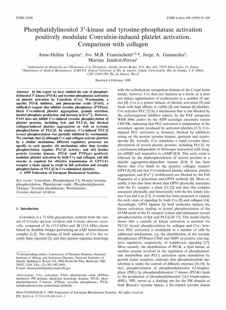
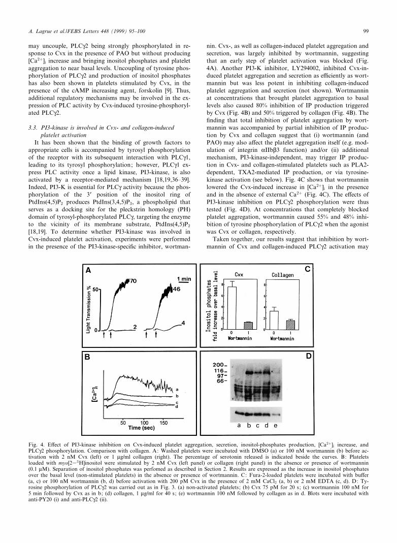
Aggregation of washed platelets induced by an optimumconcentration of Cvx (2 nM) was inhibited by pretreatmentof platelets with PAO (Fig. 1A). The e¡ect of PAO is dosedependent, with an IC50 of 0.4 WM. PAO also inhibited colla-gen-induced platelet aggregation (Fig. 1A), while thrombin-induced platelet aggregation was not signi¢cantly reduced by
PAO at concentrations up to 2.5 WM, in agreement with pre-vious reports [21,22]. As platelet aggregation, dense-granulerelease induced by Cvx or by collagen was inhibited byPAO we have thus investigated the e¡ect of PAO on Cvx-induced IPs production. Inhibition of Cvx- and collagen-in-duced platelet aggregation by PAO was accompanied by morethan 65% and 45% inhibition of IP production, respectively(Fig. 1B). Next, the e¡ects of PAO on the Cvx-induced in-crease in Ca2� were studied. Convulxin induces Ca2� increasein the presence and absence of external Ca2� (Fig. 1C) indi-cating that Cvx induces intracellular Ca2� mobilization as wellas Ca2� entry, in agreement with previous results [6,8]. In thepresence of 0.1 WM PAO, Cvx-induced increase in [Ca2�]i wasdecreased by about 25%, while it was completely blocked by1 WM PAO. These results indicate that an early step in thesignal transduction pathway induced by Cvx is susceptible toPAO. Furthermore, at this point of our study, the observationthat PAO similarly inhibited platelet responses triggered byCvx or collagen favored the hypothesis that Cvx and collageninduce platelet activation by a common pathway.
3.2. PAO does not inhibit Cvx-induced protein tyrosinephosphorylation
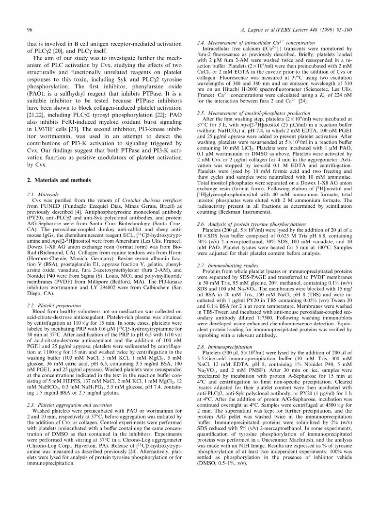
The inhibition by PAO of Cvx-induced [Ca2�]i increase in-dicates that an early step of the signaling cascade triggered byCvx was blocked. Indeed, the observation that inositol-phos-phates production was impaired by PAO, suggested that thiscompound might inhibit PLC activation or activity. In fact,PAO has previously been reported to inhibit tyrosine phos-phorylation of PLCQ2 in collagen- and FcQRIIA-stimulatedplatelets [22]. Since Cvx induces tyrosine phosphorylation ofseveral proteins including PLCQ2 and Syk, we have studiedthe e¡ect of PAO on these responses. Non-stimulated plateletscontain constitutively tyrosine phosphorylated proteins, inparticular proteins of 55^60 kDa. Platelet pretreatment with1 WM PAO alone slightly increased the intensity of some pro-teins (Fig. 2A,B), probably by inhibiting constitutively acti-vated PTPases. Fig. 2A shows that whole platelet proteintyrosyl phosphorylation triggered by Cvx was increased bypretreatment with PAO at concentrations that also attenuatedplatelet aggregation, suggesting that the inhibitory e¡ect ofPAO is associated with inhibition of PTPases. In contrast,the intensity of the protein tyrosine phosphorylation triggeredby collagen was not modi¢ed in the presence of 1 WM PAO(Fig. 2B), con¢rming previous reports and providing evidenceto suggest that a di¡erent site from PTPase, yet to be identi-¢ed, may be blocked by PAO in collagen-stimulated platelets[22].
The e¡ects of PAO on the intensity and number of tyrosine-phosphorylated proteins in Western blots of Cvx-stimulatedplatelets may be explained by a change in the equilibriumbetween the phosphorylation/dephosphorylation reactions to-ward the phosphorylation reaction. Although our results donot reveal the PAO inhibitory mechanism of Cvx-inducedplatelet activation, they do support the contention thatPTPase activation has a positive modulatory function on plat-elet activation [25^28]. Indeed, dephosphorylation of Src-likekinases that are involved in signaling by Cvx and collagen[13,14], results in the catalytic activation of Src-related kinases[29]. More recently, it has also been shown that PAO blocksFcQRI-induced myeloid oxidant signaling together with anincrease of the intensity and number of tyrosyl-phospho-
FEBS 21808 29-3-99
Fig. 1. E¡ect of tyrosine phosphatase inhibitor, phenylarsine oxide(PAO) on Cvx-induced platelet aggregation, secretion, inositol-phos-phates production, and [Ca2�]i increase. Comparison with collagen.A: Washed platelets were incubated with bu¡er (a) or in the pres-ence of 0.2 (b), 0.5 (c), or 1 WM PAO (d). Aggregation was initiated(arrow) by 2 nM Cvx (left panel) or 1 Wg/ml collagen (right panel).The percentage of serotonin released in indicated beside the curves.B: E¡ect of PAO on Cvx- and collagen-induced inositol-phosphatesproduction. Washed platelets loaded with myo[233H]inositol werestimulated by 2 nM Cvx (left panel) or 1 Wg/ml collagen (right pan-el) in the absence of presence of PAO (1 WM). Separation of inositolphosphates was performed as described in Section 2. Results are ex-pressed as the increase in inositol phosphates over the basal level(non-stimulated platelets) in the absence or presence of PAO. C: Ef-fect of PAO on Cvx-induced calcium mobilization fura 2-loadedplatelets were preincubated with bu¡er (a), or PAO 0.1 WM (b) or1 WM (c). Calcium increase was induced by 2 nM Cvx in the pres-ence of 2 mM CaCl2 (left panel) or 2 mM EDTA (right panel).
A. Lagrue et al./FEBS Letters 448 (1999) 95^100 97
rylated proteins in U937IF cells [23]. It is of interest that thisevent is accompanied by an augmentation of the interactionof the adaptor protein Cbl with other phosphoproteins,among them SHC and CRKL. Since Cvx also phosphorylatesCbl in addition to syk, PLCQ2, the Fc receptor Q chain, andthe 36^38 kDa adaptor proteins [8^10], it is plausible to sug-gest that some of these proteins bind to each other and thatthis association should be modulated by the level of tyrosylphosphorylation and hence, by PTPases. In other words, ty-rosine dephosphorylation is required for e¡ective transmissionof GPVI signaling to result in full platelet activation by Cvxand at least in part, by collagen.
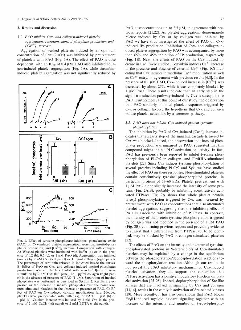
In an attempt to determine the site of inhibition of Cvx-and collagen-induced platelet activation by PAO, we investi-gated the e¡ects of this inhibitor on tyrosine phosphorylationof Syk and PLCQ2 by immunoprecipitation assays (Fig.3A,B). Although GPVI ligation by both collagen [22] andCvx induces a PAO-insensitive tyrosyl phosphorylation ofthe tyrosine kinase Syk (Fig. 3B), PLCQ2 tyrosyl phosphoryl-
ation induced by collagen [22], but not by Cvx, is inhibited byPAO (Fig. 3A). Di¡erences in signals triggered by Cvx andcollagen have been previously reported. In fact, we haveshown that inhibition of Cvx-, but not collagen-induced plat-elet responses by PAO is accompanied by an increase in PTP.In addition, PLCQ2 tyrosyl phosphorylation triggered by Cvxoccurs as early as 10 s after the Cvx addition to platelets [8,9],whereas PLCQ2 is tyrosyl phosphorylated by collagen at latertime points [30,31]. Moreover, piceatannol, a Syk inhibitor,blocks platelet aggregation and protein tyrosine phosphoryla-tion triggered by collagen, whereas it produced only a partialinhibition of Cvx-induced platelet aggregation [10]. Takinginto consideration that collagen may interact with several re-ceptors at the platelet surface (GPIV, integrin K2L1, p65) [32^35] while Cvx-induced platelet activation appears to occurmainly through GPVI ligation [6,10], it is not surprisingthat some di¡erences exist at the level of the signaling path-ways and their regulation. In addition, our results indicatethat tyrosine phosphorylation of PLCQ2 and PLC activity
FEBS 21808 29-3-99
Fig. 3. E¡ect of PAO on PLCQ2 and Syk phosphorylation triggered by Convulxin or collagen. A: PLCQ2 was immunoprecipitated from plate-lets incubated with bu¡er (a, b, d) or PAO 1 WM (c, e) and activated by 2 nM Cvx (b, c) or 1 Wg/ml collagen (d, e). Blots were incubated withanti-PY20 (i) and anti PLCQ2 (ii). B: Syk was immunoprecipitated from platelets incubated with bu¡er (a, b, d) or 1 WM PAO (c, e, f) beforeactivation with Cvx (b, c) or collagen (d^f). Blots were incubated with anti-PY20 (i) and anti-Syk (ii). The intensity of the phosphorylatedband (i) in d^f is proportional to the amount of protein (Syk) in the gel (ii). As a control, in f, the content of platelet protein loaded is 2.5times higher than in d.
Fig. 2. E¡ect of PAO on platelet protein tyrosine phosphorylation induced by Convulxin or collagen. A: Washed platelets were incubated withbu¡er (a, c) or PAO 1 WM (b, d) before activation by 2 nM Cvx for 20 s (lanes c, d). B: Washed platelets were incubated with bu¡er (a, c) orPAO 1 WM (b, d) before activation by 1 Wg/ml collagen for 40 s (lanes c, d). In A and B platelets were lysed and proteins analyzed by immu-noblotting with PY20. The same number of platelets (107) was present in all the wells.
A. Lagrue et al./FEBS Letters 448 (1999) 95^10098
may uncouple, PLCQ2 being strongly phosphorylated in re-sponse to Cvx in the presence of PAO but without producing[Ca2�]i increase and bringing inositol phosphates and plateletaggregation to near basal levels. Uncoupling of tyrosine phos-phorylation of PLCQ2 and production of inositol phosphateshas also been shown in platelets stimulated by Cvx, in thepresence of the cAMP increasing agent, forskolin [9]. Thus,additional regulatory mechanisms may be involved in the ex-pression of PLC activity by Cvx-induced tyrosine-phosphoryl-ated PLCQ2.
3.3. PI3-kinase is involved in Cvx- and collagen-inducedplatelet activation
It has been shown that the binding of growth factors toappropriate cells is accompanied by tyrosyl phosphorylationof the receptor with its subsequent interaction with PLCQ1,leading to its tyrosyl phosphorylation; however, PLCQ1 ex-press PLC activity once a lipid kinase, PI3-kinase, is alsoactivated by a receptor-mediated mechanism [18,19,36^39].Indeed, PI3-K is essential for PLCQ activity because the phos-phorylation of the 3P position of the inositol ring ofPtdIns(4,5)P2 produces PtdIns(3,4,5)P3, a phospholipid thatserves as a docking site for the pleckstrin homology (PH)domain of tyrosyl-phosphorylated PLCQ, targeting the enzymeto the vicinity of its membrane substrate, PtdIns(4,5)P2
[18,19]. To determine whether PI3-kinase was involved inCvx-induced platelet activation, experiments were performedin the presence of the PI3-kinase-speci¢c inhibitor, wortman-
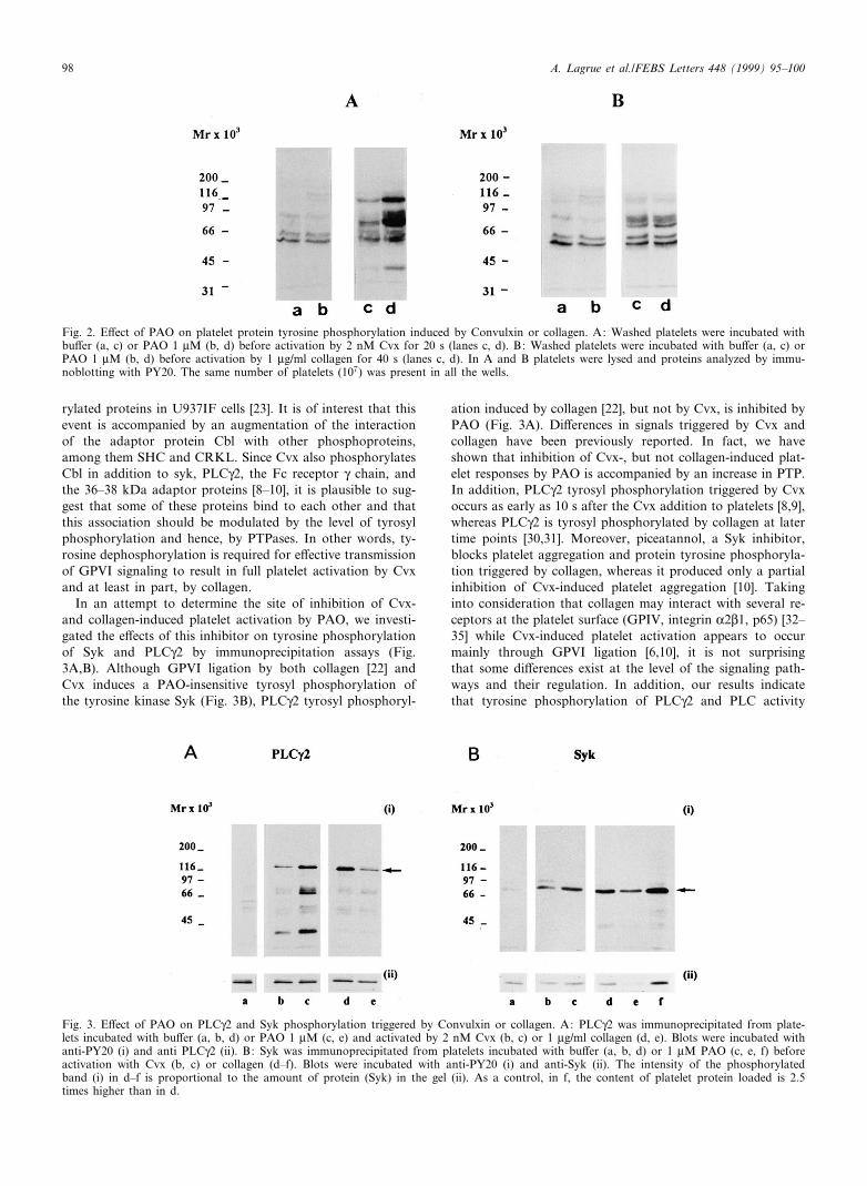
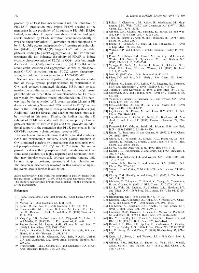
nin. Cvx-, as well as collagen-induced platelet aggregation andsecretion, was largely inhibited by wortmannin, suggestingthat an early step of platelet activation was blocked (Fig.4A). Another PI3-K inhibitor, LY294002, inhibited Cvx-in-duced platelet aggregation and secretion as e¤ciently as wort-mannin but was less potent in inhibiting collagen-inducedplatelet aggregation and secretion (not shown). Wortmanninat concentrations that brought platelet aggregation to basallevels also caused 80% inhibition of IP production triggeredby Cvx (Fig. 4B) and 50% triggered by collagen (Fig. 4B). The¢nding that total inhibition of platelet aggregation by wort-mannin was accompanied by partial inhibition of IP produc-tion by Cvx and collagen suggest that (i) wortmannin (andPAO) may also a¡ect the platelet aggregation itself (e.g. mod-ulation of integrin KIIbL3 function) and/or (ii) additionalmechanism, PI3-kinase-independent, may trigger IP produc-tion in Cvx- and collagen-stimulated platelets such as PLA2-dependent, TXA2-mediated IP production, or via tyrosine-kinase activation (see below). Fig. 4C shows that wortmanninlowered the Cvx-induced increase in [Ca2�]i in the presenceand in the absence of external Ca2� (Fig. 4C). The e¡ects ofPI3-kinase inhibition on PLCQ2 phosphorylation were thustested (Fig. 4D). At concentrations that completely blockedplatelet aggregation, wortmannin caused 55% and 48% inhi-bition of tyrosine phosphorylation of PLCQ2 when the agonistwas Cvx or collagen, respectively.
Taken together, our results suggest that inhibition by wort-mannin of Cvx and collagen-induced PLCQ2 activation may
FEBS 21808 29-3-99
Fig. 4. E¡ect of PI3-kinase inhibition on Cvx-induced platelet aggregation, secretion, inositol-phosphates production, [Ca2�]i increase, andPLCQ2 phosphorylation. Comparison with collagen. A: Washed platelets were incubated with DMSO (a) or 100 nM wortmannin (b) before ac-tivation with 2 nM Cvx (left) or 1 Wg/ml collagen (right). The percentage of serotonin released is indicated beside the curves. B: Plateletsloaded with myo[233H]inositol were stimulated by 2 nM Cvx (left panel) or collagen (right panel) in the absence or presence of wortmannin(0.1 WM). Separation of inositol phosphates was perfomed as described in Section 2. Results are expressed as the increase in inositol phosphatesover the basal level (non-stimulated platelets) in the absence or presence of wortmannin. C: Fura-2-loaded platelets were incubated with bu¡er(a, c) or 100 nM wortmannin (b, d) before activation with 200 pM Cvx in the presence of 2 mM CaCl2 (a, b) or 2 mM EDTA (c, d). D: Ty-rosine phosphorylation of PLCQ2 was carried out as in Fig. 3. (a) non-activated platelets; (b) Cvx 75 pM for 20 s; (c) wortmannin 100 nM for5 min followed by Cvx as in b; (d) collagen, 1 Wg/ml for 40 s; (e) wortmannin 100 nM followed by collagen as in d. Blots were incubated withanti-PY20 (i) and anti-PLCQ2 (ii).
A. Lagrue et al./FEBS Letters 448 (1999) 95^100 99
proceed by at least two mechanisms. First, the inhibition ofPI(3,4,5)P3 production may impair PLCQ2 docking at themembrane in the proximity of its substrate PI(4,5)P2 [18,19].Indeed, a number of papers have shown that the biologicale¡ects mediated by PI3-K activation occur independently oftyrosine phosphorylation; (i) activation of phospholipase C-Qby PI(3,4,5)P3 occurs independently of tyrosine phosphoryla-tion [40^42], (ii) PI(3,4,5,)P3 triggers Ca2� in£ux in rabbitplatelets, leading to platelet aggregation [43], (iii) wortmannintreatment did not in£uence the ability of PDGF to inducetyrosine phosphorylation of PLCQ1 in COS-1 cells but largelydecreased Ins(1,4,5)P3 production [19], (iv) FcQRIIA medi-ated-platelet secretion and aggregation, as well as phospholi-pase C (PLC) activation, but not PLCQ2 tyrosyl phosphoryl-ation, is abolished by wortmannin or LY294002 [40].
Second, since we observed partial but reproducible inhibi-tion of PLCQ2 tyrosyl phosphorylation by wortmannin inCvx- and collagen-stimulated platelets, PI3-K may be alsoinvolved in an alternative pathway leading to PLCQ2 tyrosylphosphorylation other than, or complementary to, Fc recep-tor Q chain-Syk-mediated tyrosyl phosphorylation. This path-way may be the activation of Bruton's tyrosine kinase, a PHdomain containing Src-related PTK related to PLCQ2 activa-tion in the B cell [20] and in collagen-activated platelets [44].Additional tyrosine kinases and adaptor molecules may alsobe involved in this event. Finally, the ¢nding that the p85subunit of PI3-K associates with the Fc receptor Q chain inplatelets stimulated with collagen and Cvx, provides an addi-tional support to the contention that PI-3K participates in theGPVI-Fc receptor Q chain collagen receptor [45].
In conclusion, our results show that the unrelated inhibitorsPAO and wortmannin similarly a¡ect platelet function inCvx-stimulated platelets by a mechanism that uncouples tyro-syl phosphorylation of PLCQ2 and PLC activity. Our resultsprovide evidence that phosphoinositide metabolism in Cvx-stimulated platelets is a highly regulated and redundant eventthat may involve cross-talk between tyrosine kinases, lipidkinases, adaptor proteins, tyrosine and lipid phosphatase.The molecular mechanism involved in this cascade of signal-ing events awaits further investigation.
Acknowledgements: This work was supported in part by grants fromthe European Community (CI1*CT940073) and University Paris 7.The authors acknowledge Brenda Rae Marshall for the preparationof the manuscript.
References
[1] Prado-Franceschi, J. and Vital-Brazil, O. (1981) Toxicon 19, 875^887.
[2] Marlas, G. (1985) Biochimie 67, 1231^1239.[3] Leduc, M. and Bon, C. (1998) Biochem. J. 333, 389^393.[4] Francischetti, I.M.B., Saliou, B., Leduc, M., Carlini, C.R., Hat-
mi, M., Randon, J., Faili, A. and Bon, C. (1997) Toxicon 35,1217^1228.
[5] Vargaftig, B.B., Prado-Franceschi, J., Chignard, M., Lefort, J.and Marlas, G. (1980) Eur. J. Pharmacol. 68, 451^464.
[6] Jandrot-Perrus, M., Lagrue, A.-H., Okuma, M. and Bon, C.(1997) J. Biol. Chem. 272, 27035^27041.
[7] Faili, A., Randon, J., Francischetti, I.M.B., Vargaftig, B.B. andHatmi, M. (1994) Biochem. J. 298, 87^91.
[8] Francischetti, I.M.B., Ghazaleh, F.A., Reis, R.A.M., Carlini,C.R. and Guimaraìes, J.A. (1998) Arch. Biochem. Biophys. 353,239^250.
[9] Francischetti, I.M.B., Carlini, C.R. and Guimaraìes, J.A. (1998)Arch. Biochem. Biophys. 354, 255^262.
[10] Polgaèr, J., Clemetson, J.M., Kehrel, B., Wiedemann, M., Mag-negnat, E.M., Wells, T.N.C. and Clemetson, K.J. (1997) J. Biol.Chem. 272, 13576^13583.
[11] Gibbins, J.M., Okuma, M., Farndale, R., Barnes, M. and Wat-son, S.P. (1997) FEBS Lett. 413, 255^259.
[12] Tsuji, M., Ezumi, Y., Arai, M. and Takayama, H. (1997) J. Biol.Chem. 38, 23528^23531.
[13] Ezumi, Y., Shindoh, K., Tsuji, M. and Takayama, H. (1998)J. Exp. Med. 188, 267^276.
[14] Watson, S.P. and Gibbins, J. (1998) Immunol. Today 19, 260^264.
[15] Poole, A., Gibbins, J.M., Turner, M., van Vugt, M.J., van deWinkel, J.G., Saito, T., Tybulewicz, V.L. and Watson, S.P.(1997) EMBO J. 16, 2333^2341.
[16] Yanaga, F., Poole, A., Asselin, J., Blake, R., Schieven, G.L.,Clark, E.A., Law, C.H. and Watson, S.P. (1995) Biochem.J. 311, 471^478.
[17] Neel, G. (1997) Curr. Opin. Immunol. 9, 405^420.[18] Rhee, S.G. and Bao, Y.S. (1997) J. Biol. Chem. 272, 15045^
15048.[19] Falasca, M., Logan, S.K., Lehto, V.P., Baccante, G., Lemmon,
M.A. and Schlessinger, J. (1998) EMBO J. 17, 414^422.[20] Takata, M. and Kurrosaki, T. (1996) J. Exp. Med. 184, 31^40.[21] Greenwalt, D.A. and Tandon, N.N. (1994) Br. J. Haematol. 88,
830^838.[22] Yanaga, F., Asselin, J., Schieven, G.L. and Watson, S.P. (1995)
FEBS Lett. 368, 377^380.[23] Erdreich-Epstein, A., Liu, M., Liu, Y. and Durden, D.L. (1997)
Exp. Cell Res. 237, 288^295.[24] Grynkiewicz, G., Poenie, M. and Tsien, R.Y. (1985) J. Biol.
Chem. 260, 3440^3450.[25] Levy-Toledano, S., Gallet, C., Nadal, F., Bryckaert, M., Ma-
clouf, J. and Rosa, J.P. (1997) Thromb. Haemost. 78, 226^233.
[26] Frangeoni, J.V., Oda, A., Smith, M., Salzman, E.W. and Neel,B.G. (1993) EMBO J. 12, 4843^4856.
[27] Ezumi, Y., Takayama, H. and Okuma, M. (1995) J. Biol. Chem.20, 11927^11934.
[28] Giuriato, S., Payrastre, B., Drayer, A.L., Plantavid, M., Wo-scholski, R., Parker, P., Erneux, C. and Chap, H. (1997) J. Biol.Chem. 272, 26857^26863.
[29] Corey, S.J. and Anderson, S.M. (1999) Blood 93, 1^14.[30] Daniel, J.L., Dangelmaier, C. and Smith, J.B. (1994) Biochem. J.
302, 617^622.[31] Blake, R.A., Schieven, G.L. and Watson, S.P. (1994) FEBS Lett.
353, 212^216.[32] Tandon, N.N., Kralisz, U. and Jamieson, G.A. (1989) J. Biol.
Chem. 264, 7576^7583.[33] Santoro, S. and Zutter, M.M. (1995) Thromb. Haemost. 74, 813^
821.[34] Chiang, T.M., Rinaldy, A. and Kang, A.H. (1997) J. Clin. Invest.
100, 514^521.[35] Ichinohe, T., Takayama, T., Ezumi, Y., Yanagi, S., Yamamura,
H. and Okuma, M. (1995) J. Biol. Chem. 270, 28029^28036.[36] Li, Z., Wahl, M., Eguinoa, A., Stephens, L.R., Hawkins, P.T.
and Witte, O.N. (1997) Proc. Natl. Acad. Sci. USA 94, 13820^13825.
[37] Rittenhouse, S.E. (1996) Blood 88, 4401^4414.[38] Klarlund, J.K., Guilherme, A., Holik, J.J., Virbasius, J.V., Chaw-
la, A. and Czech, M.P. (1998) Science 275, 1927^1930.[39] Guilherme, A., Klarlund, J.K., Krystal, G. and Czech, M.P.
(1996) J. Biol. Chem. 271, 29533^29536.[40] Gratacap, M.-P., Payrastre, B., Viala, C., Mauco, G., Plantavid,
M. and Chap, H. (1998) J. Biol. Chem. 273, 24314^24321.[41] Bae, Y.S., Cantley, L.G., Chen, C.S., Kim, S.R., Kwon, K.S. and
Rhee, S.G. (1998) J. Biol. Chem. 273, 4465^4469.[42] Rameh, L.E., Rhee, S.G., Spokes, K., Kazlauskas, A., Cantley,
L.C. and Cantley, L.G. (1998) J. Biol. Chem. 273, 23750^23757.[43] Lu, P., Wang, D. and Chen, C. (1998) Biochemistry 37, 9776^
9783.[44] Quek, L.S., Bolen, J. and Watson, S.P. (1998) Curr. Biol. 8,
1137^1140.[45] Gibbins, J.M., Briddon, S., Shutes, A., Vugt, M.J., Winkel,
J.G.J., Saito, T. and Watson, S.P. (1998) J. Biol. Chem. 273,34437^34443.
FEBS 21808 29-3-99
A. Lagrue et al./FEBS Letters 448 (1999) 95^100100