activation of the pi3k/akt pathway early during vaccinia...
TRANSCRIPT

JOURNAL OF VIROLOGY, July 2009, p. 6883–6899 Vol. 83, No. 130022-538X/09/$08.00�0 doi:10.1128/JVI.00245-09Copyright © 2009, American Society for Microbiology. All Rights Reserved.
Activation of the PI3K/Akt Pathway Early during Vaccinia andCowpox Virus Infections Is Required for both Host
Survival and Viral Replication�
Jamaria A. P. Soares,1,2†‡ Flavia G. G. Leite,1,2† Luciana G. Andrade,1,2 Alice A. Torres,1,2
Lirlandia P. De Sousa,3,4 Lucíola S. Barcelos,4,5 Mauro M. Teixeira,4 Paulo C. P. Ferreira,2Erna G. Kroon,2 Thaís Souto-Padron,6 and Claudio A. Bonjardim1,2*
Grupo de Transducao de Sinal,1 Laboratorio de Vírus,2 Departamento de Microbiologia, Setor de Patologia, Coltec/UFMG,3
Laboratorio de Imunofarmacologia, Departamento de Bioquímica e Imunologia,4 and Departamento de Fisiologia eBiofísica,5 Instituto de Ciencias Biologicas, Universidade Federal de Minas Gerais, 31270-901 Belo Horizonte,Minas Gerais, and Laboratorio de Biologia Celular e Ultraestrutura, Universidade Federal do Rio de Janeiro,
21941-590 Rio de Janeiro,6 Brazil
Received 4 February 2009/Accepted 13 April 2009
Viral manipulation of the transduction pathways associated with key cellular functions such as actinremodeling, microtubule stabilization, and survival may favor a productive viral infection. Here we show thatconsistent with the vaccinia virus (VACV) and cowpox virus (CPXV) requirement for cytoskeleton alterationsearly during the infection cycle, PBK/Akt was phosphorylated at S473 [Akt(S473-P)], a modification associatedwith the mammalian target of rapamycin complex 2 (mTORC2), which was paralleled by phosphorylation atT308 [Akt(T308-P)] by PI3K/PDK1, which is required for host survival. Notably, while VACV stimulatedAkt(S473-P/T308-P) at early (1 h postinfection [p.i.]) and late (24 h p.i.) times during the infective cycle, CPXVstimulated Akt at early times only. Pharmacological and genetic inhibition of PI3K (LY294002) or Akt (Akt-Xand a dominant-negative form of Akt-K179M) resulted in a significant decline in virus yield (from 80% to>90%). This decline was secondary to the inhibition of late viral gene expression, which in turn led to an arrestof virion morphogenesis at the immature-virion stage of the viral growth cycle. Furthermore, the cleavage ofboth caspase-3 and poly(ADP-ribose) polymerase and terminal deoxynucleotidyl transferase-mediated de-oxyuridine nick end labeling assays confirmed that permissive, spontaneously immortalized cells such as A31cells and mouse embryonic fibroblasts (MEFs) underwent apoptosis upon orthopoxvirus infection plusLY294002 treatment. Thus, in A31 cells and MEFs, early viral receptor-mediated signals transmitted via thePI3K/Akt pathway are required and precede the expression of viral antiapoptotic genes. Additionally, theinhibition of these signals resulted in the apoptosis of the infected cells and a significant decline in viral titers.
The family Poxviridae is a family of large, linear, double-stranded DNA viruses that carry out their entire life cyclewithin the cytoplasmic compartment of infected cells. Vacciniavirus (VACV) is a prototypical member of the genus Orthopox-virus, which also includes the closely related cowpox virus(CPXV) (12, 52). The genomes of these viruses are approxi-mately 200 kbp in length, with a coding capacity of approxi-mately 200 genes. The genes involved in virus-host interactionsare situated at both ends of the genome and are associatedwith the evasion of host immune defenses (1). These evasionmechanisms operate mainly extracellularly. For example, thesecretion of soluble cytokine and chemokine receptor homo-logues blocks the receptor recognition by intercepting the cog-nate cytokine/chemokine in the extracellular environment.
This mechanism facilitates viral attachment and entry into cells(1, 70). Therefore, decoy receptors for alpha interferon (IFN-�), IFN-�, IFN-�, and tumor necrosis factor alpha play animportant immunomodulatory role by affecting both the hostantiviral and apoptotic responses.
To counteract the host proapoptotic response, poxviruseshave developed a number of antiapoptotic strategies, includingthe inhibition of apoptotic signals triggered by the extrinsicpathway (those mediated by death receptors such as tumornecrosis factor and Fas ligand) or the intrinsic pathway (me-diated by the mitochondria and triggered upon viral infection)(1, 25, 70, 74). Many studies previously identified viral inhibi-tors that block specific steps of the intrinsic pathway. Theseinclude the VACV-encoded E3L, F1L, and N1L genes and themyxoma virus (MYXV)-encoded M11L gene, which block cy-tochrome c release (14, 20, 34, 39, 45, 75, 90), and the CPXV-encoded cytokine response modifier gene (CrmA) as well asthe VACV-encoded SPI-2 gene, which inhibits both caspase-1and caspase-8 (25, 58, 61, 74).
An emerging body of evidence has also highlighted the piv-otal role played by intracellular signaling pathways in Or-thopoxvirus biology (18, 48, 92). We and others have shown thatpoxvirus manipulation of signaling pathways can be virus spe-cific. For example, while both VACV and CPXV stimulate the
* Corresponding author. Mailing address: Grupo de Transducao deSinal, Laboratorio de Vírus, Departamento de Microbiologia, Institutode Ciencias Biologicas, Universidade Federal de Minas Gerais, Av.Antonio Carlos, 6627 Campus Pampulha, 31270-901 Belo Horizonte,Minas Gerais, Brazil. Phone: 55-31 3409-2752. Fax: 55-31 3443-6482.E-mail: [email protected].
† J.A.P.S. and F.G.G.L. contributed equally to this work.‡ Present address: Department of Microbiology and Molecular Ge-
netics, Medical College of Wisconsin, Milwaukee, WI.� Published ahead of print on 22 April 2009.
6883
on February 2, 2019 by guest
http://jvi.asm.org/
Dow
nloaded from

MEK/extracellular signal-regulated kinase (ERK)/EGR-1pathway during a substantial length of time of their infectivecycle, the pathway is required only for VACV replication,whereas its role in CPXV biology has yet to be identified (71).MYXV, a rabbit-specific poxvirus, also activates the MEK/ERK pathway in a mouse model of poxvirus-host interactions.However, this stimulation led to the expression of IFN-�,which consequently blocked virus replication and possibly ex-plains why MYXV has such a restricted host range (87).
Another signaling molecule associated with viral replicationis Akt kinase (also known as protein kinase B). The MYXVhost range factor M-T5 is able to reprogram the intracellularenvironment, thereby increasing human tumor cell permissive-ness to viral replication, which is directly associated with levelsof phosphorylated Akt (88). In addition, M-T5 is functionallyreplaced by the host phosphatidylinositol 3-kinase (PI3K) en-hancer A protein (92).
The transmission of intracellular signals mediated by theserine/threonine kinase Akt to downstream molecules in re-sponse to diverse stimuli such as growth factors, insulin, andhormones is dependent upon the phosphorylation of serine473 (S473-P) and threonine 308 (T308-P). This phosphoryla-tion is mediated by mammalian target of rapamycin complex 2(mTORC2) and phosphoinositide-dependent protein kinase 1(PDK1), which act as downstream effectors of the PI3K/Akt/mTORC1 pathway (2, 66). PI3Ks are a family of enzymes(classes I to III) that generate lipid second messengers by thephosphorylation of plasma membrane phosphoinositides.Class IA PI3Ks consist of a catalytic subunit (p110, comprisingthe three isoforms �, �, and �) and an adaptor/regulatorysubunit (p85, comprising the two isoforms � and �) (for adetailed review, see reference 80).
The Akt family of proteins is comprised of the three iso-forms �, �, and �, which are composed of an N-terminalpleckstrin homology domain, a central catalytic domain, and aC-terminal hydrophobic domain. Akt is recruited to the plasmamembrane through the binding of its pleckstrin homology do-main to the phosphatidylinositol 3,4,5-triphosphate (PIP3),which is a product of PI3K that is anchored to the plasmamembrane. PDK1 is also recruited to the plasma membranethrough interactions with PIP3. As both PDK1 and Akt inter-act with PIP3, PDK1 colocalizes with Akt and activates it byphosphorylating threonine 308 (T308-P) (2, 66). Following itsactivation, Akt phosphorylates a number of downstream sub-strates such as caspase-9, BAD, glycogen synthase kinase 3�(GSK-3�), and FKHR. This leads to the suppression of apop-tosis, cell growth, survival, and proliferation (11, 16, 56).
Another downstream target of PI3K/Akt is mTOR, a serine/threonine kinase that plays a central role in the regulation ofcell growth, proliferation, survival, and protein synthesis (26).mTOR kinase has recently been found to be associated withtwo functionally distinct complexes in mammalian cells, knownas mTORC1 and mTORC2 (63, 66). Although these multipro-tein complexes share molecules in common, distinct adaptorproteins are recruited into each complex: regulatory-associatedprotein of TOR (raptor) is recruited into mTORC1, whilerapamycin-insensitive companion of TOR (rictor) is recruitedinto mTORC2 (33, 64). While mTORC1 controls cell growthand protein translation and has proven to be rapamycin sen-sitive, mTORC2 regulates the actin cytoskeleton and is as-
sumed to be rapamycin insensitive, even though under condi-tions of prolonged exposure to the drug, it appears to inhibitmTORC2 assembly (29, 64, 65). Additionally, it has been dem-onstrated that mTORC2 regulates the activity of Akt throughthe phosphorylation of S473 (S473-P). S473-P appears to berequired for the full activation of Akt, since S473-P has beenshown to enhance the subsequent phosphorylation of T308 byPDK1 (66, 67, 94). Moreover, the phosphorylation of bothS473 and T308 results in a four- to fivefold increase in Aktactivity compared to T308-P by PDK1 alone (66).
The PI3K/PDK1/Akt(T308)/mTORC1 pathway regulates vi-tal cellular processes that are important for viral replicationand propagation, including cell growth, proliferation, and pro-tein translation. This pathway is particularly important for thereplication of DNA viruses, as their replication is cap depen-dent. However, the Akt signaling pathway can also negativelyaffect viral replication. The stress response downstream of Aktsignaling, including hypoxia and energy and amino acid deple-tion, inhibits mTORC1 (5, 9, 69). Therefore, DNA virusesmust overcome these constraints to translate their mRNAs.
Pharmacological disruption of the PI3K/Akt pathway withthe specific PI3K inhibitor LY294002 (2-morpholino-8-phenyl-4H-1-benzopyran-4-one) (82) has been reported to not onlyincrease the cleavage of downstream molecules associated withproapoptotic activity [e.g., poly(ADP-ribose) polymerase(PARP) and the executioner caspase-3] (38, 41) but also pro-mote microtubule stabilization, actin filament remodeling/cellmigration, and bleb formation/viral infectivity (10, 35, 49,54, 59).
Because the PI3K/Akt and PI3K/Akt/mTOR pathways in-fluence diverse cellular functions and possibly a healthy anti-viral response, usurping these pathways could support an in-crease in viral replication. In support of this, a number ofreports have demonstrated that either the PI3K/Akt or thePI3K/Akt/mTOR pathway plays a role in the replication ofmany viruses including flavivirus (38), hepatitis C virus (27),human immunodeficiency virus type 1 (93), human papilloma-virus (44, 96), respiratory syncytial virus (77), coxsackievirus B3(19), Epstein-Barr virus (17, 50, 73), human cytomegalovirus(36, 37, 72), herpes simplex virus type 1 (7, 83), varicella-zostervirus (60), Kaposi’s sarcoma-associated herpesvirus (89), ade-novirus (55), and simian virus 40 (SV40) (95). With this inmind, we also investigated whether the PI3K/Akt pathwayplayed a pivotal role in orthopoxvirus biology. In this study, weshow that the VACV- and CPXV-stimulated PI3K/Akt path-way not only contributes to the prevention of host-cell deathbut also plays a beneficial role in the viral replication cycle.
MATERIALS AND METHODS
Cell culture, antibodies, and chemicals. A spontaneously immortalized cellline (A31), which is derived from mouse BALB/c 3T3 cells, wild-type mouseembryonic fibroblasts (MEFs) (81), SV40 LT-immortalized MEFs (MEFs-LT)(kindly provided by C. Ronald Kahn, Joslin Diabetes Center), and BSC-40 cellswere cultured in Dulbecco’s modified Eagle’s medium supplemented with 7.5%(vol/vol) heat-inactivated fetal bovine serum (FBS) (Cultilab; Campinas, SaoPaulo, Brazil) and antibiotics in 5% CO2 at 37°C. After reaching 80 to 90%confluence, the medium was then changed to 1% FBS, and the cells wereincubated for 12 h. Antibodies against phospho-Akt(Ser473/Thr308); ERK1/2;PARP, which recognizes full-length PARP (116 kDa) and the large (89 kDa) andsmall (24 kDa) fragments of PARP resulting from caspase cleavage; andcaspase-3, which detects full-length caspase-3 (35 kDa) and the large fragment ofcaspase-3 resulting from cleavage (17 kDa), and cleaved caspase-3 (Asp175),
6884 SOARES ET AL. J. VIROL.
on February 2, 2019 by guest
http://jvi.asm.org/
Dow
nloaded from

which detects the large fragment of cleaved caspase-3 (17 to 19 kDa), werepurchased from Cell Signaling Technology (Beverly, MA). LY294002, a phar-macological inhibitor of PI3K; rapamycin, a pharmacological inhibitor ofmTORC1; the pancaspase inhibitor benzyloxycarbonyl-Val-Ala-Asp-fluorom-ethylketone (zVAD.fmk); the viral DNA synthesis inhibitor cytosine arabinoside(Ara C), and anti-�-actin antibody were purchased from Sigma-Aldrich (SaoPaulo, Brazil). The inhibitor of Akt, Akt-X {10-[4�-(N-diethylamino)butyl]-2-chlorophenoxazine}, and Geneticin (G418) were purchased from Calbiochem(Sao Paulo, Brazil). The specific antibodies for the viral H3L, D8L, A14L, andF18R proteins were a generous gift from B. Moss (NIAID, Bethesda, MD). Anantibody that detects CrmA (SPI-2) was obtained from D. Pickup (Departmentof Molecular Genetics and Microbiology, Duke University Medical Center,Durham, NC).
Viruses and viral infection. Wild-type VACV strain WR and CPXV strain BRwere propagated in BSC-40 cells and highly purified by sucrose gradient sedi-mentation as previously described (31). The infective form of intracellular ma-ture VACV and CPXV, which represents the majority of the infectious viralprogeny, was used to carry out the experiments presented in this study. Forexperiments involving UV-irradiated viruses, viral stocks were exposed to a UVlamp producing irradiation predominantly at 365 nm for 5 to 10 min. UV-irradiated viruses were then tested for virus infectivity. Viruses that were unableto form plaques or in which late viral gene expression could not be detected byWestern blotting were considered to be UV inactivated. Viral infections werecarried out when cell cultures reached 80 to 90% confluence. Cells were infectedin the absence of FBS at the indicated multiplicity of infection (MOI) for thetimes shown. Cells were treated with the indicated drugs for 30 min prior to viralinfection and then incubated in the continued presence of drug for the indicatedtimes.
Virus infectivity assays. A31 cells, MEFs, and MEFs-LT cells were culturedand starved as described above at a density of 5 � 105 cells per well in a six-wellculture dish and then exposed to virus. Infections of A31 cells were carried outat an MOI of 10 for 3, 6, 12, 24, 36, and 48 h either in the absence or in thepresence of LY294002 (20 �M) or at the same MOI for 12 h or 24 h either in theabsence or in the presence of rapamycin (50 nM) or Akt-X (15 �M), respectively.MEFs and MEFs-LT were infected for 24 h either in the absence or in thepresence of LY294002 as indicated above. Cultures were then washed with coldphosphate-buffered saline (PBS), and cells were disrupted by freezing and thaw-ing. Virus was collected from the supernatant of centrifuged cells and assayed forinfectivity as described previously (15). Each experiment was run in duplicate,and the results are reported as average values. The data were confirmed by atleast three independent experiments with identical results.
AKT dominant-negative cell lines. Cell lines stably expressing dominant-neg-ative Akt were generated by transfecting A31 cells with 10 �g of plasmid DNAencoding an N-terminal hemagglutinin (HA)-tagged kinase-defective Akt mu-tant [Akt(K179M)] (DN-Akt-HA), an Akt mutant in which the lysine residue atposition 179 (the ATP binding site) was changed to a methionine (22), or theempty vector (pCDNA3) using a standard calcium phosphate transfection pro-tocol. Transfected cells were then ring cloned after selection with 800 �g/mlGeneticin for at least 21 days. The expression of the mutant Akt protein wasevaluated by Western blot analysis. Cell extracts were blotted and then probedwith anti-HA or anti-Akt(S473-P) antibodies as described below.
Electron microscopy. A31 cells were infected with VACV or CPXV at an MOIof 2 either in the presence or in the absence of LY294002 (20 �M) and incubatedat 37°C for 18 or 22 h, respectively. Cells were fixed with 2.5% glutaraldehyde in0.1 M phosphate buffer (pH 7.4) for 1 h at room temperature, scraped gently, andcollected by centrifugation. The cells were then washed with cacodylate buffer,fixed with 1% osmium tetroxide, dehydrated in acetone, and then processed forconventional transmission electron microscopy. Thin sections were examinedusing a Morgagni transmission electron microscope operating at 80 kV.
Western blotting. (i) Lysate preparation. Cells were grown, starved as de-scribed above, and infected with VACV or CPXV at an MOI of 10 for the timesshown. Cells were left untreated or preincubated with the indicated inhibitor for30 min and then exposed to virus in the continued presence of the drugs asshown. Cells were then washed twice with cold PBS and lysed on ice with lysisbuffer (20 mM Tris acetate [pH 7.0], 1 mM EDTA, 1% Triton X-100, 10 mM�-glycerophosphate, 50 mM NaF, 5 mM sodium pyrophosphate, 4 �g/ml leu-peptin, 1 mM sodium orthovanadate). Lysates were scraped, collected into Ep-pendorf tubes, and centrifuged at 13,500 � g for 15 min at 4°C. Protein concen-trations were determined using a Bio-Rad assay.
(ii) Electrophoresis and immunoblotting. Forty micrograms of the cell lysateper sample was separated by electrophoresis on a 10 or 15% sodium dodecylsulfate-polyacrylamide gel and then transferred onto nitrocellulose membranesas previously described (16). Membranes were blocked at room temperature for
1 h with PBS containing 5% (wt/vol) nonfat milk and 0.1% Tween 20. Themembranes were washed three times with PBS containing 0.1% Tween 20 andthen incubated with specific rabbit or mouse polyclonal or monoclonal antibody(1:1,000 to 1:3,000) in PBS containing 5% (wt/vol) bovine serum albumin and0.1% Tween 20. After washing, the membranes were incubated with horseradishperoxidase-conjugated secondary anti-rabbit (1:3,000) or anti-mouse (1:5,000)antibody. Immunoreactive bands were visualized using an ECL detection systemas recommended by the manufacturer (GE Healthcare, Brazil).
TUNEL apoptosis assay. A31 cells were grown and starved as described aboveand then infected with VACV or CPXV at an MOI of 10 for 4 h. Cells were leftuntreated or were preincubated with LY294002 (20 �M) for 30 min and thenincubated with virus in the continued presence of the drug. Cells were then fixedwith 4% paraformaldehyde. A terminal deoxynucleotidyl transferase-mediateddeoxyuridine nick end labeling (TUNEL) assay was performed according to themanufacturer’s instructions (TdT-FragEL DNA fragmentation kit; Calbiochem).The 3�-OH ends of the fragmented nucleosomal DNA in apoptotic cells werespecifically labeled using exogenous terminal deoxynucleotidyl transferase andbiotin-labeled deoxynucleoside triphosphate. Labeled ends were then detectedusing a streptavidin-horseradish peroxidase conjugate and diaminobenzidine.Nuclei were counterstained with methyl green. At least 300 cells (in 10 randomlycaptured microscopic fields at a �600 magnification) were scored to calculate thepercentage of TUNEL-positive nuclei. The data are expressed as the apoptosisindex, which is the average percentage of apoptotic nuclei. The experiment wasperformed in triplicate and repeated two times.
Densitometric analysis. Levels of phosphorylated Akt and cleaved caspase-3were quantified using densitometric analysis software (LabImage), and the levelswere normalized to the levels of �-actin in the same sample. The changes inprotein phosphorylation and cleavage with respect to control values were esti-mated. The results were expressed as the Akt-P-, cleaved caspase-3-, or H3L-A14L-to-�-actin ratio measured in arbitrary units.
RESULTS
VACV and CPXV stimulate Akt(S473/T308) phosphoryla-tion. In order to generate new progeny, poxviruses manipulateessential host signaling pathways such as the MEK/ERK path-way (4) and the PBK/Akt pathway (88) and, consequently, theintracellular environment to allow increased viral replication.
Because early events in virus-host interactions involve alter-ations in the cytoskeleton, from which orthopoxviruses maybenefit by either virus attachment/penetration, morphogenesis,or release (43, 49, 53, 68), we initially investigated whether Aktwas phosphorylated upon infection with VACV or CPXV atS473, a phosphorylation that is mediated by mTORC2. Sincesurvival signals are also important and may benefit viral repli-cation, we also analyzed the phosphorylation of Akt at T308, arole played by PDK1 (9, 63, 66). A31 cells were infected at anMOI of 10, and cell lysates were collected from the infectedcells at 1 to 24 h postinfection (p.i.) and subjected to Westernblotting using an anti-phospho-Akt antibody to evaluate thephosphorylation status of Akt. As shown in Fig. 1A (top), bothVACV and CPXV were able to induce Akt phosphorylation.The amount of Akt-P was verified throughout the course ofVACV infection, and the highest levels of Akt-P were ob-served at early (1 h p.i.) and at late (24 h p.i.) time points (Fig.1A, lanes 11 and 14), and intermediate levels were detected at3 and 7 h p.i. (lanes 12 and 13). In contrast, infection withCPXV was found to stimulate Akt-P(S473/T308) only at earlytime points (1 to 3 h p.i.) of infection (Fig. 1A, lanes 5 to 8).
We then asked whether the virus-stimulated signal leadingto Akt-P(308) was mediated by the upstream kinase PI3K.LY294002, a pharmacological inhibitor of PI3K, inhibits PI3Kactivity through the competitive inhibition of the ATP bindingsite located on the regulatory subunit of PI3K (24, 79, 82), andthe inhibition of Akt-P(308) may result, although indirectly, in
VOL. 83, 2009 ROLE PLAYED BY PI3K/Akt IN ORTHOPOXVIRUS BIOLOGY 6885
on February 2, 2019 by guest
http://jvi.asm.org/
Dow
nloaded from

FIG. 1. VACV and CPXV induce Akt phosphorylation at S473 and T308. (A and D) A31 cells (A) and MEFs (D) were either mock infectedor infected with VACV or CPXV at an MOI of 10 for the indicated times. Infected-cell lysates were subjected to Western blotting (WB) withanti-phospho-Akt(S473-P) or anti-phospho-Akt(T308-P) antibodies (top) or with anti-� actin antibody (bottom), which was used as a loadingcontrol. (A, top) Cells were infected either in the absence (lanes 5 to 8 and 11 to 14) or in the presence (lanes 9, 10, 15, and 16) of LY294002 (LY)
6886 SOARES ET AL. J. VIROL.
on February 2, 2019 by guest
http://jvi.asm.org/
Dow
nloaded from
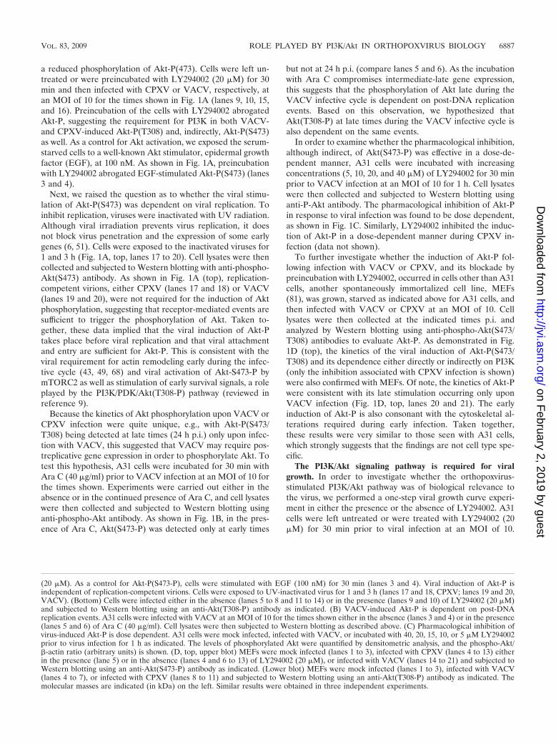
a reduced phosphorylation of Akt-P(473). Cells were left un-treated or were preincubated with LY294002 (20 �M) for 30min and then infected with CPXV or VACV, respectively, atan MOI of 10 for the times shown in Fig. 1A (lanes 9, 10, 15,and 16). Preincubation of the cells with LY294002 abrogatedAkt-P, suggesting the requirement for PI3K in both VACV-and CPXV-induced Akt-P(T308) and, indirectly, Akt-P(S473)as well. As a control for Akt activation, we exposed the serum-starved cells to a well-known Akt stimulator, epidermal growthfactor (EGF), at 100 nM. As shown in Fig. 1A, preincubationwith LY294002 abrogated EGF-stimulated Akt-P(S473) (lanes3 and 4).
Next, we raised the question as to whether the viral stimu-lation of Akt-P(S473) was dependent on viral replication. Toinhibit replication, viruses were inactivated with UV radiation.Although viral irradiation prevents virus replication, it doesnot block virus penetration and the expression of some earlygenes (6, 51). Cells were exposed to the inactivated viruses for1 and 3 h (Fig. 1A, top, lanes 17 to 20). Cell lysates were thencollected and subjected to Western blotting with anti-phospho-Akt(S473) antibody. As shown in Fig. 1A (top), replication-competent virions, either CPXV (lanes 17 and 18) or VACV(lanes 19 and 20), were not required for the induction of Aktphosphorylation, suggesting that receptor-mediated events aresufficient to trigger the phosphorylation of Akt. Taken to-gether, these data implied that the viral induction of Akt-Ptakes place before viral replication and that viral attachmentand entry are sufficient for Akt-P. This is consistent with theviral requirement for actin remodeling early during the infec-tive cycle (43, 49, 68) and viral activation of Akt-S473-P bymTORC2 as well as stimulation of early survival signals, a roleplayed by the PI3K/PDK/Akt(T308-P) pathway (reviewed inreference 9).
Because the kinetics of Akt phosphorylation upon VACV orCPXV infection were quite unique, e.g., with Akt-P(S473/T308) being detected at late times (24 h p.i.) only upon infec-tion with VACV, this suggested that VACV may require pos-treplicative gene expression in order to phosphorylate Akt. Totest this hypothesis, A31 cells were incubated for 30 min withAra C (40 �g/ml) prior to VACV infection at an MOI of 10 forthe times shown. Experiments were carried out either in theabsence or in the continued presence of Ara C, and cell lysateswere then collected and subjected to Western blotting usinganti-phospho-Akt antibody. As shown in Fig. 1B, in the pres-ence of Ara C, Akt(S473-P) was detected only at early times
but not at 24 h p.i. (compare lanes 5 and 6). As the incubationwith Ara C compromises intermediate-late gene expression,this suggests that the phosphorylation of Akt late during theVACV infective cycle is dependent on post-DNA replicationevents. Based on this observation, we hypothesized thatAkt(T308-P) at late times during the VACV infective cycle isalso dependent on the same events.
In order to examine whether the pharmacological inhibition,although indirect, of Akt(S473-P) was effective in a dose-de-pendent manner, A31 cells were incubated with increasingconcentrations (5, 10, 20, and 40 �M) of LY294002 for 30 minprior to VACV infection at an MOI of 10 for 1 h. Cell lysateswere then collected and subjected to Western blotting usinganti-P-Akt antibody. The pharmacological inhibition of Akt-Pin response to viral infection was found to be dose dependent,as shown in Fig. 1C. Similarly, LY294002 inhibited the induc-tion of Akt-P in a dose-dependent manner during CPXV in-fection (data not shown).
To further investigate whether the induction of Akt-P fol-lowing infection with VACV or CPXV, and its blockade bypreincubation with LY294002, occurred in cells other than A31cells, another spontaneously immortalized cell line, MEFs(81), was grown, starved as indicated above for A31 cells, andthen infected with VACV or CPXV at an MOI of 10. Celllysates were then collected at the indicated times p.i. andanalyzed by Western blotting using anti-phospho-Akt(S473/T308) antibodies to evaluate Akt-P. As demonstrated in Fig.1D (top), the kinetics of the viral induction of Akt-P(S473/T308) and its dependence either directly or indirectly on PI3K(only the inhibition associated with CPXV infection is shown)were also confirmed with MEFs. Of note, the kinetics of Akt-Pwere consistent with its late stimulation occurring only uponVACV infection (Fig. 1D, top, lanes 20 and 21). The earlyinduction of Akt-P is also consonant with the cytoskeletal al-terations required during early infection. Taken together,these results were very similar to those seen with A31 cells,which strongly suggests that the findings are not cell type spe-cific.
The PI3K/Akt signaling pathway is required for viralgrowth. In order to investigate whether the orthopoxvirus-stimulated PI3K/Akt pathway was of biological relevance tothe virus, we performed a one-step viral growth curve experi-ment in either the presence or the absence of LY294002. A31cells were left untreated or were treated with LY294002 (20�M) for 30 min prior to viral infection at an MOI of 10.
(20 �M). As a control for Akt-P(S473-P), cells were stimulated with EGF (100 nM) for 30 min (lanes 3 and 4). Viral induction of Akt-P isindependent of replication-competent virions. Cells were exposed to UV-inactivated virus for 1 and 3 h (lanes 17 and 18, CPXV; lanes 19 and 20,VACV). (Bottom) Cells were infected either in the absence (lanes 5 to 8 and 11 to 14) or in the presence (lanes 9 and 10) of LY294002 (20 �M)and subjected to Western blotting using an anti-Akt(T308-P) antibody as indicated. (B) VACV-induced Akt-P is dependent on post-DNAreplication events. A31 cells were infected with VACV at an MOI of 10 for the times shown either in the absence (lanes 3 and 4) or in the presence(lanes 5 and 6) of Ara C (40 �g/ml). Cell lysates were then subjected to Western blotting as described above. (C) Pharmacological inhibition ofvirus-induced Akt-P is dose dependent. A31 cells were mock infected, infected with VACV, or incubated with 40, 20, 15, 10, or 5 �M LY294002prior to virus infection for 1 h as indicated. The levels of phosphorylated Akt were quantified by densitometric analysis, and the phospho-Akt/�-actin ratio (arbitrary units) is shown. (D, top, upper blot) MEFs were mock infected (lanes 1 to 3), infected with CPXV (lanes 4 to 13) eitherin the presence (lane 5) or in the absence (lanes 4 and 6 to 13) of LY294002 (20 �M), or infected with VACV (lanes 14 to 21) and subjected toWestern blotting using an anti-Akt(S473-P) antibody as indicated. (Lower blot) MEFs were mock infected (lanes 1 to 3), infected with VACV(lanes 4 to 7), or infected with CPXV (lanes 8 to 11) and subjected to Western blotting using an anti-Akt(T308-P) antibody as indicated. Themolecular masses are indicated (in kDa) on the left. Similar results were obtained in three independent experiments.
VOL. 83, 2009 ROLE PLAYED BY PI3K/Akt IN ORTHOPOXVIRUS BIOLOGY 6887
on February 2, 2019 by guest
http://jvi.asm.org/
Dow
nloaded from

Viruses were then collected at 3, 6, 12, 24, 36, and 48 h p.i. andassayed for infectivity. The data indicate that the PI3K/Aktpathway did play a relevant role in both VACV and CPXVbiologies. A significant reduction in viral titers (�90%) wasobserved when either CPXV (Fig. 2A) or VACV (Fig. 2B)infection was carried out in the continued presence of the PI3Kinhibitor.
In order to verify that the inhibitory effect associated withLY294002 was not restricted to A31 cells, MEFs were alsoinfected with VACV or CPXV as described above. As shown inFig. 2C, LY294002 caused a significant decline in viral titers(85 to 90%), thereby demonstrating that viral inhibition is notcell type specific. Next, we investigated viral replication in the
context of a non-spontaneously-immortalized cell line such asMEFs-LT. Cells were infected with VACV or CPXV under thesame experimental conditions used for MEFs, and the titerswere then measured. Our findings revealed that virus titerswere decreased by 50% (2.0-fold) and 58% (2.4-fold), respec-tively.
Next, to rule out the possibility of a nonspecific pharmaco-logical inhibition of PI3K/Akt, we generated cell lines stablyexpressing the dominant-negative form of Akt (DN-Akt-K179M). Clones were then monitored for the expression ofHA-Akt by Western blotting using an anti-HA antibody andthe VACV stimulation of Akt(S473-P). Two representativeclones expressing DN-Akt are shown in Fig. 2D, top left (lanes
FIG. 2. The PI3K/Akt signaling pathway is required for viral replication. A31 cells were either left untreated or treated for 30 min withLY294002 (LY) (20 �M), rapamycin (50 nM), or Akt-X (15 �M) prior to virus infection and infected with CPXV or VACV at an MOI of 10 eitherin the absence or in the continued presence of LY294002 for 3, 6, 12, 24, 36, and 48 h; of rapamycin for 12 h; or of Akt-X for 24 h. (C) MEF cells,either spontaneously immortalized (MEFs) or MEFs-LT, were either left untreated or treated for 30 min with LY294002 (20 �M) prior to VACVor CPXV infection in the continued presence of inhibitor for 24 h. Viruses were then collected, and viral titers were determined. (D, top) Tworepresentative cell lines transfected with a dominant-negative form of Akt [Akt(K179M)] (DN-Akt) (lanes 2 and 3) or A31 cells transfected withthe empty vector (A31*) (lane 1) are shown. Cells were left uninfected or infected with VACV or CPXV at an MOI of 10 for 24 h as indicated.Lysates were collected and subjected to Western blotting with an HA-specific antibody (top left) or with anti-Akt(S473-P) antibody (top right). Thebottom blots were probed with �-actin as a loading control. (D, bottom) Viral infectivity assays. Data are representative of three independentexperiments with identical results (A and B) or are means of data from triplicate experiments SD (C, D [bottom], and E). **, P 0.01; ***,P 0.001. A Student’s t test was used in comparisons of A31* and DN-Akt cells (D) or cell lines untreated or treated with the indicatedpharmacological inhibitor (C and E).
6888 SOARES ET AL. J. VIROL.
on February 2, 2019 by guest
http://jvi.asm.org/
Dow
nloaded from

2 and 3), as are control cells transfected with the empty vectoronly (A31*) (lane 1), and in Fig. 2D, top right, where the levelsof Akt-P were significantly reduced upon viral infection (lanes4 and 6). The importance of Akt in viral replication was de-termined by measuring the viral titers following the infection ofcells expressing DN-Akt. The representative clones were in-fected with VACV or CPXV at an MOI of 10, and at 24 h p.i.,viruses were collected, and the titers were determined. Asshown in Fig. 2D (bottom), viral titers were significantly de-creased (�70 to 80%) in cells expressing DN-Akt, suggestingthat the PI3K/Akt pathway does play an important role in theVACV and CPXV life cycles.
To further strengthen the involvement of Akt in mediatingupstream signals after viral stimulation of PI3K, we pretreatedthe cell with the pharmacological inhibitor of Akt, Akt-X (15�M), and infected the cells at an MOI of 10 with VACV orCPXV in the continued presence of the inhibitor, and at 24 hp.i., virus was collected and assayed for infectivity. Our datashowed that the viral titers were significantly reduced by �90%(Fig. 2E). Collectively, these data strongly suggest that thePI3K/Akt pathway is beneficial for VACV and CPXV replica-tion.
Since mTORC1, the downstream target of PI3K/PDK1/Akt(T308-P), regulates key cellular events such as survival,proliferation, and translation, and because the maintenance oftranslation is important for viral cap-dependent mRNA trans-lation, we investigated whether mTORC1 could be requiredfor VACV and CPXV replication. A31 cells were left un-treated or were incubated for 30 min prior to virus infectionwith rapamycin (50 nM), a specific pharmacological inhibitorof mTORC1. Cells were infected in the continued presence ofrapamycin at an MOI of 10, and at 12 h p.i., viruses werecollected and assayed for infectivity. This period of time waschosen not only because it is the earliest time when a signifi-cant decline in virus yield was verified (Fig. 2A and B) but alsoto rule out the possibility of a nonspecific inhibition ofmTORC2 observed after a prolonged exposure of the cells torapamycin (65). Our findings demonstrate that the replicationof VACV and CPXV was only partially affected (�35% [1.5-fold decrease]) following mTORC1 inhibition (Fig. 2E). Sim-ilar levels of inhibition were also observed following incubationwith different concentrations of rapamycin (30 and 70 nM),while a higher drug concentration (100 nM) appeared to in-crease the cytotoxic effect (data not shown).
Disruption of the PI3K/Akt pathway is followed by alteredexpression levels of early and/or late viral genes. To gaininsight into the mechanism(s) underlying the decreased viralyield upon treatment with LY294002, experiments were de-signed to investigate whether early (CrmA/SPI-2) and/or late(F18R, H3L, A14L, and D8L) viral gene expression was af-fected following the preincubation of the cells with the inhib-itor. Cells were cultured either in the presence or in the ab-sence of LY294002 and infected with VACV or CPXV at anMOI of 10 for the indicated times. Cellular lysates were thencollected and subjected to Western blotting using antibodiesraised against the viral proteins CrmA/SPI-2 (Fig. 3A), F18R(Fig. 3B), H3L (Fig. 3C), A14L (Fig. 3D), and D8L (Fig. 3E).As shown in Fig. 3, viral gene expression was remarkably af-fected upon treatment with LY294002, and protein expressionwas either abrogated (Fig. 3B to D) or delayed (A and E). This
emphasizes the critical role played by the PI3K/Akt pathway inthe regulation of orthopoxvirus gene expression. It also pro-vided genetic evidence that the activation of the PI3K/Aktpathway is required for viral late gene expression. Similarly,the infection of cells expressing DN-Akt demonstrated thatboth A14L and H3L expression levels were significantly re-duced (Fig. 3G to H).
Altered viral early and/or late gene expression is accompa-nied by an arrest in orthopoxvirus morphogenesis. In order toinvestigate whether the altered expression of the viral earlyand/or late genes demonstrated in Fig. 3 was accompanied byan arrest in virion morphogenesis, cell cultures were left un-treated (Fig. 4A and C) or were pretreated with LY294002 (20�M) (B and D) and then infected with VACV (A and B) orCPXV (C and D) at an MOI of 2 for 18 and 22 h, respectively.As shown in Fig. 4, while cultures of infected cells alone (Fig.4A and C) contained the full spectrum of normal intermediatesand mature virions typically seen in virion morphogenesis, thepreincubation of cells with LY294002 (B and D) resulted in aninfectious cycle that was arrested at the immature-virion orimmature-virion-with-nucleoids stage of the virion morpho-genic cycle. Therefore, the inhibition of the PI3K/Akt pathwayresulted in an arrest that equally affected the same stages of themorphogenic cycles of both orthopoxviruses. Furthermore, thearrest in virion morphogenesis is consistent with the alteredviral gene expression induced by both the PI3K inhibitor andDN-Akt.
Viral stimulation of the PI3K/Akt pathway induces thecleavage of PARP and caspase-3. To further elucidate the roleof the PI3K/Akt pathway in regulating the survival and/orapoptosis of orthopoxvirus-infected host cells, we blocked thepathway by preincubating cells with LY294002, infected thecells with virus, and then investigated whether the cleavage ofhost proteins associated with characteristic hallmark featuresof apoptosis, such as caspase-3 and PARP, was affected. Cellswere mock infected or infected with VACV or CPXV at anMOI of 10 for 3, 6, 12, or 24 h in either the presence or theabsence of LY294002 (20 �M). Cell lysates were then har-vested and subjected to Western blotting with anti-caspase-3(Fig. 5, top) and anti-PARP (Fig. 5, middle) antibodies. Thepharmacological inhibition of the PI3K/Akt pathway resultedin the cleavage of caspase-3 and PARP in orthopoxvirus-in-fected cells (Fig. 5, top and middle, lanes 16 to 23). Thesefindings strongly suggest that the viral stimulation of the PI3K/Akt pathway has an antiapoptotic/prosurvival effect.
Inhibition of apoptosis is followed by an enhancement of thevirus-stimulated survival pathway. We have demonstratedthat the pharmacological blockade of the PI3K/Akt pathway isfollowed by a decline in levels of orthopoxvirus replication(Fig. 2). Combined with the proapoptotic data presented inFig. 5, these findings strongly suggest that, at least in part, theincreased cytopathic effects observed in the infected cells (Fig.6C and D, compare d and h with b and f) were due to thepharmacological inhibition of the antiapoptotic activity medi-ated by the PI3K/Akt pathway.
In order to further investigate this, A31 cells were prein-cubated for 30 min with increasing concentrations (10, 15,20, and 40 �M) of the pancaspase inhibitor zVAD.fmk ei-ther in the absence or in the presence of LY294002 (20 �M).Cells were then infected with virus at an MOI of 10, and at
VOL. 83, 2009 ROLE PLAYED BY PI3K/Akt IN ORTHOPOXVIRUS BIOLOGY 6889
on February 2, 2019 by guest
http://jvi.asm.org/
Dow
nloaded from

3 h p.i., cell lysates were collected and subjected to Westernblotting with anti-Akt-S473-P antibody. As shown in Fig.6A, incubation with zVAD.fmk (20 �M) prior to VACVinfection increased the levels of Akt-P (lane 4), which were
even more pronounced (1.25-fold) than the level seen withVACV infection alone (lane 1). In contrast, the pharmaco-logical inhibition of the PI3K/Akt pathway resulted in de-creased levels of Akt-P upon infection (Fig. 6A, lane 3).
FIG. 3. Inhibition of the PI3K/Akt pathway interferes with expression of viral genes. A31 cells were left untreated or treated with LY294002 (LY)(20 �M) for 30 min prior to infection with VACV or CPXV at an MOI of 10 either in the absence or in the continued presence of LY294002. (A to E)At the various time points shown, cell lysates were harvested and analyzed by Western blotting using antibodies raised against the viral proteins CrmA(SPI-2) (A), F18R (B), H3L (C), A14L (D), and D8L (E). (F) Anti-total ERK1/2 antibody was used as an internal control for protein loading. (G andH) A31 cells transfected with the empty vector (A31*) (lanes 1 to 3) or DN-Akt2 (lanes 4 to 6) cells were infected with VACV or CPXV at an MOI of10 for 24 h. Lysates were collected and subjected to Western blotting using anti-viral H3L (G) and A14L (H) antibodies. (I) Antibactin antibody was usedas an internal control for protein loading. The levels of H3L and A14L were quantified by densitometric analysis, and the H3L/�-actin ratio andA14L/�-actin ratio (arbitrary units) are shown. Molecular masses (in kDa) are indicated on the left. Data are representative of data from at least threeindependent experiments with very similar results.
6890 SOARES ET AL. J. VIROL.
on February 2, 2019 by guest
http://jvi.asm.org/
Dow
nloaded from

Remarkably, incubation with zVAD.fmk at 10, 15, 20, or 40�M reversed the levels of VACV-mediated Akt phosphory-lation in a dose-dependent manner even in the presence ofLY294002 (Fig. 6A, lanes 5 to 8). Although not as pro-nounced as those verified with VACV infection, similar re-sults were also observed when the infections were per-formed with CPXV (Fig. 6A, lanes 9 to 12).
Given that the inhibition of proapoptotic signals (by zVAD.fmk) is accompanied by enhanced host survival signals (e.g.,increased Akt-P levels) upon VACV and CPXV infection, onewould expect that under this condition, the cleavage ofcaspase-3 after exposure to zVAD.fmk would also be inhibitedin a dose-dependent manner. To investigate this hypothesis,A31 cells were incubated with 10, 15, 20, or 40 �M of zVAD.fmkfor 30 min, either in the absence or in the presence ofLY294002 (20 �M), prior to viral infection at an MOI of 10 for3 h. Cell lysates were collected and subjected to Western blot-ting with an anti-caspase-3 antibody that specifically detects
the cleaved form of caspase-3 (17 to 19 kDa). As demonstratedin Fig. 6B, the cleavage of caspase-3, as a consequence of theinhibition of the survival pathway by LY294002 (lanes 5 to 8),was inhibited in a dose-dependent manner by the antiapoptoticcompound zVAD.fmk. This would increase cell viability and,subsequently, favor viral replication. The same set of experi-ments was performed after infection with CPXV, and the re-sults were similar to those found upon VACV infection (Fig.6B, lanes 9 to 12).
With this in mind, we then investigated whether preincu-bation with zVAD.fmk was sufficient, at least in part, toreverse the increased cytopathic effect observed with theinfections carried out in the presence of LY294002. Cellswere preincubated with LY294002 (20 �M) for 30 min andthen treated with zVAD.fmk (40 �M) for an additional 30min before infection with VACV (Fig. 6C) or CPXV (Fig.6D) at an MOI of 10 for 8 or 24 h. The infected cells werethen examined by phase-contrast microscopy. Our findings
FIG. 4. Altered viral early and/or late gene expression is followed by an arrest in orthopoxvirus morphogenesis. A31 cells were infected withVACV (A and B) or CPXV (C and D) at an MOI of 2 for 18 or 22 h, respectively, either in the absence (A and C) or in the presence (B and D)of LY294002 (20 �M). Cells were then fixed and prepared for transmission electron microscopy. Electron micrographs are shown, with their scalesindicated by bars. (Inset) Detail showing the mature virus. Abbreviations: IV, immature virus; IVN, immature virus with a nucleoid; MV,intracellular mature virus; N, nucleus; M, mitochondria. Data are representative of data from at least two independent experiments with similarresults.
VOL. 83, 2009 ROLE PLAYED BY PI3K/Akt IN ORTHOPOXVIRUS BIOLOGY 6891
on February 2, 2019 by guest
http://jvi.asm.org/
Dow
nloaded from

suggest that the antiapoptotic signals induced upon expo-sure to zVAD.fmk not only decreased the cytopathic effectbut also increased cell viability in both infection models(Fig. 6C and D, compare d and h with b and f). Further-
more, while the infections carried out for 24 h in the pres-ence of LY294002 alone led to an abundant number ofdetached cells that were recovered from the supernatant,cells infected in the simultaneous presence of LY294002 and
FIG. 5. Viral stimulation of the PI3K/Akt pathway has an antiapoptotic effect on host cells. A31 cells were either mock infected (lanes 1 to 4),incubated with LY294002 (LY) (20 �M) alone (lanes 5 to 7), virus infected at an MOI of 10 (lanes 8 to 15), or incubated with LY294002 priorto virus infection for 30 min and then virus infected in the continued presence of the inhibitor (lanes 16 to 23), as indicated. At various time points,cell lysates (40 �g) were harvested and immunoblotted with anti-caspase-3 (top) or with anti-PARP (middle) antibodies to detect the cleavage ofprecursor forms. As a control for protein loading, the membrane (middle) was stripped and reprobed with an anti-ERK1/2 antibody (bottom).Molecular masses are indicated on the left. Data are representative of data from at least two independent experiments with similar results.
FIG. 6. Inhibition of apoptosis is followed by an enhancement of the virus-stimulated survival pathway. A31 cells were either infected with VACV or CPXVat an MOI of 10 for 3 h, incubated with LY294002 (LY) (20 �M) alone, or incubated simultaneously with zVAD.fmk (zVAD) prior to infection as indicated.Cell lysates were collected (40 �g) and were subjected to Western blotting with the indicated antibody. (A, top) The pancaspase inhibitor zVAD.fmk restoresthe levels of Akt-P in a dose-dependent fashion. (B, top) Inhibition of apoptosis diminishes the levels of cleaved caspase-3 in a dose-dependent manner. (A andB, bottom) An anti-�-actin antibody was used as an internal control for protein loading. Molecular masses (in kDa) are indicated on the left. The levels ofphosphorylated Akt were quantified by densitometric analysis, and the phospho-Akt/�-actin ratio (arbitrary units) is shown. (C and D) Phase-contrastmicroscopy. A31 cells were either mock infected (i), incubated with zVAD.fmk (40 �M) alone (j), incubated with LY294002 (20 �M) alone (k), or incubatedwith LY294002 or LY294002 plus zVAD.fmk as indicated prior to viral infection for 30 min and then VACV or CPXV infected (a to h) in the continued presenceof the inhibitor(s) for 8 and 24 h. Data were consistently reproduced in at least three independent experiments with very similar results.
6892 SOARES ET AL. J. VIROL.
on February 2, 2019 by guest
http://jvi.asm.org/
Dow
nloaded from

zVAD.fmk not only remained significantly more attached tothe substrate but also were recovered in the supernatant toa lesser extent (data not shown). Altogether, these dataindicate that the inhibition of proapoptotic signals, accom-panied by enhanced host survival signals and increased cellviability upon VACV or CPXV infection, plays an importantrole during the viral infective cycle.
Inhibition of viral late protein expression by LY294002 iscaspase dependent. In order to investigate whether theLY294002-mediated inhibition of viral late protein expression isdue to an acceleration of apoptosis in infected cells, we comparedthe levels of expression of the viral proteins A14L and H3L in theabsence and in the presence of zVAD.fmk. A31 cells were incu-bated with LY294002 (20 �M) for 30 min and then treated withzVAD.fmk (40 �M) for an additional 30 min prior to VACV orCPXV infection at an MOI of 10 for 24 h. Cell lysates werecollected and subjected to Western blotting with anti-A14L oranti-H3L antibodies. Remarkably, our results indicate that theinhibition of caspase-3 cleavage (Fig. 6B) and apoptosis by thegeneral pancaspase inhibitor zVAD.fmk reverse the inhibitoryeffect of LY294002 on viral A14L and H3L expression (Fig. 7).The observation that zVAD.fmk is capable of blocking caspase-3cleavage in infected cells (Fig. 6B) in association with the rever-sion of A14L and H3L expression, even in the presence ofLY294002 (Fig. 7, lanes 5 and 11), indicates that the regulatoryeffect of the PI3K/Akt pathway exerted during orthopoxvirus rep-lication is a caspase-dependent event.
Inhibition of the PI3K/Akt pathway by LY294002 results inapoptosis of infected cells. In order to firmly establish that theinhibition of the PI3K/Akt pathway by LY294002 results in theapoptosis of the infected cells, a TUNEL assay was used to mon-itor apoptosis in individual cells. A31 cells were incubated withLY294002 (20 �M) for 30 min prior to viral infection with VACVor CPXV at an MOI of 10 for 4 and 6 h, respectively. A brownprecipitate confined to the nucleus was considered to be aTUNEL-positive cell. As shown in Fig. 8A, TUNEL-positive nu-clei were barely observed in the absence of LY294002 in themock-infected cells (not shown) and in the VACV-infected(Fig.8Aa) or the CPXV-infected (Fig.8Ac) cells. However, thenumber of TUNEL-positive cells increased significantly only incultures preincubated with LY294002 and infected with VACVor CPXV (Fig. 8Ab and d). The apoptosis index was calculated,and pretreatment with LY294002 resulted in a 41% ( standarddeviation [SD]) and a 16% (SD) increase in the number ofapoptotic cells following infection with VACV or CPXV, respec-tively (Fig. 8B). To certify that the inhibition of the PI3K/Aktpathway that results in the apoptosis of A31-infected cells was nota cell-type-specific event, the same approach was used for MEFs.As demonstrated in Fig. 8Ae and f, VACV- or CPXV-infectedMEFs also underwent apoptosis upon treatment with LY294002,while apoptosis was not observed in virally infected cells in theabsence of LY294002 (data not shown). The apoptosis index forthese cells was also determined, and it was found to be similar tothat shown in Fig. 8B (data not shown). Thus, these findings not
FIG. 7. Blockade of viral late protein expression by LY294002 is caspase dependent. A31 cells were preincubated with LY294002 (20 �M) (LY)for 30 min followed by treatment with zVAD.fmk (zVAD) (40 �M) for an additional 30 min prior to infection with VACV or CPXV at an MOIof 10 for 24 h, as indicated. Cell lysates were collected (40 �g) and subjected to Western blotting using anti-A14L or -H3L antibodies as indicated.(Top and middle) The pancaspase inhibitor zVAD.fmk reverses the blockade of LY294002 upon viral late protein H3L and A14L expression (lanes5 and 11). (Bottom) Anti-�-actin antibody was used as an internal control for protein loading. The levels of H3L and A14L were quantified bydensitometric analysis, and the H3L/�-actin ratio or A14L/�-actin ratio (arbitrary units) is shown. Molecular masses are indicated on the left. Datawere consistently reproduced in at least two independent experiments with very similar results.
VOL. 83, 2009 ROLE PLAYED BY PI3K/Akt IN ORTHOPOXVIRUS BIOLOGY 6893
on February 2, 2019 by guest
http://jvi.asm.org/
Dow
nloaded from
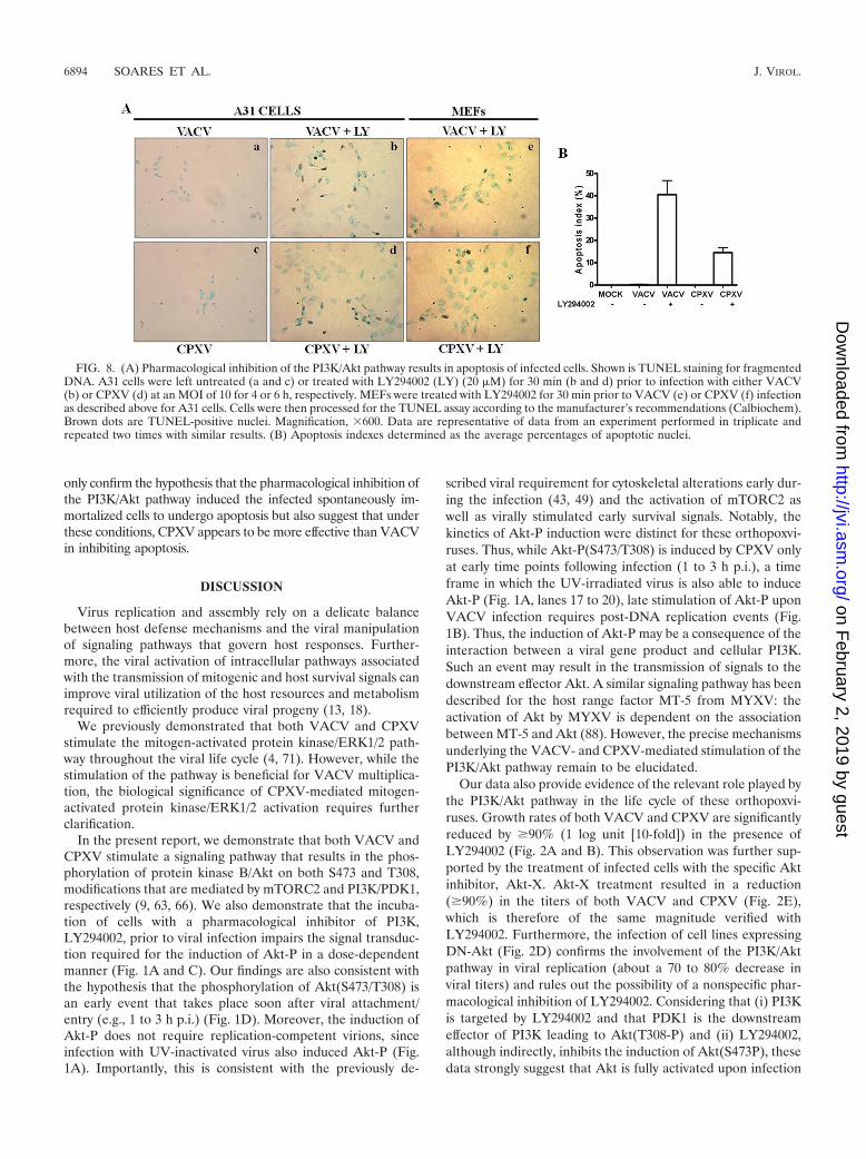
only confirm the hypothesis that the pharmacological inhibition ofthe PI3K/Akt pathway induced the infected spontaneously im-mortalized cells to undergo apoptosis but also suggest that underthese conditions, CPXV appears to be more effective than VACVin inhibiting apoptosis.
DISCUSSION
Virus replication and assembly rely on a delicate balancebetween host defense mechanisms and the viral manipulationof signaling pathways that govern host responses. Further-more, the viral activation of intracellular pathways associatedwith the transmission of mitogenic and host survival signals canimprove viral utilization of the host resources and metabolismrequired to efficiently produce viral progeny (13, 18).
We previously demonstrated that both VACV and CPXVstimulate the mitogen-activated protein kinase/ERK1/2 path-way throughout the viral life cycle (4, 71). However, while thestimulation of the pathway is beneficial for VACV multiplica-tion, the biological significance of CPXV-mediated mitogen-activated protein kinase/ERK1/2 activation requires furtherclarification.
In the present report, we demonstrate that both VACV andCPXV stimulate a signaling pathway that results in the phos-phorylation of protein kinase B/Akt on both S473 and T308,modifications that are mediated by mTORC2 and PI3K/PDK1,respectively (9, 63, 66). We also demonstrate that the incuba-tion of cells with a pharmacological inhibitor of PI3K,LY294002, prior to viral infection impairs the signal transduc-tion required for the induction of Akt-P in a dose-dependentmanner (Fig. 1A and C). Our findings are also consistent withthe hypothesis that the phosphorylation of Akt(S473/T308) isan early event that takes place soon after viral attachment/entry (e.g., 1 to 3 h p.i.) (Fig. 1D). Moreover, the induction ofAkt-P does not require replication-competent virions, sinceinfection with UV-inactivated virus also induced Akt-P (Fig.1A). Importantly, this is consistent with the previously de-
scribed viral requirement for cytoskeletal alterations early dur-ing the infection (43, 49) and the activation of mTORC2 aswell as virally stimulated early survival signals. Notably, thekinetics of Akt-P induction were distinct for these orthopoxvi-ruses. Thus, while Akt-P(S473/T308) is induced by CPXV onlyat early time points following infection (1 to 3 h p.i.), a timeframe in which the UV-irradiated virus is also able to induceAkt-P (Fig. 1A, lanes 17 to 20), late stimulation of Akt-P uponVACV infection requires post-DNA replication events (Fig.1B). Thus, the induction of Akt-P may be a consequence of theinteraction between a viral gene product and cellular PI3K.Such an event may result in the transmission of signals to thedownstream effector Akt. A similar signaling pathway has beendescribed for the host range factor MT-5 from MYXV: theactivation of Akt by MYXV is dependent on the associationbetween MT-5 and Akt (88). However, the precise mechanismsunderlying the VACV- and CPXV-mediated stimulation of thePI3K/Akt pathway remain to be elucidated.
Our data also provide evidence of the relevant role played bythe PI3K/Akt pathway in the life cycle of these orthopoxvi-ruses. Growth rates of both VACV and CPXV are significantlyreduced by �90% (1 log unit [10-fold]) in the presence ofLY294002 (Fig. 2A and B). This observation was further sup-ported by the treatment of infected cells with the specific Aktinhibitor, Akt-X. Akt-X treatment resulted in a reduction(�90%) in the titers of both VACV and CPXV (Fig. 2E),which is therefore of the same magnitude verified withLY294002. Furthermore, the infection of cell lines expressingDN-Akt (Fig. 2D) confirms the involvement of the PI3K/Aktpathway in viral replication (about a 70 to 80% decrease inviral titers) and rules out the possibility of a nonspecific phar-macological inhibition of LY294002. Considering that (i) PI3Kis targeted by LY294002 and that PDK1 is the downstreameffector of PI3K leading to Akt(T308-P) and (ii) LY294002,although indirectly, inhibits the induction of Akt(S473P), thesedata strongly suggest that Akt is fully activated upon infection
FIG. 8. (A) Pharmacological inhibition of the PI3K/Akt pathway results in apoptosis of infected cells. Shown is TUNEL staining for fragmentedDNA. A31 cells were left untreated (a and c) or treated with LY294002 (LY) (20 �M) for 30 min (b and d) prior to infection with either VACV(b) or CPXV (d) at an MOI of 10 for 4 or 6 h, respectively. MEFs were treated with LY294002 for 30 min prior to VACV (e) or CPXV (f) infectionas described above for A31 cells. Cells were then processed for the TUNEL assay according to the manufacturer’s recommendations (Calbiochem).Brown dots are TUNEL-positive nuclei. Magnification, �600. Data are representative of data from an experiment performed in triplicate andrepeated two times with similar results. (B) Apoptosis indexes determined as the average percentages of apoptotic nuclei.
6894 SOARES ET AL. J. VIROL.
on February 2, 2019 by guest
http://jvi.asm.org/
Dow
nloaded from

with these orthopoxviruses. It has also been demonstrated thatviral replication declines significantly in MEFs pretreated withLY294002, confirming the biological relevance of the PI3K/Akt pathway for these orthopoxviruses (Fig. 2C). It is worthnoting that while the pharmacological inhibition of PI3K/Akt(LY294002 and Akt-X) resulted in a �10-fold reduction inviral titers in the spontaneously immortalized cell line A31(Fig. 2A, B, and D) and that about the same level of inhibitionwas also verified with MEFs (LY294002), the decline in virusyields verified after the exposure of SV40-LT MEFs toLY294002 was just partially affected (2- to 2.4-fold reduction)(Fig. 2C). Thus, these findings suggest that the preactivationstate of the intracellular environment in the SV40-LT MEFsper se (9) is beneficial for virus replication and that pretreat-ment with LY294002 blocks only the viral increment that fol-lows the infection, which appears to reflect the partial reduc-tion observed.
In agreement with previously reported data (85), we alsodemonstrate that both VACV and CPXV require mTORC1during the infective cycle. These findings are consistent withthe viral mechanisms used to translate their cap-dependentmRNAs, a pathway that is regulated by the PI3K/PDK1/Akt(T308-P)/mTORC1 signaling pathway (reviewed in refer-ence 9). While the overall effect of LY294002 and Akt-X onviral replication is measured by a decline of �90% (�10-folddecrease) in viral titers, the blockade of mTORC1 by rapamy-cin (which affects mRNA translation) resulted in a reduction of�35% (1.5-fold decrease) in virus yield, thereby suggestingthat the other biological activities regulated by the PI3K/Aktpathway, beyond mTORC1 activation, seem to be coactivatedin order to attend the diverse viral infective demands and, thus,maximize viral replication. However, it is known that the viralE3L and K3L genes are also activated to bypass the block ofviral mRNA translation imposed by protein kinase R (12, 34,52). Remarkably, as was described previously for herpes sim-plex virus (83, 84) and human cytomegalovirus (37, 86) infec-tions, VACV also releases translation using a mechanism thatoperates downstream of mTOR. VACV requires mTORC1 tophosphorylate the eukaryotic translational repressor eIF4Ebinding protein (4E-BP). Following this phosphorylation,4E-BP is degraded by the proteasome. Indeed, it has beendemonstrated that by the inactivation of 4E-BP, VACV altersthe activity of the eIF4F complex and stimulates the accumu-lation of eIF4F components (eIF4E and eIF4G) within theviral factories, thereby facilitating viral replication (85). Whilethe mechanism employed by CPXV to release translationawaits further investigation, it is reasonable to assume that thestrategy used by CPXV might resemble the strategy describedabove for VACV. Thus, we conclude that by fully activatingAkt, VACV and CPXV facilitate and/or maximize their ownreplication.
Consistent with the viral requirement of the PI3K/Akt path-way for a productive infection, if this pathway is inhibitedupstream, viral gene expression is remarkably affected (Fig. 3).While the inhibition of PI3K/Akt appears to affect viral geneexpression to different extents, the delay observed with theCPXV induction of CrmA seems to be more pronounced thanthat observed with VACV (Fig. 3, compare lanes 9 to 12 with13 to 16). However, the delayed expression of this viral anti-apoptotic gene does not appear to affect the global CPXV
antiapoptotic mechanisms, as shown in Fig. 8B. However, themechanisms underlying this effect require further exploration.Furthermore, genetic evidence also confirms the involvementof the pathway in viral gene expression, since infection ofDN-Akt cells results in a significant decrease in H3L and A14Lexpression levels (Fig. 3G to H). Additionally, the PI3K/Aktpathway plays an important role in virion morphogenesis, asdemonstrated by the arrest that occurs at the immature-virion/immature-virion-with-nucleoid stage of the morphogenic cyclewhen the infections are carried out in the presence ofLY294002 (Fig. 4). Similarly, the functional or genetic ablationof the A14L or H3L gene also resulted in an arrest early duringvirion morphogenesis (40, 62). Taken together, these datastrongly demonstrate the beneficial role played by the PI3K/Akt pathway in both VACV and CPXV morphogenesis andgrowth.
Successful viral replication and assembly are dependentupon cell survival. Therefore, viruses have also evolved diversemechanisms to control the pathways that govern cell survival(13, 19, 21). The PI3K/Akt pathway has been demonstrated toplay a pivotal role in cell survival and proliferation (16, 78),and the inhibition of the pathway is associated with a signifi-cant decrease in host cell viability (8, 78) and virus replication(19, 30, 42, 77).
Therefore, it is reasonable to assume that VACV and CPXVstimulate the PI3K/Akt pathway to increase the viability ofinfected host cells in order to prolong the life span of the cell,which would allow more time for the virus to generate itsprogeny. As shown in Fig. 5, the inhibition of the PI3K/Aktpathway during either VACV or CPXV infection significantlyincreases the cleavage of proteins associated with the inductionof apoptosis, such as the executioner caspase-3 and PARP (19,38, 41) (Fig. 5, top and middle). This strongly suggests anantiapoptotic role for the pathway in the course of these or-thopoxvirus infections.
Our findings also demonstrate that the pancaspase inhibitorzVAD.fmk reverses the proapoptotic signals associated withthe blockade of the PI3K/Akt pathway by increasing the levelsof Akt-P (Fig. 6A). This observation, in association with datashowing that zVAD.fmk also reverses the cleavage ofcaspase-3 (Fig. 6B), reinforces the antiapoptotic role of thevirus-stimulated pathway.
It has long been known that the PI3K/Akt pathway plays acritical role in cell survival and proliferation and that its dis-ruption is associated with a significant decrease in host cellviability (8, 57). Thus, the maintenance of prosurvival andantiapoptotic signals upon viral infection is of critical impor-tance for successful viral replication. Our data demonstratethat zVAD.fmk not only decreases the cytopathic effect asso-ciated with LY294002 during infections but also appears toincrease cell adherence and viability (Fig. 6C and D), which arecritical requirements for the generation of viral progeny. Fur-thermore, the LY294002-mediated inhibition of late viral geneexpression, as demonstrated by an analysis of the A14L andH3L proteins, is a caspase-dependent process (Fig. 7). Impor-tantly, viruses have evolved diverse means to regulate viralgene expression in either a caspase-dependent (e.g., flavivi-ruses [38] and orthopoxviruses [this study]) or caspase-inde-pendent (e.g., coxsackievirus) (19) manner.
Our hypothesis that the pharmacological blockade of the
VOL. 83, 2009 ROLE PLAYED BY PI3K/Akt IN ORTHOPOXVIRUS BIOLOGY 6895
on February 2, 2019 by guest
http://jvi.asm.org/
Dow
nloaded from

PI3K/Akt pathway was, at least in part, associated with pro-survival and antiapoptotic signals (Fig. 5 to 8) was furtherstrengthened by the observation that infected cells undergoapoptosis upon exposure to LY294002, a phenomenon thatwas verified not only with A31 cells but also with MEFs (Fig.8). Therefore, receptor-mediated signals conveyed through thePI3K/Akt pathway at early times during the infection of per-missive and spontaneously immortalized cells (e.g., A31 cellsand MEFs) appear to be important to control cell survivaland/or apoptosis. Additionally, these signals should precedethe expression of viral antiapoptotic genes (CrmA, F1, E3L,and N1) because neither CPXV nor VACV seems to be capa-ble on its own of fully preventing the cells from undergoingapoptosis in the presence of LY294002 (Fig. 8). While theactivation of this pathway appears to favor VACV and CPXVreplication (Fig. 2 to 5, 7, and 8), it is remarkable that the levelsof Akt activation correlate not only with cancer progression(i.e., higher metabolic, survival, and proliferation activities)(76, 80) but also with the levels of permissiveness to infectionwith the otherwise-rabbit-specific poxvirus MYXV in a diverseset of human transformed cell lines (88, 92). Therefore, it istempting to speculate that levels of Akt-P that have beenassociated with permissive transformed cells (e.g., BSC-40 andHeLa cells) may facilitate an increased level of replication ofVACV and CPXV. In line with this assumption, it has consis-tently been verified that the viral yields following infection ofHeLa or BSC-40 cells with either VACV or CPXV are at least10-fold higher than those obtained following infection of A31or MEF cells. Not surprising, the viral yields were only partiallyaffected (approximately a twofold reduction) when the infec-tion of BSC-40 cells was carried out in the continued presenceof LY294002 or following the infection of SV40-LT-immortal-ized MEFs, which suggests that the preactivation state of Aktin these permissive lines, as well as in other transformed celllines, is sufficient to elicit the activation of downstream signal-ing that is required early during the infection (Fig. 2C and ourunpublished observations). However, this must be conclusivelyconfirmed. Since most of the relevant experiments performedin vitro to elucidate the role of apoptosis during VACV orCPXV infection were carried out with transformed cell lines(e.g., HeLa, HEK 293T, BSC-40, or Jurkat cells or the mono-cyte/macrophage cell line J774.G8) (14, 28, 39, 74, 75, 90, 91,97), their levels of Akt activation should be higher than thoseobserved in spontaneously immortalized permissive cells (e.g.,A31 cells or MEFs). Furthermore, in experiments in whichMEFs were cultured under permissive conditions for the earlyVACV-mediated PI3K/Akt signals, i.e., in the absence ofLY294004, it was demonstrated that the viral antiapoptoticgenes were necessary and sufficient to control apoptosis(75, 90).
Therefore, it was hypothesized that under this circumstance(e.g., higher levels of Akt-P), the viruses become less depen-dent on the early signals elicited by the host spontaneouslyimmortalized cell lines after viral infection, and thereby, theviral antiapoptotic repertoire could sufficiently block virus-in-duced apoptosis. This suggests that the early signals transmit-ted by the PI3K/Akt pathway upon the attachment and/orpenetration of VACV or CPXV (Fig. 1A and D) would berequired not only for cytoskeletal alterations but also for hostsurvival. Because prior phosphorylation of Akt on S473 by
mTORC2 was previously reported to be required for the phos-phorylation of Akt on T308 by PDK1 (66, 67, 94) and the earlyactivation of mTORC1 is necessary for host survival and viraltranslation (26, 85), the combination of these events, eitherdependently or independently, should provide the host (A31cells and MEFs) with the necessary means to avoid apoptosisbefore the viral antiapoptotic repertoire could be activated. Ithas long been known that orthopoxviruses have evolved severalstrategies to regulate both the extrinsic and intrinsic apoptoticpathways (25, 74). Nonetheless, our data suggest that theseviral antiapoptotic mechanisms, although necessary, are notsufficient to ensure successful viral replication (Fig. 2A to C, 3to 5, 8, and 9). This scenario thus contrasts with that of trans-formed cell lines, where higher levels of Akt activation shouldbypass the need for virus-induced survival signals, and there-fore, the virus antiapoptotic genes should be sufficient to pre-vent apoptosis.
Even though CPXV appears to be more effective thanVACV in inhibiting apoptosis, as reflected by their apoptosisindices (16% [SD] versus 41% [SD], respectively), theoverall effects of LY294002 on viral replication were similar,though not equal, for both viruses (Fig. 2 to 8). This suggeststhat the inhibition of the PI3K/Akt pathway may impact as-pects of orthopoxvirus biology other than the control of sur-vival and/or apoptosis. Indeed, the PI3K/Akt pathway regu-lates a variety of biological processes that are potentiallyimportant for viral replication, including actin remodeling, cellmigration (3, 29, 47, 59, 64), and microtubule stabilization (10,23, 54). Previous research demonstrated that usurping thesepathways would benefit orthopoxviruses in several ways: (i)transportation of the virus through the microtubules to the cellperiphery, followed by a switch from microtubule- to actin-based motility (reviewed in reference 53); (ii) transcription andtranslation of viral mRNAs (32, 46); (iii) alteration of cytoskel-etal organization and translocation of translation factors toviral factories (32, 53, 68, 85); (iv) bleb formation and apop-totic mimicry to penetrate the cell (49); and (v) phosphoryla-tion of Akt(S473) by mTORC2, which regulates the actin cy-toskeleton (9, 37, 72). Therefore, it is not surprising that thesimultaneous incubation of the infected cells with LY294002and zVAD.fmk, which protects the cells only from apoptosis,restored the viral titers only partially (�20%) compared withthe cells incubated with LY294002 alone (data not shown).This further emphasizes the global effect of the PI3K/Akt path-way on the viral life cycle beyond host survival.
In addition, it is remarkable that although VACV andCPXV belong to the same genus, they diverge in the way inwhich they manipulate cellular signaling pathways. While thestimulation of the PI3K/Akt pathway by both viruses facilitatesviral replication, the kinetics of this activation by the virusesare quite different. While VACV stimulated the pathway dur-ing the early and late phases of the infective cycle, as deter-mined by the need for viral late gene expression at 24 h p.i.(Fig. 1B), CPXV stimulation of the same pathway was re-stricted to the early phase of the viral infective cycle in bothcell lines analyzed.
In conclusion, in this report, we demonstrated that the sig-nals triggered by the PI3K/Akt pathway upon VACV andCPXV infection do play an important role in controlling cellsurvival and apoptosis. Nonetheless, it appears that the signals
6896 SOARES ET AL. J. VIROL.
on February 2, 2019 by guest
http://jvi.asm.org/
Dow
nloaded from

required for successful viral replication extend beyond survivaland apoptosis. As the late events related to the release and/orspread of orthopoxviruses are dependent upon actin dynamics,a role associated not only with mTORC2 but also with the Srcfamily of tyrosine kinases (reviewed in reference 53), it will beexciting to further investigate and compare the specific contri-butions of Akt(S473-P) and the Src family kinases to the biol-ogy of these orthopoxviruses during the late stages of theirinfective cycles. This could explain the more-pronounced effectof both LY294002 and Akt-X on CPXV replication (Fig. 2Ato C).
A schematic representation depicting the relevant findingsof this study is shown in Fig. 9. Soon after VACV and CPXVencounter a host spontaneously immortalized cell line such asA31 cells or MEFs, signals from the PI3K/Akt pathway aretriggered and are required to release translation and host sur-vival, a function associated with mTORC1, via the upstreamphosphorylation of T308 by PDK1. The activation of mTORC2
[Akt(473-P)], which is required for cytoskeletal alterations andvirus attachment and/or penetration, in conjunction with thephosphorylation of Akt on T308 via PI3K, appears to boostkinase activities and, thus, creates an intracellular environmentthat not only protects A31 cells and MEFs from undergoingapoptosis but also favors VACV and CPXV replication. Phar-macological or genetic blockade of the PI3K/Akt pathway in-terrupts early signal transmission, and under these circum-stances, the viral antiapoptotic genes are not sufficient toprevent the cells from undergoing apoptosis.
ACKNOWLEDGMENTS
We are grateful to Angela S. Lopes, Hilda M. V. Gama, Joao R. dosSantos, Andreza A. Carvalho, Gisele O. L. Rodrigues, and Jonas D.Albarnaz for their secretarial and technical assistance. We also thankH. A. Armelin and M. C. Sogayar, Department of Biochemistry, Uni-versity of Sao Paulo, Sao Paulo, Brazil; Eileen D. Adamson, TheBurnham Institute, La Jolla, CA; and Richard D. Ye, Department ofPharmacology, University of Illinois, Chicago, IL, who kindly provided
FIG. 9. Schematic representation of the VACV- and CPXV-stimulated PI3K/Akt pathway of biological relevance in spontaneously immortal-ized A31 and MEF cells. Upon attachment and/or penetration, VACV and CPXV activate Akt(S473-P) and Akt(T308-P) via mTORC2 andPI3K/PDK1, respectively. Thus, Akt is fully activated and transduces signals associated with diverse biological activities to attend viral demands,such as cytoskeleton alterations, survival, translation release, and microtubule stabilization, which appear to benefit virus penetration, morpho-genesis, and release, thereby increasing viral replication. Boxes highlight the biological consequences after pharmacological (LY294002, rapamycin,and Akt-X) and/or dominant-negative (DN-Akt) blockade of the pathways. Viral stimulation of both the PI3K/Akt pathway early during theinfective cycle and the viral antiapoptotic genes is required to impede the demise of the cells, which is followed by an increase in virus titers. Incontrast, upon the blockade of the pathway, the viral antiapoptotic genes alone are not sufficient to prevent the host cells from undergoingapoptosis, which is accompanied by a decline in virus yield.
VOL. 83, 2009 ROLE PLAYED BY PI3K/Akt IN ORTHOPOXVIRUS BIOLOGY 6897
on February 2, 2019 by guest
http://jvi.asm.org/
Dow
nloaded from

us with the A31 cell line, WT MEFs, and plasmid expressing Aktdominant-negative mutations, respectively. C. Ronald Kahn, JoslinDiabetes Center, kindly provided us with the SV40-LT immortalizedMEFs. VACV (strain WR) and CPXV (strain BR) were obtained fromC. Jungwirth, Universitat Wurzburg, Wurzburg, Germany. We alsothank L. A. Velloso (FMC-Unicamp) and J. A. Yunes-Campinas (Cen-tro I. Boldrini) for the Akt antibody (T308). We also thank BrunoBrasil for critical reading of the manuscript.
This work was supported by grants from Fundacao de Amparo aPesquisa do Estado de Minas Gerais, the Coordenadoria de Aper-feicoamento de Pessoal de Nível Superior (CAPES), the BrazilianMinistry of Culture, Science, and Technology, and the Conselho Na-cional de Desenvolvimento Científico e Tecnologico(CNPq). J.A.P.S.was a recipient of a predoctoral fellowship from the CAPES, andF.G.G.L. and L.G.A. are recipients of predoctoral fellowships from theCNPq. L.S.B. holds a PRODOC research fellowship from theCAPES. C.A.B., E.G.K., P.C.P.F., M.M.T., and T.S.-P. are recipientsof research fellowships from the CNPq.
REFERENCES
1. Alcami, A. 2003. Viral mimicry of cytokines, chemokines and their receptors.Nat. Rev. Immunol. 3:36–50.
2. Alessi, D. R., M. Andjelkovic, B. Caudwell, P. Cron, N. Morrice, P. Cohen,and B. A. Hemmings. 1996. Mechanism of activation of protein kinase B byinsulin and IGF-1. EMBO J. 15:6541–6551.
3. Amiri, A., F. Noei, S. Jeganathan, G. Kulkarni, D. E. Pinke, and J. M. Lee.2007. eEF1A2 activates Akt and stimulates Akt-dependent actin remodeling,invasion and migration. Oncogene 26:3027–3040.
4. Andrade, A. A., P. N. Silva, A. C. Pereira, L. P. De Sousa, P. C. Ferreira, R. T.Gazzinelli, E. G. Kroon, C. Ropert, and C. A. Bonjardim. 2004. The vacciniavirus-stimulated mitogen-activated protein kinase (MAPK) pathway is re-quired for virus multiplication. Biochem. J. 381:437–446.
5. Avruch, J., K. Hara, Y. Lin, M. Liu, X. Long, S. Ortiz-Vega, and K. Yon-ezawa. 2006. Insulin and amino-acid regulation of mTOR signaling andkinase activity through the Rheb GTPase. Oncogene 25:6361–6372.
6. Bablanian, R., G. Coppola, S. Scribani, and M. Esteban. 1981. Inhibition ofprotein synthesis by vaccinia virus. III. The effect of ultraviolet-irradiatedvirus on the inhibition of protein synthesis. Virology 112:1–12.
7. Benetti, L., and B. Roizman. 2006. Protein kinase B/Akt is present in acti-vated form throughout the entire replicative cycle of �US3 mutant virus butonly at early times after infection with wild-type herpes simplex virus 1.J. Virol. 80:3341–3348.
8. Bondar, V. M., B. Sweeney-Gotsch, M. Andreeff, G. B. Mills, and D. J.McConkey. 2002. Inhibition of the phosphatidylinositol 3�-kinase-AKT path-way induces apoptosis in pancreatic carcinoma cells in vitro and in vivo. Mol.Cancer Ther. 1:989–997.
9. Buchkovich, N. J., Y. Yu, C. A. Zampieri, and J. C. Alwine. 2008. TheTORrid affairs of viruses: effects of mammalian DNA viruses on the PI3K-Akt-mTOR signalling pathway. Nat. Rev. Microbiol. 6:266–275.
10. Buttrick, G. J., and J. G. Wakefield. 2008. PI3-K and GSK-3: Akt-ing to-gether with microtubules. Cell Cycle 7:2621–2625.
11. Cardone, M. H., N. Roy, H. R. Stennicke, G. S. Salvesen, T. F. Franke, E.Stanbridge, S. Frisch, and J. C. Reed. 1998. Regulation of cell death pro-tease caspase-9 by phosphorylation. Science 282:1318–1321.
12. Condit, R. C., N. Moussatche, and P. Traktman. 2006. In a nutshell: struc-ture and assembly of the vaccinia virion. Adv. Virus Res. 66:31–124.
13. Cooray, S. 2004. The pivotal role of phosphatidylinositol 3-kinase-Akt signaltransduction in virus survival. J. Gen. Virol. 85:1065–1076.
14. Cooray, S., M. W. Bahar, N. G. Abrescia, C. E. McVey, N. W. Bartlett, R. A.Chen, D. I. Stuart, J. M. Grimes, and G. L. Smith. 2007. Functional andstructural studies of the vaccinia virus virulence factor N1 reveal a Bcl-2-likeanti-apoptotic protein. J. Gen. Virol. 88:1656–1666.
15. da Fonseca, F. G., G. S. Trindade, R. L. Silva, C. A. Bonjardim, P. C.Ferreira, and E. G. Kroon. 2002. Characterization of a vaccinia-like virusisolated in a Brazilian forest. J. Gen. Virol. 83:223–228.
16. Datta, S. R., H. Dudek, X. Tao, S. Masters, H. Fu, Y. Gotoh, and M. E.Greenberg. 1997. Akt phosphorylation of BAD couples survival signals to thecell-intrinsic death machinery. Cell 91:231–241.
17. Dawson, C. W., G. Tramountanis, A. G. Eliopoulos, and L. S. Young. 2003.Epstein-Barr virus latent membrane protein 1 (LMP1) activates the phos-phatidylinositol 3-kinase/Akt pathway to promote cell survival and induceactin filament remodeling. J. Biol. Chem. 278:3694–3704.
18. de Magalhaes, J. C., A. A. Andrade, P. N. Silva, L. P. Sousa, C. Ropert, P. C.Ferreira, E. G. Kroon, R. T. Gazzinelli, and C. A. Bonjardim. 2001. Amitogenic signal triggered at an early stage of vaccinia virus infection: im-plication of MEK/ERK and protein kinase A in virus multiplication. J. Biol.Chem. 276:38353–38360.
19. Esfandiarei, M., H. Luo, B. Yanagawa, A. Suarez, D. Dabiri, J. Zhang, andB. M. McManus. 2004. Protein kinase B/Akt regulates coxsackievirus B3
replication through a mechanism which is not caspase dependent. J. Virol.78:4289–4298.
20. Everett, H., M. Barry, X. Sun, S. F. Lee, C. Frantz, L. G. Berthiaume, G.McFadden, and R. C. Bleackley. 2002. The myxoma poxvirus protein, M11L,prevents apoptosis by direct interaction with the mitochondrial permeabilitytransition pore. J. Exp. Med. 196:1127–1139.
21. Francois, F., and M. E. Klotman. 2003. Phosphatidylinositol 3-kinase regu-lates human immunodeficiency virus type 1 replication following viral entryin primary CD4� T lymphocytes and macrophages. J. Virol. 77:2539–2549.
22. Franke, T. F., S. I. Yang, T. O. Chan, K. Datta, A. Kazlauskas, D. K.Morrison, D. R. Kaplan, and P. N. Tsichlis. 1995. The protein kinase en-coded by the Akt proto-oncogene is a target of the PDGF-activated phos-phatidylinositol 3-kinase. Cell 81:727–736.
23. Fujiwara, Y., Y. Hosokawa, K. Watanabe, S. Tanimura, K. Ozaki, and M.Kohno. 2007. Blockade of the phosphatidylinositol-3-kinase-Akt signalingpathway enhances the induction of apoptosis by microtubule-destabilizingagents in tumor cells in which the pathway is constitutively activated. Mol.Cancer Ther. 6:1133–1142.
24. Galetic, I., M. Andjelkovic, R. Meier, D. Brodbeck, J. Park, and B. A.Hemmings. 1999. Mechanism of protein kinase B activation by insulin/insu-lin-like growth factor-1 revealed by specific inhibitors of phosphoinositide3-kinase—significance for diabetes and cancer. Pharmacol. Ther. 82:409–425.
25. Galluzzi, L., C. Brenner, E. Morselli, Z. Touat, and G. Kroemer. 2008. Viralcontrol of mitochondrial apoptosis. PLoS Pathog. 4:e1000018.
26. Hay, N., and N. Sonenberg. 2004. Upstream and downstream of mTOR.Genes Dev. 18:1926–1945.
27. He, Y., H. Nakao, S. L. Tan, S. J. Polyak, P. Neddermann, S. Vijaysri, B. L.Jacobs, and M. G. Katze. 2002. Subversion of cell signaling pathways byhepatitis C virus nonstructural 5A protein via interaction with Grb2 and P85phosphatidylinositol 3-kinase. J. Virol. 76:9207–9217.
28. Humlova, Z., M. Vokurka, M. Esteban, and Z. Melkova. 2002. Vaccinia virusinduces apoptosis of infected macrophages. J. Gen. Virol. 83:2821–2832.
29. Jacinto, E., R. Loewith, A. Schmidt, S. Lin, M. A. Ruegg, A. Hall, and M. N.Hall. 2004. Mammalian TOR complex 2 controls the actin cytoskeleton andis rapamycin insensitive. Nat. Cell Biol. 6:1122–1128.
30. Johnson, R. A., X. Wang, X. L. Ma, S. M. Huong, and E. S. Huang. 2001.Human cytomegalovirus up-regulates the phosphatidylinositol 3-kinase(PI3-K) pathway: inhibition of PI3-K activity inhibits viral replication andvirus-induced signaling. J. Virol. 75:6022–6032.
31. Joklik, W. K. 1962. The purification of four strains of poxvirus. Virology18:9–18.
32. Katsafanas, G. C., and B. Moss. 2007. Colocalization of transcription andtranslation within cytoplasmic poxvirus factories coordinates viral expressionand subjugates host functions. Cell Host Microbe 2:221–228.
33. Kibler, K. V., T. Shors, K. B. Perkins, C. C. Zeman, M. P. Banaszak, J.Biesterfeldt, J. O. Langland, and B. L. Jacobs. 1997. Double-stranded RNAis a trigger for apoptosis in vaccinia virus-infected cells. J. Virol. 71:1992–2003.
34. Kim, D. H., D. D. Sarbassov, S. M. Ali, J. E. King, R. R. Latek, H. Erdju-ment-Bromage, P. Tempst, and D. M. Sabatini. 2002. mTOR interacts withraptor to form a nutrient-sensitive complex that signals to the cell growthmachinery. Cell 110:163–175.
35. Kolsch, V., P. G. Charest, and R. A. Firtel. 2008. The regulation of cellmotility and chemotaxis by phospholipid signaling. J. Cell Sci. 121:551–559.
36. Kudchodkar, S. B., Y. Yu, T. G. Maguire, and J. C. Alwine. 2006. Humancytomegalovirus infection alters the substrate specificities and rapamycinsensitivities of raptor- and rictor-containing complexes. Proc. Natl. Acad. Sci.USA 103:14182–14187.
37. Kudchodkar, S. B., Y. Yu, T. G. Maguire, and J. C. Alwine. 2004. Humancytomegalovirus infection induces rapamycin-insensitive phosphorylation ofdownstream effectors of mTOR kinase. J. Virol. 78:11030–11039.
38. Lee, C. J., C. L. Liao, and Y. L. Lin. 2005. Flavivirus activates phosphatidyl-inositol 3-kinase signaling to block caspase-dependent apoptotic cell death atthe early stage of virus infection. J. Virol. 79:8388–8399.
39. Lee, S. B., and M. Esteban. 1994. The interferon-induced double-strandedRNA-activated protein kinase induces apoptosis. Virology 199:491–496.
40. Lin, C. L., C. S. Chung, H. G. Heine, and W. Chang. 2000. Vaccinia virusenvelope H3L protein binds to cell surface heparan sulfate and is importantfor intracellular mature virion morphogenesis and virus infection in vitro andin vivo. J. Virol. 74:3353–3365.
41. Lin, S. Y., H. Cui, B. Yusta, and D. D. Belsham. 2004. IGF-I signalingprevents dehydroepiandrosterone (DHEA)-induced apoptosis in hypotha-lamic neurons. Mol. Cell. Endocrinol. 214:127–135.
42. Linnemann, T., Y. H. Zheng, R. Mandic, and B. M. Peterlin. 2002. Interac-tion between Nef and phosphatidylinositol-3-kinase leads to activation ofp21-activated kinase and increased production of HIV. Virology 294:246–255.
43. Locker, J. K., A. Kuehn, S. Schleich, G. Rutter, H. Hohenberg, R. Wepf, andG. Griffiths. 2000. Entry of the two infectious forms of vaccinia virus at theplasma membrane is signaling-dependent for the IMV but not the EEV.Mol. Biol. Cell 11:2497–2511.
6898 SOARES ET AL. J. VIROL.
on February 2, 2019 by guest
http://jvi.asm.org/
Dow
nloaded from

44. Lu, Z., X. Hu, Y. Li, L. Zheng, Y. Zhou, H. Jiang, T. Ning, Z. Basang, C.Zhang, and Y. Ke. 2004. Human papillomavirus 16 E6 oncoprotein interfer-ences with insulin signaling pathway by binding to tuberin. J. Biol. Chem.279:35664–35670.
45. Macen, J. L., K. A. Graham, S. F. Lee, M. Schreiber, L. K. Boshkov, and G.McFadden. 1996. Expression of the myxoma virus tumor necrosis factorreceptor homologue and M11L genes is required to prevent virus-inducedapoptosis in infected rabbit T lymphocytes. Virology 218:232–237.
46. Mallardo, M., S. Schleich, and J. K. Locker. 2001. Microtubule-dependentorganization of vaccinia virus core-derived early mRNAs into distinct cyto-plasmic structures. Mol. Biol. Cell 12:3875–3891.
47. Manning, B. D., and L. C. Cantley. 2007. AKT/PKB signaling: navigatingdownstream. Cell 129:1261–1274.
48. McFadden, G. 2005. Poxvirus tropism. Nat. Rev. Microbiol. 3:201–213.49. Mercer, J., and A. Helenius. 2008. Vaccinia virus uses macropinocytosis and
apoptotic mimicry to enter host cells. Science 320:531–535.50. Moody, C. A., R. S. Scott, N. Amirghahari, C. A. Nathan, L. S. Young, C. W.
Dawson, and J. W. Sixbey. 2005. Modulation of the cell growth regulatormTOR by Epstein-Barr virus-encoded LMP2A. J. Virol. 79:5499–5506.
51. Moss, B. 1996. Poxviridae, p. 2637–2671. In B. N. Fields, D. M. Knipe, andP. M. Howley (ed.), Fields virology, 3rd ed., vol. 2. Lippincott-Raven Pub-lishers, Philadelphia, PA.
52. Moss, B. 2006. Poxviridae, p. 2905–2946. In D. M. Knipe, P. M. Howley, D. E.Griffin, R. A. Lamb, M. A. Martin, B. Roizman, and S. E. Straus (ed.), Fieldsvirology, 5th ed, vol. 2. Lippincott Williams & Wilkins, Philadelphia, PA.
53. Munter, S., M. Way, and F. Frischknecht. 2006. Signaling during pathogeninfection. Sci. STKE 2006:re5.
54. Onishi, K., M. Higuchi, T. Asakura, N. Masuyama, and Y. Gotoh. 2007. ThePI3K-Akt pathway promotes microtubule stabilization in migrating fibro-blasts. Genes Cells. 12:535–546.
55. O’Shea, C. C., S. Choi, F. McCormick, and D. Stokoe. 2005. Adenovirusoverrides cellular checkpoints for protein translation. Cell Cycle 4:883–888.
56. Pap, M., and G. M. Cooper. 1998. Role of glycogen synthase kinase-3 in thephosphatidylinositol 3-kinase/Akt cell survival pathway. J. Biol. Chem. 273:19929–19932.
57. Peruzzi, F., M. Prisco, M. Dews, P. Salomoni, E. Grassilli, G. Romano, B.Calabretta, and R. Baserga. 1999. Multiple signaling pathways of the insulin-like growth factor 1 receptor in protection from apoptosis. Mol. Cell. Biol.19:7203–7215.
58. Pickup, D. J., B. S. Ink, W. Hu, C. A. Ray, and W. K. Joklik. 1986. Hemor-rhage in lesions caused by cowpox virus is induced by a viral protein that isrelated to plasma protein inhibitors of serine proteases. Proc. Natl. Acad.Sci. USA 83:7698–7702.
59. Qian, Y., L. Corum, Q. Meng, J. Blenis, J. Z. Zheng, X. Shi, D. C. Flynn, andB. H. Jiang. 2004. PI3K induced actin filament remodeling through Akt andp70S6K1: implication of essential role in cell migration. Am. J. Physiol. CellPhysiol. 286:C153–C163.
60. Rahaus, M., N. Desloges, and M. H. Wolff. 2007. Varicella-zoster virusrequires a functional PI3K/Akt/GSK-3alpha/beta signaling cascade for effi-cient replication. Cell. Signal. 19:312–320.
61. Ray, C. A., R. A. Black, S. R. Kronheim, T. A. Greenstreet, P. R. Sleath, G. S.Salvesen, and D. J. Pickup. 1992. Viral inhibition of inflammation: cowpoxvirus encodes an inhibitor of the interleukin-1 beta converting enzyme. Cell69:597–604.
62. Rodriguez, J. R., C. Risco, J. L. Carrascosa, M. Esteban, and D. Rodriguez.1998. Vaccinia virus 15-kilodalton (A14L) protein is essential for assemblyand attachment of viral crescents to virosomes. J. Virol. 72:1287–1296.
63. Sarbassov, D. D., S. M. Ali, and D. M. Sabatini. 2005. Growing roles for themTOR pathway. Curr. Opin. Cell Biol. 17:596–603.
64. Sarbassov, D. D., S. M. Ali, D. H. Kim, D. A. Guertin, R. R. Latek, H.Erdjument-Bromage, P. Tempst, and D. M. Sabatini. 2004. Rictor, a novelbinding partner of mTOR, defines a rapamycin-insensitive and raptor-inde-pendent pathway that regulates the cytoskeleton. Curr. Biol. 14:1296–1302.
65. Sarbassov, D. D., S. M. Ali, S. Sengupta, J. H. Sheen, P. P. Hsu, A. F. Bagley,A. L. Markhard, and D. M. Sabatini. 2006. Prolonged rapamycin treatmentinhibits mTORC2 assembly and Akt/PKB. Mol. Cell 22:159–168.
66. Sarbassov, D. D., D. A. Guertin, S. M. Ali, and D. M. Sabatini. 2005.Phosphorylation and regulation of Akt/PKB by the rictor-mTOR complex.Science 307:1098–1101.
67. Scheid, M. P., P. A. Marignani, and J. R. Woodgett. 2002. Multiple phos-phoinositide 3-kinase-dependent steps in activation of protein kinase B. Mol.Cell. Biol. 22:6247–6260.
68. Schepis, A., B. Schramm, C. A. de Haan, and J. K. Locker. 2006. Vacciniavirus-induced microtubule-dependent cellular rearrangements. Traffic7:308–323.
69. Schwarzer, R., D. Tondera, W. Arnold, K. Giese, A. Klippel, and J. Kauf-mann. 2005. REDD1 integrates hypoxia-mediated survival signaling down-stream of phosphatidylinositol 3-kinase. Oncogene 24:1138–1149.
70. Seet, B. T., J. B. Johnston, C. R. Brunetti, J. W. Barrett, H. Everett, C.Cameron, J. Sypula, S. H. Nazarian, A. Lucas, and G. McFadden. 2003.Poxviruses and immune evasion. Annu. Rev. Immunol. 21:377–423.
71. Silva, P. N., J. A. Soares, B. S. Brasil, S. V. Nogueira, A. A. Andrade, J. C.
de Magalhaes, M. B. Bonjardim, P. C. Ferreira, E. G. Kroon, O. Bruna-Romero, and C. A. Bonjardim. 2006. Differential role played by the MEK/ERK/EGR-1 pathway in orthopoxviruses vaccinia and cowpox biology. Bio-chem. J. 398:83–95.
72. Soroceanu, L., A. Akhavan, and C. S. Cobbs. 2008. Platelet-derived growthfactor-alpha receptor activation is required for human cytomegalovirus in-fection. Nature 455:391–395.
73. Swart, R., I. K. Ruf, J. Sample, and R. Longnecker. 2000. Latent membraneprotein 2A-mediated effects on the phosphatidylinositol 3-kinase/Akt path-way. J. Virol. 74:10838–10845.
74. Taylor, J. M., and M. Barry. 2006. Near death experiences: poxvirus regu-lation of apoptotic death. Virology 344:139–150.
75. Taylor, J. M., D. Quilty, L. Banadyga, and M. Barry. 2006. The vaccinia virusprotein F1L interacts with Bim and inhibits activation of the pro-apoptoticprotein Bax. J. Biol. Chem. 281:39728–39739.
76. Testa, J. R., and A. Bellacosa. 2001. AKT plays a central role in tumorigen-esis. Proc. Natl. Acad. Sci. USA 98:10983–10985.
77. Thomas, K. W., M. M. Monick, J. M. Staber, T. Yarovinsky, A. B. Carter,and G. W. Hunninghake. 2002. Respiratory syncytial virus inhibits apoptosisand induces NF-kappa B activity through a phosphatidylinositol 3-kinase-dependent pathway. J. Biol. Chem. 277:492–501.
78. Tsuruta, F., N. Masuyama, and Y. Gotoh. 2002. The phosphatidylinositol3-kinase (PI3K)-Akt pathway suppresses Bax translocation to mitochondria.J. Biol. Chem. 277:14040–14047.
79. Vanhaesebroeck, B., and M. D. Waterfield. 1999. Signaling by distinct classesof phosphoinositide 3-kinases. Exp. Cell Res. 253:239–254.
80. Vanhaesebroeck, B., S. J. Leevers, K. Ahmadi, J. Timms, R. Katso, P. C. Driscoll,R. Woscholski, P. J. Parker, and M. D. Waterfield. 2001. Synthesis and function of3-phosphorylated inositol lipids. Annu. Rev. Biochem. 70:535–602.
81. Virolle, T., E. D. Adamson, V. Baron, D. Birle, D. Mercola, T. Mustelin, andI. de Belle. 2001. The Egr-1 transcription factor directly activates PTENduring irradiation-induced signalling. Nat. Cell Biol. 3:1124–1128.
82. Vlahos, C. J., W. F. Matter, K. Y. Hui, and R. F. Brown. 1994. A specificinhibitor of phosphatidylinositol 3-kinase, 2-(4-morpholinyl)-8-phenyl-4H-1-benzopyran-4-one (LY294002). J. Biol. Chem. 269:5241–5248.
83. Walsh, D., and I. Mohr. 2004. Phosphorylation of eIF4E by Mnk-1 enhancesHSV-1 translation and replication in quiescent cells. Genes Dev. 18:660–672.
84. Walsh, D., and I. Mohr. 2006. Assembly of an active translation initiationfactor complex by a viral protein. Genes Dev. 20:461–472.
85. Walsh, D., C. Arias, C. Perez, D. Halladin, M. Escandon, T. Ueda, R. Watanabe-Fukunaga, R. Fukunaga, and I. Mohr. 2008. Eukaryotic translation initiation factor4F architectural alterations accompany translation initiation factor redistribution inpoxvirus-infected cells. Mol. Cell. Biol. 28:2648–2658.
86. Walsh, D., C. Perez, J. Notary, and I. Mohr. 2005. Regulation of the trans-lation initiation factor eIF4F by multiple mechanisms in human cytomega-lovirus-infected cells. J. Virol. 79:8057–8064.
87. Wang, F., Y. Ma, J. W. Barrett, X. Gao, J. Loh, E. Barton, H. W. Virgin, andG. McFadden. 2004. Disruption of Erk-dependent type I interferon induc-tion breaks the myxoma virus species barrier. Nat. Immunol. 5:1266–1274.
88. Wang, G., J. W. Barrett, M. Stanford, S. J. Werden, J. B. Johnston, X. Gao,M. Sun, J. Q. Cheng, and G. McFadden. 2006. Infection of human cancercells with myxoma virus requires Akt activation via interaction with a viralankyrin-repeat host range factor. Proc. Natl. Acad. Sci. USA 103:4640–4645.
89. Wang, L., and B. Damania. 2008. Kaposi’s sarcoma-associated herpesvirusconfers a survival advantage to endothelial cells. Cancer Res. 68:4640–4648.
90. Wasilenko, S. T., L. Banadyga, D. Bond, and M. Barry. 2005. The vacciniavirus F1L protein interacts with the proapoptotic protein Bak and inhibitsBak activation. J. Virol. 79:14031–14043.
91. Wasilenko, S. T., T. L. Stewart, A. F. Meyers, and M. Barry. 2003. Vacciniavirus encodes a previously uncharacterized mitochondrial-associated inhibi-tor of apoptosis. Proc. Natl. Acad. Sci. USA 100:14345–14350.
92. Werden, S. J., and G. McFadden. 2008. The role of cell signaling in poxvirustropism: the case of the M-T5 host range protein of myxoma virus. Biochim.Biophys. Acta 1784:228–237.
93. Wolf, D., V. Witte, B. Laffert, K. Blume, E. Stromer, S. Trapp, P. d’Aloja, A.Schurmann, and A. S. Baur. 2001. HIV-1 Nef associated PAK and PI3-kinases stimulate Akt-independent Bad-phosphorylation to induce anti-apoptotic signals. Nat. Med. 7:1217–1224.
94. Yang, J., P. Cron, V. Thompson, V. M. Good, D. Hess, B. A. Hemmings, andD. Barford. 2002. Molecular mechanism for the regulation of protein kinaseB/Akt by hydrophobic motif phosphorylation. Mol. Cell 9:1227–1240.
95. Yu, Y., S. B. Kudchodkar, and J. C. Alwine. 2005. Effects of simian virus 40large and small tumor antigens on mammalian target of rapamycin signaling:small tumor antigen mediates hypophosphorylation of eIF4E-binding pro-tein 1 late in infection. J. Virol. 79:6882–6889.
96. Zhang, B., D. F. Spandau, and A. Roman. 2002. E5 protein of humanpapillomavirus type 16 protects human foreskin keratinocytes from UVB-irradiation-induced apoptosis. J. Virol. 76:220–231.
97. Zhang, P., B. L. Jacobs, and C. E. Samuel. 2008. Loss of protein kinase Rexpression in human HeLa cells complements the vaccinia virus E3L dele-tion mutant phenotype by restoration of viral protein synthesis. J. Virol.82:840–848.
VOL. 83, 2009 ROLE PLAYED BY PI3K/Akt IN ORTHOPOXVIRUS BIOLOGY 6899
on February 2, 2019 by guest
http://jvi.asm.org/
Dow
nloaded from