phosphorylation-dependent interaction of kinesin light chain 2 and the 14-3-3 protein ...
TRANSCRIPT

Phosphorylation-Dependent Interaction of Kinesin Light Chain 2 and the 14-3-3Protein†
Tohru Ichimura,*,‡ Akiko Wakamiya-Tsuruta,§ Chiharu Itagaki,§ Masato Taoka,‡ Toshiya Hayano,|
Tohru Natsume,§ and Toshiaki Isobe‡,§
Department of Chemistry, Graduate School of Science, Tokyo Metropolitan UniVersity, Hachioji-shi, Tokyo 192-0397, Japan,Integrated Proteomics System Project, Pioneer Research on Genome the Frontier, MEXT, c/o Department of Chemistry,
Graduate School of Science, Tokyo Metropolitan UniVersity, Hachioji-shi, Tokyo 192-0397, Japan, and Department of AppliedBiosystem Science, Faculty of Agriculture, Tokyo UniVersity of Agriculture and Technology, Fuchu-shi, Tokyo 183-8509, Japan
ReceiVed NoVember 13, 2001; ReVised Manuscript ReceiVed February 25, 2002
ABSTRACT: The protein 14-3-3 is a key regulator in a cell signaling pathway mediated by proteinphosphorylation. To identify the cellular targets of this protein systematically, we have employed aproteomic approach: protein components pulled down from PC 12 cells stably expressing a myc-tagged14-3-3η isoform were analyzed by means of SDS-PAGE and mass spectrometry. This procedure allowedus to identify more than 30 proteins that include various known and unknown targets of the 14-3-3 protein.Among them are several proteins in the membrane traffic pathway, such as the heavy and light chains(KHC/KIF5B and KLC2) of conventional kinesin, a heterotetrameric mechanochemical motor involvedin the ATP-dependent movement of vesicles and organelles along microtubules. Subsequent analysis showedthat 14-3-3 directly binds to kinesin heterodimers through interaction with KLC2 and that this interactionis dependent on the phosphorylation of KLC2. Studies on the interaction between 14-3-3 and KLC2 variantsexpressed in cultured cells coupled with mass spectrometric analysis proved that Ser575 is the site ofphosphorylation in KLC2 that is responsible for the in vivo interaction with the 14-3-3 protein. Thesedata add KLC2 to the growing list of 14-3-3 targets, and suggest a role of 14-3-3 in the phosphorylation-regulated cellular transport of vesicles and organelles.
Protein phosphorylation on Ser and Thr residues hastraditionally been thought of as a means to allostericallyregulate enzyme activity. However, recent studies haverevealed that Ser/Thr phosphorylation can also regulate theformation of signaling complexes through interactions be-tween specific phosphorylated sequence motifs and phos-phoSer/Thr-binding factors. Three classes of such bindingfactors have been isolated from mammalian cells. They aremitogenic proline isomerase Pin1, ubiquitin ligase Nedd4,and the highly conserved family of eukaryotic proteinscollectively called the 14-3-3 protein (for a review, see ref1).
The 14-3-3 protein comprises a family of acidic, dimericproteins consisting of at least seven distinct subunit isoforms(R/â, γ, δ/ú, ε, η, τ, and σ; where R and δ are thephosphorylated forms ofâ andú, respectively). The crystalstructures of the 14-3-3ú and -τ isoforms have revealed thatthese isoforms have a similar tertiary fold consisting of a
bundle of nine antiparallelR-helices, and form a conservedchannel or groove in each monomer (2, 3). This groove hasan amphipathic property, with a hydrophobic face on oneside and a positively charged face on the other. Structure-based mutational analysis of 14-3-3 (4), as well as cocrys-tallization of 14-3-3ú with phosphopeptide ligands (5, 6),proposed that the amphipathic groove is the ligand-bindingsite on the 14-3-3 molecules. The recently published crystalstructure of the complex between 14-3-3ú and serotoninN-acetyltransferase has supported this proposal (7).
The 14-3-3 family is implicated as key regulators indiverse intracellular signal transduction pathways (for areview, see ref8). In yeast, this protein family plays a rolein checkpoint signaling for cell division, and null mutantsare lethal (9-11). Recent genetic studies inDrosophilashowed that mutations in 14-3-3 genes disrupt Ras-mediateddifferentiation of photoreceptor R7 cells during eye develop-ment, and decrease the capacities for olfactory learning andmemory (12-14). There are currently over 70 proteins thathave been identified as targets of the 14-3-3 family, such asthose involved in a Ras/mitogen-activated protein kinase(MAPK)1 signaling cascade and in an apoptosis-signalingpathway. In many cases, the capacity of a 14-3-3 protein tobind and form a complex with a target protein depends onthe phosphorylation of the target, particularly at a specificSer residue(s). On the basis of these findings, it has beenproposed that the 14-3-3 family is a type of chaperone ormolecular scaffold that modulates the activity, conformation,
† This work was supported in part by Grants-in-Aid for ScientificResearch and by Grants for the Integrated Proteomics System Project,Pioneer Research on Genome the Frontier from MEXT of Japan.
* To whom correspondence should be addressed at the Departmentof Chemistry, Graduate School of Science, Tokyo MetropolitanUniversity, Hachioji-shi, Tokyo 192-0397, Japan. Tel: 81-426-77-2543;Fax: 81-426-77-2525. E-mail: [email protected].
‡ Department of Chemistry, Tokyo Metropolitan University.§ Integrated Proteomics System Project, MEXT.| Department of Applied Biosystem Science, Tokyo University of
Agriculture and Technology.
5566 Biochemistry2002,41, 5566-5572
10.1021/bi015946f CCC: $22.00 © 2002 American Chemical SocietyPublished on Web 04/06/2002
stability, interaction, or intracellular localization of targetproteins.
In this study, we have screened 14-3-3-binding proteinsin PC12 cells by means of proteomic techniques. Thisanalysis allowed us to identify more than 30 proteins thatinclude various known and unknown targets of the 14-3-3protein. From among them, the heavy- and light-chainsubunits (KHC/KIF5B and KLC2) of conventional kinesin(termed c-kinesin; for a review, see ref15) were selectedfor further analyses, because these subunits were found tobe major constituents of the 14-3-3 immunoprecipitate.Subsequent analyses showed that 14-3-3 directly binds toc-kinesin through interaction with KLC2 and that thisinteraction is dependent on the phosphorylation of KLC2 atSer575. These results suggest that 14-3-3 participates inphosphorylation-regulated, kinesin-mediated, vesicle trans-port processes. This finding is particularly important withrespect to the recent proposal that JNK signaling may controlkinesin motor activity (16, 17), because 14-3-3 proteins alsobind to several stress-induced MAPKKKs, such as ASK1(18), MLK2 (19), and MEKK1, -2, and -3 (20), which canactivate JNK and p38MAPK.
EXPERIMENTAL PROCEDURES
Materials.Anti-myc Sepharose beads were generated bycovalent coupling of monoclonal myc antibody 9E10 andCNBr-activated Sepharose beads (Amersham PharmaciaBiotech). The 9E10 monoclonal antibody was purified frommouse ascites by ammonium sulfate fractionation and proteinG-Sepharose chromatography. Rabbit polyclonal antibodiesagainst 14-3-3η were provided by Immuno-Biological Labo-ratories. GST-14-3-3η was produced by PCR amplificationand cDNA cloning into the pGEX-3X vector as described(21). The expressed protein was purified with glutathione-agarose beads. Phosphopeptides were synthesized and puri-fied by C18 reverse-phase high-performance liquid chroma-tography as described (21). Oligonucleotides were purchasedfrom Biotech International.
Plasmid Construction.The cDNA for myc-tagged 14-3-3η was generated by PCR using oligonucleotides 5′-AAGGAT CCT TAG TTC TCT CCC TCT CC-3′ and 5′-CAGGAT CCT CAG TTG CCT TCT CCG GC-3′ and bovine14-3-3 cDNA (pAP62, ref22). The PCR fragment wasdigested withMluI and BamHI, and then inserted into thecloning site of myc-tag vector pMUM1 (23). The myc-tagged14-3-3η cDNA was then digested withHindIII and XbaI,and inserted into mammalian expression vector pcDNA3(Invitrogen). The cDNA for myc-tagged 14-3-3η in retroviralexpression vector pLNCX5 has previously been described(24). The cDNA for Flag-tagged full-length KLC2 (termedpWK, see Figure 3) in mammalian expression vector pCMV-Tag2C (Invitrogen) was produced by subcloning anEcoRI/
SacI fragment of mouse KLC2 cDNA (a gift from Dr. L. S.B. Goldstein, University of California) into the pUC18vector. The KLC2 cDNA was digested withEcoRI andSalI,and then inserted into expression vector pCMV-Tag2C. ThecDNAs for truncation mutants of KLC2 (termed pMKN,pMKC1, and pMKC2; see Figure 3) were created by ligatingBglII/XhoI, EcoRI/FspI, andEcoRI/ApaI fragments of pWKinto theBamHI/XhoI, EcoRI/EcoRV, andEcoRI/ApaI sitesof pCMV-Tag2C, respectively. Site-directed mutagenesis wascarried out as described (24) using the following mutagenicprimers: 5′-GAG GGC CAG TCT AGC TAA CTT-3′ (forklcS575A); 5′-GGG CCG TGG CTG AGC TGG AC-3′ (forE185K); 5′-GGG CCG TGG CTG AGC TGG AC-3′ (forV180D).
PC12 Cell Culture. PC12 cells were grown on collagen-coated tissue culture plates in RPMI1640 medium supple-mented with 10% heat-inactivated donor horse and 5% fetalbovine sera, as described previously (25). Retroviral infectionwas carried out as described (24) by adding 0.5-1 mL of avirus-containing supernatant, recovered from packaging cells(Bosc23), to 50% confluent PC12 cell cultures. Infected cellswere cultured for 1 week and then subjected to selectionwith G418 for at least 1 month. Surviving cells werepolyclonally expanded and cultured (termed PC12η cells).
Immunoprecipitation and Phosphopeptide Treatment.PC12ηcells (∼2 × 10⁷ cells) were lysed in 1 mL of lysis buffercomprising 50 mM Tris-HCl (pH 7.5), 150 mM NaCl, 10%glycerol, 100 mM NaF, 10 mM EGTA, 1 mM Na3VO4, 1%Triton X-100, 5 µM ZnCl2, 2 mM phenylmethylsulfonylfluoride, 10µg/mL aprotinin, and 1µg/mL leupeptin, andthe lysate was centrifuged at 100000g for 20 min at 4°C.The supernatant was saved and cleared by incubation with100 µL of Sepharose beads for 60 min at 4°C. Immuno-precipitation was then performed by incubating the super-natant (8 mg of protein) with 100µL of anti-myc Sepharosebeads for 90 min at 4°C. The beads were washed 7 timeswith 1 mL of TNTG buffer comprising 20 mM Tris-HCl(pH 7.5), 150 mM NaCl, 10% glycerol, and 0.1% TritonX-100, and once with 1 mL of 20 mM Tris-HCl (pH 7.5),150 mM NaCl, and 0.1% Triton X-100. Proteins bound tothe immobilized 14-3-3 were dissociated from the washedbeads by incubation with 1 mM synthetic phosphopeptidescorresponding to cRaf-1 amino acids 250-265 for 120 minat 4 °C.
In-Gel Digestion and Mass Spectrometry.Protein bandsexcised from the SDS-polyacrylamide gel were placed inmicrotubes and cut into small pieces (1× 1 × 1 mm). Aprotein-free gel piece was treated in parallel as a negativecontrol throughout the process. The gel pieces were dehy-drated with 100µL of acetonitrile for 10 min, dried undervacuum, and then rehydrated on ice for 45 min in 10µL of50 mM Tris-HCl (pH 8.8) containing 25 ng/µL lysylendopeptidase (LysC). After the excess solution had beenremoved, to the gel was added to 10µL of 50 mM Tris-HCl(pH 8.8), followed by digestion at 37°C for 16 h. The peptidesolution was recovered, and the gel piece was extracted twicewith 10 µL of 50% acetonitrile in 5% formic acid. Thecombined solution was concentrated to 5µL in a vacuumconcentrator. The peptide solution was then diluted with 400µL of 0.1% trifluoroacetic acid and was added to 5µL of a0.4% (w/v) suspension of POROS R2 reversed-phase beads(Applied Biosystems). After the beads were washed twice
1 Abbreviations: KHC, kinesin heavy chain; KIF, kinesin familymember; KLC, kinesin light chain; c-kinesin, conventional kinesin;MAPK, mitogen-activated protein kinase; MAPKKKs, MAPK kinasekinases; ASK, apoptosis signal-regulating kinase; MLKs, mixed lineageSer/Thr kinases, MEKKs, MEK kinases; PAK, p21(Cdc42/Rac1)-activated protein kinase; JNK, c-Jun N-terminal kinase; JIP, JNK-interacting protein; TPR, tetra-trico peptide repeats; GST, glutathioneS-transferase; LysC, lysyl endopeptidase; PAGE, polyacrylamide gelelectrophoresis.
2 T. Ichimura and A. Wakamiya-Tsuruta, unpublished data.
Interaction of KLC2 and 14-3-3 Biochemistry, Vol. 41, No. 17, 20025567
with 400µL of 0.1% trifluoroacetic acid, the bound peptideswere recovered with 2µL of 50% methanol in 5% aceticacid. The peptide mixture was then directly applied to aquadrupole time-of-flight mass spectrometer (Q-TOF; Mi-cromass) equipped with a nanospray tip (Micromass).Proteins were identified using the Mascot program (Matrix-science). The database used for all sequences was thenonredundant protein sequence database maintained at theNational Center for Biotechnology Information (NIH). Toidentify the phosphorylation site in KLC2, the excised 354-599 fragment was in-gel-digested with trypsin or LysC, andthe resulting peptides were concentrated under vacuum. Thepeptide mixture was analyzed with the direct nano-flow LC-MS/MS system, equipped with an electrospray tip reversed-phase column and a nano-flow gradient device. The detailsof this system will be published elsewhere.
Far-Western Blotting.The KHC and KLC2 immunopre-cipitates were subjected to 7.5% SDS-PAGE and thentransferred to nitrocellulose membranes. After the membraneswere blocked with 5% skim milk in TBS-T (30 mM Tris,pH 7.5, 125 mM NaCl, 0.1% Tween 20) for 60 min at roomtemperature, they were incubated overnight at 4°C with 5µg/mL GST-14-3-3η or GST in TBS-T. The bound GSTproteins were detected by incubating the membranes withan anti-GST monoclonal antibody (Santa Cruz) for 90 min,followed by a horseradish perixidase-conjugated donkey anti-mouse antibody and ECL Western blotting detection system(Amersham Pharmacia Biotech). Phosphatase treatment wasperformed essentially according to the procedure describedby Michaud et al. (26). Briefly, an immobilized KLCmembrane (1× 1 cm) was incubated for 60 min at roomtemperature with 3 units of type VII potato acid phosphatase(Sigma) in a buffer comprised of 40 mM PIPES [piperazine-N,N′-bis(2-ethanesulfonic acid)], pH 6.0, 1 mM dithiothreitol,1 mM phenylmethylsulfonyl fluoride, 0.15 unit/mL aprotinin,and 5µg/mL leupeptin, in a final volume of 1 mL.
Transient Expression and Binding Assay.The transfectionof 293T cells was performed by the method of Chen andOkayama (27). The treatment and lysis of the cells wereperformed as described (28). Briefly, ∼5 × 10⁶ cells werelysed in 500 µL of lysis buffer, and the lysates werecentrifuged at 100000g for 20 min. The lysates were thenincubated with 20µL of anti-Flag Sepharose beads (Sigma)for 90 min at 4°C. The immunoprecipitates were washed 8times with TNTG buffer and then solubilized in SDS samplebuffer. Samples were analyzed by SDS-PAGE followed byWestern blotting using the indicated primary antibodies.
Others.SDS-PAGE and Western blotting were performedas described (24). Silver staining was carried out as describedpreviously (29) without glutaraldehyde.
RESULTS
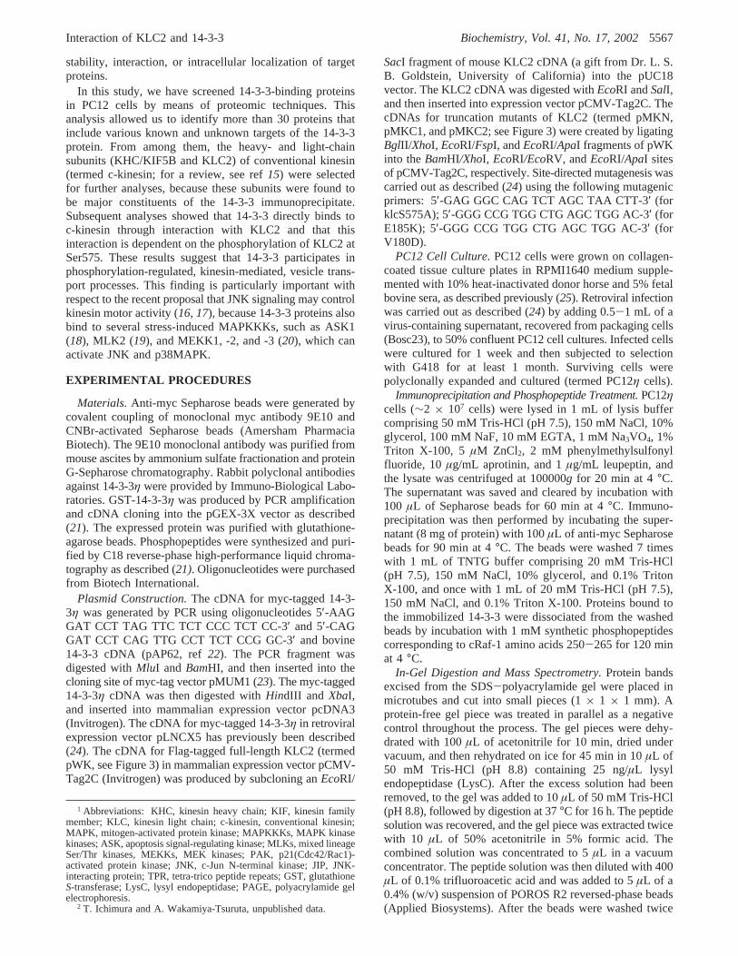
Association of 14-3-3η with c-Kinesin in PC12 Cells.Toisolate proteins that physically associate with the 14-3-3protein, we used the PC12 cell line (termed PC12η cells)stably expressing a myc-tagged 14-3-3η isoform (Figure 1A).The PC12η cells were lysed, and the expressed myc-η wasimmunoprecipitated from the lysate with anti-myc mono-clonal antibody-conjugated Sepharose beads. After the beadswere washed, synthetic phosphopeptides corresponding tothe Raf-binding site of 14-3-3 (LSQRQRSTpSTPNVHA, ref
30) were added to the beads, and the proteins bound to theimmobilized 14-3-3 were dissociated from the phosphoSer-binding groove (see also Experimental Procedures). Figure1B (lane 2) shows SDS-PAGE photographs of the dissoci-ated proteins. About 30 proteins with molecular massesbetween 35 and 300 kDa were reproducibly detected withthis procedure, while only a few proteins were detected witha control lysate of cells not expressing myc-η (lane 1),suggesting that most of the proteins were co-immunopre-cipitated with the expressed 14-3-3η. Western blotting withantibodies against several reported 14-3-3 targets showed that
FIGURE 1: Association of 14-3-3η with c-kinesin in PC12η cells.(A) Structure of a myc-tagged 14-3-3η isoform. (B) Analysis ofproteins coimmunoprecipitated with myc-14-3-3η. PC12η cellsstably expressing myc-14-3-3η were lysed, and the expressed myc-ηwas immunoprecipitated with anti-myc monoclonal antibody-conjugated Sepharose beads. Proteins bound to the immobilizedmyc-14-3-3 were dissociated with phosphorylated Raf-1 peptidesand then analyzed by SDS-PAGE (7.5% PAGE gel, silver staining,left panel, lane 2), or by Western blotting with the indicatedantibodies (right panel, lane 2). A control experiment was performedunder the same conditions with nontransfected PC12 cells (lanes1). The weights of molecular mass markers are shown in kDa, andthe positions of KHC and KLC2 are indicated by arrows. (C)Phosphopeptide-specific dissociation of c-kinesin subunits from the14-3-3 immunocomplexes. Extracts of PC12 cells (lanes 1 and 2)or PC12η cells (lanes 3 and 4) were subjected to immunoprecipi-tation as in (A) except that nonphosphorylated Raf-1 peptides wereused for the dissociation of bound proteins (lanes 2 and 4).Dissociated proteins were analyzed by Western blotting with anti-KHC and anti-KLC antibodies.
5568 Biochemistry, Vol. 41, No. 17, 2002 Ichimura et al.
this protein fraction contained the IRS-1 docking protein,PI3 kinaseR, Cdc25C phosphatase, tyrosine hydroxylase, andthree Raf kinase isozymes (Figure 1B, right panel).
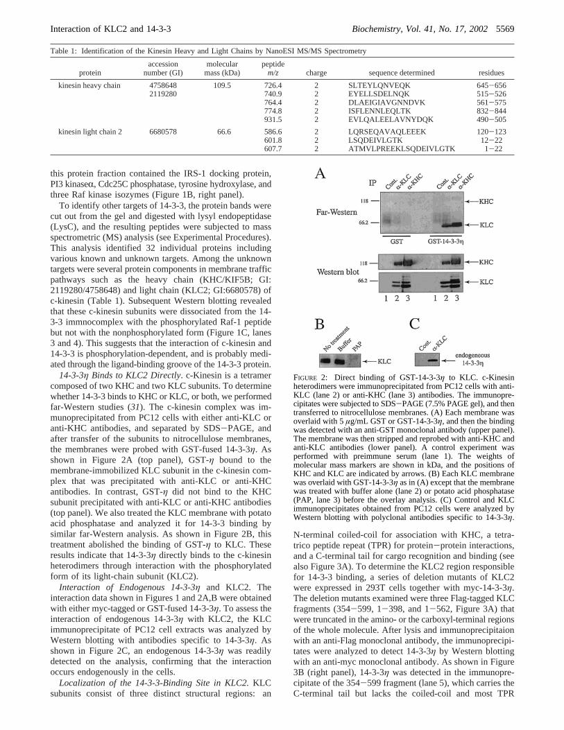
To identify other targets of 14-3-3, the protein bands werecut out from the gel and digested with lysyl endopeptidase(LysC), and the resulting peptides were subjected to massspectrometric (MS) analysis (see Experimental Procedures).This analysis identified 32 individual proteins includingvarious known and unknown targets. Among the unknowntargets were several protein components in membrane trafficpathways such as the heavy chain (KHC/KIF5B; GI:2119280/4758648) and light chain (KLC2; GI:6680578) ofc-kinesin (Table 1). Subsequent Western blotting revealedthat these c-kinesin subunits were dissociated from the 14-3-3 immnocomplex with the phosphorylated Raf-1 peptidebut not with the nonphosphorylated form (Figure 1C, lanes3 and 4). This suggests that the interaction of c-kinesin and14-3-3 is phosphorylation-dependent, and is probably medi-ated through the ligand-binding groove of the 14-3-3 protein.
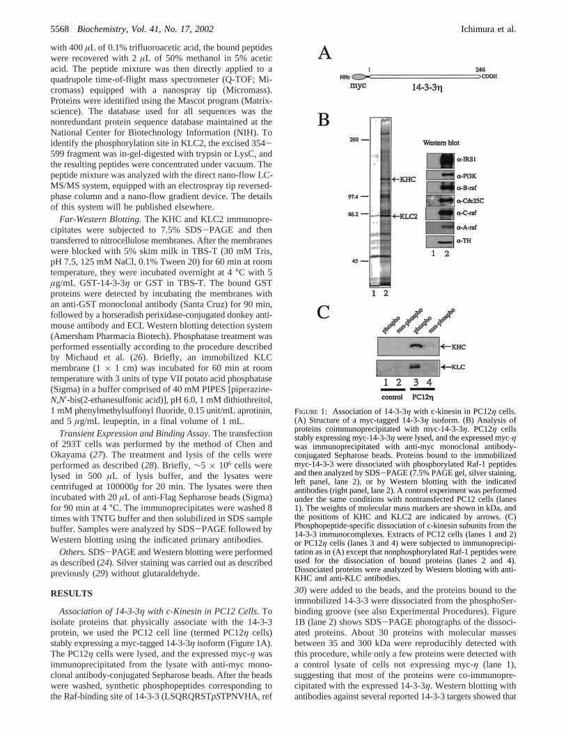
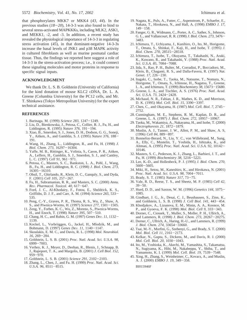
14-3-3η Binds to KLC2 Directly. c-Kinesin is a tetramercomposed of two KHC and two KLC subunits. To determinewhether 14-3-3 binds to KHC or KLC, or both, we performedfar-Western studies (31). The c-kinesin complex was im-munoprecipitated from PC12 cells with either anti-KLC oranti-KHC antibodies, and separated by SDS-PAGE, andafter transfer of the subunits to nitrocellulose membranes,the membranes were probed with GST-fused 14-3-3η. Asshown in Figure 2A (top panel), GST-η bound to themembrane-immobilized KLC subunit in the c-kinesin com-plex that was precipitated with anti-KLC or anti-KHCantibodies. In contrast, GST-η did not bind to the KHCsubunit precipitated with anti-KLC or anti-KHC antibodies(top panel). We also treated the KLC membrane with potatoacid phosphatase and analyzed it for 14-3-3 binding bysimilar far-Western analysis. As shown in Figure 2B, thistreatment abolished the binding of GST-η to KLC. Theseresults indicate that 14-3-3η directly binds to the c-kinesinheterodimers through interaction with the phosphorylatedform of its light-chain subunit (KLC2).
Interaction of Endogenous 14-3-3η and KLC2. Theinteraction data shown in Figures 1 and 2A,B were obtainedwith either myc-tagged or GST-fused 14-3-3η. To assess theinteraction of endogenous 14-3-3η with KLC2, the KLCimmunoprecipitate of PC12 cell extracts was analyzed byWestern blotting with antibodies specific to 14-3-3η. Asshown in Figure 2C, an endogenous 14-3-3η was readilydetected on the analysis, confirming that the interactionoccurs endogenously in the cells.
Localization of the 14-3-3-Binding Site in KLC2.KLCsubunits consist of three distinct structural regions: an
N-terminal coiled-coil for association with KHC, a tetra-trico peptide repeat (TPR) for protein-protein interactions,and a C-terminal tail for cargo recognition and binding (seealso Figure 3A). To determine the KLC2 region responsiblefor 14-3-3 binding, a series of deletion mutants of KLC2were expressed in 293T cells together with myc-14-3-3η.The deletion mutants examined were three Flag-tagged KLCfragments (354-599, 1-398, and 1-562, Figure 3A) thatwere truncated in the amino- or the carboxyl-terminal regionsof the whole molecule. After lysis and immunoprecipitaionwith an anti-Flag monoclonal antibody, the immunoprecipi-tates were analyzed to detect 14-3-3η by Western blottingwith an anti-myc monoclonal antibody. As shown in Figure3B (right panel), 14-3-3η was detected in the immunopre-cipitate of the 354-599 fragment (lane 5), which carries theC-terminal tail but lacks the coiled-coil and most TPR
Table 1: Identification of the Kinesin Heavy and Light Chains by NanoESI MS/MS Spectrometry
proteinaccession
number (GI)molecular
mass (kDa)peptide
m/z charge sequence determined residues
kinesin heavy chain 4758648 109.5 726.4 2 SLTEYLQNVEQK 645-6562119280 740.9 2 EYELLSDELNQK 515-526
764.4 2 DLAEIGIAVGNNDVK 561-575774.8 2 ISFLENNLEQLTK 832-844931.5 2 EVLQALEELAVNYDQK 490-505
kinesin light chain 2 6680578 66.6 586.6 2 LQRSEQAVAQLEEEK 120-123601.8 2 LSQDEIVLGTK 12-22607.7 2 ATMVLPREEKLSQDEIVLGTK 1-22
FIGURE 2: Direct binding of GST-14-3-3η to KLC. c-Kinesinheterodimers were immunoprecipitated from PC12 cells with anti-KLC (lane 2) or anti-KHC (lane 3) antibodies. The immunopre-cipitates were subjected to SDS-PAGE (7.5% PAGE gel), and thentransferred to nitrocellulose membranes. (A) Each membrane wasoverlaid with 5µg/mL GST or GST-14-3-3η, and then the bindingwas detected with an anti-GST monoclonal antibody (upper panel).The membrane was then stripped and reprobed with anti-KHC andanti-KLC antibodies (lower panel). A control experiment wasperformed with preimmune serum (lane 1). The weights ofmolecular mass markers are shown in kDa, and the positions ofKHC and KLC are indicated by arrows. (B) Each KLC membranewas overlaid with GST-14-3-3η as in (A) except that the membranewas treated with buffer alone (lane 2) or potato acid phosphatase(PAP, lane 3) before the overlay analysis. (C) Control and KLCimmunoprecipitates obtained from PC12 cells were analyzed byWestern blotting with polyclonal antibodies specific to 14-3-3η.
Interaction of KLC2 and 14-3-3 Biochemistry, Vol. 41, No. 17, 20025569

regions. However, 14-3-3η was not detected in the immu-noprecipitates of the 1-398 and 1-562 fragments (lanes 3and 4), both of which have deletions of the C-terminal tailregion. These data suggest that the C-terminal tail of KLC2is important for the observed 14-3-3 binding.
To further characterize the interaction between 14-3-3 andKLC2, we prepared two structural variants of the 14-3-3ηprotein, 14-3-3ηV180D and -E185K, in which V and Kreplaced D180 and E185, respectively. It has been reportedthat V180D is a mutant that is 100-fold less potent in theactivation of ExoS than the wild-type protein (32), and thatE185K is a dominant-negative mutant that suppresses RAS1signaling in photoreceptor formation (12). Binding experi-ments showed that both these variants did not bind to eitherthe full-length KLC2 or the 354-599 fragment, indicatingthat the observed interaction was specific and would befunctionally significant.
The C-Terminal Tail of KLC2 Contains the PhosphoSerInVolVed in 14-3-3 Binding.Although KLCs have beenshown to exist as several phosphorylated forms in cells (33),there has been no information regarding the site(s) ofphosphorylation. To identify the phosphoSer involved in 14-3-3 binding, we examined whether the expressed 354-599is indeed phosphorylated in 293T cells, particularly at itsC-terminal tail including the potential 14-3-3-binding site.The 354-599 fragment was transfected into 293T cells, and
the expressed fragment was recovered by immunoprecipi-tation. After SDS-PAGE, the 354-599 fragment was in-gel-digested with a protease, and the resulting peptides weredirectly analyzed by nano-flow LC-MS/MS (see Experimen-tal Procedures). A sequence database search with theobserved MS/MS spectra identified 34 peptides that covered∼90% of the 354-599 sequence. Of them, peptide 572-580 was found to be phosphorylated at Ser575 (Figure 4).The analysis also detected nonphosphorylated peptide 572-580 (data not shown), indicating that Ser575 is not aconstitutive site for phosphorylation in the cells. Neverthe-less, no additional phosphopeptides were found on thisanalysis. These data suggested that the phosphorylationoccurred in the C-terminal tail of the KLC2 molecule,particularly at Ser575, and that this site of phosphorylationmediated the interaction between KLC2 and 14-3-3.
To confirm this observation, a KLC2 mutant was preparedin which Ser575 was substituted by Ala (termed klcS575A).While this KLC2 mutant retained the ability to associate withthe KHC subunit, it no longer bound to 14-3-3 (Figure 5).Thus, we concluded that phosphorylation at Ser575 isnecessary and sufficient for the association of KLC2 withthe 14-3-3 protein.
DISCUSSION
Kinesins are motor proteins that utilize the energy of ATPhydrolysis to move cargo along microtubules. Among themembers of this superfamily, c-kinesin is the most ubiquitousmotor found in a variety of cells and tissues (for reviews,see refs15, 34). c-Kinesin is an elongated heterotetramercomprising two KHCs of approximately 120 kDa and twoKLCs of 60-70 kDa (35, 36). KHC contains the motordomain that generates the ATP-dependent force for move-ment along microtubules, while KLC is believed to beinvolved in vesicle binding and regulation of the motor
FIGURE 3: Analysis of the site of KLC2 responsible for the bindingwith 14-3-3. (A) Schematic illustration of the full-length and mutantforms of Flag-tagged KLC2. The relative NH2 and COOH terminiare denoted by adjacent numbers. (B) Effects of truncation mutantsof KLC2 on 14-3-3 binding. 293T cells were transfected with theindicated KLC2 constructs or the vector alone and myc-14-3-3η.The lysates (10µg of protein each) were separated by SDS-PAGE,transferred to poly(vinylidene difluoride) membranes, and thenanalyzed by Western blotting with an anti-Flag (upper) or anti-myc (lower) monoclonal antibody (left panel). The expressed KLC2and its truncation mutants were immunoprecipitated from the lysateswith anti-Flag Sepharose beads. The immunoprecipitates wereanalyzed by Western blotting with an anti-Flag (upper) or anti-myc (lower) monoclonal antibody (right panel). The weights ofmolecular mass markers are shown in kDa, and the arrow indicatesthe coprecipitated myc-14-3-3η.
FIGURE 4: Identification of Ser575 as the in vivo phosphorylationsite in KLC2. The 354-599 fragment expressed in 293 cells wasimmunoprecipitated with anti-Flag Sepharose beads. The immu-noprecipitate was subjected to SDS-PAGE, digested with trypsinor LysC, and then analyzed by LC-MS/MS (see ExperimentalProcedures). The [M+2H]2+ peptide ion withm/z ) 615.26 wasused as the parent ion for fragmentation to produce the MS/MSspectrum. The detected ions of the b- and y-ion collision series areindicated. The asterisk-denoted peaks, b(4)*, y(2)*, y(3)*, and y(8)*,represent the b(4) ion, y(2) ion, y(3) ion, and y(8) ion, respectively,which have lost their NH3 groups (17.03 Da). The mass differencebetween b(3) (315.18 Da) and b(4)* (465.15 Da), 149.97 Da, isconsistent with a phosphorylated Ser.
5570 Biochemistry, Vol. 41, No. 17, 2002 Ichimura et al.

activity of the kinesin protein complex. A large number ofbiochemical and immunocytochemical experiments haveshown that c-kinesin plays roles in a variety of cellularprocesses during both interface and mitosis. It has also beenshown that c-kinesin is required for the progress of neuro-genesis, since mutations in either the KHC or the KLC genecause defects in the number of synaptic boutons in actionpotential propagation and neurotransmitter release inDroso-phila (37, 38). Although the structure and function ofc-kinesin are thus well characterized, little is known aboutthe mechanisms that regulate motor activity and cargobinding. In this report, we have shown that 14-3-3 binds toc-kinesin through interaction with KLC2 and that thisinteraction is dependent on the phosphorylation of KLC2 inintact cells. Since 14-3-3 is a potential regulator of diversekinase-mediated processes (8), we assume that this interactionis functionally significant.
The amino acid sequences of KLCs cloned from variousorganisms indicated that the C-terminal tails exhibit notablesequence variability, while the coiled-coil and TPF regionsare highly conserved across species (39). To search forpotential targets of 14-3-3 among the KLC family, weperformed a BLAST search of the NCBI (National Centerfor Biotechnology Information) peptide sequence databaseusing the C-terminal sequence of 40 amino acids in KLC2.
This retrieval revealed that 7 out of 28 known KLCs sharedthe short unique sequence MKRA(S)*SLN(F)LN (where *Sis phosphoSer) in the C-terminal end. These include seaurchin KLC1, -2, and -3, mouse KLC3, rat KLCt, and humanKLC2 and KLC3 (Table 2), suggesting that a subset of theKLC family might also be targets of the 14-3-3 protein.
It is also known that there are a number of proteins in thekinesin superfamily that are clearly distinct from c-kinesinin structural organization and function (34). For instance,KIF1C, involved in vesicle transport between the Golgiapparatus and the endoplasmic reticulum, is a homodimer,and contains a motor domain and cargo-binding site in eachmonomer (40). This protein was recently found to be a targetof the 14-3-3 protein (41). In fact, KIF1C contains theconsensus 14-3-3-binding sequence RxxpSxP (ref29) atresidues 1089-1094. Likewise, the computer-assisted re-trieval of the NCBI database showed that at least 5 out of39 mouse members of this class of kinesin molecules sharedthe same consensus sequence for 14-3-3 binding. Thus, 14-3-3 may participate in a variety of cellular processes, whichare dependent on the phosphorylation of the kinesin super-family.
c-Kinesin binds to two distinct proteins, hsc70 and JIPs(JIP1, -2, and -3). Hsc70 is thought to be a regulator of thekinesin/vesicle interaction as it dissociates c-kinesin fromthe vesicle surface (42). JIPs are potential linkers of c-kinesinand cargo that connect c-kinesin and a transmembranelipoprotein receptor, such as ApoER2, in the vesicle mem-brane (16). c-Kinesin binds to these molecules through theTPR motifs in its light-chain subunit (16, 42). The presentstudy showed that the light-chain subunit of c-kinesin alsobinds to 14-3-3, but at a site apart from the TPR motifs, thatis, through its C-terminal tail. It should be noted that theinteraction between c-kinesin and 14-3-3 is strictly regulatedby phosphorylation at Ser575 of KLC2. Thus, it appearslikely that c-kinesin can potentially associate with hsc70 andJIP molecules, and 14-3-3 through the distinct structuralregions of KLC2.
Increasing evidence suggests cross-talk between the vesicletransport controlled by motor proteins and the stress-inducedsignaling pathway, leading to the activation of JNK and p38MAPK. For instance, Nagata et al. (19) have shown that theKIF3 motor protein associates with the mixed lineage kinasesMLK2 and MLK3, both of which can activate the JNK andp38MAPK cascade. It has also been shown that JIPs arescaffold proteins that bind to three kinase components ofthe JNK signaling pathway: JNK itself, a kinase thatphosphorylates JNK such as MKK7 or MKK4, and a kinase
Table 2: A Conserved Sequence Motif Located in the C-Terminal Tails of Known KLCsa
a The indicated sequence is common to sea urchin KLC1, -2, and -3. The numbers are for sKLC1. The accession (GI) numbers are as follows:mouse KLC2 (6680578); sea urchin KLC1-3 (161526 for sKLC1, 161528 for sKLC2, 1362661 for sKLC3); mouse KLC3 (13878552); rat KLC2(10281106); human KLC2 (13878553); and human KLC3 (13878563).b The conserved residues are highlighted. The consensus sequence is alsoindicated at the bottom.
FIGURE 5: Effect of the Ser575 mutation on 14-3-3 binding. Flag-tagged KLC2 or Flag-tagged klcS575A was coexpressed with myc-14-3-3η in 293 cells (A) or PC12 cells (B), and the expressedproteins were immunoprecipitated with anti-Flag Sepharose beads.The immunoprecipitates were separated by SDS-PAGE, transferredto poly(vinylidene difluoride) membranes, and then analyzed byWestern blotting with an anti-Flag or anti-myc monoclonal antibody.The immunoprecipitates were also analyzed by Western blottingwith anti-KHC antibodies to assess the association of KHC withklcS575A.
Interaction of KLC2 and 14-3-3 Biochemistry, Vol. 41, No. 17, 20025571
that phosphorylates MKK7 or MKK4 (43, 44). In theprevious studies (18-20), 14-3-3 was also found to bind toseveral stress-activated MAPKKKs, including MLK2, ASK1,and MEKK1, -2, and -3. In addition, a recent study hasrevealed the physiological importance of 14-3-3 in regulatingstress activation (45), in that dominant-negative 14-3-3sincrease the basal levels of JNK1 and p38 MAPK activityin cultured fibroblasts as well as murine postnatal cardiactissue. Thus, the findings we reported here suggest a role of14-3-3 in the stress-activation process; i.e., it could connectthese signaling molecules and motor proteins in response tospecific signal inputs.
ACKNOWLEDGMENT
We thank Dr. L. S. B. Goldstein (University of California)for the kind donation of mouse KLC2 cDNA, Dr. L. A.Greene (Columbia University) for the PC12 cells, and Mr.T. Shinkawa (Tokyo Metropolitan University) for the experttechnical assistance.
REFERENCES
1. Barinaga, M. (1999)Science 283, 1247-1249.2. Liu, D., Bienkowska, J., Petosa, C., Collier, R. J., Fu, H., and
Liddington, R. (1995)Nature 376, 191-194.3. Xiao, B., Smerdon, S. J., Jones, D. H., Dodson, G. G., Soneji,
Y., Aitken, A., and Gamblin, S. J. (1995)Nature 376, 188-191.
4. Wang, H., Zhang, L., Liddington, R., and Fu, H. (1998)J.Biol. Chem. 273, 16297-16304.
5. Yaffe, M. B., Rittinger, K., Volinia, S., Caron, P. R., Aitken,A., Leffers, H., Gamblin, S. J., Smerdon, S. J., and Cantley,L. C. (1997)Cell 91, 961-971.
6. Petosa, C., Masters, S. C., Bankston, L. A., Pohl, J., Wang,B., Fu, H., and Liddington, R. C. (1998)J. Biol. Chem. 273,16305-16310.
7. Obsil, T., Ghirlando, R., Klein, D. C., Ganquly, S., and Dyda,F. (2001)Cell 105, 257-267.
8. Fu, H., Subramanian, R. R., and Masters, S. C. (2000)Annu.ReV. Pharmacol. Toxicol. 40, 617-647.
9. Ford, J. C., Al-Khodairy, F., Fotou, E., Sheldrick, K. S.,Griffiths, D. J. F., and Carr, A. M. (1994)Science 265, 533-535.
10. Peng, C.-Y., Graves, P. R., Thoma, R. S., Wu, Z., Shaw, A.S., and Piwnica-Worms, H. (1997)Science 277, 1501-1505.
11. Zeng, Y., Forbes, K. C., Wu, Z., Moreno, S., Piwnica-Worms,H., and Enoch, T. (1998)Nature 395, 507-510.
12. Chang, H. C., and Rubin, G. M. (1997)Genes DeV. 11, 1132-1139.
13. Kockel, L., Vorbriiggen, G., Jackel, H., Mlodzik, M., andBohmann, D. (1997)Genes DeV. 11, 1140-1147.
14. Skoulakis, E. M. C., and Davis, R. L. (1998)Mol. Neurobiol.16, 269-284.
15. Goldstein, L. S. B. (2001)Proc. Natl. Acad. Sci. U.S.A. 98,6999-7003.
16. Verhey, K. J., Meyer, D., Deehan, R., Blenis, J., Schnapp, B.J., Rapoport, T. A., and Margolis, B. (2001)J. Cell Biol. 152,959-970.
17. Goldstein, L. S. B. (2001)Science 291, 2102-2103.18. Zhang, L., Chen, J., and Fu. H. (1999)Proc. Natl. Acad. Sci.
U.S.A. 96, 8511-8515.
19. Nagata, K., Puls, A., Futter, C., Aspenstrom, P., Schaefer, E.,Nakata, T., Hirokawa, N., and Hall, A. (1998)EMBO J. 17,149-158.
20. Fanger, G. R., Widmann, C., Porter, A. C., Sather, S., Johnson,G. L., and Vallancourt, R. R. (1998)J. Biol. Chem. 273, 3476-3483.
21. Ichimura, T., Uchiyama, J., Kunihiro, O., Ito, M., Horigome,T., Omata, S., Shinkai, F., Kaji, H., and Isobe, T. (1995)J.Biol. Chem. 270, 28515-28518.
22. Ichimura, T., Isobe, T., Okuyama, T., Takahashi, N., Araki,K., Kuwano, R., and Takahashi, Y. (1988)Proc. Natl. Acad.Sci. U.S.A. 85, 7084-7088.
23. Iida, S., Rao, P. H., Bulter, M., Corradini, P., Boccadoro, M.,Klein, B., Chaganti, R. S., and Dalla-Favera, R. (1997)Nat.Genet. 17, 226-230.
24. Itagaki, C., Isobe, T., Taoka, M., Natsume, T., Nomura, N.,Horigome, T., Omata, S., Ichinose, H., Nagatsu, T., Greene,L. A., and Ichimura, T. (1999)Biochemistry 38, 15673-15680.
25. Greene, L. A., and Tischler, A. S. (1976)Proc. Natl. Acad.Sci. U.S.A. 73, 2424-2428.
26. Michaud, N. R., Fabian, J. R., Mathes, K. D., and Morrison,D. K. (1995)Mol. Cell. Biol. 15, 3390-3397.
27. Chen, C., and Okayama, H. (1987)Mol. Cell. Biol. 7, 2745-2752.
28. Cunningham, M. E., Stephens, R. M., Kaplan, D. R., andGreene, L. A. (1997)J. Biol. Chem. 272, 10957-10967.
29. Taoka, M., Wakamiya, A., Nakayama, H., and Isobe, T. (2000)Electrophoresis 21, 1872-1879.
30. Muslin, A. J., Tanner, J. W., Allen, P. M., and Shaw, A. S,(1996)Cell 84, 889-897.
31. Bonnefoy-Berard, N., Liu, Y. C., von Willebrand, M., Sung,A., Elly, C., Mustelin, T., Yoshida, H., Ishizaka, K., andAltman, A. (1995)Proc. Natl. Acad. Sci. U.S.A. 92, 10142-10146.
32. Masters, S. C., Pederson, K. J., Zhang, L., Barbieri, J. T., andFu, H. (1999)Biochemistry 38, 5216-5221.
33. Lee, K.-D., and Hollenbeck, P. J. (1995)J. Biol. Chem. 270,5600-5605.
34. Miki, H., Setou, M., Kaneshiro, K., and Hirokawa, N. (2001)Proc. Natl. Acad. Sci. U.S.A. 98, 7004-7011.
35. Brady, S. T. (1985)Nature 317, 73-75.36. Vale, R. D., Reese, T. S., and Sheetz, M. P. (1985)Cell 42,
39-50.37. Hurd, D. D., and Saxton, W. M. (1996)Genetics 144, 1075-
1085.38. Gindhart, J. G., Jr., Desai, C. J., Beushausen, S., Zinn, K.,
and Goldstein, L. S. B. (1998)J. Cell Biol. 141, 443-454.39. Khodjakov, A., Lizunova, E. M., Minin, A. A., Koonce, M.
P., and Gyoeva, F. K. (1998)Mol. Biol. Cell 9, 333-343.40. Dorner, C., Ciossek, T., Muller, S., Moller, P. H., Ullrich, A.,
and Lammers, R. (1998)J. Biol. Chem. 273, 20267-20275.41. Dorner, C., Ullrich, A., Haring, H.-U., and Lammers, R. (1999)
J. Biol. Chem. 274, 33654-33660.42. Tsai, M.-Y., Morfini, G., Szebenyi, G., and Brady, S. T. (2000)
Mol. Biol. Cell 11, 2161-2173.43. Kelkar, N., Gupta, S., Dickens, M., and Davis, R. J. (2000)
Mol. Cell. Biol. 20, 1030-1043.44. Ito, M., Yoshioka, K., Akechi, M., Yamashita, S., Takamatsu,
N., Sugiyama, K., Hibi, M., Nakabeppu, Y., Shiba, T., andYamamoto, K. I. (1999)Mol. Cell. Biol. 19, 7539-7548.
45. Xing, H., Zhang, S., Weinheimer, C., Kovacs, A., and Muslin,A. J. (2000)EMBO J. 19, 349-358.
BI015946F
5572 Biochemistry, Vol. 41, No. 17, 2002 Ichimura et al.