phyllosticta capitalensis p. helicteres, p. sterculiae … diversity phyllosticta capitalensis, p....
TRANSCRIPT

Fungal Diversity
Phyllosticta Capitalensis, P. helicteres, P. sterculiae and other
Phyllosticta Species from Sterculiaceae
Wulandari, N. F. 1,2*
and To–anun, C.1
1Department of Nematology and Plant Pathology, Agriculture Faculty, Chiang Mai University,
50200 Chiang Mai, Thailand, 2Microbiology Division, Research Centre for Biology, Indonesian
Institute of sciences (LIPI), Jl. Raya Bogor, KM 46, Cibinong Science Centre, Cibinong, 16911,
West Java, Indonesia.
Wulandari, N. F. and To–anun, C. (2014). Phyllosticta capitalensis, P. helicteres, P. sterculiae
and other Phyllosticta species from Sterculiaceae. International Journal of Agricultural
Technology 10(1):133-146.
Abstract Phyllosticta as an important plant pathogenic genus associated with plants as
endophytes, pathogens or saprobes. Phyllosticta sterculiae occurs on Sterculia foetida
(Sterculiaceae) in the Philippines, where it causes a leaf blight. The holotype of P. sterculiae is
redescribed and compared with a Phyllosticta species collected on Sterculia monosperma in
Thailand. P. sterculiae has oblong to obovoid ascospores whereas the fungus from Thailand has
ellipsoidal ascospores that are swollen in the middle and possess polar appendages. The
comparison between other Phyllosticta species on Sterculiaceae revealed Phyllosticta species
from Thailand collection is different in ascospores dimention. Furthermore, the anamorph in
culture compare to the Thailand collections with Phyllosticta melochiae from Indonesia on host
subtrate posseses similar character. The BLAST result showed that the Thailand species is
belonging Phyllosticta capitalensis. The need of epitypification for all the Phyllosticta spp.
occurs in Sterculiaceae is needed here to clarify the species name. The Thailand fungus is
described, illustrated and discussed.
Keywords: Blast, Dothideomycetes, Indonesia, leaf blight, morphology, plant disease,
taxonomy, Thailand
Introduction
Phyllosticta is an important plant pathogenic genus with Guignardia
sexual state as previously recorded (Sivanesan, 1984; Bussaban et al., 2004;
Farr and Rossman, 2012; Hawksworth, 2012; Stevens, 1917; Van der Aa, 1973;
Van der Aa and Vanev, 2002; Rehm, 1914; Yates, 1918; Von Arx and Müller,
1954; Hyde, 1995; Benjapalakorn, 2006; Wong et al., 2012; Wulandari et al.,
2009; Wulandari et al., 2010a,b,c; Wulandari et al., 2011; McMakin, 2000;
Hennings, 1980; Sontirat et al., 1994; Crous et al., 2004; Raciborski, 1990;
Rayner, 1994; Gardner et al., 2000; Somrithipol and Hyde, 2004; Thongkontha
* Corresponding author: Wulandari, N. F.; Email: [email protected]
International Journal of Agricultural Technology 2014 Vol. 10(1):133-146
Available online http://www.ijat-aatsea.com ISSN 2630-0192 (Online)

134
et al., 2008; Wikee et al., 2011, 2012; The Royal Institute (1995), Photita et al.,
2001; Choi et al., 1999). Species occur as endophytes, pathogens and saprobes
on a wide range of plant families such as, Agavaceae (Hawksworth, 2012; Van
der Aa and Vanev, 2002; Rehm, 1914; Wulandari et al., 2010b; Crous et al.,
2004; Wikee et al., 2011) Arecaceae (palms) (Hawksworth, 2012; Van der Aa
and Vanev, 2002; Rehm, 1914; Sivanesan, 1984; McMakin, 2000; Crous et al.,
2004). Musaceae (Sivanesan, 1984; Stevens, 1917; Yates, 1918; 1994; Crous et
al., 2004; Raciborski, 1990). Orchidaceae (Sivanesan, 1984; Hawksworth, 2012;
an der Aa and Vanev, 2002; Sontirat et al., 1994), Pandanaceae Rehm (1914),
Zingiberaceae (Bussaban et al., 2004; Van der Aa, 1973; Rehm, 1914)
(monocotyledons), and dicotyledons: Acanthaceae Rehm (1914),
Caesalpiniaceae Rehm (1914), Custutaceae Rehm (1914), Dioscoreaceae
(Sivanesan, 1984; Hawksworth, 2012; Van der Aa and Vanev, 2002; Rehm
1914; Thongkontha et al., 2008), Dipterocarpaceae Rehm (1914), Fabaceae
(Hawksworth, 2012; Van der Aa and Vanev, 2002; Rehm, 1914; Wulandari et
al., 2010b; Crous et al., 2004), Rubiaceae (Sivanesan, 1984; Hawksworth, 2012
Van der Aa and Vanev, 2002; Rehm, 1914; Wulandari et al., 2010a,b,
Wulandari et al., 2011) and Rutaceae (Sivanesan, 1984; Hawksworth, 2012
Van der Aa and Vanev, 2002; Rehm, 1914; Wulandari et al., 2010a,b,
Wulandari et al., 2011). We are studying the genus Phyllosticta in Northern
Thailand and in this paper report Phyllosticta species on Sterculiaceae.
The Sterculiaceae compise large trees native to China and Thailand
(http://www.quisqualis.com/tvPEFC2P063.html) and widely introduce into
Southern China, Sumatra (Indonesia) and Taiwan. Theobroma cacao is an
important genus in this family and is used for cocoa production. Sterculia
monosperma Vent. is known as Chinese cheshnut, noble battle tree, or as pheng
pok in Thai (Somrithipol and Hyde, 2004) and its starch granules may be used
as thickening in chili sauce (Wong et al., 2012). Melochia umbellata is known
as Melochia (Somrithipol and Hyde, 2004), the wood is used for handycraft and
tool (Somrithipol and Hyde, 2004). Sterculia foetida known as Sumrong [Thai]
and it has unpleasent aroma of the flower (Hennings, 1980).
Phyllosticta melochiae, P. helicteres and P. sterculiae are Phyllosticta
species known from Sterculiaceae. Phyllosticta melochia was found in Delhi,
Borneo, Indonesia (Rehm, 1914) with specific zona leaf spot, P. helicteres was
found on Helicteres jamaicensis in Puerto Rico with target spot (Van der Aa,
1973) and Phyllosticta sterculiae was reported on Sterculia foetida in the
Philippines, where it causes leaf blight (Yates, 1918). In this study we made
collections of Phyllosticta species from Sterculia monosperma in Thailand. The
species found was distinct from P. sterculiae and P. helicteres and it is
described, illustrated and discussed. For comparative purposes we also
International Journal of Agricultural Technology 2014, Vol. 10 (1): 133-146
135
examined the type material of P. sterculiae and P. helicteres. BLAST result
were also used to support the finding. By using BLAST, it can compare
nucleotide sequence to a whole sequence library and will gave the high
sequence similarity (http://blast.ncbi.nlm.nih.gov/Blast.cgi).
Materials and methods
Type specimens of sexual state of Phyllosticta sterculiae were borrowed
from ILL (USA) and S (Sweden), P. helicteres from BPI (USA) and NY (USA)
while fresh collections on Sterculia monosperma were made in Chiang Rai
Province, Northern Thailand. Fungi were isolated by single spore method as
described in (Choi et al., 1999). Morphological character were recorded using a
Nikon 80i microscope with Tarosof program for measuring spores and camera
Lucida attachment for line drawing. Strains were isolated onto Difco malt
extract agar (MEA) and growth rate was measured at 24o C in triplicate; colour
nomenclature and pigmentation follow Rayner (Gardner et al., 2000). Holotype
specimens Myco Bank number were provided by MycoBank website
(Raciborski, 1990).
Results
Hawksworth (2012) reported that can be only one fungus represent one
name can be applied to all the state of certain fungi. This paper will treated all
Guignardia name into Phyllosticta name. The isotype of P. sterculiae from
Philippines and P. helicteres from Puerto Rico were reexamined and it is
redescribed and illustrated here. Collections of Phyllosticta species on Sterculia
monosperma from Chiang Rai Province in Thailand differed from P. sterculiae
and P. helicteres, from BLAST (Basic Local Alignment Search Tool,
http://blast.ncbi.nlm.nih.gov/Blast.cgi) the fungus is recognize as Phyllosticta
capitalensis. All Phyllosticta species occurs on Sterculiaceae will be described
and illustrated below.
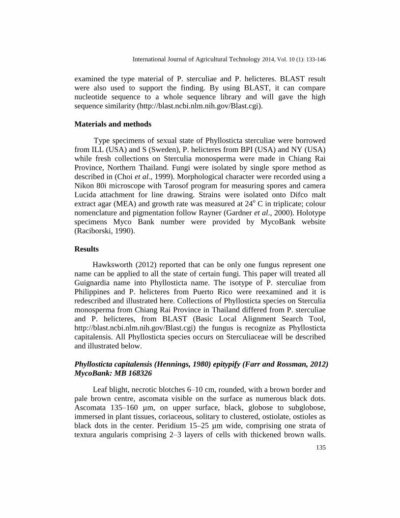
Phyllosticta capitalensis (Hennings, 1980) epitypify (Farr and Rossman, 2012)
MycoBank: MB 168326
Leaf blight, necrotic blotches 6–10 cm, rounded, with a brown border and
pale brown centre, ascomata visible on the surface as numerous black dots.
Ascomata 135–160 µm, on upper surface, black, globose to subglobose,
immersed in plant tissues, coriaceous, solitary to clustered, ostiolate, ostioles as
black dots in the center. Peridium 15–25 µm wide, comprising one strata of
textura angularis comprising 2–3 layers of cells with thickened brown walls.
136
Pseudoparaphyses not observed. Asci 50–91 × 9–14 µm ( = 69 × 12 µm, n =
20), 8–spored, bitunicate, fissitunicate, broadly cylindrical to cylindro–clavate,
rounded at the apex, where the diameter is 7–9 µm, ocular chamber 3–6 µm
high, tapering gradually to a 6–8 × 5–7 µm long pedicel attached to the basal
peridium. Ascospores 14–19 × 5–7 µm ( = 17 × 6 µm, n = 20), biseriate or
occasionally overlapping uniseriate, ellipsoidal, swollen in the centre, flattened
on one side when viewed from above, hyaline–greenish, 1–celled, coarse–
guttulate, smooth–walled, with a 6–8 × 5–7 µm long mucilaginous appendage
at each end.
Cultural characteristics: Colonies on MEA colonies reaching 4.1 mm in 1
week. Flat, slightly raised, irregular to lobate edge, black–olivaceous to black
above and black–olivaceous reverse.
Pycnidia 39–68 µm diameter, 90–103 um high, singly, black, globose to
elongate, immersed in media. Peridium 15–17 µm in diameter. Conidiogenous
cells 7–15 × 2–4 µm ( = 12 × 3 µm, n = 20), holoblastic, determinate, discrete,
rarely integrated, hyaline, cylindrical to doliiform cells lining the pycnidial
locule. Conidia 7–11 × 5–7 µm ( = 10 × 6 µm, n = 20), hyaline–greenish, 1–
celled, coarse–guttulate, smooth–walled, globose, ellipsoidal, clavate or
obclavate, with an obtuse apex, sometimes truncate at the base, surrounded by
1–2 µm thick mucilaginous sheath which persists at maturity and in some
spores with a single, hyaline, curved or straight, 1–5 µm long appendage.
Habitat: On living leaves of Sterculia monosperma Vent. (Sterculiaceae)
causing leaf blotch/leaf blight.
Known Distribution: Thailand.
Material examined: Thailand, Chiang Rai, on leaves of Sterculia
monosperma, 20 November 2009, N. F. Wulandari, NFW 249 (MFLU10 0292;
MFLUCC 0340, living culture) teleomorph only present; ibid., 17 December
2009, NFW 266 (MFLU10 0293) teleomorph only present; ibid., 21 December
2009, N. F. Wulandari, NFW 308 (MFLU10 0294) teleomorph only present.
International Journal of Agricultural Technology 2014, Vol. 10 (1): 133-146
137
Fig. 1a. Phyllosticta capitalensis (MFLU10 0292) a. Leaf blight (arrowed) on leaf. b.
Appearance of ascomata on the host surface. c. Section of ascoma on the leaf. d. Peridium of
textura angularis comprising 2–3 layers of cells with thickened angular brown walls. e–f. Asci
with ocular chamber. g–n. Ascospores with bipolar mucilaginous appendages, rounded at the
base and pointed at the apex. Scale bars: b = 100 µm, c–d = 20 µm, e–f = 25 µm, g–n = 10 µm.

138
Fig. 1b. Phyllosticta capitalensis (MFLU10 0292) line drawing. o. Section of ascoma in the
leaf (darkened area is fungal cells, arrowed) p. Asci. q. Immature ascus. r. Ascospores

International Journal of Agricultural Technology 2014, Vol. 10 (1): 133-146
139
Fig. 2a. Phyllosticta helicteres (BPI 598377, isotype) a. Target spot (arrowed) on the leaf. b.
Appearance of ascomata on the host surface. c. Peridium comprising one strata of textura
angularis comprising 2–3 layers of cells with thickened brown walls. d–h. Asci. i–j.
Ascospores. Scale bars: a = 3 mm, b = 100 μm, c–h = 10 μm.

140
Fig. 2b. Phyllosticta helicteres (BPI 598377, isotype) line drawing. k. Section of ascoma in the
leaf (darkened area are fungal cells in arrowed) l. Asci. m. Ascospores without mucilaginous
sheath.
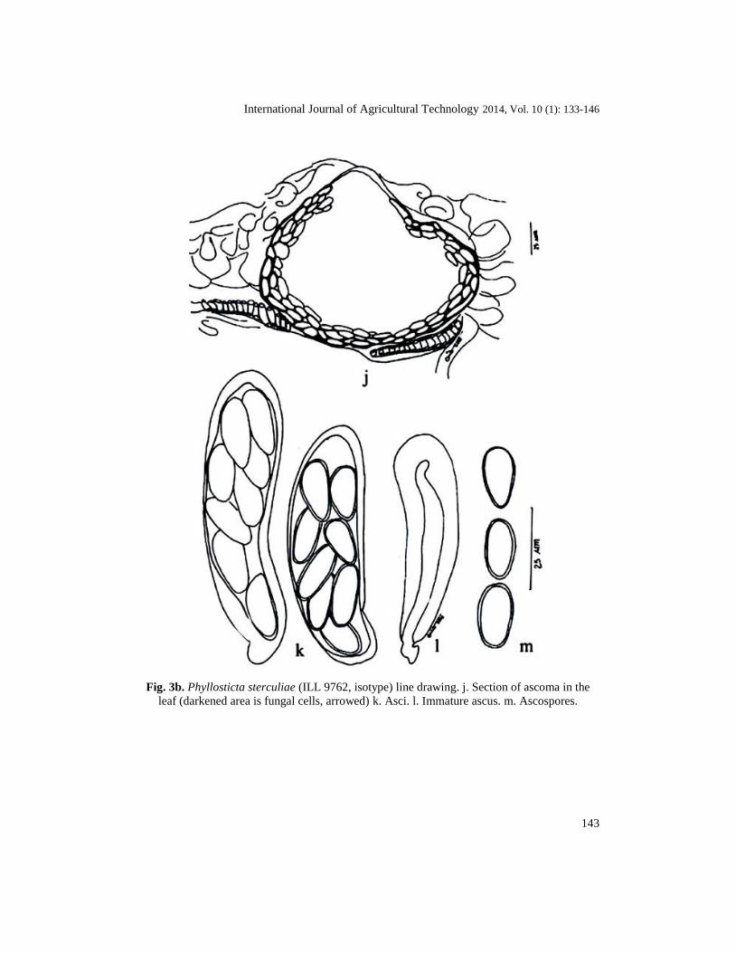
Phyllosticta sterculiae (Rehm) Wulandari & K. D. Hyde, comb. nov. =
Guignardia sterculiae (Rehm, 1914). MycoBank: MB 528760
Ascomata 110–165 µm diameter, 105–155 µm high, on upper and lower
leaf surface, black, globose to subglobose, immersed in plant tissues. Peridium
International Journal of Agricultural Technology 2014, Vol. 10 (1): 133-146
141
13–25 µm wide, one strata of textura angularis comprising 1–2 layers of cells
with thickened brown angular walls. Pseudopharaphyses not observed. Asci
55–105 × 19–25 µm ( = 78 × 21 µm, n = 10), 8–spored, bitunicate,
cylindrical to cylindro–clavate, rounded at the apex, where the diameter is 14–
16 µm, tapering gradually to a 6–19 × 6–8 µm pedicel attached to the basal
peridium. Ascospores 15–21 × 8–13 µm ( = 18 × 9 um, n = 20), uniseriate to
biseriate, ellipsoidal, oblong, or obovoid when viewed in any plane, hyaline–
greenish, 1–celled, coarse–guttulate, smooth–walled, without mucilaginous
appendages.
Habitat: On dead leaves of Sterculia foetidae (Sterculiaceae) causing leaf
blight.
Known Distribution: Philippines.
Material examined: Philippines, Luzon, Laguna, Los Baños on leaves of
Sterculia foetidae, September 1914, C.F Baker, Fungi Malayana No. 31,
Philippines (ILL 9762, isotype; F 10723, holotype) teleomorph only present.
Notes: This species differs from Phyllosticta helicteres in having bigger
ascospores and longer asci, 15–21 × 8–13 µm; 55–105 × 19–25 µm for P.
sterculiae and 14–18 × 5–9 μm; 50–94 × 8–20 μm for P. helicteres.
Furthermore, the ascopsores shape is also differ respectively, ellipsoidal,
oblong to obovoid for P. sterculiae and ellipsoidal widest 2/5 near the apex for
P. helicteres.
Phyllosticta melochiae (Van der Aa, 1973; Van der Aa and Vanev, 2002;
Yates, 1918) MycoBank: MB 519218From the original reference:
Pycnidia 45–90 µm diameter, singly, black, globose to elongate,
immersed in media. Peridium 15–17 µm in diameter. Conidiogenous cells 7–12
× 5–7 µm, some of the cell sometimes reduced. Conidia 7–12 × 5–7 µm,
hyaline–greenish, 1–celled, coarse–guttulate, smooth–walled, obovoidal,
ovoidal, slightly globose, with an truncate base when young, broadly rounded
apically, surrounded by thick mucilaginous sheath containing a large number of
coarse guttulate, with an apical appendage.
Habitat: On living leaves of Melochia umbellata
(Sterculiaceae/Buettneriaceae)
causing leaf spot.
Known Distribution: Indonesia
Note: The examination of two Phyllosticta spp. on the same genus of
Melochia found that those two species differ. Phyllosticta melochiae from
Melochia umbellata possesses smaller conidia than Phyllosticta sp. from
Melochia sp. (Van der Aa and Vanev, 2002). This result showed there might be

142
two different species of Phyllosticta occur on Melochia spp. (Sterculiaceae).
This Phyllosticta species cause zona spot on the leaf.
Fig. 3a. Phyllosticta sterculiae (ILL 9762, isotype). a–b. Appearance of ascomata on host
surface. c–d. Peridium of textura angularis comprising 1–2 layers of cells with thickened
angular brown walls. e–f. Asci. g–i. Ascospores obovoid when viewed in any plane. Scale
bars. b = 100 µm, c–d = 10 µm, e–f = 18 µm, g–i = 10 µm.
International Journal of Agricultural Technology 2014, Vol. 10 (1): 133-146
143
Fig. 3b. Phyllosticta sterculiae (ILL 9762, isotype) line drawing. j. Section of ascoma in the
leaf (darkened area is fungal cells, arrowed) k. Asci. l. Immature ascus. m. Ascospores.
144
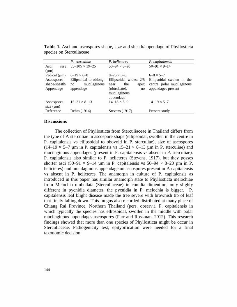
Table 1. Asci and ascospores shape, size and sheath/appendage of Phyllosticta
species on Sterculiaceae
P. sterculiae P. helicteres P. capitalensis
Asci size
(µm)
Pedicel (µm)
55–105 × 19–25
6–19 × 6–8
50–94 × 8–20
8–26 × 3–6
50–91 × 9–14
6–8 × 5–7
Ascospores
shape/sheath/
Appendage
Ellipsoidal to oblong,
no mucilaginous
appendage
Ellipsoidal widest 2/5
near the apex
(obtrullate), no
mucilaginous
appendage
Ellipsoidal swolen in the
centre, polar mucilaginous
appendages present
Ascospores
size (µm)
15–21 × 8–13 14–18 × 5–9 14–19 × 5–7
Reference Rehm (1914) Stevens (1917) Present study
Discussions
The collection of Phyllosticta from Sterculiaceae in Thailand differs from
the type of P. sterculiae in ascospore shape (ellipsoidal, swollen in the centre in
P. capitalensis vs ellipsoidal to obovoid in P. sterculiae), size of ascospores
(14–19 × 5–7 µm in P. capitalensis vs 15–21 × 8–13 µm in P. sterculiae) and
mucilaginous appendages (present in P. capitalensis vs absent in P. sterculiae).
P. capitalensis also similar to P. helicteres (Stevens, 1917), but they posses
shorter asci (50–91 × 9–14 µm in P. capitalensis vs 50–94 × 8–20 µm in P.
helicteres) and mucilaginous appendage on ascospores present in P. capitalensis
vs absent in P. helicteres. The anamorph in culture of P. capitalensis as
introduced in this paper has similar anamorph state to Phyllosticta melochiae
from Melochia umbellata (Sterculiaceae) in conidia dimention, only slighly
different in pycnidia diameter, the pycnidia in P. melochia is bigger. P.
capitalensis leaf blight disease made the tree severe with brownish tip of leaf
that finaly falling down. This fungus also recorded distributed at many place of
Chiang Rai Province, Northern Thailand (pers. observ.). P. capitalensis in
which typically the species has ellipsoidal, swollen in the middle with polar
mucilaginous appendages ascospores (Farr and Rossman, 2012). This research
findings showed that more than one species of Phyllosticta might be occur in
Sterculiaceae. Pathogenicity test, epitypification were needed for a final
taxonomic decision.

International Journal of Agricultural Technology 2014, Vol. 10 (1): 133-146
145
Acknowledgements
Nilam Wulandari acknowledges herbaria BPI, ILL, NY and S for loaning the type
specimens. Mae Fah Luang University and CBS are thanked for used of laboratory facilities.
Eric McKenzie and Kevin D. Hyde thank you for valuable comments on manuscript. Rampai
Kodsueb thanked for comment and suggestion on photoplate of fungi. Samantha Karunarathna
thanked for valuable book references. The Mushroom Research Foundation is thanked for a
PhD scholarship. P. W. Crous, CBS, the Netherlands also thanked for partially funded this
research.
References
Benjapalakorn, M. (2006). Physicochemical properties of chestnut Sterculia monosterma Vent.
flour and starch. (Master’s Thesis). Chulalongkkorn University, Thailand.
Bussaban, B., P. Lumyong, E. H. C. McKenzie, K. D. Hyde and S. Lumyong (2004). Fungi on
Zingiberaceae (ginger), in Thai Fungal Diversity,Thailand. pp. 189-195.
Choi, Y., K. D. Hyde and W. W. H. Ho, (1999). Single spore isolation of fungi. Fungal
Diversity. pp. 29–38.
Crous, P. W. W. Gams, J. A. Stalpers, V. Robert and G. Stegehuis. (2004). MycoBank: an
online initiative to launch mycology into the 21st century. Studies in Mycology 50:19-
22.
Farr, D. F. and A. Y. Rossman (2012). Fungal Databases, Systematic Mycology and
Microbiology Laboratory, ARS, USDA. Retrieved from http://nt.ars–
grin.gov/fungaldatabases.
Gardner S., P. Sidisunthorn and V. Anusarnsunthorn (2000). A Field Guide to Forest Trees of
Northern Thailand, KobfaiPublishing Project, Bangkok.
Hawksworth, D. L. (2012). Managing and coping with names of pleomorphic fungi in a period
of transition. IMA Fungus and Mycosphere 3:143–155.
Hennings, P. (1908). Fungi S. Paulenses IV a cl. Puttemans collecti, Hedwigia 48:1-20.
Hyde, K. D. (1995). Fungi from palms. XX. The Genus Guignardia, Sydowia 47:180–198.
McMakin P. D. (2000). Field Guide to the Flowering Plants of Thailand, White Lotus Co. Ltd.,
Bangkok.
Photita, W., S. Lumyong, P. Lumyong and K. D. Hyde (2001). Endophytic fungi of wild
banana (Musa acuminata) at Doi Suthep Pui National Park, Thailand, Mycological
Research 105:1508–1513.
Raciborski (1909). Parasitische Algen und Pilze Javas. Bulletin International. Academie des
Sciences due Cracovie Classe des Mathematiques et Naturalles. Serie B. Sciences
Naturreles. Serie B. Sciences Naturreles 3:1–388.
Rayner R.W. (1994). A Mycological Colour Chart Commonwealth Mycological Institute. Kew,
Surrey, U.K.
Rehm, H. (1914). Ascomycetes philippinenses V Leaflets of Philippine. Botany 6:2191–2237.
Sivanesan, A. (1984). The bitunicate ascomycetes and their anamorph. pp. 164–176.
Somrithipol, S. and Hyde, K. D. (2004). Plant Pathogens in Thai Fungal Diversity (Ed..B.G.
Jones, M. Tanticharoen and K. D Hyde), BIOTEC, Thailand. 20 pp.
Sontirat, P., P. Pitakpriwan, T. Khamhangridthiroong, W. Choobamroong and U. Kueprakone,
(1994). Host Index of Plant Diseases in Thailand 3rd
edition. Mycology Section, Plant
Pathology and Microbiology Division, Department of Agriculture, Bangkok, Thailand.
146
Stevens, F. L. (1917). Porto Rican fungi, old and new. Transaction of the Illinois Academy of
Science 10:162–218.
The Royal Institute (1995). The Taxonomy of Plant. Retrieved from
http://www.rspg.or.th/plants_data/plantdat/sterculi/smonos_2.htm.
Thongkontha S., Lumyong, S., McKenzie, E. H. C. and Hyde, K. D. (2008). Fungal saprobes
and pathogens occurence on tissue of Dracaena loureiri and Pandanus spp. Fungal
Diversity 30:149–179.
Van der Aa, H. A. (1973). Studies in Phyllosticta I. Studies in Mycology 5:1–110.
Van der Aa, H. A. and S. Vanev (2002). A revision of the species described in Phyllosticta
Centraalbureau voor Schimmelcultures, Utrecht, The Netherlands. pp. 1–510.
Von Arx, J. A. and Müller, E. (1954). Die Gattungen der amerosporen Pyrenomyceten.
Schweiz 11:151-153.
Wikee, S., Udayanga, D., Crous, P. W., Chukeatirote, E., McKenzie, E. H. C. (2001).
Phyllosticta an overview of current status of species recognition. Fungal Diversity
51:43–61.
Wikee, S., Wulandari, N. F., McKenzie, E. H. C. and Hyde, K. D. (2012). Phyllosticta
ophiopogonis sp. nov. from Ophiopogon japonicus (Liliaceae). Saudi Journal of
Biological Sciences 19:13–16.
Wong, M. H., Crous, P. W., Henderson, J., Groenewald, J. Z. and Drenth, A.. (2012).
Phyllosticta species associated with freckle disease of banana. Fungal Diversity 56:173-
187.
Wulandari, N. F., To–anun, C. and Hyde, K. D. (2010c). Guignardia morindae frog eye– leaf
spotting disease of Morinda citrifolia (Rubiaceae). Mycosphere 1:325–331.
Wulandari, N. F., To–anun, C., Hyde, K. D., Duong, L. M., De Gruyter, J., Meffert, J. P.,
Groenewald, J. Z., and Crous, P. W. (2009). Phyllosticta citriasiana sp nov., the causes
of Citrus tan spot of Citrus maxima (Pamelo). Fungal Diversity 34:23–39.
Wulandari, N. F., To–anun, C., Crous, P. W. and Hyde, K. D. (2010a). Guignardia/Phyllosticta
from northern Thailand. Proceedings of the International Conference of Association
Tropical Biodiversity Conservation (ATBC), Bali, Indonesia. 149 pp.
Wulandari, N. F., To–anun, C., McKenzie, E. H. C. and Hyde, K. D. (2011). Guignardia
bispora and G.ellipsoidea spp nov and other Guignardia species from palms (Arecaceae).
Mycosphere 2:115–128.
Wulandari, N. F., To–anun, C., Cai, L., Abd–Elsalam, K. A. and Hyde, K. D. (2010b).
Guignardia Phyllosticta species on banana. Cryptogamie Mycologie 31:403–418.
Yates, H. S. (1918). Fungi from British North Borneo. Philippine Journal of Science 13:233–
240.
(Received 15 October 2013; accepted 12 January 2014)