physicomechanical properties of a zinc-reinforced glass ... · physicomechanical properties of a...
TRANSCRIPT

11
Journal of Oral Science, Vol. 56, No. 1, 11-16, 2014
Original
Physicomechanical properties of a zinc-reinforced glass ionomer restorative material
Sarah S. Al-Angari1,2), Anderson T. Hara3), Tien-Min Chu1), Jeffrey Platt1),
George Eckert4), and N. Blaine Cook5)
1) Division of Dental Biomaterials, Department of Restorative Dentistry,
Indiana University School of Dentistry, Indianapolis, IN, USA 2) Division of Operative Dentistry, Department of Restorative Dentistry, King Saud University, Riyadh, KSA
3) Department of Preventive and Community Dentistry, Indiana University School of Dentistry, Indianapolis, IN, USA
4) Department of Biostatistics, Indiana University School of Medicine, Indianapolis, IN, USA 5) Division of Operative Dentistry, Department of Restorative Dentistry, Indiana University School of Dentistry,
Indianapolis, IN, USA
(Received September 13, 2013; Accepted November 30, 2013)
Abstract: We compared a zinc-reinforced glass ionomer restorative material (ChemFil Rock) with three commercially available glass ionomer cements (GICs), namely, Fuji IX GP Extra, Ketac Molar Quick Aplicap, and EQUIA Fil, with respect to fracture toughness, microhardness, roughness, and abrasive wear. Fracture toughness (KIC) was tested
other commercially available GICs, ChemFil Rock had intermediate fracture toughness, the lowest microhardness, and the greatest change in surface roughness. (J Oral Sci 56, 11-16, 2014)
Keywords: glass ionomer; wear; hardness; fracture
toughness. according to ISO 13586 (n = 10). Hardness, rough- ness, and abrasive wear were also tested (n = 9). Data were analyzed using the Wilcoxon rank-sum test with adjustment for multiple comparisons (α = 0.05). As compared with the other GICs ChemFil Rock exhibited a greater increase in surface roughness (P < 0.05) and lower microhardness (P < 0.01). The wear resistance of ChemFil Rock was comparable to that of the other GICs (P > 0.05). ChemFil Rock had significantly lower fracture toughness as compared with EQUIA Fil (P = 0.01) and significantly higher fracture toughness as compared with the other GICs (P < 0.02). In conclusion, as compared with the three
Correspondence to Dr. Sarah S. Al-Angari, Division of Dental Biomaterials, Department of Restorative Dentistry, Indiana University School of Dentistry, 1121 W Michigan St, Indianapolis, IN 46202, USA Fax: +1-317-278-4900 E-mail: [email protected]
doi.org/10.2334/josnusd.56.11 DN/JST.JSTAGE/josnusd/56.11
Introduction Glass ionomer cements (GICs) were first launched in Europe in 1975 (1) and first marketed in the United States in 1977. Since then, the composition of GICs has been modified to improve their mechanical properties, resulting in the many GIC materials available today. Conventional GICs are used by dentists because of their biocompatibility, low cytotoxicity (2), fluoride release, and limited microleakage (3). However, they also have less-desirable physical and mechanical properties such as poor polishability, susceptibility to dehydration and moisture contamination during initial setting (4), and low fracture toughness and flexural strength (5).
GICs are recommended in situations such as Class I, II, III, and V restorations in primary teeth, Class III and V restorations in permanent teeth, interim therapeutic restorations, and in atraumatic restorative technique.
A zinc-reinforced glass ionomer (ZRGI) restorative

12
Material Knoop hardness (KHN, kg/mm2 )
Surface loss (µm) Roughness change (R , µm)
Fracture toughness (K , MPa·m1/2)
ChemFil Rock 52.39 ± 2.67c 4.69 ± 1.23a,b 0.79 ± 0.14a 0.99 ± 0.07b Fuji IX GP Extra 66.86 ± 5.36a 5.21 ± 1.48a 0.10 ± 0.98a,b 0.80 ± 0.04c Ketac Molar Quick Aplicap 62.53 ± 2.91a,b 3.79 ± 2.82ab 0.62 ± 0.60a,b 0.85 ± 0.09c EQUIA Fil 58.64 ± 2.01b 5.72 ± 1.04a 0.14 ± 0.46b 1.21 ± 0.23a Premise Composite 45.44 ± 2.87d 3.07 ± 0.93b 0.68 ± 0.97ab N/A
.
Table 1 Materials, codes, descriptions, manufacturers, mixing and setting times, and batch numbers of materials tested
Material Code Description Manufacturer Mixing time (s) Setting time (min) Batch ChemFil Rock CFR Zinc-reinforced glass ionomer Dentsply 15 6:00 1105000887/1106000636 Fuji IX GP Extra FIX Packable glass ionomers GC America 10 2:30 1112101 Ketac Molar Quick Aplicap KM Packable glass ionomers ESPE 10 3:30 471469 EQUIA Fil EF Resin-coated glass ionomer cement GC America 10 2:30 1204241 Premise Composite PC Nanofilled hybrid composite resin Kerr N/A N/A 4442265
Table 2 Mean values and standard deviations for Knoop hardness, surface loss, roughness, and fracture toughness
a IC
*Values with the same superscript letters are not significantly different (P > 0.05).
material (ChemFil Rock, Dentsply Caulk) was recently introduced to improve flexural strength (6), hardness, wear resistance, and fracture toughness. However, few studies have evaluated these properties. The aim of this study was therefore to compare a ZRGI restorative mate- rial with three commercially available GICs in relation to fracture toughness, microhardness, surface roughness, and abrasive wear.
Materials and Methods
Experimental design This study investigated restorative material as the single experimental factor, using a completely randomized design in two independent phases. In phase 1 (surface properties), specimens (n = 9) of ChemFil Rock (CFR) and three other commercially available conventional GICs—Fuji IX GP Extra (FIX), Ketac Molar Quick Aplicap (KM), and EQUIA Fil (EF) — and a resin-based composite (control), Premise (PC), were tested for surface roughness (Ra), surface loss (µm), and microhardness (KHN) (Table 1). In phase 2 (fracture toughness), GIC specimens (n = 10) were prepared and tested for fracture toughness (MPa·m1/2). Color shade A2 was selected for all materials.
Phase 1 (test of surface characteristics) Specimen preparation The sequence of specimen (n = 9) preparation and testing followed a previously determined randomiza- tion schedule. Each material was mixed according to the manufacturers’ instructions, injected into circular metal molds (ø = 5 mm; height 2 mm), covered with a
Mylar strip and a glass slide, and allowed to set for the recommended time (Table 1). Resin surface sealant was applied to EF specimens and light-cured for 20 s. PC was syringed into metal molds and covered with a Mylar strip, after which the resin was polymerized using a Demi light-curing unit (Kerr, Danbury, CT, USA) with a light output of 625 mW/cm2 for 40 s. The power of the curing unit was measured using a radiometer (Demetron; Kerr) to ensure that light output exceeded 400 mW/cm2
Specimens were maintained in 100% relative humidity at 37°C for 20 min (7,8) and embedded in acrylic resin (Varidur; High Performance Mounting Kit; Buehler, Lake Bluff, IL, USA) to facilitate mounting in the testing devices. Specimen surfaces were wet-polished using a sequence of 500, 1,200, 2,400, and 4,000 grit silicon carbide paper (9), then immersed in distilled water at 37°C for 24 h (6,8-10).
Measurement of baseline surface roughness and surface loss The specimens were scanned by a 3-D optical profilom- eter (Proscan 2000, Scantron Industrial Products Ltd., Taunton, UK) using the S5/03 chromatic sensor. Two areas were scanned. First, roughness was measured in a square (0.5 × 0.5 mm) located at the center of the spec- imen (step size, 0.01 × 0.01; number of steps, 100). Then the entire specimen surface (3 × 3 mm) was scanned to measure wear (step size, 0.1 × 0.1; number of steps, 60). All scanning was completed at a frequency of 100 Hz with full sensor speed (100%).
13
K
Ic
Ic
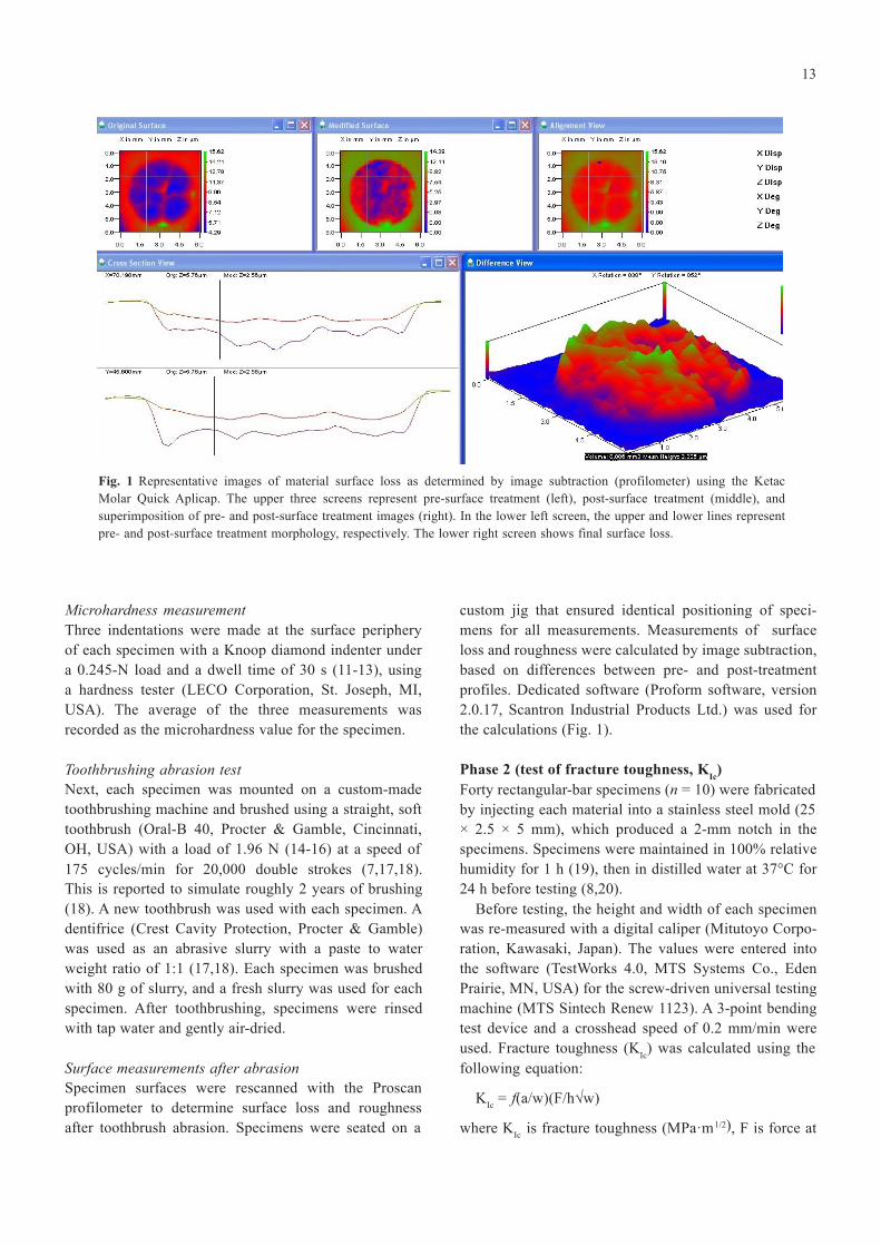
Fig. 1 Representative images of material surface loss as determined by image subtraction (profilometer) using the Ketac Molar Quick Aplicap. The upper three screens represent pre-surface treatment (left), post-surface treatment (middle), and superimposition of pre- and post-surface treatment images (right). In the lower left screen, the upper and lower lines represent pre- and post-surface treatment morphology, respectively. The lower right screen shows final surface loss.
Microhardness measurement Three indentations were made at the surface periphery of each specimen with a Knoop diamond indenter under a 0.245-N load and a dwell time of 30 s (11-13), using a hardness tester (LECO Corporation, St. Joseph, MI, USA). The average of the three measurements was recorded as the microhardness value for the specimen.
Toothbrushing abrasion test Next, each specimen was mounted on a custom-made toothbrushing machine and brushed using a straight, soft toothbrush (Oral-B 40, Procter & Gamble, Cincinnati, OH, USA) with a load of 1.96 N (14-16) at a speed of 175 cycles/min for 20,000 double strokes (7,17,18). This is reported to simulate roughly 2 years of brushing (18). A new toothbrush was used with each specimen. A dentifrice (Crest Cavity Protection, Procter & Gamble) was used as an abrasive slurry with a paste to water weight ratio of 1:1 (17,18). Each specimen was brushed with 80 g of slurry, and a fresh slurry was used for each specimen. After toothbrushing, specimens were rinsed with tap water and gently air-dried.
Surface measurements after abrasion
custom jig that ensured identical positioning of speci- mens for all measurements. Measurements of surface loss and roughness were calculated by image subtraction, based on differences between pre- and post-treatment profiles. Dedicated software (Proform software, version 2.0.17, Scantron Industrial Products Ltd.) was used for the calculations (Fig. 1).
Phase 2 (test of fracture toughness, KIc) Forty rectangular-bar specimens (n = 10) were fabricated by injecting each material into a stainless steel mold (25 × 2.5 × 5 mm), which produced a 2-mm notch in the specimens. Specimens were maintained in 100% relative humidity for 1 h (19), then in distilled water at 37°C for 24 h before testing (8,20).
Before testing, the height and width of each specimen was re-measured with a digital caliper (Mitutoyo Corpo- ration, Kawasaki, Japan). The values were entered into the software (TestWorks 4.0, MTS Systems Co., Eden Prairie, MN, USA) for the screw-driven universal testing machine (MTS Sintech Renew 1123). A 3-point bending test device and a crosshead speed of 0.2 mm/min were used. Fracture toughness (KIc) was calculated using the following equation:
Specimen surfaces were rescanned with the Proscan profilometer to determine surface loss and roughness after toothbrush abrasion. Specimens were seated on a
= f(a/w)(F/h√w)
where K is fracture toughness (MPa·m
1/2), F is force at
14
Ic
the beginning of crack propagation (N), a is crack length (mm), h is specimen thickness (mm), w is specimen width (mm), and f(a/w) is the fracture geometry factor, calculated as:
6α½ [1.99 – α(1- α)(2.15 – 3.93α + 2.7α²)] / [(1+2α)(1-α)3/2]
Statistical methods The Wilcoxon rank-sum test was used to compare groups for differences in Knoop hardness, abrasive wear, surface roughness (R ), and fracture toughness (K ). The Sidak
However, there was no significant difference between FIX and KM (P > 0.05).
Discussion
The addition of zinc oxide particles is the essential modification of CFR, which also contains a high-molec- ular-weight acrylic acid polymer. The manufacturer maintains that inclusion of zinc oxide enhances the setting reaction and increases strength, while retaining similar methods of clinical application and working time as compared with regular GICs.
a Ic adjustment was used to control for multiple pair-wise group comparisons. All statistical analyses were carried out at a 5% significance level.
Results
Phase 1 Knoop hardness Among the tested materials, FIX had the highest hard- ness value (66.86 KHN) and PC had the lowest value (45.44 KHN). The hardness value was significantly higher for FIX than for EF (P = 0.004). The hardness was significantly lower for CFR than for EF, KM, and FIX (P ≤ 0.008). The hardness of PC was significantly lower than that of the other groups (P = 0.006 CFR, P = 0.004 FIX, KM and EF).
Toothbrush abrasion The mean average surface loss for EF, FIX, CFR, KM, and PC was 5.72 µm, 5.21 µm, 4.69 µm, 3.79 µm, and 3.7 µm, respectively. The values for EF and FIX were significantly higher than that for PC (P = 0.004 and P = 0.046, respectively). However, there was no significant difference between KM and the other groups (P > 0.05).
Roughness Mean change in Ra in CFR, PC, KM, EF, and FIX was 0.79 µm, 0.68 µm, 0.62 µm, 0.14 µm, and 0.10 µm, respectively. Values for roughness change were signifi- cantly or marginally higher for CFR than for EF and FIX (P = 0.006 and P = 0.060, respectively). However, there was no significant difference between PC and the other tested materials (P > 0.05).
Phase 2 Fracture toughness The KIc for EF was the highest among the tested GICs and was significantly higher than the values for CFR, FIX, and KM (P = 0.013, P = 0.001, P = 0.001, respectively).
A nanofilled hybrid composite (Premise) was included as a control group in the Phase 1 study (microhardness, wear, and surface roughness testing) because there were no obvious differences between the various GIC groups in our preliminary pilot study. No significant difference was found in the hardness values of FIX and KM, which were harder than the other materials tested. These find- ings are consistent with those of previous studies (6,12). EF was significantly harder than CFR, as was noted in another study (6). Microhardness was significantly lower for PC than for the GICs, which accords with the findings of previous studies (21,22). The relatively low hardness of CFR in this study could be due to filler size and morphology (11) or to insufficient dispersion of zinc and glass particles.
Abrasive wear did not significantly differ among the GICs tested in the present study, which confirms the findings of previous investigations (15,23). However, the abrasive wear of all GICs was significantly or margin- ally greater than the values noted in the tested nanofilled hybrid composite (Premise). Due to their composition, GICs exhibit more wear than composites. Their acid- base reaction results in a matrix comprising an ionically cross-linked polyalkenoate network, which is weaker than the matrix of composites strengthened by fillers and 2-hydroxyethyl methacrylate polymer chains (24).
Attraction of dental plaque to roughened restorations is a concern since it may increase the risk of secondary caries (18). The surfaces of carefully polished dental restorations can be compromised by subsequent home care, including toothbrushing. Most studies of the effects of toothbrushing and polishing on dental restorations concluded that restoration surfaces are smoother before polishing or toothbrushing and tend to increase in rough- ness afterward (10,15,25,26).
In the current study, CFR exhibited the greatest change in surface roughness among GICs, perhaps due to differ- ences in composition. Previous studies concluded that
The K for CFR was significantly higher than the values the surface roughness of GICs is affected by filler size, for FIX and KM (P = 0.001 and P = 0.022, respectively). shape, distribution, and number of particles in the matrix

15
(10). In this study, the fracture toughness of GICs was tested
after 24 h, to ensure assessment at the peak strength of the materials (27). Fracture toughness was significantly greater for EF than for the other GICs; CFR had the second highest fracture toughness. The high fracture toughness of EF may be due to the resin coat applied to the surface. In addition, as compared with CFR, EF has a wider ranges of clinical uses in high-stress–bearing areas and cases requiring buildup. The relative high fracture toughness of CFR could be due to formation of zinc-polycarboxylate complexes during the setting reaction (6). Additionally, the incorporation of itaconic acid as a comonomer in CFR might increase the flexural and tensile strength of GIC (28). Another explanation for the relative high fracture toughness of CFR is its small mean particle size as compared with other conventional GICs (11). Our data on the fracture toughness of KM are consistent with those from a previous study (29). In summary, we conclude that, as compared with the other tested GICs, CFR had intermediate fracture toughness and comparable abrasive wear but inferior surface rough- ness and hardness characteristics.
Acknowledgments
The authors thank Dentsply, GC America, and 3M ESPE for donating the investigated materials.
References
1. Nicholson JW (1998) Chemistry of glass-ionomer cements: a review. Biomaterials 19, 485-494.
2. Costa CA, Ribeiro AP, Giro EM, Randall RC, Hebling J (2011) Pulp response after application of two resin modi- fied glass ionomer cements (RMGICs) in deep cavities of prepared human teeth. Dent Mater 27, e158-170.
3. Cehreli SB, Tirali RE, Yalcinkaya Z, Cehreli ZC (2013) Microleakage of newly developed glass carbomer cement in primary teeth. Eur J Dent 7, 15-21.
4. McCabe JF, Jones PA, Wilson HJ (1979) Some properties of a glass ionomer cement. Br Dent J 146, 279-281.
5. Topbasi B, Öveçoglu ML, Türkmen C (2003) Flexural strength and fracture surface characterization of glass-ionomer cements stored in water. Oral Health Dent Manag 2, 18-26.
6. Zoergiebel J, Ilie N (2013) Evaluation of a conventional glass ionomer cement with new zinc formulation: effect of coating, aging and storage agents. Clin Oral Investig 17, 619-626.
7. Momoi Y, Hirosaki K, Kohno A, McCabe JF (1997) In vitro toothbrush-dentifrice abrasion of resin-modified glass iono- mers. Dent Mater 13, 82-88.
8. Zhao J, Weng Y, Xie D (2009) In vitro wear and fracture toughness of an experimental light-cured glass-ionomer cement. Dent Mater 25, 526-534.
9. Heintze SD, Forjanic M, Ohmiti K, Rousson V (2010)
Surface deterioration of dental materials after simulated toothbrushing in relation to brushing time and load. Dent Mater 26, 306-319.
10. Bala O, Arisu HD, Yikilgan I, Arslan S, Gullu A (2012) Evaluation of surface roughness and hardness of different glass ionomer cements. Eur J Dent 6, 79-86.
11. Xie D, Brantley WA, Culbertson BM, Wang G (2000) Mechanical properties and microstructures of glass-ionomer cements. Dent Mater 16, 129-138.
12. Bonifácio CC, Kleverlaan CJ, Raggio DP, Werner A, de Carvalho RC, van Amerongen WE (2009) Physical- mechanical properties of glass ionomer cements indicated for atraumatic restorative treatment. Aust Dent J 54, 233-237.
13. Raggio DP, Bonifácio CC, Bönecker M, Imparato JC, Gee AJ, Amerongen WE (2010) Effect of insertion method on knoop hardness of high viscous glass ionomer cements. Braz Dent J 21, 439-445
14. Frazier KB, Rueggeberg FA, Mettenburg DJ (1998) Compar- ison of wear-resistance of Class V restorative materials. J Esthet Dent 10, 309-314.
15. Rios D, Honôrio HM, de Araújo PA, Machado MA (2002) Wear and superficial roughness of glass ionomer cements used as sealants, after simulated toothbrushing. Pesqui Odontol Bras 16, 343-348.
16. de Paula AB, Fucio SB, Ambrosano GM, Alonso RC, Sardi JC, Puppin-Rontani RM (2011) Biodegradation and abrasive wear of nano restorative materials. Oper Dent 36, 670-677.
17. Tanoue N, Matsumura H, Atsuta M (2000) Analysis of composite type and different sources of polymerization light on in vitro toothbrush/dentifrice abrasion resistance. J Dent 28, 355-359.
18. Tanoue N, Matsumura H, Atsuta M (2000) Wear and surface roughness of current prosthetic composites after toothbrush/ dentifrice abrasion. J Prosthet Dent 84, 93-97.
19. Yamazaki T, Schricker SR, Brantley WA, Culbertson BM, Johnston W (2006) Viscoelastic behavior and fracture toughness of six glass-ionomer cements. J Prosthet Dent 96, 266-272.
20. Moshaverinia A, Brantley WA, Chee WW, Rohpour N, Ansari S, Zheng F et al. (2010) Measure of microhardness, fracture toughness and flexural strength of N-vinylcaprolactam (NVC)-containing glass-ionomer dental cements. Dent Mater 26, 1137-1143.
21. Kim KH, Ong JL, Okuno O (2002) The effect of filler loading and morphology on the mechanical properties of contempo- rary composites. J Prosthet Dent 87, 642-649.
22. Blackham JT, Vandewalle KS, Lien W (2009) Properties of hybrid resin composite systems containing prepolymerized filler particles. Oper Dent 34, 697-702.
23. Peutzfeldt A, García-Godoy F, Asmussen E (1997) Surface hardness and wear of glass ionomers and compomers. Am J Dent 10, 15-17.
24. Davidson CL (2006) Advances in glass-ionomer cements. J Appl Oral Sci 14, Suppl, 3-9.
25. Carvalho FG, Fucio SB, Paula AB, Correr GM, Sinhoreti

16
MA, Puppin-Rontani RM (2008) Child toothbrush abrasion effect on ionomeric materials. J Dent Child 75, 112-116.
26. Kakuta K, Wonglamsam A, Goto S, Ogura H (2012) Surface textures of composite resins after combined wear test simu- lating both occlusal wear and brushing wear. Dent Mater J 31, 61-67.
27. Lucas ME, Arita K, Nishino M (2003) Toughness, bonding and fluoride-release properties of hydroxyapatite-added glass
ionomer cement. Biomaterials 24, 3787-3794. 28. Moshaverinia A, Roohpour N, Darr JA, Rehman IU (2009)
Synthesis and characterization of a novel N-vinylcapro- lactam-containing acrylic acid terpolymer for applications in glass-ionomer dental cements. Acta Biomater 5, 2101-2108.
29. Bonilla ED, Mardirossian G, Caputo AA (2000) Fracture toughness of various core build-up materials. J Prosthodont 9, 14-18.