physiology of muscle and nerve - university of baghdad semister2014... · tissue lecture 1 dr....
TRANSCRIPT
Physiology of muscle and nerve
Dr. Hanan Luay

Introduction to the excitable tissue
Lecture 1
Dr. Hanan Luay

Objectives:
Upon completion of this lecture ,the student will be able to:
1-Define excitable tissues .
2- Identify the ionic basis of the
resting membrane potential.
3- Describe the importance of Na-K
pump.

What is irritability? An ability of all living tissues to
respond to stimuli (either external or internal environment)
What is excitability?
An ability of specialized cells to respond to certain stimuli
by producing electrical signals known as action potential at
its membrane

Muscle and nerve are excitable tissues. They are excitable because they have electrical phenomenon i.e. they are
polarized.
The neurons are excitable cells specialized for reception,
integration and transmission of nerve impulses.
What are 2 basic properties of excitable cell membranes?
1. The membranes have an electrical excitability across the membrane,and may transmit an impulse along the membrane 2. The membranes contain a variety of ion channels (pores) that may be opened or closed, allowing specific ions to flow across.

The electrical phenomena of the nerve cells:


Resting membrane potential
Membrane potential difference (transmembrane voltage) that exists when cell membranes of excitable tissues are not producing an action
potential (at rest)

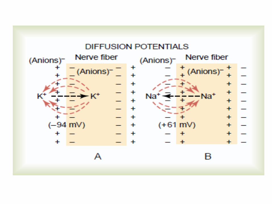
The ionic basis of the resting membrane potential:
1-The contribution of the K ion diffusion potential. 2-The contribution of the Na ion diffusion potential through the nerve membrane. 3-The contribution of the Na_ K ion pump.

Na- K pump
((Guyton,2012


Equilibrium potential of an ion is the value of transmembrane voltage at which the electric force generated by diffusional movement of the ion down its concentration gradient becomes equal to the molecular force of that diffusion.
Nernst equation. For example, for potassium ions will be as follows E (K+) =_ (2.3 RT) log [K +] o OR = - 61 log concentration inside ZF [K+]i concentration outside Goldman equation is used in cell membrane physiology to
determine the equilibrium potential across a cell's membrane taking into account all of the ions that are permeable through that membrane .
•

Nerve structure and function
Lecture 2

1- Define myline sheath and its origin. 2-Recognize the functional and structural organization of the neuron. 3-Compare between the mylinated and the unmylinated nerve fibers. 4-Differentiate between the types of neural communication ? 5-Classify the nerve fibers according to their basic properties and factors affecting each type
:Objectives

The neurons in general are composed of 3 major parts: 1-The soma. 2- Dendrites (2-7). Conduct towards the cell body. The soma and the dendrites form a large area which is specialized for reception. 3- Single axon, it conducts away from the cell body. (Axon hillock, axon knob)

Nerve cells are secretory cells,
but they differ from other
secretory cells in that the
secretory zone is generally at the
end of the axon, far from the cell
body.

TYPES OF NEURONS: Structurally divided into;

Functionally they are divided in
to:
1-Sensory
2- motor and
3- interneuron.

The process of myline sheath forming (mylination) involves the following:
The Schwann cell membrane first envelops the axon.
The cell rotates around the axon many times laying down multiple layers of
Schwann cell membrane.


This sheath has a main role in conduction because; it increases the velocity of conduction. In the CNS the mylination is done by other cells
which are called the oligodendrocytes.
•

Functional organization of the neuron:
1- The receptor zone or dendritic zone. 2- The initial segment zone. 3- The axonal zone. 4- The nerve ending zone.

Axoplasmic transport:
• Fast one (400mm/day).
• Slower axoplasmic flow (6-10 mm/day).
• Slowest axoplasmic flow (3-5mm/day).
Retrograde transport (200mm/day)

In the CNS there is microglia, oligodendrocytes and astrocyte. The astrocytes support the CNS and transport substances between the neurons and the blood vessels. Oligodendrocytes produce the myline sheath. Microglia, provide support and phagocyte bacteria.

Communication of cells inside the human body
At cellular level, communication is based on
Electrical & Chemical signalling

Neural communication
The neurons communicate with each other by 2 types of communication:
1-The electronic potential (generator potential): Local, nonpropagated potentials called synaptic, generator, or electrotonic potentials.
2- The action potential (nerve impulse). is a
propagated disturbance used to send information for long distances without any loss of energy.


NERVE FIBER TYPES & FUNCTION:
Conduction differs due to:
1 -The difference in diameter:
2- Presence of myline sheath
So the nerve fibers are classified into different types by 2 systems of classifications:
A –General system: 3 types according to the peaks produced during compound action potential:
A, B, and C groups, further subdividing the A group into α(the fastest), β, γ, and δ(the slowest) fibers.
B- Numerical system (Ia, Ib, II, III, IV).

Fiber Type Function Fiber Diameter(μm) Conduction Velocity(m/s) Spike Duration ms Absolute refractory period
A Proprioception; 12-2 70-120
somatic motor
α
B Touch, pressure 5-12 30-70 0.4-0.5 0.4-1
γ Motor to muscle spindles 3-6 15-30
Δ Pain, cold, touch 2-5 12-30
B Preganglionic autonomic <3 3-15 1.2 1.2
C
Dorsal root
Pain, temperature,
some mechano-reception,
reflex responses 0.4-1.2 0.5-2 2 2
Sympathetic
Postganglionic sympathetics 0.3-1.3 0.7-2.3 2 2
A and B fibers are myelinated; C fibers are unmyelinated.

Number Origin Fiber Type Ia Muscle spindle, annulospinal ending. A α Ib Golgi tendon organ. A α II Muscle spindle flower-spray ending touch, pressure. A β III Pain and cold receptors; some touch receptors. A δ IV Pain, temperature, and other receptors. Dorsal root C

when we give anesthesia, there will be loss of sensation first. During hypoxia there will be loss of autonomic function first then motor actions, then sensation
Susceptibility to: Most Susceptible Intermediate Least Susceptible
Hypoxia B A C
Pressure A B C
Local anesthetics C B A

Action potential ( ionic basis,properties)
Lecture 3

Objectives: 1-Define action potential.
2-List the successive stages of nerve action potential and recognize their properties. 3-Draw a diagram showing action potential stages.
4-Differentiate between a single axon and a mixed nerve properties.
5- Describe the effect of changing ions concentrations on membrane potential
6-Describe the properties of action potential.
7-Compare the conduction of nerve impulse between mylinated and non mylinated fibers.

Nerve signals (are coded information)
transmitted by action potentials which
are rapid changes in the membrane
potential in response to stimulus that
spread rapidly along the nerve fiber
membrane.
Nerve action potential:

The successive stages in action
potential are as follows:
1-The resting stage. The membrane is polarized. There is brief irregular deflection of the baseline,
called the stimulus artifact. 2-The latent period (latency). Is also an isoptential state, the membrane here is still polarized. It is the
interval starting from the beginning of stimulation to the beginning of potential changes.

LA Latent period is Proportional to 1-The distance
between the stimulus site and the recording site 2- Inversely to the conduction speed of the
axon.

3-The depolarization stage. - Opening of Na ion channels.
-The potential increases rapidly in the positive direction.
-There will be overshooting beyond the zero level.
The overshooting not exceeds 35mv, and Na ion will not reach its equilibrium potential because:
-The concentration gradient for Na ions will be reduced.
-The direction of the electrical gradient will be reversed.
-The Na ion channels will close or inactivated rapidly.

4-The repolarization stage.
Na ion channels begin to close and the K ion channels open.
The sharp rise and the rapid fall of the action potential is called the spike potential
The last 30% of repolarization which is slow is called after depolarization.
The membrane potential will not stop at the resting membrane potential (-70 mv), but reduces further 1-2 mv below it, then return to the resting potential after a period of 40 ms, this period called after hyperpolarization.


Decreasing the external Na+ concentration
↓
Decrease the action potential
Increasing the external K+ concentration
↓
Decreases the resting potential i.e becomes less negative ( threshold is less).
Decrease in extracellular Ca2+ concentration
↓
Increases the excitability of nerve and muscle cells

Orthodromic & Antidromic Conduction In a living animal, impulses normally pass in one
direction only, i.e, from synaptic junctions or receptors along axons to their termination.
Any antidromic impulses that are set up fail to pass
the first synapse they encounter and die out at that point.

Properties of the action potential:
1 – Threshold. the minimal intensity of the stimulus required to excite the nerve and to produce action potential.
2- Self –reinforcement (regeneration). the action potential has the same size and shape without any energy loss.
3- The all or none law. The action potential fails to occur if the stimulus is subthreshold in magnitude (the none part), and it occurs if the stimulus is at or above threshold intensity.Further increases in the intensity of a stimulus produce no increment in the action potential.

4-The refractory period (RP),absolute and relative. it is the time or interval during which the axon or nerve fiber is incapable of
firing a second action potential when a second stimulus is applied.
Absolute RP. Relative RP.

5-Propagation of action potential. -Current sink. the positive charges from the area in front and behind the action potential will flow into the area of negativity represented by the action potential

-Salutatory conduction

6- Accommodation to slow depolarization (failure to fire despite high voltage):
Slowly rising currents fail to fire the nerve because the nerve adapts to the applied stimulus.

Electrotonic Potentials, Local Response, & Firing Level: -Produced by subthreshold stimuli. -Rises sharply and decays exponentially with time. -It is proportional to the magnatitude of the stimulus. -Its spread is graded. -It could be cathodal or anodal.

Energy source and production in the nerve fiber:
70% of the energy is used to maintain polarization of the membrane by the action of Na+-K+ ATPase. During maximal activity, the metabolic rate of the nerve doubles the
normal.

The mixed nerve: Peripheral nerves in mammals are made up of many axons
bound together With subthreshold stimuli, none of the axons are stimulated. When the stimuli are of threshold intensity, axons with low
thresholds fire and a small potential change is observed. As the intensity of the stimulating current is increased, the axons
with higher thresholds are also discharged. IT DOES NOT OBEY THE ALL OR NONE LAW. Supramaximal stimuli produces no further increase in the size of
the observed potential.

Compound Action Potentials Appearance of multiple peaks in the action
potential.

Synapses
Lecture 4

Objectives:
1- Define synapse? And illustrate the
basic structure of synapses.
2- Describe the neuromuscular junction and
the sequence of events during
neuromuscular transmission.
3- identify the types of synaptic
transmission.
4- Determine the properties of synaptic
transmission.

Transmission: JunctionalSynaptic &
Junction is the connection between a nerve cell and
another (muscle fiber or gland). Synapse is the connection between 2 nerve cells. Each nerve ending makes a junction with the
muscle fiber near its midpoint, called the neuromuscular junction.
The nerve fiber with its branching plus the thickened muscle surface is called the motor end plate.


.
The NMJ consists of: - Axon terminal. -Synaptic gutter or synaptic trough. -The synaptic space or synaptic cleft. -Subneural clefts, Acetylecholine (Ach) receptors

Neuromuscular transmission miniature end plate potential.
1- Action potential spreads over the terminal; →
channels open and allow calcium ions to diffuse
to the interior of the nerve terminal.
2- The calcium ions ,exert an attractive influence
on the acetylcholine vesicles, drawing them to
the neural membrane adjacent to the dense bars.

3-The vesicles then fuse with the neural
membrane and empty their acetylcholine
into the synaptic space by the process of
exocytosis (Botulinum and Tetanus toxins
block the transmitter release).
4- Ach will diffuse to the synaptic cleft and
binds with the receptors, causing
activation of the Na and K ionic channels
resulting in local depolarization and firing
level is reached ,and action potential is
initiated.

Destruction of the Released Acetylcholine by Acetyl
:cholinesterase 1-Most of the acetylcholine is destroyed by the enzyme acetylcholinesterase, into choline and acetate (physostigmine). 2- A small amount of acetylcholine diffuses out of the synaptic space.

Mysthemia Gravis :an autoimmune disease in which there is sever voluntary muscle weakness. Antibodies that destroy the Ach receptors
Lambert-Eaton syndrome. muscle weakness is caused by antibodies against one of the Ca2+ channels in the nerve endings at the neuromuscular junction.

SYNAPTIC TRANSMISSION:
The synapse consists of:
-Presynaptic terminal (axon, dendrite).
-Postsynaptic terminal (axon ,dendrite, soma)
-Synaptic cleft. Space between 1 and 2.


The transmission in the synapse is of 3 types: 1- Chemical transmission: The impulse in the presynaptic axon causes secretion of a neurotransmitter such as acetylcholine or serotonin. 2-Electrical transmission: The membranes of the presynaptic and postsynaptic neurons come close together, and gap junctions formed. 3- Both electrical and chemical transmission.

Properties of synaptic transmission: 1- One way conduction: the transmitter is only on one side, the impulse can go in one direction only.
2- Convergence & Divergence:
3- The post synaptic potential is an electronic potential.

4-Postsynaptic potential could be excitatory or inhibitory.
If the neurotransmitter (like Ach, epinephrine and norepinephrine) causes opening of Na ion channels (or Ca ion channels) → depolarization occur (excitatory postsynaptic potential (EPSP). If it opens K ion or Cl ion channels → hyperpolarization (IPSP), (like glycine, GABA (Gamma Amino Butyric Acid).

The synapse inhibition is of 2 types: --Direct postsynaptic inhibition: inhibition affecting the neuron directly by generating IPSP, through interneuron secreting a transmitter that hyperpolarizes the postsynaptic neuron by increasing Cl ion influx or K efflux.
--Presynaptic inhibition: mediated by thick interneuron
ends on the excitatory ending of the presynaptic neuron where they form axo-axonal synapse.

5- Summation of the postsynaptic potential: 2 types of summation: Spatial:When activity is present in more than one synaptic knob at the same time. Temporal: if repeated afferent stimuli cause postsynapyic potential, before previous one have decayed.

6- Fatigue of synapses: Repeated stimulation at high rate for long time cause impairment of the synaptic transmission .this occurs due to: 1-Mainly depression of the stores of the neurotransmitters. 2-Inactivation of the postsynaptic receptors. 3-Slow built up of Ca ions.

6- Synaptic delay: When an impulse reaches the
presynaptic terminals, there is an interval
of at least 0.5 ms, the synaptic delay,
before a response is obtained in the
postsynaptic neuron..

Some drugs affecting NMJ: Drugs stimulating the muscle fiber, by Ach like action:
Methacholine,Nicotine(produce a state of muscle spasm ,because not destroyed by cholinesterase).
Drugs that stimulate the NMJ by inactivating cholinesterase: Neostigmine,Physostigmine, nerve gas, organophosphate
insecticide. May be lethal because it also cause muscle spasm especially if involves the laryngeal muscles.
Drugs that blocks transmission of the NMJ: Curariform, blocks the action of Ach on the receptors and prevents the passage of nerve impulse from the nerve ending to the muscle

SYNAPTIC PLASTICITY & LEARNING
Short- and long-term changes in synaptic function can occur as a result of the history of discharge at a synapse; ie, synaptic conduction can be strengthened or weakened on the basis of past experience. These changes are of great interest because they obviously represent forms of learning and memory .They can be presynaptic or postsynaptic in location.

few examples of important neurotransmitter actions:
Glutamate is used at the great majority of fast excitatory synapses in the brain and spinal cord.
GABA is used at the great majority of fast inhibitory synapses in virtually every part of the brain. Many sedative/tranquilizing drugs act by enhancing the effects of GABA. Correspondingly glycine is the inhibitory transmitter in the spinal cord.

is distinguished as the transmitter Acetylcholineconnecting neuromuscular junctionat the
motor nerves to muscles. The paralytic arrow-acts by blocking transmission at curarepoison
these synapses. Acetylcholine also operates in many regions of the brain, but using different types of receptors.

Dopamine has a number of important functions in the brain. It plays a critical role in the reward system, but dysfunction of the dopamine system is also implicated in Parkinson's Disease and schizophrenia.
Serotonin has a number of important functions that are difficult to describe in a unified way, including regulation of mood, sleep/wake cycles, and body temperature. It is released during sunny weather, and also when eating chocolate.
Substance P responsible for transmission of pain from certain sensory neurons to the central nervous system.