molecular anatomy of the hair cell's ribbon synapse
TRANSCRIPT

Cellular/Molecular
Molecular Anatomy of the Hair Cell’s Ribbon Synapse
Revathy C. Uthaiah and A. J. HudspethHoward Hughes Medical Institute and Laboratory of Sensory Neuroscience, The Rockefeller University, New York, New York 10065
Hearing depends on reliable and temporally precise neurotransmission by cochlear hair cells. The wide dynamic range and high sensi-tivity with which these cells encode acoustic stimuli are associated with a presynaptic specialization termed the presynaptic dense bodyor synaptic ribbon. Apposed to the presynaptic density, this spherical or flattened structure tethers a layer of synaptic vesicles and isthought to facilitate their exocytotic fusion. Although defining the molecular constituents of the hair cell’s synaptic ribbon shouldcontribute to our understanding of neurotransmitter release at this synapse, accomplishing this task has been slowed by the difficulty ofobtaining sufficient amounts of starting material for protein analysis from hair cells. We isolated synaptic material from chickencochleas, purified synaptic ribbons with specific immunological reagents, and identified the associated proteins by tandem mass spec-trometry. Purification of the ribbons revealed a predominant composition of C-terminal-binding proteins, especially ribeye, in associa-tion with the small GTPase Rab3, which is possibly involved in attaching vesicles to the ribbon. In comparison with the components ofconventional synapses and of retinal ribbon synapses, we observed that certain regulatory proteins are excluded from the hair cell’ssynapse. Using antisera against several of the novel proteins and membrane-trafficking components that we had identified, we docu-mented their localization in isolated hair cells. Our results indicate that the ribbon synapses of hair cells display modifications to thepresynaptic machinery that are associated with the high-fidelity transmission of acoustic signals to the brain.
IntroductionSynaptic transmission by the sensory receptors of the visual, au-ditory, and vestibular systems is characterized by tonic andgraded neurotransmitter release that requires sustained highrates of exocytosis (Roberts et al., 1991; Lenzi and von Gersdorff,2001; Sterling and Matthews, 2005). The presynaptic active zonesof these glutamatergic synapses are marked by synaptic ribbons,flattened or ovoid electron-dense structures 100 –1000 nm in di-ameter. Numerous synaptic vesicles are tethered by fine filamentsto each ribbon.
The speed with which acoustic stimuli are encoded at the haircell’s ribbon synapse plays a key role in determining auditoryrepresentations in the CNS. Neurotransmitter release in hair cellsexhibits fast kinetics, allowing acoustic stimuli to be encodedwith a temporal resolution of �1 ms (Nouvian et al., 2006).Measurements of hair-cell membrane capacitance and turnoverrates (Parsons et al., 1994; Moser and Beutner, 2000; Khimich etal., 2005; Rutherford and Roberts, 2006) and recordings from affer-ent neurons (Glowatzki and Fuchs, 2002; Keen and Hudspeth, 2006;Rutherford and Roberts, 2006, 2009; Li et al., 2009) suggest
that the ribbon synapse is optimized to release large amountsof neurotransmitter continuously and indefinitely. This ex-ceptional rate of exocytosis places high demands on the syn-apse to replenish the active site with vesicles, a role thought tobe played by the synaptic ribbon.
Despite their functional differences, ribbon synapses and con-ventional synapses have molecular similarities. The expression ofsoluble N-ethylmaleimide-sensitive factor (NSF) attachmentprotein (SNAP) receptors (SNAREs) is highly conserved. Syn-taxin 1, SNAP25, and vesicle-associated membrane protein(VAMP1, synaptobrevin 1) occur at hair-cell synapses (Safied-dine and Wenthold, 1999), and syntaxin 3, SNAP25, and VAMP3(cellubrevin, synaptobrevin-3) are found at retinal synapses (VonKriegstein et al., 1999; Lenzi and von Gersdorff, 2001). However,all ribbon synapses appear to lack the vesicle-associated synapsins1 and 2. The hair-cell synapse lacks complexins (Strenzke et al.,2009), synaptophysins 1 and 2, and synaptotagmins 1 and 2(Safieddine and Wenthold, 1999), proteins that are abundantlyexpressed at both conventional synapses and retinal ribbonsynapses.
The specialized components of ribbon synapses have beendescribed in the retina (tom Dieck et al., 2005), in which ribbonscontain three members of the C-terminal-binding protein(CtBP) family of transcriptional repressors: CtBP1, CtBP2, andits alternative splice form ribeye (Magupalli et al., 2008; Schmitz,2009). Two proteins have been identified as binding partners ofribeye in photoreceptor ribbons: munc119, a prenyl-bindingprotein (Alpadi et al., 2008), and IQ-ArfGEF (BRAG1), aguanine-nucleotide exchange factor for ADP-ribosylation factors(Katsumata et al., 2009). Cytomatrix proteins such as piccolo,Rab3-interacting molecule 1 (RIM1), and KIF3A are also associ-ated with the ribbon, and active-zone scaffolding molecules such
Received Feb. 25, 2010; revised July 10, 2010; accepted July 19, 2010.This work was supported by Grant DC000241 from the National Institutes of Health. R.C.U. was a recipient of a
Women and Science Postdoctoral Fellowship from The Rockefeller University. A.J.H. is an Investigator of HowardHughes Medical Institute. We are grateful to Ms. Y. Castellanos for assistance with the dissections and biochemicalprocedures and to Dr. K. Uryu for conducting the electron-microscopic studies. Dr. M. Oeffinger and Dr. M. P. Routoffered helpful discussions on magnetic beads, and Dr. H. Yu provided assistance with mass spectrometry. We thankthe members of our research group, especially Dr. J. A. N. Fisher, Dr. E. C. Keen, Mr. L. Kowalik, and Dr. S. Lagier, forcomments on this manuscript.
Correspondence should be addressed to A. J. Hudspeth, Howard Hughes Medical Institute and Laboratory ofSensory Neuroscience, The Rockefeller University, 1230 York Avenue, New York, NY 10065-6399. E-mail:[email protected].
DOI:10.1523/JNEUROSCI.1014-10.2010Copyright © 2010 the authors 0270-6474/10/3012387-13$15.00/0
The Journal of Neuroscience, September 15, 2010 • 30(37):12387–12399 • 12387

as bassoon, RIM2, ERC2 (CAST1), andmunc13-1 tether the ribbon to the presyn-aptic membrane (tom Dieck et al., 2005).
Understanding of the molecular com-position of the hair cell’s ribbon synapsehas remained limited (Nouvian et al.,2006; LoGiudice and Matthews, 2009),mostly because of the effort required inharvesting sufficient cochlear material forprotein analysis. We have used biochemi-cal and proteomic approaches to isolatepresynaptic components from chickencochleas and identified the proteins bytandem mass spectrometry (MS–MS).
Materials and MethodsAnimals and organs. White Leghorn chickens(Gallus gallus) two weeks of age were killedwith CO2 and decapitated. We dissected theircochleas in chick saline solution consisting ofthe following (in mM): 154 NaCl, 6 KCl, 5.6CaCl2, 2.3 MgCl2, 8 D-glucose, and 5 HEPES,pH 7.4. The specimens were immediately fro-zen in liquid nitrogen and preserved at �80°Cuntil use. Cochlear and utricular hair cells wereisolated from mice (Mus musculus; strainC57B/6) 1 to 2 weeks of age (He and Dallos,2000). Mouse and chicken brains were isolatedfrom animals 2 weeks of age and stored at�80°C after homogenization in radioimmu-noprecipitation assay (RIPA) lysis solutioncontaining 150 mM NaCl, 1% Igepal-40, 1%sodium deoxycholate, 0.1% SDS, and 25 mM
tris(hydroxymethyl)aminomethane at pH 7.6.Mouse retinal lysate was purchased (Anaspec).To produce chicken retinal lysate, we dissectedneural retinas from pigment epithelia and ho-mogenized them in RIPA solution.
Antibodies. Antisera and antibodies againstthe following proteins were purchased fromthe indicated sources: calcium/calmodulin-dependent serine protein kinase (CASK),CtBP1, CtBP2, dynamin, munc13, munc18,N-cadherin, neuroligin, Rab3, RIM1, synapsin 1,synaptotagmin 1, syntaxin 4, syntaxin 6, tomo-syn, VAP-33, valosin-containing protein (VCP),and �-catenin were from BD Transduction Lab-oratories; actin, KIF3A, nonmuscle myosin heavychain (MYH9), NF68, NF160, NF200, NSF,SNAP23, syntaxin 1, syntaxin 7, �-tubulin,�-tubulin, chicken spectrin, and PRPF39 werefrom Sigma Immunochemicals; ERC2, piccolo,RIM2, synaptojanin, and VAMP3 werefrom Synaptic Systems; bassoon, NIPSNAP1,SNAP25, and VAMP1 were from Abcam;CaBP4, synaptophysin 1, �-SNAP, and �-SNAPwere from Santa Cruz Biotechnology; GFAP andSec13 were from Novus Biologicals; Ca2�/calmodulin-dependent kinase II (CaMKII) wasfrom Cell Signaling Technology; clathrin heavychain was from Affinity BioReagents; com-plexin 1 and complexin 2 were from MilliporeBioscience Research Reagents; SV2 was fromthe Developmental Hybridoma Bank; glycer-aldehyde phosphate dehydrogenase (GAPDH)was from Millipore; cysteine string protein(CSP) and VAMP2 were from Stressgen; and
80
60
kDa
20
40
50
40
110120
1,500
X g
solub
le
1,500
X g
insolu
ble
20,00
0 X g
solub
le
20,00
0 X g
insolu
ble
47,00
0 X g
solub
le
47,00
0 X g
insolu
ble
20,00
0 X g
solub
le
20,00
0 X g
insolu
ble
200,0
00 X
g so
luble
200,0
00 X
g ins
oluble
sucro
se 20
0-40
0 mM
sucro
se 40
0-60
0 mM
sucro
se 60
0-80
0 mM
sucro
se pe
llet
ribeye
CtBP2
VAMP2
syntaxin 1
Retina
B
20
80
6050
40
110120 ribeye
CtBP2
VAMP2
lysate
1,500
X g
solub
le
47,00
0 X g
solub
le
47,00
0 X g
insolu
ble
20,00
0 X g
solub
le
20,00
0 X g
insolu
ble
200,0
00 X
g so
luble
200,0
00 X
g ins
oluble
sucro
se pe
llet
Cochlea
1,500
X g
insolu
ble
Cochleas homogenized in buffered sucrose
soluble insoluble
insolublesoluble
47,000 X g spin for 15 min
Resuspended in 40 mM sucrose
Cell lysis by hypoosmotic shock for 45 min
1,500 X g spin for 15 min
20,000 X g spin for 30 min
insolublesoluble
200,000 X g spin for 60 min
insolublesoluble Sucrose-density-gradient centrifugation100,000 X g for 120 min
A
800 mM
600 mM
400 mM
200 mM200-400 mM sucrose boundary
400-600 mM sucrose boundary
600-800 mM sucrose boundary
pellet
Figure 1. Fractionation of presynaptic proteins A, A flow chart describes the purification of the presynaptic material fromthe retina and cochlea by differential and sucrose-gradient centrifugation. B, Immunoblotting delineates the fractionationof the ribbon proteins ribeye and CtBP2, the vesicle protein VAMP2, and the membrane SNARE syntaxin 1 in the retina andcochlea.
12388 • J. Neurosci., September 15, 2010 • 30(37):12387–12399 Uthaiah and Hudspeth • Ribbon Synapses
endophilin was from Zymed Laboratories. Additional details are pro-vided in supplemental Table 1 (available at www.jneurosci.org as supple-mental material).
Isolation of presynaptic proteins. The presynaptic components fromchicken cochleas and bovine retinas were purified using a standard pro-tocol (Huttner et al., 1983) with a few modifications. Approximately2000 chicken cochleas were dissected and cryomilled with Mixer MillsMM301 (Retsch) and homogenized with a Kontes mortar and pestle(Daigger) in a solution containing 320 mM sucrose, 1 mM magnesiumacetate, 0.5 mM calcium acetate, and 1 mM sodium bicarbonate at pH 7.2.The homogenate was centrifuged at 1500 � g for 15 min to pellet largecell bodies, red blood cells, and myelin. The supernatant was removed,transferred to a fresh tube, and centrifuged again at 20,000 � g for 30min. The crude synaptosomal pellet was resuspended in a hypotonicsolution containing 6 mM tris(hydroxymethyl)aminomethane maleate atpH 8.1, homogenized with 10 strokes of a Dounce homogenizer (Pyrex;Corning) in the presence of protease inhibitors, and incubated on ice for45 min with intermittent vortexing to ensure cell lysis. The crude synap-tosome fraction was centrifuged at 47,000 � g for 15 min in a SW55 TiRotor (Beckman) to remove large membrane components. The super-natant was collected and small membrane components and synaptic ves-icles were pelleted by centrifugation at 200,000 � g in a SW28 rotor(Beckman) for 60 min. A solution of 320 mM sucrose, CompleteProtease Inhibitor Cocktail (Roche), and 10 mM HEPES at pH 7.4 wasadded to the pellets and the mixture was homogenized using a smallglass Wheaton mortar with a Teflon pestle (Pyrex; Corning) to dis-solve all the membrane pieces. The crude vesicle suspension was di-luted into 200 mM sucrose, then layered on top of a step gradientcontaining 200, 400, 600, and 800 mM sucrose and centrifuged at100,000 � g for 120 min. The proteins fractionated between the su-crose layers were collected. All these samples and the 47,000 � g pelletsample were separated by denaturing SDS-PAGE and stained withcolloidal Coomassie Blue (Pierce). One millimeter bands were cutfrom the gel for mass-spectrometric analysis.
To purify presynaptic components from the bovine retina, we usedeight retinas and followed the procedure described above.
All chemicals were obtained from Sigma-Aldrich unless otherwise specified.
SDS PAGE and immunoblotting. Sampleswere solubilized in NuPAGE LDS samplebuffer and NuPAGE reducing agent, incubatedat 100°C, and loaded on linear-gradient 4 –12%Bis-Tris gels (Invitrogen). Samples were subse-quently electrotransferred to polyvinylidenefluoride membranes (Immobilon-P; Milli-pore) with the Trans-Blot SD semidry transferapparatus (Bio-Rad). Immunocomplexes werevisualized on film with horseradish peroxi-dase-conjugated secondary antibodies and en-hanced chemiluminescence detection (ECLPlus; GE Healthcare). Chemiluminescence wasdetected with Classic Blue AutoradiographyFilms (MidSci) developed in a Konica SRX101-A Processor.
Liquid chromatography-tandem mass spec-trometry. Bands 1 mm in length were excisedfrom SDS-PAGE gels and subjected to in-geltrypsin digestion and peptide extraction(Kumarathasan et al., 2005). In brief, the proteinswere reduced with 10 mM dithiothreitol, alky-lated with 55 mM iodoacetamide, and digestedovernight at 37°C with sequencing-grade modi-fied trypsin (Promega) in ammonium bicar-bonate buffer. The digested products wereextracted twice with 0.1% trifluoroacetic acid,50% acetonitrile, and 1% trifluoroacetic acid.The extracted mixture was dried by Speed-Vacand redissolved in 10 ml of 0.1% trifluoroaceticacid. The extracts were subjected to liquidchromatography-tandem mass spectrometry
(LC-MS/MS) analysis. The pool of peptides was first separated in areverse-phase liquid chromatography column with the Dionex U3000Capillary/nano-HPLC system at a flow rate of 250 ml/min, and thendirectly introduced by electrospray into a LTQ-Orbitrap mass spectrom-eter (Thermo Fisher Scientific) operated in data-dependent scan mode.A fused-silica capillary column 75 mm in internal diameter and 100 mmin length (Upchurch Scientific) was packed with C-18 resin (300 A; 5mm; Varian). Mobile phase solvent A contained 0.1% formic acid andmobile phase solvent B contained 99.9% acetonitrile and 0.1% formicacid. Gradient runs for 60 min were performed in solvent B with a flowrate of 250 nl/min. The mass-acquisition method involved one mass-spectrometric scan followed by six tandem scans in the ion trap. Theinitial scan was acquired for mass-to-charge (m/z) ratios of 400 to 1600.The six most intense peaks from each scan were selected in the ion trapfor further fragmentation and tandem mass spectrometry.
The results were analyzed and the peptides as well as the parentproteins for chicken and bovine experiments were identified in theNational Center for Biotechnology Information nonredundant data-base, NCBInr, choosing Chordata, with the MASCOT software searchalgorithm (MatrixScience) and the following parameters: missed cleav-age, 1; fixed modification, carbamidomethylation; variable modification,oxidation of methionine. The maximum error tolerance was 20 ppm formass-spectrometric scans and 1.2 Da for tandem scans. We used strin-gent criteria for protein identification: a protein was designated as a “hit”only if it matched at least two distinct peptides with an ion score of at least40. A single-peptide match was considered if the peptide yielded goodtandem mass spectra and matched only one protein. The ion score, whichis given by �10 � log( P), in which P is the probability that the observedmatch is a random event, indicates how well a spectrum matches a par-ticular peptide. Individual ion scores exceeding 38 indicate identity orextensive homology at the level p � 0.05. For proteins matching the samesets of peptides, only the protein with the greatest percentage of peptiderepresentation was selected.
Isolation of hair cells and immunocytochemistry. The dissected chicken’scochlea was pinned at both ends, and the tegmentum vasculosum was
Synaptic fractionate from cochleas
A B
Synaptic fractionatefrom retinas
Percentage (%)0 5 10 15 20 25 30 35 40
Vesicle-transport proteins
Metabolic enzymes
Intracellular-organelle enzymes
Chaperones
Cytoskeleton
Nucleus and transcription
Cochlea-specific and others
Isolation from cochleas
D
Percentage (%)0 5 10 15 20 25 30 35 40
Vesicle-transport proteins
Metabolic enzymes
Intracellular-organelle enzymes
Chaperones
Cytoskeleton
Nucleus and transcription
Retina-specific and others
Isolation from retinas
C
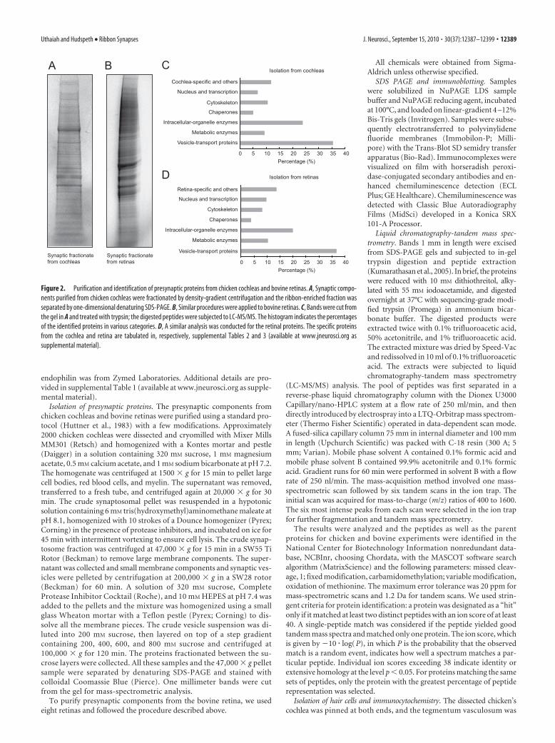
Figure 2. Purification and identification of presynaptic proteins from chicken cochleas and bovine retinas. A, Synaptic compo-nents purified from chicken cochleas were fractionated by density-gradient centrifugation and the ribbon-enriched fraction wasseparated by one-dimensional denaturing SDS-PAGE. B, Similar procedures were applied to bovine retinas. C, Bands were cut fromthe gel in A and treated with trypsin; the digested peptides were subjected to LC-MS/MS. The histogram indicates the percentagesof the identified proteins in various categories. D, A similar analysis was conducted for the retinal proteins. The specific proteinsfrom the cochlea and retina are tabulated in, respectively, supplemental Tables 2 and 3 (available at www.jneurosci.org assupplemental material).
Uthaiah and Hudspeth • Ribbon Synapses J. Neurosci., September 15, 2010 • 30(37):12387–12399 • 12389

removed with fine forceps. The basilar pa-pilla and tectorial membrane were separatedfrom the rest of the tissue and placed in freshchick saline solution. The sample was tritu-rated 10 times through a trimmed 200 �l pi-pette tip to separate the tectorial membraneand free individual cells. Isolated cells were al-lowed to settle onto coverslips coated with 1mg/ml concanavalin A. The cells were fixed for20 min with 4% formaldehyde in 10 mM PBS atpH 7.4, permeabilized for 15 min with 0.05%Triton X-100 in PBS, and blocked for 30 minwith 0.05% Triton X-100 in PBS with 1% nor-mal serum from the species (goat, mouse, orrabbit) in which the primary antiserum wasraised. The cells were incubated overnight withprimary antibody and for 120 min with fluoro-phore-coupled secondary antibody, with threeintermittent washes in a blocking buffer fol-lowing each incubation. For labeling of fila-mentous actin, Alexa Fluor 568-phalloidin(Invitrogen) was added at a concentration of50 ng/ml; for staining nuclei, 4�,6�-diamidino-2-phenylindole (Invitrogen) was included at aconcentration of 1 �g/ml throughout thesecondary-antibody incubation. The cells weremounted in Vectashield and imaged with alaser-scanning confocal microscope (FluoviewFV-1000; Olympus).
Immunoprecipitation. Antibodies were cou-pled to magnetic M-270 Epoxy Dynabeads(Dynal; Invitrogen) with an optimized versionof the protocol suggested by the manufacturer.The beads were washed twice with 1 ml of 100mM sodium phosphate buffer at pH 7.4, with a10 min period of shaking between the washes.Antibodies were added at a ratio of 20 �g permilligram of beads in 100 mM sodium phos-phate and 1 M ammonium sulfate, and thenincubated overnight on a shaker at 30°C. Thecoupled magnetic beads were washed sequen-tially as follows: in 100 mM sodium phosphate,pH 7.4; in 100 mM glycine-HCl, pH 2.5; in 10mM tris(hydroxymethyl)aminomethane, pH8.8; in 100 mM triethylamine; thrice in PBS;twice in PBS containing 0.5% Triton X-100;and twice in PBS. The beads were then stored at4°C in PBS and 0.02% sodium azide.
Beads were washed three times with lysisbuffer just before use. The cryomilled cochlearpowder was thawed and synaptosomes wereisolated as described above. Samples were sus-pended for 30 min at 4°C in a solution contain-ing 110 mM potassium acetate, 100 mM NaCl, 2mM MgCl2, 1% Igepal-40, 0.1% Tween 20, 1mM PMSF, Complete Protease Inhibitor Cock-tail (Roche), and 20 mM HEPES, pH 7.4. Aftercentrifugation at 15,000 � g for 30 min, the solu-ble fraction was added to the antibody-coupledmagnetic beads. Immunoaffinity purificationswere achieved by slow mixing at 4°C for 30–120min. The magnetic beads were collected using aDynal magnet (Invitrogen) and washed rapidlyapproximately six times with the same solution.The isolated protein complex was then elutedfrom the beads for 20 min at room temperaturein aqueous 500 mM NH4OH and 0.5 mM EDTA.The resulting supernatant was frozen in liquidnitrogen and left to dry overnight by vacuum
A
VAMP1
MCMB CB CR CC20
VAP-3330
MCMB CB CR CC
NSF
20
30
MCMB CB CR CC
VAMP2
MRMB MC CB CR CC20
VAMP3
MB MC CB CR CC20
60
80
MCMB CB CR CC
ChickenMouse
syntaxin 1
syntaxin 7
syntaxin 6
SNAP23
syntaxin 4
SNAP25
20
ChickenMouse
40
30
kDa
MCMB CB CR CC
30
40
20
30
40MRMB MC CB CR CC
MB CB CR CCMC
30
MR MC CB CR CCMB
40
30
MCMB CB CR CC
30CB CR CCMCMRMB
syntaxin 1 SNAP25 MergePhalloidin
syntaxin 1 VAMP3 MergePhalloidin
syntaxin 6 SNAP25 MergePhalloidin
VAMP2 SNAP25 MergePhalloidin
B C D
E F G
H I J
L MK
kDa
α-SNAP/β-SNAP
Figure 3. ExpressionofneuronalandnonneuronalSNAREs. A, Immunoblotanalysiswasconductedonlysatesfrommousebrains(MB),retinas (MR), and cochleas (MC), and on chicken brains (CB), retinas (CR), and cochleas (CC). We examined the expression of: the neuronalSNAREssyntaxin1,SNAP25,andVAMP2;thenonneuronalSNAREssyntaxin4,syntaxin6,syntaxin7,SNAP23,VAMP1,VAMP2,andVAMP3;the SNARE-associated protein VAP-33; and the SNARE-dissociation components �-SNAP, �-SNAP, and ATPase NSF. The antisera againstsyntaxin 6, SNAP23, and ATPase NSF identify the respective proteins in all tissues tested. The antisera against syntaxin 4, syntaxin 7, andVAP-33 labeled only the chicken tissues, whereas the antisera against VAMP1, VAMP3, �-SNAP, and �-SNAP labeled only the mousetissues. The protein loading in each lane was standardized to a loading control such as �-tubulin or GAPDH. B–M, Immunofluorescencelocalization of SNAREs in single confocal images of isolated chicken hair cells. The labeling with individual antibodies is shown in grayscale,and the merged panels are displayed in color. The hair bundles are labeled cyan with phalloidin. B–D, The t-SNARE syntaxin 1 is expressedatthetwotraffickinghotspotsofthehaircell,belowthecuticularplateandatthebasolateralend.SNAP25ispredominantlyassociatedwiththe cell membrane. The yellow spots in D show the overlay between the t-SNAREs, which can form a binary complex. E–G, The vesicleSNARE VAMP2 shows a diffuse pattern of labeling throughout the hair cell exclusive of the hair bundle. There is some colocalization ofVAMP2 with SNAP25 at the membrane. H–J, Syntaxin 1 occurs diffusely throughout the cytoplasm, whereas VAMP3 is concentratedbeneath the hair bundle. K–M, Syntaxin 6 and SNAP25 overlap partially in their expression at the hair cell’s base. Scale bars: 2 �m.
12390 • J. Neurosci., September 15, 2010 • 30(37):12387–12399 Uthaiah and Hudspeth • Ribbon Synapses
centrifugation. The pellet was resuspended in sample buffer and subjected toSDS-PAGE and LC-MS/MS analysis.
Ribeye was immunoprecipitated with either of two reagents. We useda murine monoclonal antibody (BD Transduction Laboratories) againstthe C-terminal B-domain of CtBP2, which is shared by ribeye. A rabbitpolyclonal antiserum was raised against the purified N-terminalA-domain of ribeye. For this purpose, a cDNA encoding the A-domain ofchicken ribeye (amino acids 1–553) was cloned in a pET28a � vector(Novagen) and transformed into Escherichia coli strain BL21 (DE3). Cul-tures in logarithmic phase, at an optical density at 600 nm of 0.6 – 0.8,were induced with 0.5 mM isopropyl-�-D-thiogalactopyranoside for pro-tein expression overnight at 16°C. The soluble protein was purifiedthrough its hexahistidine tag by affinity chromatography on nickel ni-trilotriacetic acid beads (Qiagen).
Immunoelectron microscopy. Dissected cochleas were fixed overnightwith 4% paraformaldehyde and 0.1% glutaraldehyde in PBS at pH 7.4.The tissue was treated with 0.5% H2O2 and 0.1% sodium borohydride;blocked with 3% bovine serum albumin, 0.1% cold-water fish-skin gel-atin, and 0.1% saponin; and incubated with antiserum against ribeye,CtBP1, CtBP2, Rab3, or syntaxin 1. The preparation was then incubatedwith biotinylated secondary antibody, and the immunocomplex wasvisualized by the avidin-biotin-peroxidase complex method (VectorLaboratories) with diaminobenzidine as a chromogen and silver en-hancement (Galvin et al., 1999). After postfixation with 1% osmiumtetroxide, the tissue was dehydrated through a graded series of ethanolconcentrations and embedded in EMBed812. Thin sections were cut andexamined under an electron microscope (Tecnai Spirit G2; FEI).
ResultsOur objective was to isolate and characterize the synaptic com-ponents of cochlear hair cells by using biochemical purificationof presynaptic material and immunoprecipitation of ribeye-
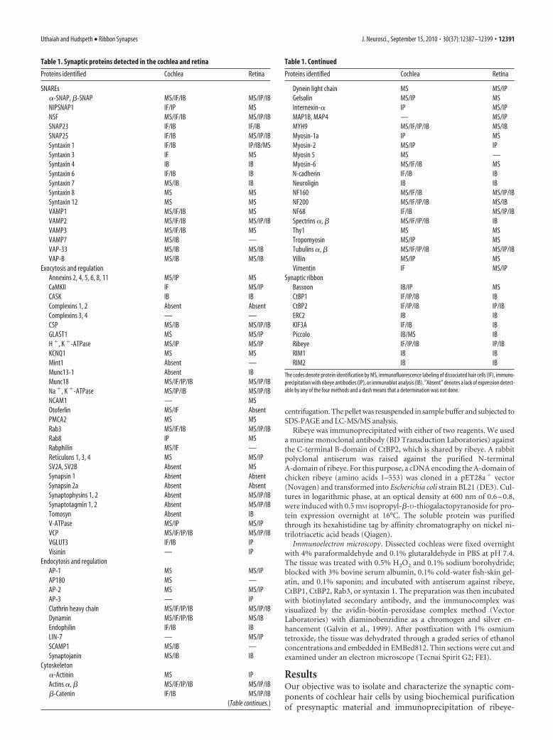
Table 1. Synaptic proteins detected in the cochlea and retina
Proteins identified Cochlea Retina
SNAREs�-SNAP, �-SNAP MS/IF/IB MS/IP/IBNIPSNAP1 IF/IP MSNSF MS/IF/IB MS/IP/IBSNAP23 IF/IB IF/IBSNAP25 IF/IB MS/IP/IBSyntaxin 1 IF/IB IP/IB/MSSyntaxin 3 IF MSSyntaxin 4 IB IBSyntaxin 6 IF/IB IBSyntaxin 7 MS/IB IBSyntaxin 8 MS MSSyntaxin 12 MS MSVAMP1 MS/IF/IB MSVAMP2 MS/IF/IB MS/IP/IBVAMP3 MS/IF/IB MSVAMP7 MS/IB —VAP-33 MS/IB MS/IBVAP-B MS/IB MS/IB
Exocytosis and regulationAnnexins 2, 4, 5, 6, 8, 11 MS/IP MSCaMKII IF MS/IPCASK IB IBComplexins 1, 2 Absent AbsentComplexins 3, 4 — —CSP MS/IB MS/IP/IBGLAST1 MS MS/IPH �, K �-ATPase MS/IP MS/IPKCNQ1 MS MSMint1 Absent —Munc13-1 Absent IBMunc18 MS/IF/IP/IB MS/IP/IBNa �, K �-ATPase MS/IP/IB MS/IP/IBNCAM1 — MSOtoferlin MS/IF AbsentPMCA2 MS MSRab3 MS/IF/IB MS/IP/IBRab8 IP MSRabphilin MS/IF —Reticulons 1, 3, 4 MS MS/IPSV2A, SV2B Absent MSSynapsin 1 Absent AbsentSynapsin 2a Absent AbsentSynaptophysins 1, 2 Absent MS/IP/IBSynaptotagmin 1, 2 Absent MS/IP/IBTomosyn Absent IBV-ATPase MS/IP MS/IPVCP MS/IF/IP/IB MS/IP/IBVGLUT3 IF/IB IPVisinin — IP
Endocytosis and regulationAP-1 MS MS/IPAP180 MS —AP-2 MS MS/IPAP-3 — IPClathrin heavy chain MS/IF/IP/IB MS/IP/IBDynamin MS/IF/IP/IB MS/IBEndophilin IF/IB IBLIN-7 — MS/IPSCAMP1 MS/IB —Synaptojanin MS/IB IB
Cytoskeleton�-Actinin MS IPActins �, � MS/IF/IP/IB MS/IP/IB�-Catenin IF/IB MS/IP/IB
(Table continues.)
Table 1. Continued
Proteins identified Cochlea Retina
Dynein light chain MS MS/IPGelsolin MS/IP MSInternexin-� IP MS/IPMAP1B, MAP4 — MS/IPMYH9 MS/IF/IP/IB MS/IBMyosin-1a IP MSMyosin-2 MS/IP IPMyosin 5 MS —Myosin-6 MS/IF/IB MSN-cadherin IF/IB IBNeuroligin IB IBNF160 MS/IF/IB MS/IP/IBNF200 MS/IF/IP/IB MS/IBNF68 IF/IB MS/IP/IBSpectrins �, � MS/IF/IP/IB IBThy1 MS MSTropomyosin MS/IP MSTubulins �, � MS/IF/IP/IB MS/IP/IBVillin MS/IP MSVimentin IF MS/IP
Synaptic ribbonBassoon IB/IP MSCtBP1 IF/IP/IB IBCtBP2 IF/IP/IB IP/IBERC2 IB IBKIF3A IF/IB IBPiccolo IB/MS IBRibeye IF/IP/IB IP/IBRIM1 IB IBRIM2 IB IB
The codes denote protein identification by MS, immunofluorescence labeling of dissociated hair cells (IF), immuno-precipitation with ribeye antibodies (IP), or immunoblot analysis (IB). �Absent� denotes a lack of expression detect-able by any of the four methods and a dash means that a determination was not done.
Uthaiah and Hudspeth • Ribbon Synapses J. Neurosci., September 15, 2010 • 30(37):12387–12399 • 12391

containing protein complexes. Proteinsisolated in this manner were identified bymass spectrometry. A subset of theseproteins was selected to analyze theirsite-specific localization by immunocy-tochemical examination of isolated haircells, and their size was analyzed by im-munoblotting on cochlear lysates.
Fractionation of presynaptic proteinsfrom ribbon synapsesTo isolate presynaptic proteins fromchicken cochleas, we began by using astandard purification protocol used forsynaptosomes from the CSN. Because weneeded large amounts of tissue, as was es-tablished for synaptosome isolation in theCNS, this constituted a major experimen-tal effort. As indicated in the flowchart(Fig. 1A), the presynaptic material wasfirst purified by differential centrifugationand then by density-gradient centrifuga-tion based on sedimentation velocity. Thecochlear synaptosome preparations mayhave included vesicle proteins from the at-tached afferent and efferent terminals andfrom other cell types (Corwin and Warchol,1991) in addition to hair cells.
Immunoblot analysis of the samplesfrom the sequential steps of differentialand gradient centrifugation disclosed thepresence of the vesicle protein VAMP2(synaptobrevin 2) and ribeye in the sedi-mented pellet of the sucrose gradient fromboth cochlear and retinal fractions (Fig.1B). Electron-microscopic analysis of thissample showed the labeling for ribeye inthe electron-dense structures (supplemen-tal Fig. 1, available at www.jneurosci.org assupplemental material). We therefore sug-gest that this fraction comprises synapticribbons and some tethered vesicles. Thefractionated proteins were collected, sepa-rated by denaturing-gel electrophoresis(Fig. 2A,B), and subjected to LC-MS/MS.
The cochlea and retina both expressednumerous proteins in several functionalclasses (Fig. 2C,D; supplemental Tables 2, 3,available at www.jneurosci.org as supple-mental material). As expected, we identifiedmany proteins that are expressed in the pre-synaptic region, which we classify as ves-icle and membrane transport proteins,accounting for �35% of the total in each organ. Among thesewe found in both organs a significant number of proteins thateffect or modulate synaptic exocytosis: SNAREs and SNAREregulators; endocytotic components; proteins involved in in-tracellular membrane trafficking; ion channels, transporters,and pumps; small GTPases and their regulators; and Ca 2�-and lipid-binding proteins (supplemental Tables 2, 3, avail-able at www.jneurosci.org as supplemental material). We alsoisolated molecular chaperones and regulators of cytoskeletaldynamics, both of which are essential for vesicle transport.
Apart from synaptic proteins, we identified in both tissues anabundance of enzymes involved in glycolysis, the citric acid cycle,and oxidative phosphorylation. We also observed proteins fromintracellular organelles such as the endoplasmic reticulum, Golgiapparatus, peroxisomes, and mitochondria. A few nuclear pro-teins and some cochlea- and retina-specific proteins were en-riched in our preparations.
Synaptosomes purified from the synapses in the CNS frac-tionate at the 200 – 400 mM sucrose interface (Takamori et al.,2006). Immunoblotting of retinal and cochlear fractions revealed
A
30
synaptotagmin 1
synapsin 1
MRMB MC CB CR CC
CSP
MB CB CR CCMC
SV2
MB CB CR CCMC
MR
Rab3
MB MR CB CR CCMC
GAPDH
MB MC CB CR CC
ChickenMouse
synaptophysin 1 MRMB MC CB CR CC
tomosynCB CR CCMB MC
CaBP4
MB MC CB CR CC
munc18
MB MC CB CR CCMR
CASK
MB MC CB CR CCMR
munc13
MRMB CB CR CCMC
complexin 1/2
80
5060
30
7060
80
20
30
120
6080
100
20
30
3040
40
2030
100120
20
30
60
50
80100120
80
220
100120
30
20
MB MC CB CR CC
MRMB MC CB CR CC
B
synaptojanin
ChickenMouse
clathrin
MB MC CB CR CC
dynamin
endophilin
100120
220
100120
220
10080
4050
MB MC CB CR CC
MB MC CB CR CC
MRMB CB CR CCMC
C
actin
ChickenMouse
N-cadherin
MB MC CB CR CC
GFAP
MB MC CB CR CC
NF68
NF160
NF200
MB MC CB CR CCMR
neuroligin
50
40
120100
80
100
4050
60
60
120100 80
4050
220
120100
80100120
MB MC CB CR CCMR
MB MC CB CR CCMR
MB MC CB CR CCMR
MB MC CB CR CC
MB MC CB CR CC
MB MC CB CR CC
kDa kDa
kDa
Figure 4. Expression studies of exocytotic, endocytotic, and cytoskeletal proteins. Immunoblot analysis was conducted onlysates from mouse brains (MB), retinas (MR), and cochleas (MC), and from chicken brains (CB), retinas (CR), and cochleas (CC).Some of the antisera, such as those against synapsin 1 and synaptophysin 1, recognized only mouse lysates; the antisera againstclathrin, neurofilament-L (NF68), and �-tubulin recognized only chicken material. The antiserum against synaptophysin 1 recog-nized a protein of a size other than that expected for synaptophysins. A, Analysis of proteins involved in exocytosis documents thatmunc18, the multidomain scaffolding protein CASK, and the small GTPase Rab3 are expressed in all tissues tested. We alsoobserved wide expression of the Ca 2�-binding protein CaBP4 and CSP. The vesicle-priming regulators complexins 1 and 2,munc13-1, and tomosyn were excluded from hair-cell lysates. Although cochlear lysates from the chicken lacked synaptotagmin 1,we observed faint signals for synapsin 1, SV2, and synaptotagmin 1 in murine cochlear samples, possibly because of contaminationfrom respectively afferent and efferent terminals. B, The endocytotic proteins dynamin, clathrin, endophilin, and synaptojaninoccur in the cochlea. C, Cochlear lysates contained actin, tubulins, and neurofilament proteins as well as adhesion proteins such asN-cadherin, �-catenin, and neuroligin.
12392 • J. Neurosci., September 15, 2010 • 30(37):12387–12399 Uthaiah and Hudspeth • Ribbon Synapses

relatively large amounts of the vesicle-associated membrane pro-tein VAMP2 at this interface, in addition to the sucrose pelletdescribed above, and some expression in the other fractions (Fig.1B). Immunoelectron microscopy confirmed the presence ofVAMP2 at the 200 – 400 mM sucrose interface in the cochlea (sup-plemental Fig. 2, available at www.jneurosci.org as supplementalmaterial). This difference in distribution could be caused by thepresence of the ribbon. Hence, these various sucrose-gradientsamples originating from the cochleas were subjected to tandemmass spectrometry to analyze the fractionation pattern of thevarious synaptic proteins (supplemental Tables 4 – 6, available atwww.jneurosci.org as supplemental material). We also analyzedthe 47,000 � g differential centrifugation pellet (supplementalTable 7, available at www.jneurosci.org as supplemental mate-rial), since we observed ribeye expression in immunoblotting.Interestingly, these results indicated also a broad distribution forthe cochlea-specific protein otoferlin in the 400 – 600 mM and600 – 800 mM sucrose interfaces and in the pellet (supplementalTables 4 – 6, available at www.jneurosci.org as supplementalmaterial).
Although we identified a large number of proteins during thefractionation of presynaptic material, we set out to study in detailthe expression analysis of the crucial SNARE molecules, theirregulators (Sudhof and Rothman, 2009), and the various exocytotic,endocytotic, cytoskeletal, and other proteins that are involved inregulating neurotransmitter release (Garner et al., 2000).
Expression analysis of SNARE proteinsA key event in membrane fusion and neurotransmitter releaseis the association of the v-SNARE VAMP2 in the vesicle mem-brane with the target SNAREs (t-SNAREs) syntaxin 1 andSNAP25 in the target plasmalemma. In our cochlear fraction-ation experiments, we isolated VAMP2 and other SNAREssuch as syntaxin 7, syntaxin 8, syntaxin 12, VAMP3, and teta-nus neurotoxin-insensitive VAMP (TI-VAMP, VAMP7). Syn-aptic fractionation of the retina revealed syntaxin 3, SNAP25,VAMP2, and the endocytotic proteins syntaxin 8 and VAMP3.NSF, �-SNAP, �-SNAP, and �-SNAP were found in both tis-sues (supplemental Tables 2, 3, available at www.jneurosci.orgas supplemental material).
To validate the expression of SNAREs in the cochlea, we mea-sured the expression of proteins, including some not identified inthe proteomic analysis, by immunoblotting of cochlear lysates.We compared the results with those of brain and retinal lysatesfrom the mouse and chicken. We documented the expression ofneuronal SNAREs, albeit with only a weak signal for SNAP25 inthe cochlea, as well as of nonneuronal SNAREs and the SNARE-dissociation proteins NSF and �-SNAP or �-SNAP (Fig. 3A).
By immunofluorescence labeling of isolated chicken hair cells,we inquired whether the SNAREs occur in the synaptic portionsof hair cells. Like VGLUT3, VAMP2 should be distributedthroughout the cytosol (Obholzer et al., 2008; Seal et al., 2008).SNAP25 and syntaxin 1 would be expected to occur at the plasmamembrane, potentially clustering at the ribbon sites. We ob-served syntaxin 1 labeling at both the bases and apices of hair cells(Fig. 3B–D). SNAP25 labeling, on the other hand, was concen-trated along the plasmalemma. Although both immunoreactivi-ties display broad distributions, we observed overlap in thelabeling patterns of these two proteins especially at the base of thehair cell (Fig. 3D). Note that we also observe syntaxin 1 labelingoutside the cell, possibly from the residual efferent terminals (Fig.3B–D). VAMP2 was distributed diffusely throughout the hair cellexclusive of the hair bundle (Fig. 3E–G). The endosomal SNARE
17 17
95
55
72
130
250
28
36
ribeye and CtBP2 IP(250 cochleas)
* ribeye* ribeye
* PRPF39* PRPF39
* CtBP1* actin* Hsp40/EF-Tu
* IgG/complement/ Hsp27
95
55
72
130
250
28
36
17
ribeye IP (800 cochleas)
95
55
72
130
250
28
36
ribeye IP (5 bovine retinas)
A B C
ChickenMouse
ribeye
CtBP240
5060
80100120
MB MC CB CR CCMR
CtBP140
50
30
MB MC CBMR CR CC
bassoon220
MRMB MC CB CR CC
piccolo
5060
80
100MRMB MC CB CR CC
ERC280
100120
MR
KIF3A80100120
MB MC CB CR CC
RIM1220
100120
RIM2220
100120
MCMB CB CR CC
MRMB MC CB CR CC
MB MC CB CR CC
D kDa
kDa kDa kDa
Figure 5. Purification of synaptic ribbons. A–C, Coomassie-stained SDS-PAGE bandsfrom coimmunoprecipitation experiments and protein identification by LC-MS/MS. A, Animmunoprecipitation on 250 cochleas with B-domain antibodies captures CtBP2, ribeye,and a large number of other proteins (Table 2). B, Use of the A-domain antiserum on alysate from 800 cochleas isolated only a modest number of proteins that are labeledalongside the bands in the gel. C, Use of the A-domain antiserum on synaptosomes pre-pared from five bovine retinas revealed a large number of vesicle proteins (Table 3). D,Immunoblot analysis showed the expression of ribbon proteins and ribbon-associatedmolecules in lysates from mouse brains (MB), retinas (MR), and cochleas (MC), and fromchicken brains (CB), retinas (CR), and cochleas (CC). The core ribbon proteins of the CtBPfamily, CtBP1, CtBP2, and ribeye, are abundantly expressed. Cochlear lysates also includedthe known ribbon-associated proteins ERC2, KIF3A, RIM1, RIM2, bassoon, and piccolo.
Uthaiah and Hudspeth • Ribbon Synapses J. Neurosci., September 15, 2010 • 30(37):12387–12399 • 12393
VAMP3 occurred just below the base of the hair bundle, creatinga necklace-like pattern of labeling (Fig. 3H–J). Syntaxin 6 local-ized to the apical and basolateral regions (Fig. 3K–M), the twoproposed areas of membrane trafficking in the hair cell. Table 1provides a detailed comparative analysis of the expression of syn-aptic proteins identified from the cochlea and retina by massspectrometry after fractionation and immunoprecipitation andthose that were analyzed further by immunocytochemistry andimmunoblotting.
Analysis of proteins expressed in the presynaptic regionIn our fractionations of the cochlea and retina, we isolated severalproteins that are involved in the steps leading to synaptic-vesiclerelease and regulation (Table 1; supplemental Tables 2, 3, available atwww.jneurosci.org as supplemental material). Many were expressedin both the cochlea and retina, such as CaMKII, munc18,NIPSNAP1, Rab3, Thy1, VAMP-associated proteins (VAP-33 andVAP-B), neural cell-adhesion molecule 1 (NCAM1), VCP (p97ATPase), vacuolar ATPase, H�, K�-ATPase, Na�, K�-ATPase,plasma-membrane Ca2� ATPase 2 (PMCA2), excitatory amino-acid transporter 1 (EAAT1, GLAST1), and K� channel KCNQ1.The Ca2�- and phospholipid-binding annexins and the endoplas-mic reticulum-associated reticulon family of trafficking proteinswere particularly abundant in both the tissues.
To validate the expression of the identified molecules, and toconfirm the expression of some synaptic proteins that were not iden-tified in the proteomic analysis, we conducted immunoblotting ex-periments. We observed in both tissues the multidomain scaffoldingprotein CASK, the vesicle-priming protein munc18, the smallGTPase Rab3, the Ca2�-binding protein CaBP4, and CSP (Fig. 4A).Also present in the cochlea were the endocytotic proteins AP-1,AP-2, clathrin, dynamin, endophilin, secretory-associated mem-brane protein 1 (SCAMP1), and synaptojanin (Fig. 4B). Both thecochlear and the retinal fractions and immunoblots on tissue lysatesdisplayed high expression levels of cytoskeletal proteins such as actin,actin-binding proteins, tubulin, dynein light chain, and neurofil-
amins, as well as of adhesion proteins such as N-cadherin, �-catenin,and neuroligin (Fig. 4C). Both the tissues contained large amounts ofMYH9, �-spectrin, and �-spectrin.
Fractionation revealed notable differences between the pro-tein complements of the cochlea and retina. Otoferlin was one ofthe most abundant synaptic proteins isolated from the cochleabut was absent from the retina. Synapsins 1 and 2, synaptophysin1, synaptogyrin, synaptoporin, and SV2s were not apparent in thecochlear fractions (Table 1). Except for synapsins, all of thesevesicle proteins were isolated from the retinal fractions. However,faint immunoblot signals for synaptophysin 1, synapsin 1, andSV2 were observed in cochlear tissue lysates (Fig. 4A), suggestingthat the labeling stems from residual afferent and efferent termi-nals in our preparation. Consistent with this explanation, weobserved no specific labeling when we sought these proteins byimmunocytochemistry of isolated hair cells (data not shown).Fractionation experiments and immunoblots on tissue lysatesfrom the cochlea confirmed the absence of the regulatory pro-teins synaptotagmin 1, complexins 1 and 2, munc13-1, and to-mosyn, all of which were expressed in brain and retinal lysates(Fig. 4A). Some synaptic proteins were found only in the cochlea:the Ca 2� sensor otoferlin, Ca 2�-binding synaptotagmin 7, theregulatory proteins �-synuclein and syntaphilin, the cytomatrixprotein piccolo, and the endocytotic components synaptojanin 2and SCAMP1 (Table 1; supplemental Tables 2, 3, available atwww. jneurosci.org as supplemental material).
We conclude that many synaptic proteins are conserved be-tween ribbon synapses and conventional synapses. Several im-portant regulatory proteins are absent from ribbon synapses,though, and we observe that the composition of ribbon synapsesin hair cells differs from that in the retina.
Coimmunoprecipitation of ribeye-containingprotein complexesTo investigate the molecular constitution of the synaptic ribbon,we purified ribeye and its binding partners by co-immuno-
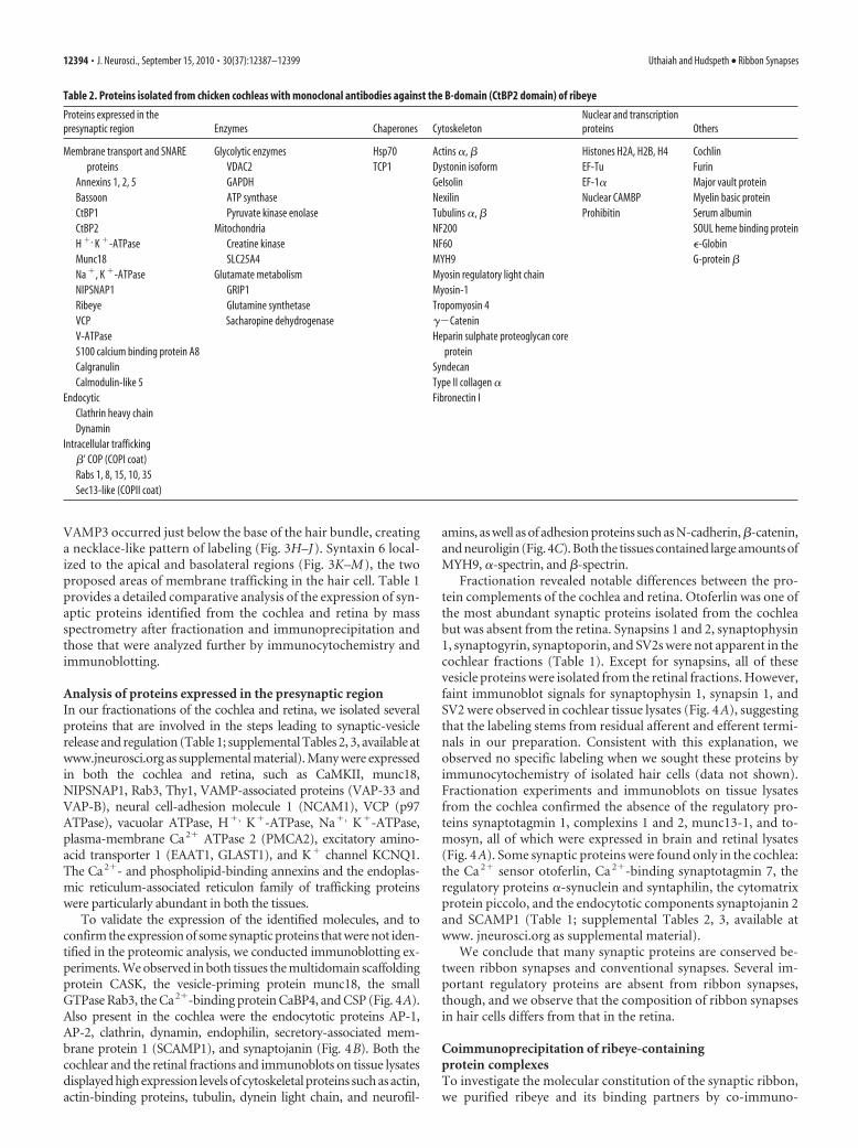
Table 2. Proteins isolated from chicken cochleas with monoclonal antibodies against the B-domain (CtBP2 domain) of ribeye
Proteins expressed in thepresynaptic region Enzymes Chaperones Cytoskeleton
Nuclear and transcriptionproteins Others
Membrane transport and SNAREproteins
Annexins 1, 2, 5BassoonCtBP1CtBP2H �, K �-ATPaseMunc18Na �, K �-ATPaseNIPSNAP1RibeyeVCPV-ATPaseS100 calcium binding protein A8CalgranulinCalmodulin-like 5
EndocyticClathrin heavy chainDynamin
Intracellular trafficking�’ COP (COPI coat)Rabs 1, 8, 15, 10, 35Sec13-like (COPII coat)
Glycolytic enzymesVDAC2GAPDHATP synthasePyruvate kinase enolase
MitochondriaCreatine kinaseSLC25A4
Glutamate metabolismGRIP1Glutamine synthetaseSacharopine dehydrogenase
Hsp70TCP1
Actins �, �Dystonin isoformGelsolinNexilinTubulins �, �NF200NF60MYH9Myosin regulatory light chainMyosin-1Tropomyosin 4��CateninHeparin sulphate proteoglycan core
proteinSyndecanType II collagen �Fibronectin I
Histones H2A, H2B, H4EF-TuEF-1�Nuclear CAMBPProhibitin
CochlinFurinMajor vault proteinMyelin basic proteinSerum albuminSOUL heme binding protein�-GlobinG-protein �
12394 • J. Neurosci., September 15, 2010 • 30(37):12387–12399 Uthaiah and Hudspeth • Ribbon Synapses

precipitation with either monoclonal antibodies directed againstthe B-domain (CtBP2 domain) or a polyclonal antiserum againstthe A-domain of ribeye (Fig. 5). Immmunoblots indicate specificimmunoprecipitation of both ribeye and CtBP2 in the B-domainimmunoprecipitation (supplemental Fig. 3A, available at www.jneurosci.org as supplemental material) but of ribeye alone in theA-domain immunoprecipitation (supplemental Fig. 3B, avail-able at www.jneurosci.org as supplemental material). Althoughwe cannot yet confirm that we have purified intact synaptic rib-bons, we have successfully purified ribeye-containing proteincomplexes and further examined the associated proteins. Immu-noprecipitation with B-domain antibodies isolated a large num-ber of proteins (Fig. 5A, Table 2). In contrast, the A-domainantiserum yielded only a small complement of proteins (Fig. 5B).From these experiments, we inferred that the A-domain anti-serum is a more specific reagent than the B-domain antibody.
Although we used 800 cochleas, we purified only a small quan-tity of ribeye and its interacting proteins with the A-domain an-tiserum. We therefore elected to use the bovine retina as a sourceof greater amounts of ribeye and its partners. From this organ wepurified a large number of synaptic proteins, including synapto-tagmin 1, synaptophysin 1, VAMP2, CSP, Rab3a, Rab3B, andRab3D, SNAP25, munc18-1, NSF, AP-1, clathrin heavy chain,CaMKII, and vacuolar ATPase (Fig. 5C, Table 3). Isolation of
vesicle proteins indicated that the ribbons remained intact andthat the synaptic vesicles were attached to the ribbon during ourpurification procedure. Several cytoskeletal proteins such as ac-tin, tubulin, spectrin, neurofilament-H (NF200), neurofila-ment-M (NF160), and �-internexin were also identified. We didnot, however, consistently isolate the previously describedribbon-associated proteins RIM1, piccolo, KIF3A, ERC2, andbassoon (Fig. 5B, Tables 2, 3). Even in our fractionation experi-ments, we isolated piccolo only from the cochlea and bassoononly from the retina (Table 1). We therefore tested for the pres-ence of these proteins in cochlear lysates by immunoblotting andcompared these results with those obtained from brain and reti-nal lysates (Fig. 5D). As expected, we observed expression of allthe ribbon-associated proteins in every examined organ with theexception of ribeye in the brain.
We next examined the localization of the isolated proteinswithin hair cells to determine whether they colocalized with syn-aptic ribbons. Many of the vesicle transport proteins were highlyexpressed in the basolateral portions of hair cells. We observedonly partial colocalization of the ribbons with the SNAREs,SNARE-dissociation ATPase NSF (supplemental Fig. 4, availableat www.jneurosci.org as supplemental material), the endocytoticproteins clathrin and endophilin (supplemental Fig. 5, availableat www.jneurosci.org as supplemental material), and cytoskeletal
Table 3. Proteins isolated from bovine retinas with a polyclonal antiserum against the A-domain of ribeye
Proteins expressed in the presynaticregion Enzymes Chaperones Cytoskeleton
Nuclear and transcriptionproteins Others
Membrane-transport and SNAREproteins
Annexin 6CaMKIICSPmunc18-1Na �, K �-ATPaseNCAM1NSFRab3a, 3B, 3DRibeyeSNAP25Synaptophysin 1Synaptotagmin 1VAMP2VAP-33V-ATPase, lysosomalV-ATPaseVCPSLCA1 (GLAST)Unc-51-like kinase 2
EndocyticAP-1AP-2Clathrin heavy chain
Intracellular traffickingRabs 1, 4, 15, 33, 35, 37Rap1Reticulons 1, 3A, 4Brain-abundant membrane-attached
protein1Lipid binding
Apolipoprotein OFatty acid synthasePEBP1
Glycolytic enzymesGAPDHGlucose phosphate isomeraseHexokinase 1Phosphoglycerate kinasePhosphoglycerate mutasePyruvate kinase 3Triose-phosphate isomerase� Enolase
Mitochondria and citric acid cycleAconitase 2ATP synthaseCreatine kinaseCytochrome c oxidaseF1-ATPaseLactate dehydrogenaseMalate dehydrogenaseMitofilinNADH dehydrogenaseOxoglutarate carrierPhosphofructokinaseSLC25A4, SLC25A5, SLC25A6,
SLC25A12SLC13Succinate dehydrogenase
transketolaseUbiquinone-cytochrome c reductaseVDACs 1, 2, 3
Glutamate metabolismGlutamine synthetaseGlutamine oxaloacetic transaminaseAspartate aminotransferase
Hsp1Hsp40Hsp27Hsp70Hsp90ER chaperones
CalnexinRibophorin
Actins �, �Dihydropyrimi-dinase-like 2Matrin 3Spectrin �Tubulins �, �NF68NF160GFAPInternexin-�Fibronectin IVimentin 1/2
hnRNP A2/B1hnRNP KsnRNP SmD1Histone H2A, H4NucleoporinCohesin1 homologProhibitin 2
Blue-sensitive opsinG proteins �, �PeripherinPhosducinRetinal GPCRRetinaldehyde-binding
proteinRetinol-binding proteinRhodopsinRod outer segment-binding
proteinS-arrestinTransducins �, �UbiquitinErythrocytic membrane
protein band 4.1-like 2Haemoglobin �Serum albumin precursorBasiginGlutathione transferasecGMP- specific
phosphodiesterase14-3-3 �
Uthaiah and Hudspeth • Ribbon Synapses J. Neurosci., September 15, 2010 • 30(37):12387–12399 • 12395
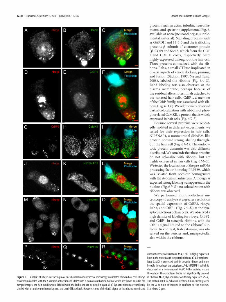
proteins such as actin, tubulin, neurofila-ments, and spectrin (supplemental Fig. 6,available at www.jneurosci.org as supple-mental material). Signaling proteins suchas GAPDH and 14-3-3 and the traffickingproteins � subunit of coatomer protein(�-COP) and Sec13, which form the COPI and COP II coats, respectively, werehighly expressed throughout the hair cell.Three proteins colocalized with the rib-bons. Rab3, a small GTPase implicated indiverse aspects of vesicle docking, priming,and fusion (Sudhof, 1997; Ng and Tang,2008), labeled the ribbons (Fig. 6A–C).Rab3 labeling was also observed at theplasma membrane, perhaps because ofthe residual afferent terminals attached tothe isolated hair cells. CtBP1, a memberof the CtBP family, was associated with rib-bons (Fig. 6D,E). We additionally observedpartial colocalization with ribbons of phos-phorylated CaMKII, a protein that is widelyexpressed in hair cells (Fig. 6G–I).
Because several proteins were repeat-edly isolated in different experiments, wetested for their expression in hair cells.NIPSNAP1, a nonneuronal SNAP25-likeprotein, showed strong labeling through-out the hair cell (Fig. 6 J–L). The endocy-totic protein dynamin was also diffuselydistributed. We conclude that these proteinsdo not colocalize with ribbons, but arehighly expressed in hair cells (Fig. 6M–O).We tested the localization of the pre-mRNAprocessing factor homolog PRPF39, whichwas isolated from cochlear homogenateswith the A-domain antiserum. Although asexpected strong labeling was apparent in thenucleus (Fig. 6P–R), no colocalization withribbons was observed.
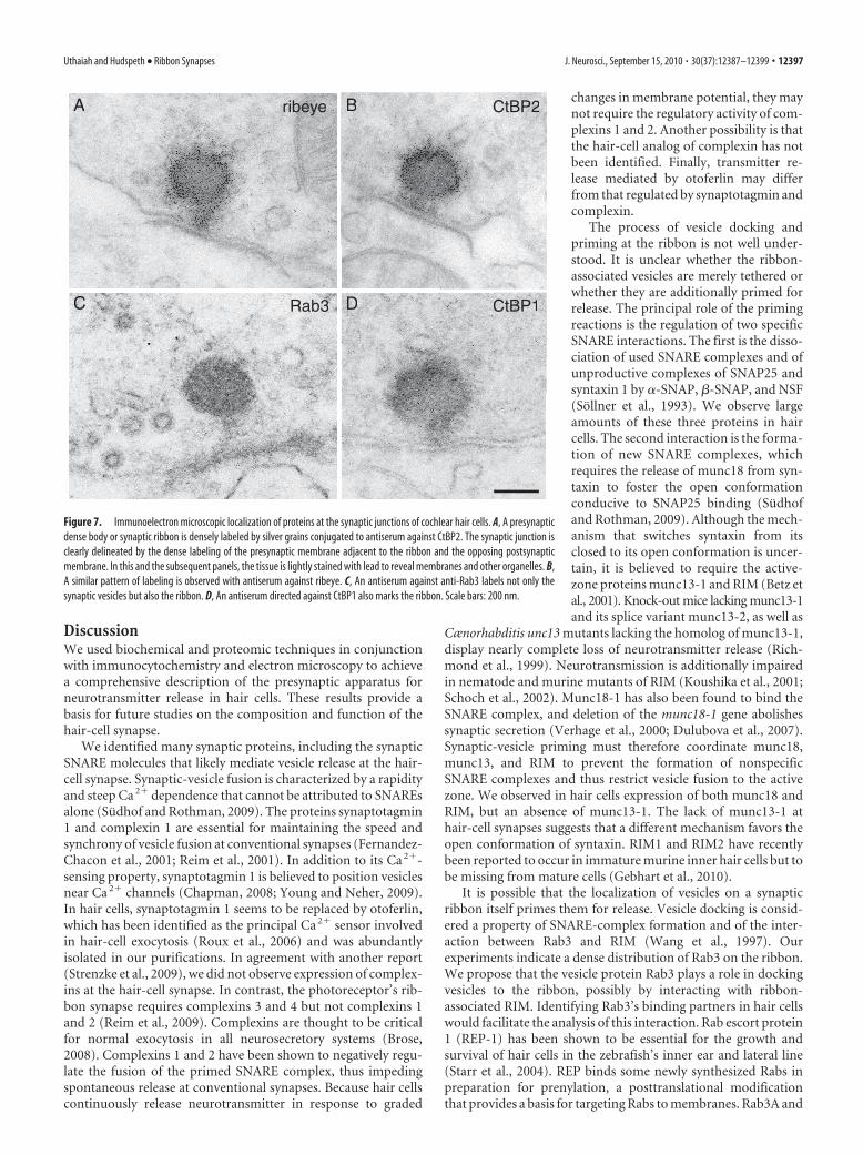
We performed immunoelectron mi-croscopy to analyze at a greater resolutionthe spatial expression of CtBP2, ribeye,Rab3, and CtBP1 (Fig. 7A–D) at the syn-aptic junctions of hair cells. We observed ahigh density of labeling for ribeye, CtBP2,and CtBP1 in synaptic ribbons, with theCtBP1 signal limited to the ribbons’ sur-faces. In contrast, Rab3 staining was ob-served on the vesicles and, unexpectedly,also within the ribbons.
Rab3BribeyeA MergePhalloidin
C
ribeyeJ NIPSNAP1K MergePhalloidin
L
ribeyeM dynaminN MergePhalloidin
O
ribeyeP PRPF39Q MergePhalloidin
R
CtBP1ED ribeye MergePhalloidin
F
G CtBP2 CaMKIIH MergePhalloidin
I
Figure 6. Analysis of ribeye-interacting molecules by immunofluorescence microscopy on isolated chicken hair cells. Ribeyewas immunolabeled with the A-domain antiserum and CtBP2 with B-domain antibodies, both of which are shown as red in themerged images; the hair bundles were labeled with phalloidin and are depicted in cyan. A–C, Synaptic ribbons are uniformlylabeled with an antiserum directed against the small GTPase Rab3. However, some of the Rab3 signal at the plasma membrane
4
does not overlay with ribbons. D–F, CtBP1 is highly expressedboth in the nucleus and in synaptic ribbons. G–I, Phosphory-lated CaMKII is expressed both in synaptic ribbons and morebroadly throughout the cytoplasm. J–L, NIPSNAP1, which isdescribed as a nonneuronal SNAP25-like protein, occursthroughout the cytoplasm but is not significantly presentin ribbons. M–O, Dynamin is also diffusely expressed. P–R,The protein PRPF39, which is identified in cochlear lysatesby the A-domain antiserum, is confined to the nucleus.Scale bars: 2 �m.
12396 • J. Neurosci., September 15, 2010 • 30(37):12387–12399 Uthaiah and Hudspeth • Ribbon Synapses
DiscussionWe used biochemical and proteomic techniques in conjunctionwith immunocytochemistry and electron microscopy to achievea comprehensive description of the presynaptic apparatus forneurotransmitter release in hair cells. These results provide abasis for future studies on the composition and function of thehair-cell synapse.
We identified many synaptic proteins, including the synapticSNARE molecules that likely mediate vesicle release at the hair-cell synapse. Synaptic-vesicle fusion is characterized by a rapidityand steep Ca 2� dependence that cannot be attributed to SNAREsalone (Sudhof and Rothman, 2009). The proteins synaptotagmin1 and complexin 1 are essential for maintaining the speed andsynchrony of vesicle fusion at conventional synapses (Fernandez-Chacon et al., 2001; Reim et al., 2001). In addition to its Ca 2�-sensing property, synaptotagmin 1 is believed to position vesiclesnear Ca 2� channels (Chapman, 2008; Young and Neher, 2009).In hair cells, synaptotagmin 1 seems to be replaced by otoferlin,which has been identified as the principal Ca 2� sensor involvedin hair-cell exocytosis (Roux et al., 2006) and was abundantlyisolated in our purifications. In agreement with another report(Strenzke et al., 2009), we did not observe expression of complex-ins at the hair-cell synapse. In contrast, the photoreceptor’s rib-bon synapse requires complexins 3 and 4 but not complexins 1and 2 (Reim et al., 2009). Complexins are thought to be criticalfor normal exocytosis in all neurosecretory systems (Brose,2008). Complexins 1 and 2 have been shown to negatively regu-late the fusion of the primed SNARE complex, thus impedingspontaneous release at conventional synapses. Because hair cellscontinuously release neurotransmitter in response to graded
changes in membrane potential, they maynot require the regulatory activity of com-plexins 1 and 2. Another possibility is thatthe hair-cell analog of complexin has notbeen identified. Finally, transmitter re-lease mediated by otoferlin may differfrom that regulated by synaptotagmin andcomplexin.
The process of vesicle docking andpriming at the ribbon is not well under-stood. It is unclear whether the ribbon-associated vesicles are merely tethered orwhether they are additionally primed forrelease. The principal role of the primingreactions is the regulation of two specificSNARE interactions. The first is the disso-ciation of used SNARE complexes and ofunproductive complexes of SNAP25 andsyntaxin 1 by �-SNAP, �-SNAP, and NSF(Sollner et al., 1993). We observe largeamounts of these three proteins in haircells. The second interaction is the forma-tion of new SNARE complexes, whichrequires the release of munc18 from syn-taxin to foster the open conformationconducive to SNAP25 binding (Sudhofand Rothman, 2009). Although the mech-anism that switches syntaxin from itsclosed to its open conformation is uncer-tain, it is believed to require the active-zone proteins munc13-1 and RIM (Betz etal., 2001). Knock-out mice lacking munc13-1and its splice variant munc13-2, as well as
Cænorhabditis unc13 mutants lacking the homolog of munc13-1,display nearly complete loss of neurotransmitter release (Rich-mond et al., 1999). Neurotransmission is additionally impairedin nematode and murine mutants of RIM (Koushika et al., 2001;Schoch et al., 2002). Munc18-1 has also been found to bind theSNARE complex, and deletion of the munc18-1 gene abolishessynaptic secretion (Verhage et al., 2000; Dulubova et al., 2007).Synaptic-vesicle priming must therefore coordinate munc18,munc13, and RIM to prevent the formation of nonspecificSNARE complexes and thus restrict vesicle fusion to the activezone. We observed in hair cells expression of both munc18 andRIM, but an absence of munc13-1. The lack of munc13-1 athair-cell synapses suggests that a different mechanism favors theopen conformation of syntaxin. RIM1 and RIM2 have recentlybeen reported to occur in immature murine inner hair cells but tobe missing from mature cells (Gebhart et al., 2010).
It is possible that the localization of vesicles on a synapticribbon itself primes them for release. Vesicle docking is consid-ered a property of SNARE-complex formation and of the inter-action between Rab3 and RIM (Wang et al., 1997). Ourexperiments indicate a dense distribution of Rab3 on the ribbon.We propose that the vesicle protein Rab3 plays a role in dockingvesicles to the ribbon, possibly by interacting with ribbon-associated RIM. Identifying Rab3’s binding partners in hair cellswould facilitate the analysis of this interaction. Rab escort protein1 (REP-1) has been shown to be essential for the growth andsurvival of hair cells in the zebrafish’s inner ear and lateral line(Starr et al., 2004). REP binds some newly synthesized Rabs inpreparation for prenylation, a posttranslational modificationthat provides a basis for targeting Rabs to membranes. Rab3A and
ribeye CtBP2B
Rab3C CtBP1
A
D
ribeye CtBP2B
Rab3C CtBP1
A
D
Figure 7. Immunoelectron microscopic localization of proteins at the synaptic junctions of cochlear hair cells. A, A presynapticdense body or synaptic ribbon is densely labeled by silver grains conjugated to antiserum against CtBP2. The synaptic junction isclearly delineated by the dense labeling of the presynaptic membrane adjacent to the ribbon and the opposing postsynapticmembrane. In this and the subsequent panels, the tissue is lightly stained with lead to reveal membranes and other organelles. B,A similar pattern of labeling is observed with antiserum against ribeye. C, An antiserum against anti-Rab3 labels not only thesynaptic vesicles but also the ribbon. D, An antiserum directed against CtBP1 also marks the ribbon. Scale bars: 200 nm.
Uthaiah and Hudspeth • Ribbon Synapses J. Neurosci., September 15, 2010 • 30(37):12387–12399 • 12397

Rab3D are able to bind REP and are modified by geranylgerany-lation (Pavlos et al., 2005; Baron and Seabra, 2008). Detailedanalysis of the Rab3-interacting proteins could elucidate theribbon-vesicle interaction. We have identified some Rab3 effec-tors, such as Rab3 GAP, rabphilin 3A, and the neuron-specificGDP-dissociation inhibitor � (Rab GDI�), all of which have beenshown to regulate synaptic transmission (Sudhof, 1997; Takai etal., 2001). A large-scale genetic screen to identify proteins neces-sary for bruchpilot localization to the presynaptic densities orT-bars of the Drosophila neuromuscular junction revealed thatRab3 is involved in the recruitment of presynaptic release pro-teins at individual active zones (Graf et al., 2009). This propertyand its localization within the ribbons suggest that Rab3 has syn-aptic functions apart from vesicle docking at the active zone.
Several proteins involved in the localization of vesicles to theactive zone are excluded from ribbon synapses. Synapsin has sofar been absent from all ribbon synapses. At conventional syn-apses, synapsins tether synaptic vesicles to each other and to anactin-based cytoskeletal meshwork, securing a reserve pool ofvesicles in the vicinity of the active zone (Greengard et al., 1993).Perturbation of synapsin function leads to disruption of the re-serve pool and an increase in synaptic depression, suggesting thatthe synapsin-dependent cluster of vesicles is required for sus-tained release of neurotransmitter during high levels of neuronalactivity. The absence of synapsins at ribbon synapses suggests thatthe mechanisms for vesicle clustering and mobilization in theseterminals differ from those of conventional synapses. Synapto-physin was also absent from our hair-cell preparations. Becausethe synaptophysin–synaptobrevin complex is critical for syna-ptic-vesicle maturation at conventional synapses (Becher et al.,1999), the ribbon may compensate for the absence of these pro-teins by tethering and immobilizing a large pool of vesicles nearthe active zone.
In fractionation experiments, we isolated multiple nonneuro-nal SNAREs that are thought to occur either in the Golgi appara-tus or in early or late endosomes. For example, we observedexpression of SNARE molecules involved in endosomal fusion,suggesting that synaptic vesicles communicate with endosomalintermediates in their life cycle. Our localization studies showedlabeling of several exocytotic and endocytotic proteins not only inthe basolateral but also in the apical regions of hair cells. Novelmembrane structures have been found previously in the apicaland basal compartments of inner hair cells (Spicer et al., 1999),suggesting specialized membrane trafficking in these cells. Usinga method for the purification of synaptic proteins that has beenused previously for the purification of rat-brain vesicles (Taka-mori et al., 2006), we isolated a large number of synaptic proteinsfrom the hair-cell synapse. However, we also extracted proteinsoriginating from intracellular organelles, a result that could sig-nal the isolation of all hair-cell vesicles by our procedure. Thismay explain the strong expression of �-COP (COP I coat pro-tein) and Sec13 (COP II coat protein) in our fractionationexperiments.
Some of the proteins we identified have also been isolatedfrom hair bundles (Shin et al., 2007) and may therefore representcontaminants expressed at high levels throughout the hair cell.These proteins include cytoskeletal proteins such as actins, tubu-lins, and their chaperone t-complex polypeptide-1 (TCP1);brain-specific creatine kinase; peripheral membrane proteinssuch as annexin-A5 and phosphatidyl-ethanolamine bindingprotein (PEBP); and proteins involved in Ca 2� sequestration andremoval such as parvalbumin 3, calmodulin, calretinin, andPMCA2. Also commonly identified were histones H2A and H4,
molecular chaperones such as Hsp70 and GRP74, mitochondrialproteins such as ATP synthase � subunit and voltage-dependentanion-selective channel protein 2 (VDAC2), and enzymes in-volved in energy metabolism.
In summary, we confirmed using various methodologies thatmany proteins described at other synapses also occur at the rib-bon synapses of hair cells. We established the presence of theneuronal SNAREs necessary for vesicle release as well as of otherSNAREs involved in intracellular trafficking. We noted not onlythe presence of hair cell-specific proteins but also the exclusion ofvarious vesicle-regulatory components. We identified many traf-ficking proteins localized to the apex or the base of the hair cell.Although we cannot demonstrate definitively the presence of allthe proteins identified, our results provide a basis for future mo-lecular analysis of ribbon synapses. Finally, we confirmed that thehair cell’s synaptic ribbon is composed primarily of the CtBPfamily of proteins and is associated with the GTPase Rab3, aprotein that may play a role in docking vesicles to the ribbon.
ReferencesAlpadi K, Magupalli VG, Kappel S, Koblitz L, Schwarz K, Seigel GM, Sung
CH, Schmitz F (2008) RIBEYE recruits Munc119, a mammalian or-tholog of the Caenorhabditis elegans protein unc119, to synaptic ribbonsof photoreceptor synapses. J Biol Chem 283:26461–26467.
Baron RA, Seabra MC (2008) Rab geranylgeranylation occurs preferentiallyvia the pre-formed REP-RGGT complex and is regulated by geranylgera-nyl pyrophosphate. Biochem J 415:67–75.
Becher A, Drenckhahn A, Pahner I, Margittai M, Jahn R, Ahnert-Hilger G(1999) The synaptophysin-synaptobrevin complex: a hallmark of synap-tic vesicle maturation. J Neurosci 19:1922–1931.
Betz A, Thakur P, Junge HJ, Ashery U, Rhee JS, Scheuss V, Rosenmund C,Rettig J, Brose N (2001) Functional interaction of the active zone pro-teins Munc13-1 and RIM1 in synaptic vesicle priming. Neuron30:183–196.
Brose N (2008) For better or for worse: complexins regulate SNARE func-tion and vesicle fusion. Traffic 9:1403–1413.
Chapman ER (2008) How does synaptotagmin trigger neurotransmitter re-lease? Annu Rev Biochem 77:615– 641.
Corwin JT, Warchol ME (1991) Auditory hair cells: structure, function, de-velopment, and regeneration. Annu Rev Neurosci 14:301–333.
Dulubova I, Khvotchev M, Liu S, Huryeva I, Sudhof TC, Rizo J (2007)Munc18-1 binds directly to the neuronal SNARE complex. Proc NatlAcad Sci U S A 104:2697–2702.
Fernandez-Chacon R, Konigstorfer A, Gerber SH, Garcia J, Matos MF,Stevens CF, Brose N, Rizo J, Rosenmund C, Sudhof TC (2001) Synap-totagmin I functions as a calcium regulator of release probability. Nature410:41– 49.
Galvin JE, Uryu K, Lee VM, Trojanowski JQ (1999) Axon pathology in Par-kinson’s disease and Lewy body dementia hippocampus contains alpha-,beta-, and gamma-synuclein. Proc Natl Acad Sci U S A 96:13450 –13455.
Garner CC, Kindler S, Gundelfinger ED (2000) Molecular determinants ofpresynaptic active zones. Curr Opin Neurobiol 10:321–327.
Gebhart M, Juhasz-Vedres G, Zuccotti A, Brandt N, Engel J, TrockenbacherA, Kaur G, Obermair GJ, Knipper M, Koschak A, Striessnig J (2010)Modulation of Cav1.3 Ca 2� channel gating by Rab3 interacting molecule.Mol Cell Neurosci 44:246 –259.
Glowatzki E, Fuchs PA (2002) Transmitter release at the hair cell ribbonsynapse. Nat Neurosci 5:147–154.
Graf ER, Daniels RW, Burgess RW, Schwarz TL, DiAntonio A (2009) Rab3dynamically controls protein composition at active zones. Neuron64:663– 677.
Greengard P, Valtorta F, Czernik AJ, Benfenati F (1993) Synaptic vesiclephosphoproteins and regulation of synaptic function. Science 259:780 –785.
He DZ, Dallos P (2000) Properties of voltage-dependent somatic stiffness ofcochlear outer hair cells. J Assoc Res Otolaryngol 1:64 – 81.
Huttner WB, Schiebler W, Greengard P, De Camilli P (1983) Synapsin I(protein I), a nerve terminal-specific phosphoprotein. III. Its associationwith synaptic vesicles studied in a highly purified synaptic vesicle prepa-ration. J Cell Biol 96:1374 –1388.
12398 • J. Neurosci., September 15, 2010 • 30(37):12387–12399 Uthaiah and Hudspeth • Ribbon Synapses
Katsumata O, Ohara N, Tamaki H, Niimura T, Naganuma H, Watanabe M,Sakagami H (2009) IQ-ArfGEF/BRAG1 is associated with synaptic rib-bons in the mouse retina. Eur J Neurosci 30:1509 –1516.
Keen EC, Hudspeth AJ (2006) Transfer characteristics of the hair cell’s af-ferent synapse. Proc Natl Acad Sci U S A 103:5537–5542.
Khimich D, Nouvian R, Pujol R, tom Dieck S, Egner A, Gundelfinger ED,Moser T (2005) Hair cell synaptic ribbons are essential for synchronousauditory signalling. Nature 434:889 – 894.
Koushika SP, Richmond JE, Hadwiger G, Weimer RM, Jorgensen EM, NonetML (2001) A post-docking role for active zone protein Rim. Nat Neu-rosci 4:997–1005.
Kumarathasan P, Mohottalage S, Goegan P, Vincent R (2005) An optimizedprotein in-gel digest method for reliable proteome characterization byMALDI-TOF-MS analysis. Anal Biochem 346:85– 89.
Lenzi D, von Gersdorff H (2001) Structure suggests function: the case forsynaptic ribbons as exocytotic nanomachines. Bioessays 23:831– 840.
Li GL, Keen E, Andor-Ardo D, Hudspeth AJ, von Gersdorff H (2009) Theunitary event underlying multiquantal EPSCs at a hair cell’s ribbon syn-apse. J Neurosci 29:7558 –7568.
LoGiudice L, Matthews G (2009) The role of ribbons at sensory synapses.Neuroscientist 15:380 –391.
Magupalli VG, Schwarz K, Alpadi K, Natarajan S, Seigel GM, Schmitz F(2008) Multiple RIBEYE–RIBEYE interactions create a dynamic scaffoldfor the formation of synaptic ribbons. J Neurosci 28:7954 –7967.
Moser T, Beutner D (2000) Kinetics of exocytosis and endocytosis at thecochlear inner hair cell afferent synapse of the mouse. Proc Natl Acad SciU S A 97:883– 888.
Ng EL, Tang BL (2008) Rab GTPases and their roles in brain neurons andglia. Brain Res Rev 58:236 –246.
Nouvian R, Beutner D, Parsons TD, Moser T (2006) Structure and functionof the hair cell ribbon synapse. J Membr Biol 209:153–165.
Obholzer N, Wolfson S, Trapani JG, Mo W, Nechiporuk A, Busch-NentwichE, Seiler C, Sidi S, Sollner C, Duncan RN, Boehland A, Nicolson T (2008)Vesicular glutamate transporter 3 is required for synaptic transmission inzebrafish hair cells. J Neurosci 28:2110 –2118.
Parsons TD, Lenzi D, Almers W, Roberts WM (1994) Calcium-triggeredexocytosis and endocytosis in an isolated presynaptic cell: capacitancemeasurements in saccular hair cells. Neuron 13:875– 883.
Pavlos NJ, Xu J, Riedel D, Yeoh JS, Teitelbaum SL, Papadimitriou JM, Jahn R,Ross FP, Zheng MH (2005) Rab3D regulates a novel vesicular traffickingpathway that is required for osteoclastic bone resorption. Mol Cell Biol25:5253–5269.
Reim K, Mansour M, Varoqueaux F, McMahon HT, Sudhof TC, Brose N,Rosenmund C (2001) Complexins regulate a late step in Ca 2�-dependent neurotransmitter release. Cell 104:71– 81.
Reim K, Regus-Leidig H, Ammermuller J, El-Kordi A, Radyushkin K,Ehrenreich H, Brandstatter JH, Brose N (2009) Aberrant function andstructure of retinal ribbon synapses in the absence of complexin 3 andcomplexin 4. J Cell Sci 122:1352–1361.
Richmond JE, Davis WS, Jorgensen EM (1999) UNC-13 is required for syn-aptic vesicle fusion in C. elegans. Nat Neurosci 2:959 –964.
Roberts WM, Jacobs RA, Hudspeth AJ (1991) The hair cell as a presynapticterminal. Ann N Y Acad Sci 635:221–233.
Roux I, Safieddine S, Nouvian R, Grati M, Simmler MC, Bahloul A, PerfettiniI, Le Gall M, Rostaing P, Hamard G, Triller A, Avan P, Moser T, Petit C(2006) Otoferlin, defective in a human deafness form, is essential forexocytosis at the auditory ribbon synapse. Cell 127:277–289.
Rutherford MA, Roberts WM (2006) Frequency selectivity of synaptic exo-cytosis in frog saccular hair cells. Proc Natl Acad Sci U S A 103:2898 –2903.
Rutherford MA, Roberts WM (2009) Spikes and membrane potential oscil-lations in hair cells generate periodic afferent activity in the frog sacculus.J Neurosci 29:10025–10037.
Safieddine S, Wenthold RJ (1999) SNARE complex at the ribbon synapses ofcochlear hair cells: analysis of synaptic vesicle- and synaptic membrane-associated proteins. Eur J Neurosci 11:803– 812.
Schmitz F (2009) The making of synaptic ribbons: how they are built andwhat they do. Neuroscientist 15:611– 624.
Schoch S, Castillo PE, Jo T, Mukherjee K, Geppert M, Wang Y, Schmitz F,Malenka RC, Sudhof TC (2002) RIM1alpha forms a protein scaffoldfor regulating neurotransmitter release at the active zone. Nature415:321–326.
Seal RP, Akil O, Yi E, Weber CM, Grant L, Yoo J, Clause A, Kandler K, NoebelsJL, Glowatzki E, Lustig LR, Edwards RH (2008) Sensorineural deafnessand seizures in mice lacking vesicular glutamate transporter 3. Neuron57:263–275.
Shin JB, Streijger F, Beynon A, Peters T, Gadzala L, McMillen D, Bystrom C,Van der Zee CE, Wallimann T, Gillespie PG (2007) Hair bundles arespecialized for ATP delivery via creatine kinase. Neuron 53:371–386.
Sollner T, Bennett MK, Whiteheart SW, Scheller RH, Rothman JE (1993) Aprotein assembly-disassembly pathway in vitro that may correspond tosequential steps of synaptic vesicle docking, activation, and fusion. Cell75:409 – 418.
Spicer SS, Thomopoulos GN, Schulte BA (1999) Novel membranous struc-tures in apical and basal compartments of inner hair cells. J Comp Neurol409:424 – 437.
Starr CJ, Kappler JA, Chan DK, Kollmar R, Hudspeth AJ (2004) Mutation ofthe zebrafish choroideremia gene encoding Rab escort protein 1 devas-tates hair cells. Proc Natl Acad Sci U S A 101:2572–2577.
Sterling P, Matthews G (2005) Structure and function of ribbon synapses.Trends Neurosci 28:20 –29.
Strenzke N, Chanda S, Kopp-Scheinpflug C, Khimich D, Reim K, BulankinaAV, Neef A, Wolf F, Brose N, Xu-Friedman MA, Moser T (2009)Complexin-I is required for high-fidelity transmission at the endbulb ofheld auditory synapse. J Neurosci 29:7991– 8004.
Sudhof TC (1997) Function of Rab3 GDP-GTP exchange. Neuron18:519 –522.
Sudhof TC, Rothman JE (2009) Membrane fusion: grappling with SNAREand SM proteins. Science 323:474 – 477.
Takai Y, Sasaki T, Matozaki T (2001) Small GTP-binding proteins. PhysiolRev 81:153–208.
Takamori S, Holt M, Stenius K, Lemke EA, Grønborg M, Riedel D, Urlaub H,Schenck S, Brugger B, Ringler P, Muller SA, Rammner B, Grater F, HubJS, De Groot BL, Mieskes G, Moriyama Y, Klingauf J, Grubmuller H,Heuser J, Weiland F, Jahn R (2006) Molecular anatomy of a traffickingorganelle. Cell 127:831– 846.
tom Dieck S, Altrock WD, Kessels MM, Qualmann B, Regus H, Brauner D,Fejtova A, Bracko O, Gundelfinger ED, Brandstatter JH (2005) Molec-ular dissection of the photoreceptor ribbon synapse: physical interactionof Bassoon and RIBEYE is essential for the assembly of the ribbon com-plex. J Cell Biol 168:825– 836.
Verhage M, Maia AS, Plomp JJ, Brussaard AB, Heeroma JH, Vermeer H,Toonen RF, Hammer RE, van den Berg TK, Missler M, Geuze HJ, SudhofTC (2000) Synaptic assembly of the brain in the absence of neurotrans-mitter secretion. Science 287:864 – 869.
Von Kriegstein K, Schmitz F, Link E, Sudhof TC (1999) Distribution ofsynaptic vesicle proteins in the mammalian retina identifies obligatoryand facultative components of ribbon synapses. Eur J Neurosci 11:1335–1348.
Wang Y, Okamoto M, Schmitz F, Hofmann K, Sudhof TC (1997) Rim is aputative Rab3 effector in regulating synaptic-vesicle fusion. Nature388:593–598.
Young SM Jr, and Neher E (2009) Synaptotagmin has an essential functionin synaptic vesicle positioning for synchronous release in addition to itsrole as a calcium sensor. Neuron 63:482– 496.
Uthaiah and Hudspeth • Ribbon Synapses J. Neurosci., September 15, 2010 • 30(37):12387–12399 • 12399