pitfalls and remedies in pet/ct imaging for rt...
TRANSCRIPT

Pitfalls and Remedies in PET/CT
imaging for RT planning
Tinsu Pan, Ph.D.
M.D. Anderson Cancer Center
The University of Texas

Outlines
• Background
• Average CT (< 1 mSv) to reduce mis-alignment of PET and CT
– Automatic processing without operator interaction
• Two different implementations
– Average CT w/o gating (this talk)
– Average CT w/ gating (GE/Siemens/Philips)
• Summary

PET/CT applications at M.D.Anderson
• Majority for diagnosis and staging of cancer
• One PET/CT scan per patient prior to treatment
Diagnostic Imaging Radiation Oncology

Free-breathing or breath-hold in CT
of PET/CT ?

X-ray tube PET det.
PET det.CT det.
Helical CT data acquisition
X-ray tube PET det.
PET det.CT det.
X-ray tube PET det.
PET det.CT det.
X-ray tube PET det.
PET det.CT det.
X-ray tube PET det.
PET det.CT det.
X-ray tube PET det.
PET det.CT det.
X-ray tube PET det.
PET det.CT det.
X-ray tube PET det.
PET det.CT det.
X-ray tube PET det.
PET det.CT det.
X-ray tube PET det.
PET det.CT det.
X-ray tube PET det.
PET det.CT det.

Breathing Artifacts
Protocol: 16x0.625 mm, 0.8 s gantry rotation, pitch 1.375:1
Speed: 13.75 mm/0.8 s or 17.2 mm/s

Breathing artifacts to physiological info
Breath cycle= 80.3/(13.75/0.8)= 4.67 s
Heart rate= (21/(13.75/0.8)) -1*60= 49 bpm

Registration of Free-Breathing CT
and PET maybe like

Scout Helical CT
End-inspiration (FB)
End-expiration (FB)
Mid-expiration
(BH)
X-ray on
Misalignment in breathing states
0 20 40 60 80 100 120 (sec)
Pan et al, JNM, 2005

Mis-matched PET-CT data sets
Mismatch 1:
CT diaphragm position
lower than PET
Mismatch 2:
CT diaphragm position
higher than PET

Deep inspiration breath-
hold PET/CT

Deep inspiration breath-hold (3 min)
Nehmeh et al, JNM, 07
FB PET/CT
DIBH PET/CT
5 sec DBH CT
9 x 20 sec DIBH PET

SUVmax= 3.6
SUVmax= 8.1
Torizuka et al, JNM, 09
FB PET/CT
DIBH PET/CT
Deep inspiration breath-hold (20 s)

4D PET/CT(better with list-mode, and challenging to
perform in the clinic)

Respiratory gating devices
Varian (Optical) Philips (air bellows)Anzai (Pressure)
Infrared reflector
Infrared camera
Flat tabletop
Infrared reflector
Radiation Oncology Diagnostic Imaging

4D PET patient study
SUV=5.0 SUV=8.5

Near end-expiration
PET/CT

Near end-expiration PET
Daouk, et al, ’08

Average CT (ACT) < 1 mSv
Data acquired at high temporal resolution and
averaged over one breath cycle
Attenuation correction, RT dose calculation, IGRT

Computer
Sorting
Respiratory
Trace
RPM
Monitoring system
CT Scanner Cine CT images
4D-CT
Pan, et al, Med Phys ‘04
4D-CT

RPM
Computer
Sorting
Respiratory
TraceTracking system
CT Scanner Cine CT images
4D-CT
MIP(mip) AveragePan, et al, JNM ‘05
Average CT (ACT) < 1 mSv

Soft tissue (400,40) 40% SUVmax
Lung (1000,-700) 20% SUVmax
Tumor contouring with MIP CT

X-ray tube PET det.
PET det.CT det.
Cine CT Data Acquisition
X-ray tube PET det.
PET det.CT det.
X-ray tube PET det.
PET det.CT det.
X-ray tube PET det.
PET det.CT det.
X-ray tube PET det.
PET det.CT det.
X-ray tube PET det.
PET det.CT det.
X-ray tube PET det.
PET det.CT det.
X-ray tube PET det.
PET det.CT det.
X-ray tube PET det.
PET det.CT det.
X-ray tube PET det.
PET det.CT det.
X-ray tube PET det.
PET det.CT det.
Average CT 5 mGy (0.87 mSv per 14 cm)

Average CT is better than slow CT(2 adjacent CT slices of 2.5 mm apart)
4-s slow CT 4-s slow CT
Average CTAverage CT
Pan, et al, Med Phys ‘06

Helical CT
ACT
Helical CT w/o thoraxHelical CT w ACT
Creating combined CT
Pan, et al, Med Phys ‘06

Average CT for RT dose calculation

Clinical examples of
average CT

Clinical Studies
NSCLC
Esophageal
+57%
+56%

Lung tumor 1
SUV=10.8 SUV=13.7 (+27%)

SUV=3.9
Lung tumor 2
SUV=3.7 (- 5%)

SUV=3.4
Lung tumor or lymph node
SUV=4.6
SUV=4.3(+26%)
SUV=7.5(+62%)

Lung tumor

Liver lesion

Kidney or liver uptake

Lung or liver lesion?

Average CT
Lung lesion or liver lesion?

Colorectal cancer

Average CT
Colorectal cancer

Inside the liver?
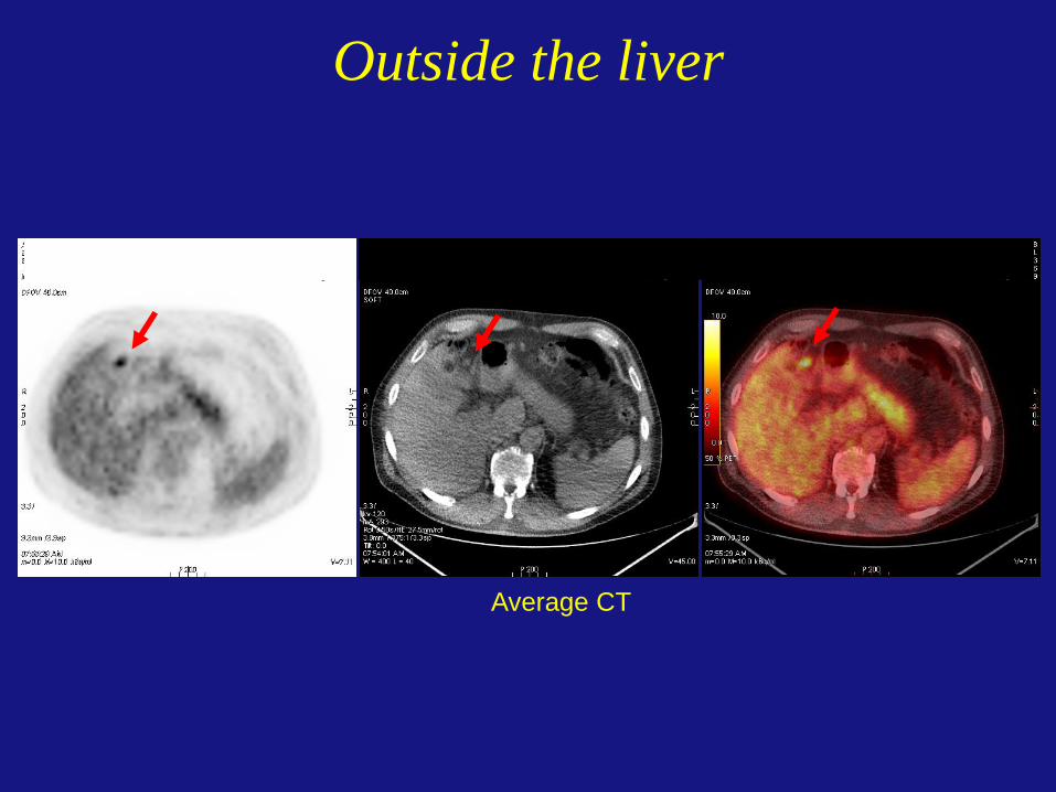
Outside the liver
Average CT

SUV=2.6
Tumor and cardiac imaging
HCT PETHCT
SUV=5.0
ACT PETACT

HCT- misregistration
Average CT in cardiac PET
Pan et al, Med. Phys. 2006
ACT

PET/CT scan indicated a positive response to induction chemo with HCT.
The patient had a negative response to the chemo with ACT.
Improve the restaging after chemo
Pan, et al, Med Phys ‘08

Impact on treatment planning
Previous GTV was outlined based on CT and clinical PET without motion correction.
New GTV was redefined based on the correct information from PET with ACT.
Old GTV
New GTV
Pan, et al, Med Phys ‘08

Oncology PET/CT dose estimate
1 mCi = 3.7 x 10⁷ dps = 3.7 x 10⁷ bequerel (Bq)
Injection dose: 10 mCi per patient
Radiation dose: 7 mSv from PET
5 to 10 mSv from CT
< 0.5 to 1 mSv from ACT
Radiation dose: 3.6 mSv from the environment

Why are we not using
average CT?
Complexity, Workflow and Cost

RPM
Computer
Sorting
Respiratory
TraceTracking system
CT Scanner Cine CT images
4D-CT
MIP(mip) AveragePan, et al, JNM ‘05
Average CT (ACT) < 1 mSv

4D-CT Workflow
Computer
CT Console
CT or PET/CT
Console
AW and Advantage 4D-CTRPM
Respiratory
Signal
Cine CT images
Treatment
Planning
System
4D-CT
Average CT (ACT)
PET & CT
Controls

Cine CT Workflow without Gating
CT Console
CT or PET/CT
Console
Cine CT images
Treatment
Planning
System
Average CT (ACT)
PET & CT
MIP
Transparent to the user


Automatically generated

Summary
• Respiratory gating PET can improve quantification, but is challenging to be performed in the clinic
• Average CT can improve registration of CT and PET, is used for dose calculation and IGRT in radiation therapy
• MIP CT can assist tumor contouring
• Workflow and efficiency can be improved, and cost can be reduced