pkc theta/beta and cyld are antagonistic partners in the...
TRANSCRIPT

LUND UNIVERSITY
PO Box 117221 00 Lund+46 46-222 00 00
PKC theta/beta and CYLD Are Antagonistic Partners in the NF kappa B and NFATTransactivation Pathways in Primary Mouse CD3(+) T Lymphocytes
Thuille, Nikolaus; Wachowicz, Katarzyna; Hermann-Kleiter, Natascha; Kaminski, Sandra;Fresser, Friedrich; Lutz-Nicoladoni, Christina; Leitges, Michael; Thome, Margot; Massoumi,Ramin; Baier, GottfriedPublished in:PLoS ONE
DOI:10.1371/journal.pone.0053709
Published: 2013-01-01
Link to publication
Citation for published version (APA):Thuille, N., Wachowicz, K., Hermann-Kleiter, N., Kaminski, S., Fresser, F., Lutz-Nicoladoni, C., ... Baier, G.(2013). PKC theta/beta and CYLD Are Antagonistic Partners in the NF kappa B and NFAT TransactivationPathways in Primary Mouse CD3(+) T Lymphocytes. PLoS ONE, 8(1). DOI: 10.1371/journal.pone.0053709
General rightsCopyright and moral rights for the publications made accessible in the public portal are retained by the authorsand/or other copyright owners and it is a condition of accessing publications that users recognise and abide by thelegal requirements associated with these rights.
• Users may download and print one copy of any publication from the public portal for the purpose of privatestudy or research. • You may not further distribute the material or use it for any profit-making activity or commercial gain • You may freely distribute the URL identifying the publication in the public portal

PKCh/b and CYLD Are Antagonistic Partners in the NFkBand NFAT Transactivation Pathways in Primary MouseCD3+ T LymphocytesNikolaus Thuille1, Katarzyna Wachowicz1, Natascha Hermann-Kleiter1, Sandra Kaminski1,
Friedrich Fresser1, Christina Lutz-Nicoladoni1, Michael Leitges2, Margot Thome3, Ramin Massoumi4,
Gottfried Baier1*
1 Department of Pharmacology and Genetics, Medical University of Innsbruck, Innsbruck, Austria, 2 The Biotechnology Centre of Oslo, Oslo, Norway, 3 Department of
Biochemistry, University of Lausanne, Lausanne, Switzerland, 4 Department of Laboratory Medicine, Lund University, Malmo, Sweden
Abstract
In T cells PKCh mediates the activation of critical signals downstream of TCR/CD28 stimulation. We investigated themolecular mechanisms by which PKCh regulates NFkB transactivation by examining PKCh/b single and double knockoutmice and observed a redundant involvement of PKCh and PKCb in this signaling pathway. Mechanistically, we define aPKCh-CYLD protein complex and an interaction between the positive PKCh/b and the negative CYLD signaling pathwaysthat both converge at the level of TAK1/IKK/I-kBa/NFkB and NFAT transactivation. In Jurkat leukemic T cells, CYLD isendoproteolytically processed in the initial minutes of stimulation by the paracaspase MALT1 in a PKC-dependent fashion,which is required for robust IL-2 transcription. However, in primary T cells, CYLD processing occurs with different kineticsand an altered dependence on PKC. The formation of a direct PKCh/CYLD complex appears to regulate the short-termspatial distribution of CYLD, subsequently affecting NFkB and NFAT repressional activity of CYLD prior to its MALT1-dependent inactivation. Taken together, our study establishes CYLD as a new and critical PKCh interactor in T cells andreveals that antagonistic PKCh/b-CYLD crosstalk is crucial for the adjustment of immune thresholds in primary mouse CD3+
T cells.
Citation: Thuille N, Wachowicz K, Hermann-Kleiter N, Kaminski S, Fresser F, et al. (2013) PKCh/b and CYLD Are Antagonistic Partners in the NFkB and NFATTransactivation Pathways in Primary Mouse CD3+ T Lymphocytes. PLoS ONE 8(1): e53709. doi:10.1371/journal.pone.0053709
Editor: Colin Combs, University of North Dakota, United States of America
Received September 14, 2012; Accepted December 3, 2012; Published January 15, 2013
Copyright: � 2013 Thuille et al. This is an open-access article distributed under the terms of the Creative Commons Attribution License, which permitsunrestricted use, distribution, and reproduction in any medium, provided the original author and source are credited.
Funding: This work was supported by grants from the FWF Austrian Science Fund (SFB-021, MCBO-DK, P23537, and P25044), funds from the Austrian BM:WF andthe European Community Program SYBILLA under grant agreement HEALTH-F4-2008-201106. The funders had no role in study design, data collection andanalysis, decision to publish, or preparation of the manuscript.
Competing Interests: The authors have declared that no competing interests exist.
* E-mail: [email protected]
Introduction
The central role of PKCh in signal transduction pathways
during an adaptive immune response has extensively focused on
the exact biochemical mechanisms of PKCh function (reviewed in
[1–3]). A recent study by Kong et al. identified the structural
requirement in PKCh for its localization to the immunological
synapse as a prerequisite for activation of downstream signaling
[4]. Several transcription factors essential for the T cell activation
response (i.e. NFkB, AP1, and NFAT) are regulated by PKCh[5,6]. In vivo analysis of PKCh 2/2 mice revealed the importance
of PKCh for Th2- [7] and Th17-mediated immune responses [8,9]
but not for host-protective antiviral responses [10]. Nevertheless,
despite a profound understanding of the cellular role of PKCh,
little is known about its molecular function, specifically the effector
proteins downstream of PKCh during T cell activation.
Ubiquitylation and deubiquitylation are established posttrans-
lational mechanisms for regulating immune responses, as well as
the development and activation of immune cells. The tumor
suppressor gene CYLD encodes an evolutionary conserved and
ubiquitously expressed protein of approximately 120 kDa and was
originally discovered as gene mutated in familial cylindromatosis,
an autosomal dominant inherited disease characterized by the
development of multiple benign skin tumors, principally on the
head and neck [11]. Functionally it is a deubiquitylating enzyme
(DUB) which removes mainly K63-linked polyubiquitin chains
from several specific substrates, influencing in a negative way the
activation status and/or spatial distribution of these target proteins
in different signaling pathways. Numerous studies both in vitro
and in vivo provided us with new insights in its established
function as an important negative regulator of inflammatory
responses, by counteracting the aberrant activation of NFkB
signaling: Cyld2/2 animals spontaneously develop intestinal
inflammation and autoimmune symptoms due to the constitutive
activation of the TAK1/IKK/IkBa axis [12,13]; the study of Lim
et al. described a CYLD dependent negative NFkB regulation
during bacteria induced lung inflammation in mice via deubiqui-
tylation of TRAF6 and TRAF7 [14]; moreover, the same scientific
group showed that Cyld knockout mice are protected from
Streptococcus pneumonia infection and lethality via a negative
crosstalk with p38 MAPK [15]; a synergistic crosstalk between the
E3 ligase Itch and CYLD for TAK1 inactivation and termination
of tumor necrose factor dependent inflammatory signaling was
recently described [16].
PLOS ONE | www.plosone.org 1 January 2013 | Volume 8 | Issue 1 | e53709
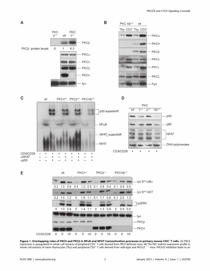
Figure 1. Overlapping roles of PKCh and PKCb in NFkB and NFAT transactivation processes in primary mouse CD3+ T cells. (A) PKCbexpression is upregulated in whole cell extracts of peripheral CD3+ T cells derived from PKCh-deficient mice. (B) The PKC isoform expression profile inwhole cell extracts of naive thymocytes (Thy) and peripheral CD3+ T cells derived from wild-type and PKCh/b2/2 mice. PKCh/b inhibition leads to an
PKCh/ß and CYLD Signaling Crosstalk
PLOS ONE | www.plosone.org 2 January 2013 | Volume 8 | Issue 1 | e53709

CYLD plays also an essential role in regulating T cell
development and activation. Cyld-deficient mice show a delayed
thymocyte development due to a constitutively K48-ubiquitylated
and degraded LCK protein [17]. In addition, Cyld-deficient T cells
are hyperresponsive to TCR/CD28 stimulation and CYLD has
been firmly established as negative regulator of NFkB and JNK
activation in response to antigen receptor activation in T cells
[12,13,18].
In the current study, we defined physiologically redundant roles
for the PKCh and PKCb isotypes in TCR/CD28-dependent
NFkB and NFAT transactivation by examining PKCh/b single
and double knockout mouse lines. Additionally, we provide
experimental evidence that a constitutive interaction of PKChwith CYLD apparently leads to CYLD sequestration that affects
the transactivation of the critical transcription factors NFkB and
NFAT. Therefore, the results described here elucidate some
aspects of PKCh and PKCb function during TCR activation and
the processes that modulate CYLD function upstream of NFkB
and NFAT activation in primary CD3+ T lymphocytes.
Materials and Methods
MicePKCh/b knockout mice are viable, fertile and were generated by
crossing PKCh [5] and PKCb [19] single knockout mice. The
generation of Cyld-deficient mice was described previously [20]. All
mice were on a C57Bl/6 background and housed (under SPF
conditions) at the mouse facility of the Medical University of
Innsbruck. All animal experiments were performed in accordance
with the Austria ‘‘Tierversuchsgesetz’’ (BGBI. Nr. 501/1988
i.d.g.F.) and have been granted by the Bundesministerium fur
Bildung, Wissenschaft und Kultur (bm:bwk).
Plasmids and ReagentsStrep-HA-tagged PKCh and Cyld cDNAs (full-length and
R324A mutant) were cloned into pEF-Neo. Vectors expressing
full-length Flag-tagged wild-type CYLD or N- or C- terminally
truncated forms of CYLD (encoding residues 1–212, 318–956 and
587–986 of CYLD) were described previously [21].
The pan-PKC low molecular weight inhibitor LMWI [22] was
provided by NYCOMED GmbH, and the tetrapeptide inhibitor
z-VRPR-fmk (MALT1 LMWI) was a gift from Dr. Margot
Thome.
Cell Culture and TransfectionsJurkat-TAg cells [23] (a kind gift from G.R. Crabtree, Stanford
University, CA) were maintained in RPMI medium supplemented
with 10% FCS (Life Technologies, Inc.) and antibiotics. Transient
transfection of cells with 20 mg of plasmids encoding GFP, wild-
type Cyld or a cleavage-resistant R324A Cyld mutant was
performed by electroporation with a BTX-T820 Electro Square
Porator (ITC, Biotech, Heidelberg, Germany) apparatus under
predetermined optimal conditions: 26107 cells at 450 V/cm and
five pulses of 99 ms.
HEK293T cells were cultured in Dulbecco’s Modified Eagle’s
Medium supplemented with 10% FCS, 2 mM L-glutamine, and
100 mg/ml penicillin–streptomycin. HEK293T cells were trans-
fected using MetafecteneTM transfection reagent according to the
manufacturer instructions.
Primary human T cells were purified from PBMCs (isolated by
standard Hypaque–Ficoll separation from whole blood samples)
with the Pan T Cell Isolation Kit (Miltenyi Biotec) according to the
manufacturer instructions.
Primary mouse CD3+ T cells were purified from pooled spleens
and lymph nodes with mouse T cell enrichment columns (R&D
Systems). T cell populations were typically 95% CD3+ as
determined by staining and flow cytometry.
increased NF-kB and NFAT transactivation defect in T cells. (C) The nuclear extracts of resting and stimulated (overnight) wild-type, PKCh2/2, PKCb2/2
and PKCh/b2/2 CD3+ T cells were probed for DNA binding to radio-labeled probes containing NFkB and NFAT binding site sequences, as indicated.One representative experiment of three is shown. (D) Impaired nuclear import of p50, p65 and NFAT in activated PKCh/b2-deficient T cells. Nuclearextracts of resting and stimulated (overnight) wild-type, PKCh2/2, PKCb2/2 and PKCh/b2/2 CD3+ T cells were probed for p65, p50 and NFAT usingimmunoblot assays. DNA polymerase served as the loading control. One representative experiment of three is shown. (E) Effect of PKCh/b inhibitionon proximal phosphorylation events after a brief stimulation. Western blot analysis was performed with cytosolic extracts from wild-type, PKCh2/2,PKCb2/2 and PKCh/b2/2 CD3+ T cells. CD3+ T cells were stimulated with anti-CD3/anti-CD28 and probed at different time points for thephosphorylation status of (p)S-32 IkBa, (p)S-473 AKT and (p)ERK1/2, as indicated. Fyn served as loading control. One representative experiment ofthree is shown. Protein phosphorylation levels were relatively quantitated by densitometric analysis. Numbers beneath bands indicate fold changecompared to wt control after normalization to FYN.doi:10.1371/journal.pone.0053709.g001
Table 1. Flow cytometric analyses of the cellularity of thethymus, spleen and lymph nodes from wild-type and PKCh/b2/2 mice.
Thymus CD3+ CD19+ CD4+ CD8+ CD4+CD8+
wt 17,0562,29 0,0960,05 5,2161,50 2,1161,47 89,7662,63
PKCh/b2/2 11,0863,51 0,2160,07 2,9360,05 1,4660,71 93,9561,90
Spleen CD3+ CD19+ CD4+ CD8+ CD4+CD8+
wt 22,8468,57 66,86616,6317,1664,59 9,8962,52 0,9060,98
PKCh/b2/2 26,5963,79 63,3765,24 14,9762,33 13,7061,92 0,8860,72
Lymphnodes CD3+ CD19+ CD4+ CD8+ CD4+CD8+
wt 53,3160,56 37,9262,28 36,4960,30 20,8160,34 1,5160,73
PKCh/b2/2 53,1461,31 39,8460,37 31,7461,91 24,5063,27 1,0960,37
Surface expression of CD3, CD4, CD8 and CD19 were measured by flowcytometry; the relative fluorescence intensities are indicated as a percentage ofpositive cells. The results shown are the mean6SE of three independentexperiments.doi:10.1371/journal.pone.0053709.t001
Table 2. Absolute cell numbers of thymic populations fromwild-type and PKCh/b2/2 mice.
CD3high CD4+ CD8+ CD4+CD8+
wt 27,161,3 8,261,6 3,362,0 143,8616,9
PKCh/b2/2 17,262,5 4,760,9 2,260,7 150,6629,6
Absolute cell numbers of thymic populations (x106). The results shown are themean6SE of three independent experiments.doi:10.1371/journal.pone.0053709.t002
PKCh/ß and CYLD Signaling Crosstalk
PLOS ONE | www.plosone.org 3 January 2013 | Volume 8 | Issue 1 | e53709

Analysis of Proliferative Response and IL-2 CytokineProduction
For in vitro proliferation, 56105 T cells in 200 ml proliferation
medium (RPMI supplemented with 10% FCS, 2 mM L-glutamine
and 50 units/ml penicillin/streptomycin) were added in duplicate
to 96-well plates precoated with anti-CD3 antibody (clone 2C11,
5 mg/ml) and soluble anti-CD28 (1 mg/ml; BD Bioscience) was
added. For TCR-independent T cell stimulation, 10 ng/ml
Phorbol 12,13-dibutyrate (PDBu) and 125 ng/ml of the calcium
ionophore ionomycin were added to the media. Cells were
harvested on filters after a 64 h stimulation period, pulsed with
H3-thymidine (1 mCi/well) in the final 16 h and the incorporation
of H3-thymidine was measured with a Matrix 96 direct b counter
system.
For short time stimulation, cells were activated by the addition
of anti-CD3 and anti-CD28 (or PDBu and ionomycin), both in
soluble form. For crosslinking, anti-hamster IgG1 (clone HIG-632)
was used.
IL-2 production in mouse CD3+ T cells after antibody
stimulation was determined by BioPlex technology (BioRad
Laboratories) from the supernatant.
Western Blot AnalysisCells were lysed in ice-cold lysis buffer [5 mM Na3VO4, 5 mM
NaP2P, 5 mM NaF, 5 mM EDTA, 150 mM NaCl, 50 mM Tris
(pH 7.3), 2% NP-40, 50 mg/ml aprotinin and leupeptin] and
centrifuged at 15,0006g for 15 min at 4uC. Protein lysates were
subjected to immunoblotting using antibodies against NFATc1
(Affinity Bioreagents), (p)S473 AKT, (p)ERK, ERK, (p)T183/
Figure 2. PKCh and PKCb synergistically regulate TAK1 and JNK activation. (A) Defective Tak1 and JNK activation in PKCh/b-deficient CD3+
cells. Cytosolic extracts of PDBu- and ionomycin-stimulated wild-type and PKCh/b2/2 CD3+ T cells were probed for the phosphorylation status ofTAK1, JNK and ERK1/2, as indicated. Actin served as loading control. One representative experiment of three is shown. (B) PKC enzymatic activityinfluences TAK1 and JNK activation status. The cytosolic extracts of PDBu- and ionomycin-stimulated, pan-PKC LMWI pretreated, or untreated controlwild-type CD3+ T cells were probed for the phosphorylation status of Tak1, JNK and ERK1/2, as indicated. Actin served as loading control. Onerepresentative experiment of three is shown.doi:10.1371/journal.pone.0053709.g002
PKCh/ß and CYLD Signaling Crosstalk
PLOS ONE | www.plosone.org 4 January 2013 | Volume 8 | Issue 1 | e53709
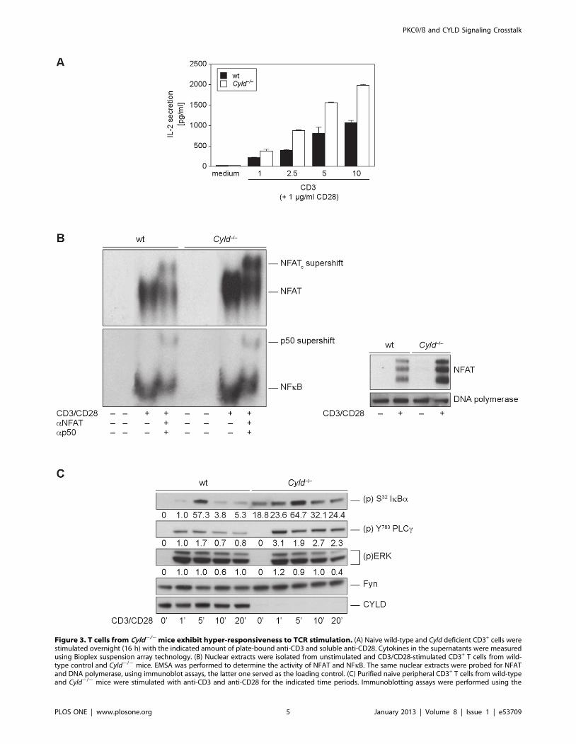
Figure 3. T cells from Cyld2/2 mice exhibit hyper-responsiveness to TCR stimulation. (A) Naive wild-type and Cyld deficient CD3+ cells werestimulated overnight (16 h) with the indicated amount of plate-bound anti-CD3 and soluble anti-CD28. Cytokines in the supernatants were measuredusing Bioplex suspension array technology. (B) Nuclear extracts were isolated from unstimulated and CD3/CD28-stimulated CD3+ T cells from wild-type control and Cyld2/2 mice. EMSA was performed to determine the activity of NFAT and NFkB. The same nuclear extracts were probed for NFATand DNA polymerase, using immunoblot assays, the latter one served as the loading control. (C) Purified naive peripheral CD3+ T cells from wild-typeand Cyld2/2 mice were stimulated with anti-CD3 and anti-CD28 for the indicated time periods. Immunoblotting assays were performed using the
PKCh/ß and CYLD Signaling Crosstalk
PLOS ONE | www.plosone.org 5 January 2013 | Volume 8 | Issue 1 | e53709

Y185 JNK, JNK, p50, p65, (p)Y-783 PLCc1, (p)T184/T187Tak1,
Tak1 (all from Cell Signaling), PKCb, PKCh (both from BD
Transduction Laboratories), actin, Cyld (E10), DNA polymerase
and Fyn (all from Santa Cruz Biotechnology). The Cyld antibody
recognizing the NH2-terminal region of Cyld was described
previously [20].
Co-immunoprecipitation AnalysisFor co-immunoprecipitation, 16107 primary mouse T cells (or
16106 transiently transfected HEK293T cells) were lysed in
400 ml of immunoprecipitation buffer [5 mM Na3VO4, 5 mM
NaP2P, 5 mM NaF, 5 mM EDTA, 150 mM NaCl, 1% NP-40,
50 mM Tris (pH 7.4), 50 mg/ml aprotinin and leupeptin]. Lysates
were precleared for 1 h at 4uC. Immunoprecipitation was
performed at 4uC overnight using 2 mg of the relevant antibodies.
Thereafter, lysates were incubated with protein G Sepharose
(Amersham-Pharmacia, Vienna) [for Streptactin IP, Streptactin
Beads were used] for 1 h at 4uC, extensively washed in lysis buffer,
resolved on an SDS–PAGE and immunostained for the relevant
protein.
Gel Mobility Shift AssaysNuclear extracts were harvested from 16107 cells according to
standard protocols. Briefly, purified CD3+ T cells were washed in
PBS and resuspended in 10 mM HEPES (pH 7.9) 10 mM KCl,
0.1 mM EDTA, 0.1 mM EGTA, 1 mM DTT and protease
inhibitors. Cells were incubated on ice for 15 min. NP-40 was
added to a final concentration of 0.6%, cells were vortexed
vigorously, and the mixture was centrifuged for 5 min. The
nuclear pellets were washed twice and resuspended in 20 mM
HEPES (pH7.9), 0.4 M NaCl, 1 mM EDTA, 1 mM EGTA, and
1 mM DTT and protease inhibitors, and the tube was rocked for
30 min at 4uC. After centrifugation for 10 min, the supernatant
was collected. Extracted proteins (2 mg) were incubated in binding
buffer with [32P]-labeled, double-stranded oligonucleotide probes
(NFkB: 59-GCC ATG GGG GGA TCC CCG AAG TCC-39;
NFAT: 59-GCC CAA AGA GGA AAA TTT GTT TCA TAC
AG-39) (Nushift; Active Motif). In each reaction, 36105 c.p.m. of
labeled probe was used, and the band shifts were resolved on 5%
polyacrylamide gels. All experiments were performed at least three
times with similar outcomes.
Flow CytometrySingle-cell suspensions from the spleen, lymph node and thymus
were prepared and incubated for 30 min on ice in staining buffer
(PBS containing 2% fetal calf serum and 0.2% NaN3) with FITC,
PE or APC antibody conjugates. Surface marker expression was
analyzed using a FACScanTM cytometer (Becton Dickinson & Co.,
Mountain View, CA) and CellQuestProTM software according to
standard protocols. Antibodies against murine CD3, CD4, and
CD8 were obtained from Caltag Laboratories; CD19, CD69,
CD44, and CD25 were obtained from BD PharMingen.
Retroviral Transduction of Primary Mouse T cellsThe packaging cell line platE was transfected with a pMX
retroviral vector encoding an EGFP-Cyld fusion cDNA. Approx-
imately 36 h later, supernatants were collected and used directly to
infect 24 h to 48 h preactivated CD3+ cells using spin inoculation
(1 h, 20006g, 32uC), followed by a 5–6 h incubation period at
37uC. Infected cells were washed, resuspended in full supplement-
ed medium and incubated for an additional 48 h to 72 h. From
these cultures, GFP-expressing cells were analyzed using confocal
microscopy to track the subcellular distribution of the protein of
interest.
Monitoring CYLD Localization Using ConfocalMicroscopy
CD3+ cells from wild type and PKCh/b knockout mice that were
transduced with a retrovirus expressing an EGFP-CYLD were not
stimulated or PDBu- and ionomycin-stimulated, transferred to a
polylysine-coated slide and fixed with 2% paraformaldehyde. After
permeabilization (0.1% TritonX-100 in PBS) and a blocking step
(5% goat serum in PBS), the cells were stained with Alexa595-
CTB (for lipid raft staining) and TOPRO 3 (Nucleus) (Molecular
Probes). Immunofluorescence was analyzed with a Zeiss LSM 510
confocal laser scanning microscope and Zeiss LSM software v3.2.
Statistical AnalysisDifferences between genotypes were analyzed using the
unpaired Student’s t test.
Results
Overlapping Roles of PKCh and PKCb in NFkB and NFATTransactivation Processes in Primary Mouse CD3+ T cells
Studies using targeted gene disruption defined a critical role for
PKCh in the activation of the IL-2 promoter in the NFkB and
Ca2+/NFAT pathways [5,6]. Surprisingly, the phenotypic char-
acterization of PKCh-deficient T cells revealed a strong upregula-
tion of PKCb protein levels in PKCh single knockout T cells
(Fig. 1A). To investigate potentially compensatory and overlapping
roles of these two PKC family members in T cell activation
processes, PKCh/b double knockout mice were generated. These
mice were viable, fertile and breed at normal Mendelian ratios.
The null mutations for PKCh and PKCb were confirmed by PCR
and immunoblotting of whole cell lysates from naive thymocytes
and peripheral CD3+ T cells (Fig. 1B).
Flow cytometric analysis of thymocyte populations in PKCh/
b2/2double knockout mice revealed a slightly diminished
percentage of CD3-, CD4- and CD8-positive cells, comparable
to the PKCh single knockout phenotype and in agreement with
previous research [24,25], which might indicate an involvement of
PKCh in the positive selection process during thymocyte
development. Nevertheless, in the periphery, PKCh/b2/2 mice
revealed no gross differences in the distribution of CD3-, CD4-,
CD8-positive cells, leading to the conclusion that the concomitant
loss of PKCh and PKCb did not additively affect T cell development
(Table 1+ Table 2).
Examination of the stimulation-dependent upregulation of
CD25, CD69 and CD44 surface markers on CD4+ and CD8+
subsets revealed no gross differences in the total percentage of
positive cells between the genotypes, but the total protein amount
per cell, monitored by median fluorescence intensity, was strongly
reduced in PKCh/b2/2 and to an intermediate extend in PKC
singly-deficient T cells. These data might indicate a possible defect
in the upregulation of both the IL-2 receptor chain alpha (CD25)
and the activation marker CD69 in PKC-deficient T cells in both
CD4+ and CD8+ T cells (Fig. S1).
indicated phospho-specific and pan-antibodies. Protein phosphorylation levels were relatively quantitated by densitometric analysis. Numbersbeneath bands indicate fold change compared to wt control after normalization to FYN.doi:10.1371/journal.pone.0053709.g003
PKCh/ß and CYLD Signaling Crosstalk
PLOS ONE | www.plosone.org 6 January 2013 | Volume 8 | Issue 1 | e53709

PKCh/ß and CYLD Signaling Crosstalk
PLOS ONE | www.plosone.org 7 January 2013 | Volume 8 | Issue 1 | e53709

In contrast to the relatively normal T-cell development
observed, the T-cell response of peripheral T cells after TCR
stimulation was affected by the single and simultaneous loss of
PKCh and PKCb. The H3-thymidine uptake and IL-2 secretion
response of PKCh/b-deficient T cells stimulated with anti-CD3
and with or without anti-CD28-activated did not significantly
exacerbate the defects already observed in the absence of PKChalone (Fig. S2A–C). To exclude the proliferative defects being
caused by deregulated apoptosis, we analyzed the activation-
induced cell death (AICD) of CD4+ and CD8+ T-cell blasts
derived from wild-type and double knockout animals using CD3
engagement in vitro; in addition also the Fas ligand induced cell
death was monitored, but no enhanced apoptotic responses of
PKCh/b2/2 were detected (Fig. S3A–B).
However, analysis of the pathways leading to IL-2 transcription
revealed additively reduced binding of NFkB and NFAT to DNA
in PKCh/b double-deficient CD3+ T cells after CD3/CD28
stimulation (Fig. 1C). Immunoblot analysis of nuclear extracts
demonstrated that the weaker DNA binding of NFkB and NFAT
was due to the reduced nuclear entry of two NFkB subunits, p50
and p65, and NFAT upon stimulation (Fig. 1D). Activation of
NFkB involves the phosphorylation of I-kBa by IKKb and its
subsequent proteasomal degradation. Consistent with the additive
affect on NFkB translocation, the double knockout showed a
weaker I-kBa phosphorylation after stimulation with CD3/CD28.
Also the activation of the Map kinase pathway was partially
affected by PKCh/b deficiency, visible through a reduced ERK
phosphorylation, whereas the activation of Akt/PKB was normal
(Fig. 1E).
PKCh and PKCb Synergistically Regulate TAK1 and JNKActivation
In agreement with the defective IKK/I-kBa axis, PKCh/b2/2
CD3+ cells revealed a drastic activation defect in TGF b activated
kinase 1 (TAK1), which is known to be a key regulator of IKKbsignaling. The loss of both PKC isotypes appears to be required to
abolish the signal, because TAK1 activation levels were similar
between the wild-type and PKC single knockout T cells (not
shown). Additionally, the JNK signal was attenuated by the
targeted disruption of PKCh and PKCb, whereas ERK1/2
activation was only marginally affected (Fig. 2A). Similar outcomes
were observed in PDBu- and ionomycin-stimulated primary
mouse wild-type T cells pretreated with 500 nM of a PKC-
specific low molecular weight inhibitor (PKC LMWI). The
stronger effect of the pharmacological pan-PKC inhibitor on
MAP kinase activation can be best explained by its established
inhibition of additional PKC family members next to PKCh and
PKCb (Fig. 2B).
Cyld2/2 T cells Show a Hyperactive Phenotype in NFkBand NFAT Transactivation Responses
Since the deubiquitinating enzyme CYLD has been shown to be
a negative regulator of Tak1 [12] and JNK signaling [26], we
investigated a possible link between CYLD and the PKCh/bisotypes in NFkb and NFAT driven IL-2 upregulation.
Despite of an observed thymocyte maturation defect, recent
work on T cell signaling in Cyld2/2 mice demonstrated hyper-
responsiveness to TCR stimulation by constitutive activation of
NFkB [12]. We confirmed these results, as we also observed that
Cyld2/2 T cells showed elevated activation-induced IL-2 responses
(Fig. 3A). This hyper-responsive IL-2 secretion correlated with an
increase of NFkB DNA binding to the IL-2 promoter in the
nuclear fractions of stimulated Cyld-deficient T cells (Fig. 3B) and
hyper-phosphorylated I-kBa levels in the cytosol compared to
wild-type controls (Fig. 3C). Interestingly, and in accordance with
a previous publication [27], we determined that CYLD also acts as
a negative modulator of the NFAT pathway. The examination of
NFAT transactivation using immunoblot and EMSA technology
revealed increased nuclear translocation and subsequent binding
of NFAT to DNA in Cyld-deficient cells (Fig. 3B). Our EMSA
result was confirmed by the elevated activation status of
phospholipase Cc1, which has been identified as a key regulator
of Ca2+/Calcineurin/NFAT signaling. However, ERK signaling
was not affected by the loss of Cyld (Fig. 3C).
Association of CYLD with PKChConsidering the reciprocal phenotypes of Cyld- and PKCh/b-
deficient T cells involving NFAT and NFkB transactivation we
investigated a potential direct interaction between this enzymes.
Interestingly and indeed, we identified a physical and functional
PKCh-CYLD interaction in the cytosol of primary T cells. The co-
immunoprecipitation analysis of CYLD and the PKCh isotype
from cell extracts of unstimulated and CD3/CD28-activated
peripheral CD3+ cells revealed that PKCh and CYLD physically
associate in a complex in resting conditions (Fig. 4A). Next, we
mapped the PKCh interaction domain in the CYLD protein by
co-transfection of HEK293T cells with a vector encoding PKChand with vectors expressing full-length Flag-tagged wild-type or N-
and C- terminally truncated forms of CYLD (encoding residues 1–
212, 318–956 and 587–986 of CYLD). The CYLD pull down with
a specific Flag antibody revealed increased binding of PKCh to
CYLD mutants containing the deubiquitinase domain (Fig. 4B).
We observed identical results when the co-immunoprecipitation
was performed to precipitate PKCh. A strep-tagged PKChconstruct was co-transfected with the Flag-tagged CYLD con-
structs. A GFP control for each CYLD construct was included to
identify unspecific binding to the Streptactin-beads. PKChprecipitation confirmed that the C-terminal part of CYLD is
necessary for complex formation between the two interacting
protein (Fig. 4C).
In Jurkat Cells, CYLD is Cleaved by MALT1 in a PKC-dependent Mechanism
When Jurkat cells overexpressing an N-terminally HA-tagged
CYLD construct were stimulated for 30 min with PDBu and
ionomycin, a CYLD fragment of approximately 40 kDa was
Figure 4. Association of CYLD with PKCh. (A) CYLD directly interacts with PKCh in primary mouse CD3+ cells. The complex is formedconstitutively and is not affected by TCR activation. One representative experiment of three is shown. The C-terminal region of CYLD is important forinteraction with PKCh. (B) Co-immunoprecipitation of PKCh using CYLD pulldown. Increased binding of PKCh to the C-terminus of CYLD was shown inHEK293T cells transiently co-transfected with vectors encoding PKCh (PEFneo) and a full-length Flag-tagged wild-type CYLD, N- or C-terminallytruncated forms of CYLD (residues 1–212, 318–956 and 587–986 of CYLD). Untransfected and GFP-transfected controls were included. Onerepresentative experiment of three is shown. A schematic representation depicts the CAP-Gly and peptidase domains in wild-type and truncationmutants of CYLD. (C) Co-immunoprecipitation of CYLD using PKCh pulldown. A strep-tagged full-length PKCh construct was co-transfected with Flag-tagged CYLD constructs into HEK293T cells. As previously, the importance of the C-terminal region of CYLD for binding is shown. GFP controls foreach CYLD construct were included. One representative experiment of three is shown.doi:10.1371/journal.pone.0053709.g004
PKCh/ß and CYLD Signaling Crosstalk
PLOS ONE | www.plosone.org 8 January 2013 | Volume 8 | Issue 1 | e53709

Figure 5. CYLD is endoproteolytically processed in the Jurkat cell line and in primary human T cells. (A) The essential role of both thecatalytic activity of PKC and MALT1 for activation-dependent CYLD cleavage. Jurkat cells transfected with HA-tagged Cyld vectors were activated byPDBu and ionomycin in the presence of PKCh and MALT1 pharmacological inhibitors. After 20 min, cells were lysed and fractionated into membrane(m), cytosolic (c) and nonsoluble (ns) fractions. CYLD and its clipping product were present in (c), inhibition of PKCh and MALT1 activity blocks CYLDcleavage. (B) CYLD processing occurred also under endogenous conditions in stimulated primary human T cells isolated from whole blood samples;
PKCh/ß and CYLD Signaling Crosstalk
PLOS ONE | www.plosone.org 9 January 2013 | Volume 8 | Issue 1 | e53709
detected using the anti-HA antibody. Because the administration
of PDBu mimics TCR signaling by activating PKC family
members, we wanted to identify a role for PKC in this cleavage
event. Therefore, Jurkat cells were pretreated with the specific
pan-PKC pharmacological inhibitor, which resulted in the
disappearance of this fragment (Fig. 5A). This finding emphasized
that the endoproteolytic cleavage of CYLD is PKC dependent. We
also isolated primary T cells from human whole blood and
analyzed the CYLD processing under endogenous conditions. In
addition, human T cells were treated with the pan-PKC inhibitor
to investigate the PKC dependency in this process. Comparable to
the results with Jurkat cells, CYLD underwent a stimulation
dependent processing also in primary human T cells, which could
be blocked by PKC inhibition. The generation of a 40 kDa NH2-
terminal and a 70 kDa C-terminal cleavage fragment was
confirmed via the use of NH2- and C-terminus recognizing
specific CYLD antibodies (Fig. 5B).
Because Coornaert et al. showed that the paracaspase MALT1
directly cleaved the deubiquitinating protein A20 to generate a
fragment with a smaller molecular size in stimulated T cells [28],
we asked if MALT1 was also responsible for the cleavage of CYLD
by treating Jurkat cells with the tetrapeptide inhibitor z-VRPR-
fmk, which has been shown to inhibit specifically the MALT1
protease activity [29]. As a result, cells treated with the MALT1
inhibitor showed a reduced CYLD cleavage after activation
(Fig. 5A).
Based on the size and the molecular weight of the CYLD N-
terminal 40 kDa proteolytic fragment, we identified the cleavable
arginine residue at position 324 in the human CYLD protein and
generated a Cyld mutant with alanine substituted for arginine at
position 324 (Cyld-R324A). Next, we investigated if this mutant
was cleavable when overexpressed in Jurkat cells or was resistant to
proteolysis. Wild-type CYLD was processed after stimulation,
whereas the mutant could no longer be cleaved (Fig. 5C). This led
to permanent inhibition of the NFkB pathway by the inactivation
resistant CYLD mutant and result in slightly diminished
phosphorylation of I-kBa. The MAPK pathway was not affected
by the expression of uncleavable CYLD.
Because NFkB is important for IL-2 upregulation in activated T
cells, we tested the influence of the protease-resistant Cyld mutant
on IL-2 transactivation using an IL-2-promoter-dependent lucif-
erase assay. The diminished NFkB signal observed by immunoblot
was correlated with impaired IL-2 transcription (Fig. 5D). This
provides experimental evidence that CYLD processing, which
leads to an inactivation of its repressor function within a positive
feedback loop, is an important prerequisite for robust IL-2
activation. Consistent with our investigation of CYLD cleavage
at arginine 324 in the Jurkat tumor cell line, Staal et al.
independently published that TCR-induced JNK activation
required CYLD proteolysis by MALT1 [18]. Nevertheless, our
findings extend the function of CYLD cleavage to NFkB
activation. To identify the physiological role of this candidate
process, we examined primary CD3+ T cells derived from wild-
type and knockout mice.
PKC-dependence, Cleavage Site and Kinetics of CYLDCleavage Differ in Primary Mouse T cells
Stimulation induced a CYLD fragment not only in human cells
but also in primary mouse T cells. Interestingly activation of
primary mouse T cells by CD3 with or without CD28
costimulation generated a NH2-terminal CYLD fragment of
approximately 25 kDa, smaller in size than the human fragment
(Fig. 6A), implicating a different cleavage site in the mouse CYLD
protein. Although we did not determine the exact cleavage site,
arginine 235 was the best candidate for the cleavage site.
Importantly, the fragment was first detectable after 4 h of
stimulation, indicating different kinetics leading to CYLD
inactivation in primary mouse cells. Unexpectedly, PKCh/b-
deficient T cells showed normal CYLD processing after stimula-
tion, implicating additional protein kinases in this process (Fig. 6B).
As a consequence, the activation defects of the TAK1/IKK axis in
PKCh/b-deficient T cells cannot solely be explained by CYLD
inactivation.
Alternatively, the existence of a constitutive CYLD/PKChcomplex might suggest that PKCh, which shows activation-
dependent subcellular translocation, is important for removing
CYLD from its NFkB-related targets and attenuates the negative
regulatory function of CYLD, enabling feedback control of NFkB
activation. To address this hypothesis, we analyzed the subcellular
distribution of a retrovirally introduced mutant CYLD-GFP fusion
protein in unstimulated and stimulated wild-type and PKCh/b-
deficient T cells with confocal microscopy. CD3+ T cells from both
genotypes were retrovirally infected with a CYLD-GFP fusion
construct, and the colocalization of CYLD-GFP with lipid rafts
was monitored with Cholera Toxin B. Interestingly, CYLD
colocalization with lipid rafts was strongly diminished in PKCh/b-
deficient T cells compared to control cells, suggesting PKC-
dependent CYLD membrane shuttling. A statistically significant
decrease in CYLD translocation in double knockout cells was
detected in unstimulated cells, whereas after 15 min of PDBu and
ionomycin stimulation, CYLD translocation to the membrane was
observed in both wild-type and knockout cells (Fig. 6C).
Discussion
Numerous studies emphasize PKChs key role as a regulator of
NFkB and Ca2+/NFAT signaling in T cells downstream of the
TCR [5,6,30–32]. The PKCb isotype is also expressed in T cells.
However, PKCb-deficient primary mouse T cells have a fairly
normal activation response [33], although Volkov et al. estab-
lished a major role for PKCb in LFA1-dependent T-cell
locomotion [34,35].
Our recent work defines a redundant role for both the novel
PKCh variant and the classical isotype PKCb in the NFkB and
NFAT signaling pathways. T cells isolated from PKCh/b-deficient
mice had a stronger impairment in NFkB and NFAT nuclear
entry and DNA binding compared to CD3+ T cells from control
and single knockout mice. Impaired TAK1 activation in double-
deficient T lymphocytes is the best candidate for restricted IKK/
IkBa signaling. Functional redundancy of PKCh with other
members of the PKC family in NFkB and/or NFAT activation
has been shown in previous studies. For instance, stimulation-
PKC dependency of this process was verified by pan-PKC LMWI treatment. The generation of a 40 kDa NH2-terminal and a 70 kDa C-terminal cleavagefragment was confirmed via the use of NH2- and C-terminus recognizing specific CYLD antibodies. (C) Mapping of the R234 cleavage site in CYLDusing site-directed mutagenesis. The cleavage-resistant CYLD mutant R324A, when overexpressed in Jurkat cells, is not cleaved and leads to anincreased suppression of NFkB-driven signals (as detected by the decreased phosphorylation of IkBa. (D) PKC- and MALT1-induced proteolysis atR234 appears critical for complete IL-2 promoter transactivation, shown by an IL-2 promoter luciferase assay.doi:10.1371/journal.pone.0053709.g005
PKCh/ß and CYLD Signaling Crosstalk
PLOS ONE | www.plosone.org 10 January 2013 | Volume 8 | Issue 1 | e53709

Figure 6. CYLD cleavage in primary mouse T cells shows a different kinetic. (A) CYLD cleavage in primary mouse cells has different kinetics.The activation of primary mouse T cells by CD3 with or without CD28 costimulation leads to the formation of an NH2-terminal CYLD fragment of
PKCh/ß and CYLD Signaling Crosstalk
PLOS ONE | www.plosone.org 11 January 2013 | Volume 8 | Issue 1 | e53709

dependent colocalization of atypical PKCf/i with PKCh in the
lipid raft fraction of T lymphocytes leads to cooperation of these
isotypes in modulating the NFkB signaling pathway [36]. The
collaborative activity of PKCh and PKCa in the NFAT pathway
was examined in a PKCa/h double knockout mouse strain.
Compared to PKCa and PKCh single-deficient T cells, double-
deficient CD3+ cells showed additively reduced IL-2 secretion
levels correlated with strongly impaired nuclear translocation and
DNA binding of NFAT after stimulation. Of note, the PKCa/hdouble knockout mice showed an impaired alloimmune response,
leading to significantly prolonged allograft survival in heart
transplantation experiments [37].
Similar to phosphorylation, K63 ubiquitination is a reversible
process that influences protein activity, trafficking and signaling
complex assembly. The removal of ubiquitin chains is mediated by a
family of deubiquitinases, of which the cylindromatous gene
product CYLD and the Tumor necrosis factor a-induced protein
3, also called A20, is currently receiving broad scientific attention.
Both CYLD and A20 have been implicated as a modulator of the
activity of NFkB-related molecules, such as NEMO (IKKc),
TRAF2 and TRAF6 [38–40]. Both enzymes overlap functionally
by targeting a similar set of substrates, which was explained by the
different expression pattern of A20 and CYLD. A20 function
depends on its transcriptional upregulation, whereas CYLD is
constitutively expressed, influencing the different time windows of
NFkB activation differently. However, a constitutive expression and
activity pattern requires a posttranslational regulatory mechanism
to inactivate the repressor during signal-induced NFkB signaling.
The cleavage-dependent inactivation of a deubiquitinase as a
posttranslational regulatory mechanism in activated T cells was first
described by Coornaert et al. [28], in which A20 was defined as a
MALT1 substrate, which upon antigen receptor engagement
undergoes cleavage for functional NFkB signaling. In our study,
we showed that PKCh and CYLD are constitutively bound in a
physical complex in the cytosol of primary mouse CD3+ cells. Direct
crosstalk between CYLD and a PKC family member has not been
described to date; therefore, we aimed to elucidate the biological
relevance of this protein-protein interaction in T-cell signaling by
examining genetic knockout mouse models in combination with
selective pharmacological inhibitors. The reciprocal phenotypes of
T-cell signaling pathways in Cyld2/2 and PKCh/b2/2 mice
prompted us to analyze the activity of key molecules linked to
NFkB and NFAT transactivation to uncover a regulatory mecha-
nism to address the modulation of TAK1 activity In agreement with
Koga et al. [27], who demonstrated negative regulation of NFAT
activity by CYLD via the TAK1/MKK3/6/p38a/b axis, our
experimental data clearly attest to CYLD involvement in NFAT
activity modulation downstream of TCR signaling.
Our results show that PKC, particularly the PKCh/b isotypes,
can influence CYLD repressive activity in different ways. In the
human Jurkat leukemic T-cell line, PKC enzymatic activity was
important for rapid MALT1-dependent CYLD processing, which is
required for TCR-linked NFkB transactivation and leads to
functional IL-2 induction. The requirement for MALT1-mediated
CYLD cleavage for intact JNK signaling downstream of the TCR
has been previously described [18]. Thus, proteolytic inactivation of
CYLD affected IL-2 transactivation via the JNK/AP1 pathway;
here, we provide experimental evidence that NFkB activity is also
specifically dependent on CYLD cleavage, subsequently modulating
IL-2 signals. Of note, we independently confirmed arginine 324 as
the CYLD cleavage site. Recently, caspase 8 has been shown to
cleave CYLD at aspartate 215 in Jurkat cells following TNFastimulation, generating a pro-survival signal to save the cells from
necrotic cell death [41]. Additionally, phosphorylation of CYLD
was found to downregulate CYLD activity: transient phosphoryla-
tion by IKK in a serine cluster just upstream of the TRAF2 binding
site, attenuates DUB function [42]. However, NFkB itself can
regulate CYLD expression in a negative feedback loop [43].
In primary T cells isolated from PKCh/b deficient mice, CYLD
was processed to the same extent as in wild-type control cells.
Additionally, the kinetics of CYLD cleavage was different in
mouse T cells compared to Jurkat cells, starting approximately 4
hours after stimulation, later then the rapid response through
TAK1 activation. The different requirement for PKC and the
altered kinetics in the mouse system led to the analysis of the
stimulation-dependent spatial and temporal organization of the
PKCh/CYLD complex using immunofluorescence microscopy.
Interestingly, we found decreased CYLD lipid raft localization in
PKCh/b-deficient T cells under resting conditions, likely affecting
activation-induced signaling.
ConclusionWe observed a direct functional connection between the positive
PKCh/b and the negative CYLD signaling pathways that fine-tune
TCR/CD28-induced signaling responses. Our findings suggest the
following scenario: PKCh/b are the essential kinases in a
physiological signaling cascade that is necessary to counteract
CYLD-mediated repression of NFkB and NFAT transactivation.
This direct and physical antagonistic crosstalk between the PKC-
derived signals and the CYLD-derived signals might represent one
mechanism of how antigen-receptor-dependent fine-tuning of the
amplitude of T lymphocyte activation is processed.
Supporting Information
Figure S1 Effect of PKCh/b deficiency on CD25, CD44,and CD69 surface expression. T cells were stimulated for
16 h by CD3/CD28 ligation and the surface expression of CD25,
CD44, and CD69 for CD4+ and CD8+ subsets were measured by
flow cytometry. The relative fluorescence intensities are indicated
as the median fluorescence intensity. The results shown are the
mean6SE of three independent experiments.
(TIF)
Figure S2 Proliferative and cytokine secretion respons-es of PKCh/b CD3+ T cells. (A, B) Proliferative responses of
PKCh/b and PKCh-deficient CD3+ T cells were analyzed in
comparison to wild-type littermate controls. After incubation using
different stimulatory conditions (antibodies or BALB/C spleno-
cytes), cells were analyzed using standard procedures for
thymidine incorporation. (C) IL-2 cytokine secretion by knockout
CD3+ T cells was analyzed in comparison to wild-type littermate
controls. After stimulation with anti-CD3 with or without soluble
approximately 25 kDa. In stark contrast to the rapid kinetics in Jurkat cells, the fragment was first detected after 4 h of stimulation. (B) Primary mouseT cells from PKCh/b2/2 mice showed normal stimulation-dependent CYLD cleavage, comparable to wild-type control mice. (C) Analysis of thesubcellular distribution of a retrovirally introduced CYLD-GFP fusion mutant in unstimulated and stimulated wild-type and PKCh/b-deficient T cells.(green): the co-localization with Cholera Toxin B stained lipid rafts (red) was monitored using confocal microscopy. Nuclei are stained in blue.Quantification of CYLD-lipid rafts co-localization is shown in the bars in the right panel and reveals a statistically significant decrease in CYLDtranslocation in unstimulated double knockout cells in comparison to wt control cells. *p,0.05; **p,0.01; ***p,0.001.doi:10.1371/journal.pone.0053709.g006
PKCh/ß and CYLD Signaling Crosstalk
PLOS ONE | www.plosone.org 12 January 2013 | Volume 8 | Issue 1 | e53709

anti-CD28, supernatants were analyzed for IL-2 concentration
using Bioplex suspension array technology. One representative
experiment of three is shown.
(TIF)
Figure S3 Activation-induced cell death (AICD) of CD4+
and CD8+ T cell blasts derived from double knockoutanimals was not increased compared to cells from singleknockout littermates. (A, B) AICD was induced by different
concentrations of anti-CD3 for 8 hours. The results shown are the
means of three independent experiments.
(TIF)
Acknowledgments
We are grateful to N. Haas and N. Krumbock for providing animal care
and technical assistance.
Author Contributions
Conceived and designed the experiments: NT GB. Performed the
experiments: NT KW NHK SK FF CLN. Analyzed the data: NT KW
SK. Contributed reagents/materials/analysis tools: ML MT RM. Wrote
the paper: NT GB.
References
1. Hayashi K, Altman A (2007) Protein kinase C theta (PKCtheta): a key player in
T cell life and death. Pharmacol Res 55: 537–544.
2. Marsland BJ, Kopf M (2008) T-cell fate and function: PKC-theta and beyond.Trends Immunol 29: 179–185.
3. Sedwick CE, Altman A (2004) Perspectives on PKCtheta in T cell activation.Mol Immunol 41: 675–686.
4. Kong KF, Yokosuka T, Canonigo-Balancio AJ, Isakov N, Saito T, et al. (2011) Amotif in the V3 domain of the kinase PKC-theta determines its localization in
the immunological synapse and functions in T cells via association with CD28.
Nat Immunol 12: 1105–1112.5. Pfeifhofer C, Kofler K, Gruber T, Tabrizi NG, Lutz C, et al. (2003) Protein
kinase C theta affects Ca2+ mobilization and NFAT cell activation in primarymouse T cells. J Exp Med 197: 1525–1535.
6. Sun Z, Arendt CW, Ellmeier W, Schaeffer EM, Sunshine MJ, et al. (2000) PKC-
theta is required for TCR-induced NF-kappaB activation in mature but notimmature T lymphocytes. Nature 404: 402–407.
7. Marsland BJ, Soos TJ, Spath G, Littman DR, Kopf M (2004) Protein kinase Ctheta is critical for the development of in vivo T helper (Th)2 cell but not Th1
cell responses. J Exp Med 200: 181–189.8. Salek-Ardakani S, So T, Halteman BS, Altman A, Croft M (2005) Protein kinase
Ctheta controls Th1 cells in experimental autoimmune encephalomyelitis.
J Immunol 175: 7635–7641.9. Tan SL, Zhao J, Bi C, Chen XC, Hepburn DL, et al. (2006) Resistance to
experimental autoimmune encephalomyelitis and impaired IL-17 production inprotein kinase C theta-deficient mice. J Immunol 176: 2872–2879.
10. Giannoni F, Lyon AB, Wareing MD, Dias PB, Sarawar SR (2005) Protein kinase
C theta is not essential for T-cell-mediated clearance of murine gammaherpes-virus 68. J Virol 79: 6808–6813.
11. Bignell GR, Warren W, Seal S, Takahashi M, Rapley E, et al. (2000)Identification of the familial cylindromatosis tumour-suppressor gene. Nat Genet
25: 160–165.12. Reiley WW, Jin W, Lee AJ, Wright A, Wu X, et al. (2007) Deubiquitinating
enzyme CYLD negatively regulates the ubiquitin-dependent kinase Tak1 and
prevents abnormal T cell responses. J Exp Med 204: 1475–1485.13. Zhang J, Stirling B, Temmerman ST, Ma CA, Fuss IJ, et al. (2006) Impaired
regulation of NF-kappaB and increased susceptibility to colitis-associatedtumorigenesis in CYLD-deficient mice. J Clin Invest 116: 3042–3049.
14. Lim JH, Jono H, Koga T, Woo CH, Ishinaga H, et al. (2007) Tumor suppressor
CYLD acts as a negative regulator for non-typeable Haemophilus influenza-induced inflammation in the middle ear and lung of mice. PLoS One 2: e1032.
15. Lim JH, Stirling B, Derry J, Koga T, Jono H, et al. (2007) Tumor suppressorCYLD regulates acute lung injury in lethal Streptococcus pneumoniae
infections. Immunity 27: 349–360.16. Ahmed N, Zeng M, Sinha I, Polin L, Wei WZ, et al. (2011) The E3 ligase Itch
and deubiquitinase Cyld act together to regulate Tak1 and inflammation. Nat
Immunol 12: 1176–1183.17. Reiley WW, Zhang M, Jin W, Losiewicz M, Donohue KB, et al. (2006)
Regulation of T cell development by the deubiquitinating enzyme CYLD. NatImmunol 7: 411–417.
18. Staal J, Driege Y, Bekaert T, Demeyer A, Muyllaert D, et al. (2011) T-cell
receptor-induced JNK activation requires proteolytic inactivation of CYLD byMALT1. EMBO J 30: 1742–1752.
19. Leitges M, Schmedt C, Guinamard R, Davoust J, Schaal S, et al. (1996)Immunodeficiency in protein kinase cbeta-deficient mice. Science 273: 788–791.
20. Massoumi R, Chmielarska K, Hennecke K, Pfeifer A, Fassler R (2006) Cyld
inhibits tumor cell proliferation by blocking Bcl-3-dependent NF-kappaBsignaling. Cell 125: 665–677.
21. Wickstrom SA, Masoumi KC, Khochbin S, Fassler R, Massoumi R (2010)CYLD negatively regulates cell-cycle progression by inactivating HDAC6 and
increasing the levels of acetylated tubulin. EMBO J 29: 131–144.22. Hermann-Kleiter N, Thuille N, Pfeifhofer C, Gruber T, Schafer M, et al. (2006)
PKCtheta and PKA are antagonistic partners in the NF-AT transactivation
pathway of primary mouse CD3+ T lymphocytes. Blood 107: 4841–4848.
23. Northrop JP, Ullman KS, Crabtree GR (1993) Characterization of the nuclear
and cytoplasmic components of the lymphoid-specific nuclear factor of activated
T cells (NF-AT) complex. J Biol Chem 268: 2917–2923.24. Gruber T, Pfeifhofer-Obermair C, Baier G (2010) PKCtheta is necessary for
efficient activation of NFkappaB, NFAT, and AP-1 during positive selection ofthymocytes. Immunol Lett 132: 6–11.
25. Morley SC, Weber KS, Kao H, Allen PM (2008) Protein kinase C-theta isrequired for efficient positive selection. J Immunol 181: 4696–4708.
26. Reiley W, Zhang M, Sun SC (2004) Negative regulation of JNK signaling by the
tumor suppressor CYLD. J Biol Chem 279: 55161–55167.27. Koga T, Lim JH, Jono H, Ha UH, Xu H, et al. (2008) Tumor suppressor
cylindromatosis acts as a negative regulator for Streptococcus pneumoniae-induced NFAT signaling. J Biol Chem 283: 12546–12554.
28. Coornaert B, Baens M, Heyninck K, Bekaert T, Haegman M, et al. (2008) T cell
antigen receptor stimulation induces MALT1 paracaspase-mediated cleavage ofthe NF-kappaB inhibitor A20. Nat Immunol 9: 263–271.
29. Rebeaud F, Hailfinger S, Posevitz-Fejfar A, Tapernoux M, Moser R, et al.(2008) The proteolytic activity of the paracaspase MALT1 is key in T cell
activation. Nat Immunol 9: 272–281.30. Kingeter LM, Schaefer BC (2008) Loss of protein kinase C theta, Bcl10, or
Malt1 selectively impairs proliferation and NF-kappa B activation in the CD4+T cell subset. J Immunol 181: 6244–6254.
31. Matsumoto R, Wang D, Blonska M, Li H, Kobayashi M, et al. (2005)
Phosphorylation of CARMA1 plays a critical role in T Cell receptor-mediatedNF-kappaB activation. Immunity 23: 575–585.
32. Wang D, Matsumoto R, You Y, Che T, Lin XY, et al. (2004) CD3/CD28
costimulation-induced NF-kappaB activation is mediated by recruitment ofprotein kinase C-theta, Bcl10, and IkappaB kinase beta to the immunological
synapse through CARMA1. Mol Cell Biol 24: 164–171.33. Thuille N, Gruber T, Bock G, Leitges M, Baier G (2004) Protein kinase C beta is
dispensable for TCR-signaling. Mol Immunol 41: 385–390.34. Volkov Y, Long A, Kelleher D (1998) Inside the crawling T cell: leukocyte
function-associated antigen-1 cross-linking is associated with microtubule-
directed translocation of protein kinase C isoenzymes beta(I) and delta.J Immunol 161: 6487–6495.
35. Volkov Y, Long A, McGrath S, Ni Eidhin D, Kelleher D (2001) Crucialimportance of PKC-beta(I) in LFA-1-mediated locomotion of activated T cells.
Nat Immunol 2: 508–514.
36. Gruber T, Fresser F, Jenny M, Uberall F, Leitges M, et al. (2008) PKCthetacooperates with atypical PKCzeta and PKCiota in NF-kappaB transactivation of
T lymphocytes. Mol Immunol 45: 117–126.37. Gruber T, Hermann-Kleiter N, Pfeifhofer-Obermair C, Lutz-Nicoladoni C,
Thuille N, et al. (2009) PKC theta cooperates with PKC alpha in alloimmuneresponses of T cells in vivo. Mol Immunol 46: 2071–2079.
38. Brummelkamp TR, Nijman SM, Dirac AM, Bernards R (2003) Loss of the
cylindromatosis tumour suppressor inhibits apoptosis by activating NF-kappaB.Nature 424: 797–801.
39. Kovalenko A, Chable-Bessia C, Cantarella G, Israel A, Wallach D, et al. (2003)The tumour suppressor CYLD negatively regulates NF-kappaB signalling by
deubiquitination. Nature 424: 801–805.
40. Trompouki E, Hatzivassiliou E, Tsichritzis T, Farmer H, Ashworth A, et al.(2003) CYLD is a deubiquitinating enzyme that negatively regulates NF-kappaB
activation by TNFR family members. Nature 424: 793–796.41. O’Donnell MA, Perez-Jimenez E, Oberst A, Ng A, Massoumi R, et al. (2011)
Caspase 8 inhibits programmed necrosis by processing CYLD. Nat Cell Biol 13:
1437–1442.42. Reiley W, Zhang M, Wu X, Granger E, Sun SC (2005) Regulation of the
deubiquitinating enzyme CYLD by IkappaB kinase gamma-dependent phos-phorylation. Mol Cell Biol 25: 3886–3895.
43. Jono H, Lim JH, Chen LF, Xu H, Trompouki E, et al. (2004) NF-kappaB isessential for induction of CYLD, the negative regulator of NF-kappaB: evidence
for a novel inducible autoregulatory feedback pathway. J Biol Chem 279:
36171–36174.
PKCh/ß and CYLD Signaling Crosstalk
PLOS ONE | www.plosone.org 13 January 2013 | Volume 8 | Issue 1 | e53709