placentation in the anteaters myrmecophaga tridactyla and tamandua tetradactyla
TRANSCRIPT
Mess et al. Reproductive Biology and Endocrinology 2012, 10:102http://www.rbej.com/content/10/1/102
RESEARCH Open Access
Placentation in the anteaters Myrmecophagatridactyla and Tamandua tetradactyla(Eutheria, Xenarthra)Andrea M Mess1*, Phelipe O Favaron1, Christiane Pfarrer2, Christine Osmann3, Allan PF Melo1,Rosangela F Rodrigues1, Carlos E Ambrósio4, Estela Bevilacqua5 and Maria A Miglino1
Abstract
Background: Since Xenarthra are serious candidates for being basal to Eutheria, their characteristics, e.g. theplacental system, influence perceptions of evolution. However, in the subgroup containing the anteaters, data arevery limited. The present study aims to elucidate the nature of the feto-maternal interface in the anteater placentaand to interpret these data within an evolutionary context.
Methods: Placentas of two species were investigated with histology, immunohistochemistry and transmissionelectron microscopy.
Results: Remnants of the maternal vessel endothelium were absent, resulting in a fully haemochorial barrierthroughout the placenta. Two structurally different parts, the villous and trabecular areas were complex andintermingled. In particular, the trabeculae which consisted of cellular, proliferative trophoblast, associated withconnective tissue, were attached to the decidua. The villi contained fetal capillaries and hypertrophiedmesenchymal cells that occured near the surface near the end of gestation. The surface of the villi consisted of flat,syncytial trophoblast, interspersed with proliferative trophoblast cells.
Conclusions: Based on fundamental differences between anteaters and armadillos, we inferred that placentalevolution was more complex than previously thought. The haemochorial pattern of anteaters was likely an ancientcondition of xenarthrans. Consequently, villous placentation may be attributed, at least in part, by convergentevolution, but was also characterized by some features that were widespread among xenarthrans.
Keywords: Evolution, Vermilingua, Trophoblast, Interhaemal barrier, Villous placenta
BackgroundXenarthra is a group of eutherian mammals that evolvedin South America since the mid Paleocene and subse-quently radiated successfully [1-3]. Three distinct groupsevolved: Cingulata comprises armadillos (Dasypodidae),Pilosa include sloths (Bradypodidae and Megalonychidae)and anteaters (Vermilingua: Myrmecophagidae andCyclopedidae) [4-6]. The latter had a specialized, elon-gated rostrum, prominent claws and long gestationperiods; they were solitary, crepuscular and inhabited
* Correspondence: [email protected] of Surgery, Faculty of Veterinary Medicine and Animal Science,University of Sao Paulo, Av. Prof. Dr. Orlando Marques de Paiva, 87, CidadeUniversitária, São Paulo SP, CEP 05508-270, BrazilFull list of author information is available at the end of the article
© 2012 Mess et al.; licensee BioMed Central LtCommons Attribution License (http://creativecreproduction in any medium, provided the or
grasslands and other habitats of Central South America[7-10]. Xenarthra represents a supraordinal clade ofEutheria. Since they are serious candidates for being basalto Eutheria [11-13], their character conditions influenceperceptions of eutherian evolution [14]. In particular, pla-cental characters vary among xenarthrans [15-19]. Placen-tation has been well characterized in armadillos [20-26];they have villous and haemochorial placentas formed by apeculiar, partly invasive interaction with maternal vessels[25-27]. In contrast sloths have lobulated, labyrinthineand endotheliochorial placentas [28-30]. Anteaters areregarded as being similar to armadillos. Consequently, anarmadillo-like pattern is regarded to represent the ancientcondition of Xenarthra, resulting in evolutionary transfor-mations on the stem lineage of sloths [31]. However, data
d. This is an Open Access article distributed under the terms of the Creativeommons.org/licenses/by/2.0), which permits unrestricted use, distribution, andiginal work is properly cited.
Mess et al. Reproductive Biology and Endocrinology 2012, 10:102 Page 2 of 7http://www.rbej.com/content/10/1/102
on anteater placentation are limited to an early stage[32] and delivered placentas [33] of the giant anteaterMyrmecophaga tridactyla, approximately 10 stagesfrom early- to mid-gestation of the lesser anteaterTamandua tetradactyla [34] as well as a single, latestage of the two-toed anteater Cyclopes didactyla [35].Important aspects are unresolved, i.e. the degree oftrophoblast invasion, development and fine structureof the trabecular area, contribution of fetal or mater-nal tissues to them as well as the presence or absenceof cellular trophoblast in the villi at term [33]. The ob-jective of the present study was to use histology,immunohistochemistry and transmission electron mi-croscopy to characterize similarities and differencesamong xenarthrans and to interpret these data in anevolutionary context.
MethodsTissue collectionMaterial from Myrmecophaga tridactyla, acquiredfrom a road-killed animal in Brazil, represented midgestation (approximately 100–110 days [36,37]). Threedelivered placentas were obtained from the breedinggroup at Dortmund Zoo, Germany. A near-term stagefrom Tamandua tetradactyla that was more advancedthan those described by Becher [34] was derived fromthe zoological park in Ilha Solteira, Brazil. This re-search was approved by the Ethical Committee at theFaculty of Veterinary Medicine and Animal Science ofthe University of Sao Paulo.
Histology and immunohistochemistryMaterial for histology, fixed in 10% formalin in 0.1 Mphosphate buffer or Bouin’s solution, was embedded inparaplast, sectioned at 5 μm in an automatic micro-tome (Leica RM 2155, Nussloch, Germany), andstained with haematoxylin and eosin, Masson’s tri-chrome, toluidine blue and the periodic acid Schiff re-action (PAS). Immunohistochemistry (for details see[26,38]) for vimentin was done to detect mesenchymalcells, including remnants of the maternal endotheliumand stromal decidua (mouse monoclonal anti-humanantibody; RTU-VimV9; 1:300; Novacastra; Wetzlar,Germany), α-smooth muscle actin that similarly la-beled vessel walls (1:400; Clone 1A4; Dako Cytoma-tion; Carpinteria, California, USA), cytokeratin toidentify epithelial tissues including trophoblast (rabbitpolyclonal antibody; wide spectrum screening N1512;1:100; Dako) and as proliferation marker a mousemonoclonal antibody to human anti-PCNA (prolifera-tion cell nuclear antigen; clone PC10; 1:300; Sigma;St. Louis, USA). Sections were subjected to endogen-ous peroxidase blockage, non-specific binding wasblocked [38], incubated with the primary antibodies
overnight at 4°C in a humid chamber, and rinsed in PBS.A biotinylated secondary antibody and streptavidin-HRP(Dako) were applied for 30 min each, followed by rinsingwith PBS. Detection was done with Fast Red TR/NaphtholAS-MX (F4523, Sigma) or DAB and substrate chromogensystem (Dako) for 2 min, counterstained with haematoxy-lin and eosin and mounted in FaramontW (Dako). Negativecontrols used a goat anti-Mouse IgG (AP308F, 1:500;-Chemicon International Temecula, California, USA) in lieuof primary antibody. Slides were examined with anOlympus BX40 microscope with Zeiss KS400 imageanalysis system.
Transmission electron microscopySamples for TEM were fixed in 2.5% glutaraldehydein cacodylate buffer, post-fixed in 2% phosphate-buffered osmium tetroxide at ph 7.4 for 2 h, embed-ded in Spurr’s Resin and sectioned with an auto-matic ultramicrotome (Ultracut R, Leica). Semi-thinsections (400 nm) were stained with toluidine blue.Ultrathin sections (90 nm) were contrasted with 2%uranyl acetate and 0.5% lead citrate and studied inan electron microscope (Morgagni 268D, FEI Com-pany, The Netherlands; Mega View III camera, SoftImaging System, Germany).
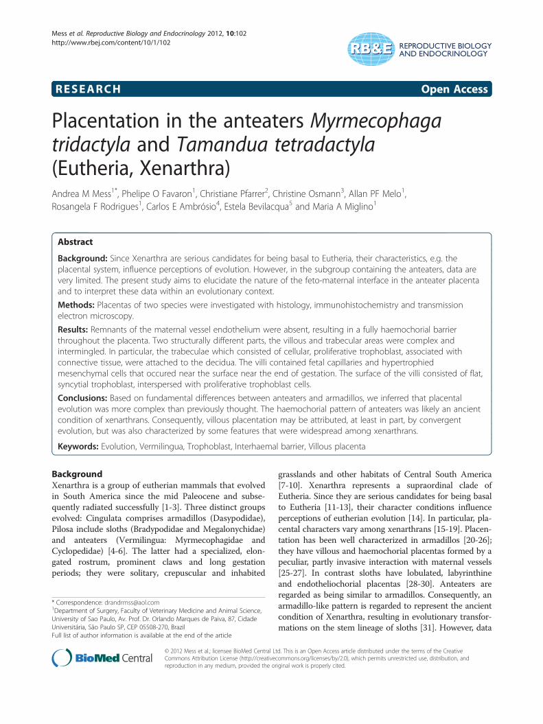
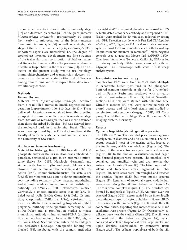
ResultsMyrmecophaga tridactyla: mid gestation placentaThe CRL was 7 cm. The extended placenta was approxi-mately 6 cm in diameter and 1 to 1.5 cm thick. The con-ceptus occupied most of the uterine cavity, located atthe fundic area, which was lobulated (Figure 1A). Thesurface of the conceptus was gelatinous and opaque(Figure 1B). In the amnion, vascularisation had begunand fibrinoid plaques were present. The umbilical cordcontained one umbilical vein and two arteries thatentered the placenta (Figure 1C). The latter consisted ofvillous and trabecular areas, organized in lobes(Figure 1D). Both areas were intermingled and reachedthe decidua (Figure 1D,E), but were mostly separate(Figure 1F). Remnants of maternal vessel endotheliumwere absent along the villi and trabeculae (Figure 2A).The villi were complex (Figure 1D). Their surface wasformed by trophoblast (Figure 2A,B). An outer layer wassyncytial (Figure 2C,D), accompanied by an inner, locallydiscontinuous layer of cytotrophoblast (Figure 2B,C).The barrier was thin in parts (Figure 2D). Inside the villi,connective tissue, hypertrophied mesenchymal cells andcapillaries were present (Figure 2A-D). Occasionally, ca-pillaries were near the surface (Figure 2D). The villi wereconfluent with the trabeculae (Figure 2A), whichconsisted of cellular trophoblast with large nuclei andliquid droplets, sourrounded by connective tissue(Figure 2A,E). The cellular trophoblast of both the villi
Figure 1 Myrmecophaga tridactyla, mid gestation placenta. (A,B) Macroscopic anatomy. Uterus (U) with lobulated structure, an extendedchorioallantoic placenta (CP), areas of gelatinous appearance (arrows), and a single embryo (E). (C) Haematoxylin and eosin. One umbilical vein(uv) and two arteries (ua) entered the placenta from the chorionic plate (chor plat). (D) Haematoxylin and eosin. Intermingling of villous (villi) andtrabeculae (trab) areas, bathed in maternal blood (arrows). (E) Haematoxylin and eosin. Both villi and trabeculae reached the decidua (dec). (F)Cytokeratin-positive trophoblast of trabeculae (arrows) attached to the decidua that was cytokeratin-negative.
Mess et al. Reproductive Biology and Endocrinology 2012, 10:102 Page 3 of 7http://www.rbej.com/content/10/1/102
and trabeculae was proliferative (Figure 2F); the tips ofthe villi were particularly active. In addition, mesenchymalcells and capillary endothelium were positive (Figure 2F).
Myrmecophaga tridactyla: term placentasAll term placentas were discoidal. The umbilical cordwas prominent, with one vein and two arteries. Insidethe disc, both villous and trabecular areas were present
(Figure 3A). The decidua was thin. On comparison tomid-gestation, the villous region had increased complex-ity and volume. The projections of the villi were inter-mingled with the trophoblast of the trabeculae, but didnot reach the decidua (Figure 3A). The villi had abundantfibers, connective tissue and enlarged mesenchymal cells,and were well vascularized (Figure 3B,C). The capillarieswere near the surface (Figure 3C). The trophoblastic
Figure 2 Myrmecophaga tridactyla, mid gestation placenta. (A) Vimentin. Villi with positive connective tissue, enlarged mesenchymal cells(mc) and fetal capillaries (cap). Cellular (ct) and syncytial (syn) trophoblast were immunonegative, as well as trophoblast of trabeculae (trab).Remnants of the maternal vessel endothelium were absent. (B) Toluidine blue. Villi with two layers of trophoblast and hypertrophiedmesenchymal cells. (C,D) TEM. The interhaemal barrier along the intervillous space (ivs) was thin and syncytial. Trophoblast cells occurred. Fetalcapillaries with endothelium (endo) were near the surface (E) TEM. Trabeculae had solid strands of cellular trophoblast with large nuclei andliquid droplets (ld) and connective tissue (con tiss). (F) PCNA. Proliferation activity was high in trophoblast cell clusters of villi and trabeculae(white arrows). Also, proliferation occurred in hypertrophied mesenchymal cells and endothelia of the villi (black arrows).
Mess et al. Reproductive Biology and Endocrinology 2012, 10:102 Page 4 of 7http://www.rbej.com/content/10/1/102
surface of the villi was syncytial and thin; however, therewere single trophoblast cells towards the interior(Figure 3C). The trabeculae consisted of cellular tropho-blast with limited syncytial areas and connective tissue(Figure 3D). At the placental base, the trabeculae were nearthe decidua, but only occasionally trophoblast cells invadedthe surface (Figure 3E). The trophoblast cells in the tips ofthe villi and the trabeculae were proliferating (Figure 3F).
Tamandua tetradactyla: late gestation or near-termplacentaThe CRL was approximately 12 cm and the discoidal pla-centa was 10 cm in diameter (Figure 4A). The conceptus
occupied approximately 75% of the fundic area of theuterine cavity. Gelatinous tissue covered the surface ofthe conceptus (Figure 4A). A thin amniotic membranecovered the placenta. The umbilical cord was 11 cm. Itcontained one umbilical vein and two arteries that com-plexly branched at the chorionic plate (Figure 4A). Theplacenta was organized into lobes. The villi were inter-mingled with trabeculae. Remnants of maternal endothe-lium were absent (Figure 4B,C). Villi were lined bysyncytiotrophoblast with some cytotrophoblast and con-tained connective tissue, fetal capillaries, and hypertro-phied mesenchymal cells. Capillaries were near thesurface (Figure 4B). The villi were connected to the
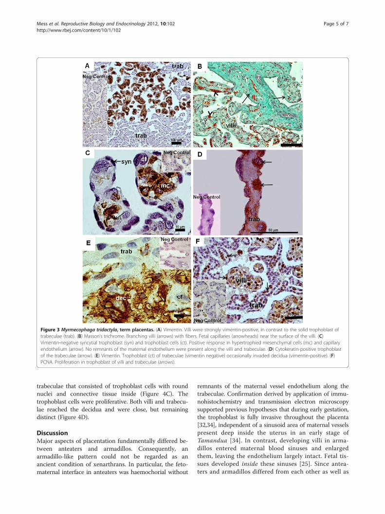
Figure 3 Myrmecophaga tridactyla, term placentas. (A) Vimentin. Villi were strongly vimentin-positive, in contrast to the solid trophoblast oftrabeculae (trab). (B) Masson’s trichrome. Branching villi (arrows) with fibers. Fetal capillaries (arrowheads) near the surface of the villi. (C)Vimentin-negative syncytial trophoblast (syn) and trophoblast cells (ct). Positive response in hypertrophied mesenchymal cells (mc) and capillaryendothelium (arrow). No remnants of the maternal endothelium were present along the villi and trabeculae. (D) Cytokeratin-positive trophoblastof the trabeculae (arrow). (E) Vimentin. Trophoblast (ct) of trabeculae (vimentin negative) occasionally invaded decidua (vimentin-positive). (F)PCNA. Proliferation in trophoblast of villi and trabeculae (arrows).
Mess et al. Reproductive Biology and Endocrinology 2012, 10:102 Page 5 of 7http://www.rbej.com/content/10/1/102
trabeculae that consisted of trophoblast cells with roundnuclei and connective tissue inside (Figure 4C). Thetrophoblast cells were proliferative. Both villi and trabecu-lae reached the decidua and were close, but remainingdistinct (Figure 4D).
DiscussionMajor aspects of placentation fundamentally differed be-tween anteaters and armadillos. Consequently, anarmadillo-like pattern could not be regarded as anancient condition of xenarthrans. In particular, the feto-maternal interface in anteaters was haemochorial without
remnants of the maternal vessel endothelium along thetrabeculae. Confirmation derived by application of immu-nohistochemistry and transmission electron microscopysupported previous hypotheses that during early gestation,the trophoblast is fully invasive throughout the placenta[32,34], independent of a sinusoid area of maternal vesselspresent deep inside the uterus in an early stage ofTamandua [34]. In contrast, developing villi in arma-dillos entered maternal blood sinuses and enlargedthem, leaving the endothelium largely intact. Fetal tis-sues developed inside these sinuses [25]. Since antea-ters and armadillos differed from each other as well as
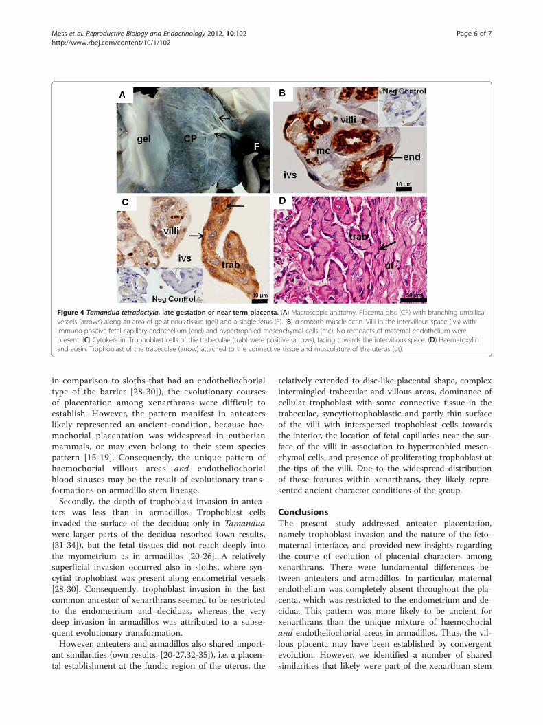
Figure 4 Tamandua tetradactyla, late gestation or near term placenta. (A) Macroscopic anatomy. Placenta disc (CP) with branching umbilicalvessels (arrows) along an area of gelatinous tissue (gel) and a single fetus (F). (B) α-smooth muscle actin. Villi in the intervillous space (ivs) withimmuno-positive fetal capillary endothelium (end) and hypertrophied mesenchymal cells (mc). No remnants of maternal endothelium werepresent. (C) Cytokeratin. Trophoblast cells of the trabeculae (trab) were positive (arrows), facing towards the intervillous space. (D) Haematoxylinand eosin. Trophoblast of the trabeculae (arrow) attached to the connective tissue and musculature of the uterus (ut).
Mess et al. Reproductive Biology and Endocrinology 2012, 10:102 Page 6 of 7http://www.rbej.com/content/10/1/102
in comparison to sloths that had an endotheliochorialtype of the barrier [28-30]), the evolutionary coursesof placentation among xenarthrans were difficult toestablish. However, the pattern manifest in anteaterslikely represented an ancient condition, because hae-mochorial placentation was widespread in eutherianmammals, or may even belong to their stem speciespattern [15-19]. Consequently, the unique pattern ofhaemochorial villous areas and endotheliochorialblood sinuses may be the result of evolutionary trans-formations on armadillo stem lineage.Secondly, the depth of trophoblast invasion in antea-
ters was less than in armadillos. Trophoblast cellsinvaded the surface of the decidua; only in Tamanduawere larger parts of the decidua resorbed (own results,[31-34]), but the fetal tissues did not reach deeply intothe myometrium as in armadillos [20-26]. A relativelysuperficial invasion occurred also in sloths, where syn-cytial trophoblast was present along endometrial vessels[28-30]. Consequently, trophoblast invasion in the lastcommon ancestor of xenarthrans seemed to be restrictedto the endometrium and deciduas, whereas the verydeep invasion in armadillos was attributed to a subse-quent evolutionary transformation.However, anteaters and armadillos also shared import-
ant similarities (own results, [20-27,32-35]), i.e. a placen-tal establishment at the fundic region of the uterus, the
relatively extended to disc-like placental shape, complexintermingled trabecular and villous areas, dominance ofcellular trophoblast with some connective tissue in thetrabeculae, syncytiotrophoblastic and partly thin surfaceof the villi with interspersed trophoblast cells towardsthe interior, the location of fetal capillaries near the sur-face of the villi in association to hypertrophied mesen-chymal cells, and presence of proliferating trophoblast atthe tips of the villi. Due to the widespread distributionof these features within xenarthrans, they likely repre-sented ancient character conditions of the group.
ConclusionsThe present study addressed anteater placentation,namely trophoblast invasion and the nature of the feto-maternal interface, and provided new insights regardingthe course of evolution of placental characters amongxenarthrans. There were fundamental differences be-tween anteaters and armadillos. In particular, maternalendothelium was completely absent throughout the pla-centa, which was restricted to the endometrium and de-cidua. This pattern was more likely to be ancient forxenarthrans than the unique mixture of haemochorialand endotheliochorial areas in armadillos. Thus, the vil-lous placenta may have been established by convergentevolution. However, we identified a number of sharedsimilarities that likely were part of the xenarthran stem
Mess et al. Reproductive Biology and Endocrinology 2012, 10:102 Page 7 of 7http://www.rbej.com/content/10/1/102
species pattern. In conclusion, an armadillo-like patternshould not be regarded as ancient condition of xenar-thrans, because their placental evolution was more com-plex than previously established.
Competing interestsThe authors declare that they have no competing interests.
Authors' contributionsMAM devised the study and participated in its design. AMM analyzed thematerial and wrote the manuscript, supported by POF and CP. All otherauthors were involved in the acquisition and procession of this rare material.All authors read and approved the final manuscript.
AcknowledgementsThis project was supported by CNPq and FAPESP. We thank members of theUniversity of Sao Paulo, Brazil, for technical support. We warmly thank Roseand John Kastelic for help with the English.
Author details1Department of Surgery, Faculty of Veterinary Medicine and Animal Science,University of Sao Paulo, Av. Prof. Dr. Orlando Marques de Paiva, 87, CidadeUniversitária, São Paulo SP, CEP 05508-270, Brazil. 2Institute of Anatomy,University of Veterinary Medicine Hannover, Bischofsholer Damm 15,30173 Hannover, Germany. 3Zoo Dortmund, Mergelteichstr. 80,44225 Dortmund, Germany. 4Department of Basic Science, Faculty of AnimalSciences and Food Engineering, University of Sao Paulo, Av. Duque de CaxiasNorte, 225 ZAB, Pirassununga CEP 13635-900, Brazil. 5Institute of BiomedicalSciences, University of Sao Paulo, Av. Prof. Lineu Prestes, 1524, CidadeUniversitária, São Paulo SP, CEP 05508-900, Brazil.
Received: 5 September 2012 Accepted: 16 November 2012Published: 30 November 2012
References1. Delsuc F, Douzery EJP: Recent advances and future prospects in
xenarthran molecular genetics. In The biology of the xenarthra. Edited byVizcaíno SFL, Loughry WJ. Gainesville: University Press of Florida; 2008:11–23.
2. Gaudin TJ, McDonald HG: Morphology-based investigations of thephylogenetic relationships among extant and fossil xenarthrans. In Thebiology of the xenarthra. Edited by Vizcaíno SFL, Loughry WJ. Gainesville:University Press of Florida; 2008:24–36.
3. Meredith RW, Janečka JE, Gatesy J, Ryder OA, Fisher CA, Teeling EC,Goodbla A, Eizirik E, Simão TL, Stadler T, Rabosky DL, Honeycutt RL, Flynn JJ,Ingram CM, Steiner C, Williams TL, Robinson TJ, Burk-Herrick A, WestermanM, Ayoub NA, Springer MS, Murphy WJ: Impacts of the cretaceousterrestrial revolution and KPg extinction on mammal diversification.Science 2011, 334:521–524.
4. Montgomery GG: The evoluion and ecology of armadillos, sloths, andvermilinguas. Washington: Smithsonian Institution; 1985.
5. Osmann C: Ordnung nebengelenktiere. In Zootierhaltung. Edited byPuschmann W. Frankfurt a.M: Verlag Harri Deutsch; 2004:253–265.
6. Wilson DE, Reeder DM: Mammal species of the world. Baltimore:Johns Hopkins University Press; 2005.
7. Redford KH, Eisenberg JF: Mammals of the neotropics. The southern cone. Chile,argentinia, uruguay, paraguay. Illinois: University of Chicago Press; 1992.
8. Eisenberg JF, Redford KH: Mammals of the neotropics: the central neotropics.Ecuador, Peru, Bolivia, brazil. Chicago: University of Chicago Press; 1999.
9. Redford KH: The edentates of the cerrado. Edentata 1994, 1:4–10.10. Emmons LH, Feer F: Neotropical rainforest mammals: A field guide. Chicago:
The University of Chicago Press; 1990.11. Murphy WJ, Eizirik E, Johnson WE, Zhang YP, Ryder OA, O'Brien SJ:
Molecular phylogenetics and the origins of placental mammals.Nature 2001, 409:614–618.
12. Springer MS, Stanhope MJ, Madsen O, Jong WW: Molecules consolidatethe placental mammal tree. Trends Ecol Evol 2004, 19:430–438.
13. Springer MS, Murphy WJ: Mammalian evolution and biomedicine: newviews from phylogeny. Biol Rev 2007, 82:375–392.
14. Delsuc F, Scally M, Madsen O, Stanhope MJ, Jong WW, Catzeflis FM,Springer MS, Douzery EJP: Molecular phylogeny of living xenarthrans and
the impact of character and taxon sampling on the placental treerooting. Mol Biol Evol 2002, 19:1656–1671.
15. Mess A, Carter AM: Evolutionary transformations of fetal membranecharacters in eutheria with special reference to afrotheria. J Exp Zool BMol Dev Evol 2006, 306:140–163.
16. Mess A, Carter AM: Evolution of the placenta during the early radiationof placental mammals. Comp Biochem Physiol A Comp Physiol 2007,148:769–779.
17. Wildman DE, Chen C, Erez O, Grossman LI, Goodman M, Romero R:Evolution of the mammalian placenta revealed by phylogenetic analysis.PNAS 2006, 103:3203–3208.
18. Elliot MG, Crespi BJ: Phylogenetic evidence for early hemochorialplacentation in eutheria. Placenta 2009, 30:49–67.
19. Capellini I: The evolutionary significance of placental interdigitation inmammalian reproduction: contributions from comparative studies.Placenta 2012, 33:763–768.
20. Strahl VH: Über den Bau der placenta von dasypus novemcinctus.Anat Anz 1913, 44:440–447.
21. Fernández M: Zur anordnung der embryonen und form der placentabei Tatusia novemcincta. Anat Anz 1914, 46:253–258.
22. Enders AC: Development and structure of the villous haemochorialplacenta of the nine-banded armadillo (dasypus novemcinctus).J Anat 1960, 94:34–45.
23. Enders AC: Electron microscopic observations on the villous haemochorialplacenta of the nine-banded armadillo (dasypus novemcinctus). J Anat 1960,94:205–215.
24. Adamoli VC, Cetica PD, Merani MS, Solari AJ: Comparative morphologicplacental types in dasypodidae (chaetophractus villosus, cabassouschacoensis, Tolypeutes matacus and dasypus hybridus). Biocell 2001,25:17–22.
25. Enders AC: Placentation in armadillos, with emphasis on development ofthe placenta in polyembryonic species. In The biology of the xenarthra.Edited by Vizcaíno SFL, Loughry WJ. Gainesville: University Press of Florida;2008:172–180.
26. Rezende LC, Barbeito CG, Mess A, Favaron PO, Miglino MA: Thefetomaternal interface in the placenta of three species of armadillos(eutheria, xenrthra, dasypodidae). Reprod Biol Endocrinol 2012, 10:38.
27. Enders AC, Carter AM: The evolving placenta: different developmentalpaths to a hemochorial relationship. Placenta 2012, 33:S92–S98.
28. Turner W: The placenta in the sloth. J Anat Physiol 1879, 14:147–148.29. King BF, Pinheiro PB, Hunter RL: The fine structure of the placental
labyrinth in the sloth, bradypus tridactylus. Anat Rec 1982, 202:15–22.30. Enders AC, Carter AM: The evolving placenta: convergent evolution of
variations in the endotheliochorial relationship. Placenta 2012,33:319–326.
31. Carter AM, Mess AM: Conservation of placentation during the tertiaryradiation of mammals in south America. J Morphol, . In press.
32. Walls EW: Myrmecophaga jubata: an embryo with placenta. J Anat 1939,73:311–317.
33. Benirschke K: Giant anteater, Myrmecophaga tridactyla. 2007.http://placentation.ucsd.edu.
34. Wislocki GB: On the placentation of the two-toed anteater (Cyclopesdidactylus). Anat Rec 1928, 39:69–79.
35. Becher H: Placenta und uterusschleimhaut von Tamandua tetradactya(Myrmecophaga). Gegenbaurs Morphol Jahrb 1931, 67:381–458.
36. Schauerte N: Untersuchungen zur zyklus- und graviditätsdiagnostik beimgroßen ameisenbären (myrmecophaga tridactyla). Gießen: Justus-Liebig-Universität, PhD Thesis; 2005.
37. Schauerte N, Osmann C: Reproducao de tamanduas em cativeiro.In Manutencao de tamanduas em cativeiro. Edited by Miranda F.;2012:134–145.
38. Oliveira MF, Favaron PO, Ambrosio CE, Miglino MA, Mess A: Chorioallantoicand yolk sac placentation in thrichomys laurentinus (echimyidae) and theevolution of hystricognath rodents. J Exp Zool B Mol Dev Evol 2012,318:13–25.
doi:10.1186/1477-7827-10-102Cite this article as: Mess et al.: Placentation in the anteatersMyrmecophaga tridactyla and Tamandua tetradactyla (Eutheria,Xenarthra). Reproductive Biology and Endocrinology 2012 10:102.