plant tissue culture training manual - … tissue culture is a practice used to propagate plants...
TRANSCRIPT
PLANT TISSUE CULTURE TRAINING MANUAL
On PLANT TISSUE CULTURE
WITH SPECIAL REFERENCE TO ORCHID MICROPROPAGATION (A Programme under the auspices of SIAS– Centre for Scientific Research)
24 th and 25 th January 2017
Course Director Dr. Sahaya Shibu B
Course Co-Ordinators Dr. Servin Wesley P Dr. Manjusha Sayd
Organized by
DEPARTMENT OF BIOTECHNOLOGY SAFI INSTITUTE OF ADVANCED STUDY (SIAS)
(THE ACADEMIC VENTURE OF SOCIAL ADVANCEMENT FOUNDATION OF INDIA) (Affiliated to the University of Calicut)
VAZHAYOOR, RAMANATTUKARA, MALAPPURAM KERALA- 673 633
TISSUE CULTURE TRAINING MANUAL
January 2017
Published by Department of Biotechnology SAFI Institute of Advanced Study (SIAS), Vazhayoor, Malappuram, Kerala-673 633
Editors
Dr. Sahaya Shibu B, Assistant Professor, Department of Biotechnology, SIAS . Dr. Manjusha Sayd, Assistant Professor, Department of Biotechnology, SIAS. DTP Works Agila Gopinath, IV th Semester MSc Arsha .V.P, IV th Semester MSc
Published by SAFI Institute of Advanced Study (SIAS), Vazhayoor Phone: 0483-2833594 Email: [email protected]
Copyright ©2017: SAFI Institute of Advanced Study (SIAS), Vazhayoor, Malappuram, Kerala-673 633
ABOUT THE INSTITUTION
SAFI Institute of Advanced Study (SIAS), affiliated to the University of Calicut is the academic wing of Social Advancement Foundation of India is the center for research and higher education. The Institute has been dedicated to the nation by Mr. M. Hamid Ansari Hon’ble Vice President of India on 11th November 2011. Along with the management, a dynamic team of Teachers and Scientists contribute significantly to the fulfillment of the academic programs. The campus accommodates a main academic as well as administrative block, a library which has been fully digitalized, a cultural center and hostel facilities for boys and girls. SAFI is committed to generating skilled manpower capable of providing leadership and direction at the cutting edge of science and technology particularly in life sciences. SAFI envisages the setting up of a deemed university in due course. The institute is committed to serve the economically and socially weaker students. SIAS- CSR SIAS- Centre for Scientific Research (SIAS-CSR) is an extended research wing of School of Biosciences. It has been constituted with Dr. K.V. Ahamed Bavappa as the Chairman. The center is engaged in various research programs which can be categorized under the major areas viz, Medicinal & Medical Biotechnology (MMB), Food Biotechnology (FB), Industrial Microbiology (IM), Environmental Biotechnology (EB), Plant Biotechnology (PB), Marine Biotechnology and Bioinformatics etc . About the Workshop The workshop on PLANT TISSUE CULTURE WITH SPECIAL REFERANCE TO MICRO PROPAGATION OF ORCHID aspires to emphasize the wide-ranging techniques (media preparation, sterilization, disinfecting and culture inoculation, asymbiotic seed germination, organogenesis, multiple shoot induction, rooting and hardening) involved in the propagation of Orchids. The workshop is designed to provide the above-mentioned techniques through seminars by eminent faculty from various research institutes and also through hands on practical sessions. Plant Tissue Culture is a practice used to propagate plants under sterile conditions, often used to produce clones of a plant. Different techniques in Plant Tissue culture offer advantages over traditional methods of propagation which includes the production of multiple clones of plants in the absence of seeds or pollinators necessary to produce seeds and mature plants. Also, it helps to produce plants in the sterile condition that results in disease-free plant production.
Prof. (Dr.) P.V. Basheer Ahammed, Principal, SAFI Institute of Advanced Study, Rasiya Nagar, Vazhayoor East P.O. Malappuram Dist. -673633, Kerala, India.
I am extremely happy to write this message because we have been able to conduct
Two days National workshop on “PLANT TISSUE CULTURE WITH SPECIAL REFERANCE
TO MICRO PROPAGATION OF ORCHID ” under the auspices of the SIAS–Centre for
Scientific Research (SIAS–CSR) which we established under the umbrella of Safi Institute of
Advanced Study (SIAS) about 3 years back. SIAS–CSR has been involved in active research
since its inception, thanks to the energetic and whole hearted participation of all the staff. I
wish all well for the training and I hope that the deliberations will pave the way and give
direction for future research activities in vital areas of Plant Biotechnology including Crop
improvement, Ornamental Plant Propagation and all other human welfare programs.
Sd/-
FOREWARD
We are extremely happy in bringing out the PLANT TISSUE CULTURE MANUAL
FOR MICRO PROPAGATION OF ORCHID. We hope that this will contribute, at least in a
little way, to the fast developing Biotechnology with special reference to Ornamental and
Medicinal plant propagation.
Sd/-
Prof. (Dr). P.V. Basheer Ahammed, Principal , SIAS.
Organizing committee
Prof. (Dr). Basheer Ahammed P.V., Principal, SAFI Institute of Advanced Study.
Mansoor Ali N, Officer on Special Duty, SIAS.
Convener
Dr. Servin Wesley P, Assistant Professor& Head, Department of Biotechnology, SIAS.
Organizing Secretary
Dr. B. Sahaya Shibu, Assistant Professor, Department of Biotechnology, SIAS.
Co-ordinators
Dr. Manjusha Sayd, Assistant Professor, Department of Biotechnology, SIAS.
Ms. Nafila PP, Assistant Professor, Department of Biotechnology, SIAS.
Mr. Suvanish Kumar, Assistant Professor, Department of Biotechnology, SIAS.
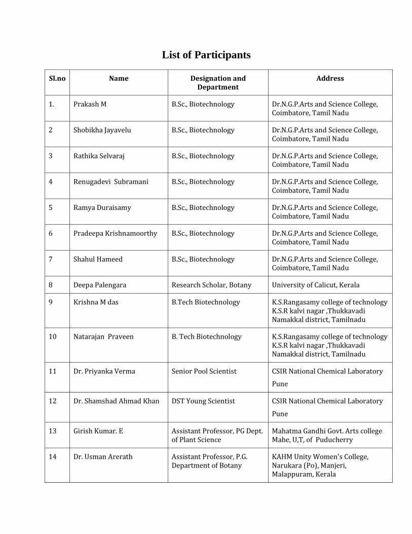
List of Participants
Sl.no Name Designation and Department
Address
1. Prakash M B.Sc., Biotechnology Dr.N.G.P.Arts and Science College, Coimbatore, Tamil Nadu
2 Shobikha Jayavelu B.Sc., Biotechnology Dr.N.G.P.Arts and Science College, Coimbatore, Tamil Nadu
3 Rathika Selvaraj B.Sc., Biotechnology Dr.N.G.P.Arts and Science College, Coimbatore, Tamil Nadu
4 Renugadevi Subramani B.Sc., Biotechnology Dr.N.G.P.Arts and Science College, Coimbatore, Tamil Nadu
5 Ramya Duraisamy B.Sc., Biotechnology Dr.N.G.P.Arts and Science College, Coimbatore, Tamil Nadu
6 Pradeepa Krishnamoorthy B.Sc., Biotechnology Dr.N.G.P.Arts and Science College, Coimbatore, Tamil Nadu
7 Shahul Hameed B.Sc., Biotechnology Dr.N.G.P.Arts and Science College, Coimbatore, Tamil Nadu
8 Deepa Palengara Research Scholar, Botany University of Calicut, Kerala
9 Krishna M das B.Tech Biotechnology K.S.Rangasamy college of technology K.S.R kalvi nagar ,Thukkavadi Namakkal district, Tamilnadu
10 Natarajan Praveen B. Tech Biotechnology K.S.Rangasamy college of technology K.S.R kalvi nagar ,Thukkavadi Namakkal district, Tamilnadu
11 Dr. Priyanka Verma Senior Pool Scientist
CSIR National Chemical Laboratory
Pune
12 Dr. Shamshad Ahmad Khan
DST Young Scientist
CSIR National Chemical Laboratory
Pune
13 Girish Kumar. E Assistant Professor, PG Dept. of Plant Science
Mahatma Gandhi Govt. Arts college Mahe, U,T, of Puducherry
14 Dr. Usman Arerath Assistant Professor, P.G. Department of Botany
KAHM Unity Women's College, Narukara (Po), Manjeri, Malappuram, Kerala
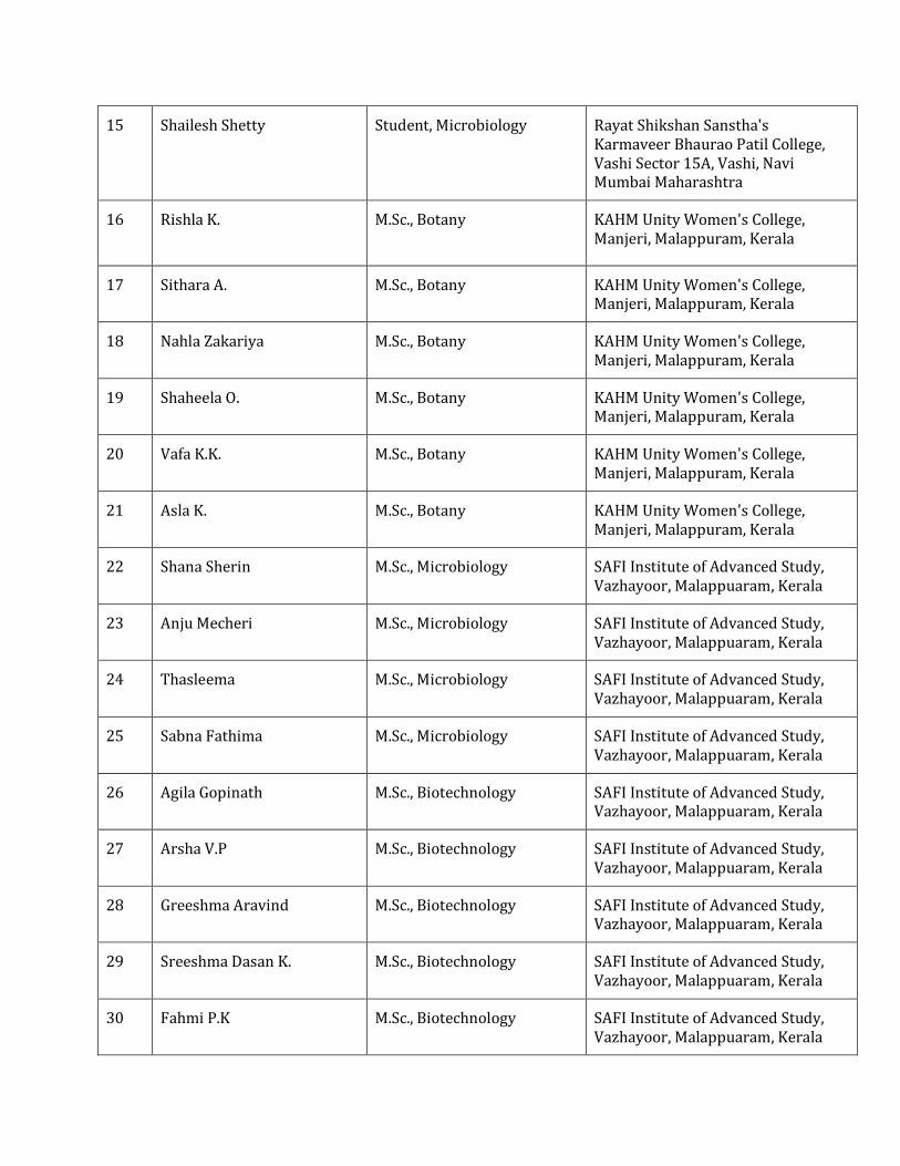
15 Shailesh Shetty
Student, Microbiology Rayat Shikshan Sanstha's Karmaveer Bhaurao Patil College, Vashi Sector 15A, Vashi, Navi Mumbai Maharashtra
16 Rishla K.
M.Sc., Botany KAHM Unity Women's College, Manjeri, Malappuram, Kerala
17 Sithara A. M.Sc., Botany KAHM Unity Women's College, Manjeri, Malappuram, Kerala
18 Nahla Zakariya M.Sc., Botany KAHM Unity Women's College, Manjeri, Malappuram, Kerala
19 Shaheela O. M.Sc., Botany KAHM Unity Women's College, Manjeri, Malappuram, Kerala
20 Vafa K.K. M.Sc., Botany KAHM Unity Women's College, Manjeri, Malappuram, Kerala
21 Asla K. M.Sc., Botany KAHM Unity Women's College, Manjeri, Malappuram, Kerala
22 Shana Sherin M.Sc., Microbiology SAFI Institute of Advanced Study, Vazhayoor, Malappuaram, Kerala
23 Anju Mecheri M.Sc., Microbiology SAFI Institute of Advanced Study, Vazhayoor, Malappuaram, Kerala
24 Thasleema M.Sc., Microbiology SAFI Institute of Advanced Study, Vazhayoor, Malappuaram, Kerala
25 Sabna Fathima M.Sc., Microbiology SAFI Institute of Advanced Study, Vazhayoor, Malappuaram, Kerala
26 Agila Gopinath M.Sc., Biotechnology SAFI Institute of Advanced Study, Vazhayoor, Malappuaram, Kerala
27 Arsha V.P M.Sc., Biotechnology SAFI Institute of Advanced Study, Vazhayoor, Malappuaram, Kerala
28 Greeshma Aravind M.Sc., Biotechnology SAFI Institute of Advanced Study, Vazhayoor, Malappuaram, Kerala
29 Sreeshma Dasan K. M.Sc., Biotechnology SAFI Institute of Advanced Study, Vazhayoor, Malappuaram, Kerala
30 Fahmi P.K M.Sc., Biotechnology SAFI Institute of Advanced Study, Vazhayoor, Malappuaram, Kerala
List of Resource Persons
Sl.No Name and Address
1 Dr. Minoo Divakaran Providence Women’s College, Kozhikode- 673009 [email protected]
2 Dr. Geetha S. Pillai Deputy Project Director Centre for Medicinal Plants Research, Arya Vaidya Sala, Kottakkal, Malappuram, Kerala-676503
3 Dr. Servin Wesley P Assistant Professor and Head, Department of Biotechnology, SIAS. [email protected]
4 Dr. B. Sahaya Shibu Assistant Professor, Department of Biotechnology, SIAS. [email protected]
5 Dr. S. Manjusha Assistant Professor, Department of Biotechnology, SIAS. [email protected]
Contents
Sl. No. Topic Titles Page No.
1 Tissue Culture - A Potential Tool For Propagation and Conservation of Orchids
1-2
2 Orchid Micropropagation- An Introduction 3
3 Medicinal Orchids and Application of Biotechnology for their Propagation, Improvement and Conservation
4-5
4 Plant Tissue Culture – Introduction and History 6-7
5 Laboratory Design for Tissue culture 8-11
6 Laboratory Requirements for Tissue culture 12-14
7 Medium preparation 15-16
8 Medium Sterilization 17-18
9 Culture Room 19
10 Tissue Culture Method- Steps 20-22
11 Inoculation , In vitro Multiplication - Callus Regeneration and Observation of Cultured Clones
20-21
12 Propagation of Cultured clone 22
13 Orchid Micropropagation- Laboratory method 23-24
Department of Biotechnology, SAFI Institute of Advanced Study (SIAS), Vazhayoor, Malappuram, Kerala.
National Workshop on Plant Tissue Culture with Special Reference to Orchid Micropropagation -2017 Page 1
1. TISSUE CULTURE - A POTENTIAL TOOL FOR PROPAGATION AND CONSERVATION OF ORCHIDS
Minoo Divakaran, Assistant Professor
Providence Women’s College, Kozhikode – 673009, Kerala. [email protected]
Orchids are nature's most extravagant and largest group of flowering plants distributed
throughout the world. With an incredible range of diversity in shape, size and color of their
flowers, they are utilized aesthetically, medicinally and as ecological indicators. Their exotic
beauty and long lasting blooming period, lead to their popularity as ornamentals, but however
many are used as herbal medicines, flavours and food, by different tribes around the world.
Except ornamental orchids, other important orchid populations are confined to their natural
habitats, facing an alarming decrease due to climatic changes leading to asynchrony in flowering
patterns, over-exploitation, population pressure and habitat destruction. At present, the orchids
also figure prominently in the Red Data Book prepared by International Union for Conservation
of Nature (IUCN). The family is now included in Appendix-II of Convention on International
Trade in Endangered Species of Wild Fauna and Flora (CITES), restricting all international trade
on the species. Dactylorhiza hatagirea, a high valued medicinal orchid from the Himalayas has
been categorized as critically endangered and listed under appendix I of CITES.
The earliest Middle East report of plant remedies is in a 4000-year-old Sumerian clay tablet
which included some orchids. For flavoring, both Vanilla and Salep are well known and widely
used long ago, the former being also known as the second most costly spice. Medicinal orchids
belong to diverse genera, viz., Bletilla, Calanthe, Coelogyne, Cymbidium, Dendrobium, etc. and
have been used in ancient Indian system of Ayurvedic medicine. Asthavarga an important
ingredient in many classical formulations viz., Chavyanprasa is reported to contain 4 species of
orchids. Dendrobium macraei is another important orchid from Ayurvedic point of view as it is
reported to be source of 'Jivanti'.
Conservation of orchids thus assumes prime significance that needs to be addressed by
both government and private sector of various nation in participation with research institutions,
community growers as well as through international collaboration. The rising threats are
addressed around the world by confluences like the International Orchid Conservation Congress
Department of Biotechnology, SAFI Institute of Advanced Study (SIAS), Vazhayoor, Malappuram, Kerala.
National Workshop on Plant Tissue Culture with Special Reference to Orchid Micropropagation -2017 Page 2
that is organized by a group of many international organizations working on orchids, every four
years
Orchids need to be conserved both in situ and ex situ. However, there is no substitution for
conservation of threatened medicinal orchid species in their natural habitat by natural
propagation method as their propagation rate is very slow. Ex situ conservation measures can be
complementary to in situ methods and they can be used as a safe alternative, against extinction.
Plant tissue culture technique has been accepted as a potential method for large scale
propagation and conservation of rare, threatened and endangered orchids. In vitro propagation
technique has become popular for cut flower industry and medicinal orchids because a
significant number of identical clones can be raised from a single protocorm or shoot tip
explants. The orchid seeds which fail to germinate naturally, due to its ex-albuminous nature, can
be germinated in vitro, so generate a large population of heterozygous segregating population,
thus leading to the accepted usage of embryo / ovule culture, seed culture for this. Embryo rescue
technique can be utilized to rescue and develop interspecific hybrids in orchids, which itself is a
very promising protocol for the floriculture industry. Reports of somatic hybridization using
protoplasts and developing interspecific hybrids between cultivated orchid species and its wild
relatives leading to introgression of desirable traits from the wild into commercial species, using
in vitro techniques, are available. Utilization of in vitro conservation methods, long term as well
as short term using cryopreservation, synthetic seeds, and other methods have helped to design a
conservation strategy for orchids. Thus plant tissue culture is definitely one of the most suitable
technologies that offers varied tools to minimize the pressure on natural population of orchids
and their sustainable utilization.
Department of Biotechnology, SAFI Institute of Advanced Study (SIAS), Vazhayoor, Malappuram, Kerala.
National Workshop on Plant Tissue Culture with Special Reference to Orchid Micropropagation -2017 Page 3
2. ORCHID MICROPROPAGATION- AN INTRRODUCTION
Dr. B. Sahaya Shibu, Assistant Professor, Department of Biotechnology, SAFI Institute of Advanced Study, Malappuram, Kerala
Orchidaceae is a highly successful family, with representatives capable of occupying
almost every ecological situation, apart from marine environments and habitats characterized by
extreme cold throughout the year. Majority of the cultivated orchids are native to tropical
countries and inhabit in humid and tropical forests of India, Srilanka, Myanmar, South China,
Thailand, Malaysia, Philippines, New Guinea, Australia, South and Central America and
Mexico. Orchids exhibit an incredible range of diversity in size, shape and colour of their
flowers and as a result, these plants have great ornamental value.
Around 10 % (3000) of the world’s total orchid species are believed to be endangered in
their native habitats. Orchids are subject to high levels of threat, through both natural and
anthropogenic causes. The greatest threat to orchid diversity is habitat loss. Clearance of natural
vegetation for ranching, monocrop agriculture, mining, logging, burning and urban development,
has decimated many orchid species. Habitat destruction triggers the loss of pollinators, other
plants and fungi on which the orchids mostly depend for their survival.
The concept of in situ conservation of orchids is wrought with many difficulties,
especially the seed biology of orchids, which is highly specialized. Orchid seeds are produced in
large numbers but are exceedingly small. A single orchid seed pod may contain nearly four
million seeds. Orchid seeds, which have been described as ‘dust seeds’, may be as small as 0.01
by 0.05 mm in dimension and weigh as little as 10-30 μg. Orchids exhibit double fertilization as
is typical of all flowering plants; however, endosperm development stops at an early stage and in
many cases definite endosperm tissues are not apparent at seed maturity. In addition, the
embryos are highly reduced. The application of in vitro seed propagation technique for orchid
conservation is a powerful tool for ex situ biodiversity conservation With non-symbiotic
germination technology, thousands of seedlings could be raised to maturity from a single seed
capsule.
Department of Biotechnology, SAFI Institute of Advanced Study (SIAS), Vazhayoor, Malappuram, Kerala.
National Workshop on Plant Tissue Culture with Special Reference to Orchid Micropropagation -2017 Page 4
3. MEDICINAL ORCHIDS AND APPLICATION OF BIOTECHNOLOGY FOR THEIR PROPAGATION, IMPROVEMENT AND CONSERVATION
Geetha S. Pillai, Deputy Project Director Centre for Medicinal Plants Research
Arya Vaidya Sala, Kottakkal, Malappuram, Kerala – 676503.
The Orchidaceae is the largest and most diverse family of flowering plants,
comprised of nearly 27,135 species belonging to 925 genera. They are most highly evolved
members represent about 30% of monocotyledons or 10% of flowering plants. Orchids
form an extremely peculiar group of plants due to the specialized morphology and
structure of flowers, their highly specialized pollination mechanism, small, thin, non-
endospermic seeds, obligate mycorrhizal association during seed germination, etc. Apart
from their ornamental value, they have recorded medicinal use too. Orchids have been
used as a source of medicine for thousands of years to treat different diseases and ailments.
Worldwide about 2% of the orchids are reported to be medicinal, but China uses 20% of its
native orchids for medicinal purposes. Orchids are used for medicinal purpose for
centuries in India, Southeast Asia, the Middle East, Africa, Europe and the Americas.
Moreover, many preparations with orchids as one of the ingredients are used as emetic,
purgative, aphrodisiac, vermifuge, cooling agent, bronchodilator, contraceptive agents, etc.
The rapid progress in orchid biotechnology has helped in determining their origin
and evolutionary trends, clarifying phylogenetic relationships, rapid clonal propagation, to
produce new varieties with novel characteristics, etc. The characterization of orchid
alkaloids has aided the discovery of novel bioactive compounds. Orchids, like other plants,
produce a large number of bioactive phytochemicals such as alkaloids, flavonoids,
carotenoids, sterols, anthocyanins, etc. It is reported that, to date, more than 2000 orchid
species have been screened for their chemical constituents and many bioactive alkaloids
have been isolated from orchids.
Techniques such as mass induction and multiplication of PLBs, large-scale seedling
production without damaging the plant or their parts, rooting and easy acclimatization
procedures are some of the achievements that have revolutionized orchid propagation.
Micropropagation of orchids is one of the highly commercialized field of biotechnology. The
Department of Biotechnology, SAFI Institute of Advanced Study (SIAS), Vazhayoor, Malappuram, Kerala.
National Workshop on Plant Tissue Culture with Special Reference to Orchid Micropropagation -2017 Page 5
overexploitation of wild orchid stocks for ornamental and medicinal purposes,
deforestation, urbanization, destruction of habitats, loss of specialized pollinators and
unauthorized trade has led to a reduction in natural populations of many orchids.
Therefore, conservation of orchids is of paramount importance to meet the demand and for
preserving this valuable group of ornamentals for posterity. A number of techniques such
as establishment of seed gene bank, slow growth conservation and cryopreservation are
available for conserving orchids. Use of biotechnological tools in orchids will lead to an
understanding of the biological, physiological, molecular, genetic and evolutionary
mechanisms of medicinal orchids.
Department of Biotechnology, SAFI Institute of Advanced Study (SIAS), Vazhayoor, Malappuram, Kerala.
National Workshop on Plant Tissue Culture with Special Reference to Orchid Micropropagation -2017 Page 6
4. AN INTRODUCTION TO PLANT TISSUE CULTURE
Plant tissue culture is a collection of techniques used to maintain or grow plant
cells, tissues or organs under sterile conditions on a nutrient culture medium of known
composition. Plant tissue culture is widely used to produce clones of a plant in a method
known as Micropropagation. The theoretical basis for plant tissue culture was proposed by
Gottlieb Haberlandt, German academy of science in 1902 on his experiments on the
culture of a single cell. The first culture was obtained by Gautheret from the cambial tissue
of Acer pseudoplantanus.
The term plant tissue culture (Micropropagation) is generally used for the aseptic
culture of cells, tissues, organs and their components under defined chemical and physical
conditions in vitro. The basic concept of the PTC, plant body can be dissected into smaller
part termed as ‘explant’ and any explants can be developed into a whole plant. It is a
central innovative area of applied plant science, including agriculture and plant
biotechnology. This technique is effective because almost all the plant cells are totipotent;
in each cell possess the genetic information and cellular machinery necessary to generate
the whole organism. Since, this technique can be used to produce a higher number of plants
that are genetically similar to a parent plant as well as to another. Two concepts, plasticity
and totipotency, are the central processes to understand the regeneration and plant cell
culture. Plants due to its longer lifespan and sessile nature have developed a greater ability
to overcome the extreme conditions. Most of the processes include in plant development
and the growth, adapt to environmental conditions. When the plant cells and tissues are
cultured in vitro, most of them are generally exhibit a very high degree of plasticity, which
allows one type of organ or tissue to be initiated from another type. Like this way the whole
plant can be subsequently regenerated. This maintenance of genetic potential is called
totipotency.
Department of Biotechnology, SAFI Institute of Advanced Study (SIAS), Vazhayoor, Malappuram, Kerala.
National Workshop on Plant Tissue Culture with Special Reference to Orchid Micropropagation -2017 Page 7
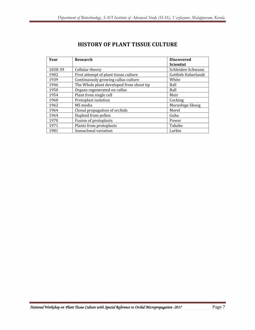
HISTORY OF PLANT TISSUE CULTURE
Year Research Discovered Scientist
1838-39 Cellular theory Schleiden-Schwann 1902 First attempt of plant tissue culture Gottlieb Haberlandt 1939 Continuously growing callus culture White 1946 The Whole plant developed from shoot tip Ball 1950 Organs regenerated on callus Ball 1954 Plant from single cell Muir 1960 Protoplast isolation Cocking 1962 MS media Murashige-Skoog 1964 Clonal propagation of orchids Morel 1964 Haploid from pollen Guha 1970 Fusion of protoplasts Power 1971 Plants from protoplasts Takebe 1981 Somaclonal variation Larkin
Department of Biotechnology, SAFI Institute of Advanced Study (SIAS), Vazhayoor, Malappuram, Kerala.
National Workshop on Plant Tissue Culture with Special Reference to Orchid Micropropagation -2017 Page 8
5. LABORATORY DESIGN FOR PLANT TISSUE CULTURE (a)Washing and storage facilities
(b) Media preparation, sterilization and storage room
(c) Aseptic area for Inoculation
(d) Transfer area for aseptic manipulations
(e) Culture rooms or incubators for maintenance of cultures under controlled conditions of temperature, light and humidity
(f) Observation or data collection area (g) Transplantation area
a. Washing Area and storage facilities This is very important for a tissue culture laboratory. It should be provided with a large sink, running hot and cold tap water, brushes of various sizes, detergent and a bucket of single distilled water for a final rinse of the washed glass goods.
A number of plastic buckets are required for soaking the glass goods to be washed. Another separate bucket with lid is also required for disposing off the used or infected media before cleaning. Only this bucket should be kept outside the room or cleaning area and should be cleaned twice-in a week. For storage of washed and dried labware, the laboratory should be provided with dustproof cupboards or storage cabinets.
b. Media Preparation, Sterilization Room and Storage Space: This part is the central section of the laboratory where most of the activities are performed i.e., media preparation and sterilization of media and glasswares needed for culture.
There should be sufficient working bench as well as storage space.
c. Aseptic Area for Inoculation:
This room should be without any window or ventilator in order to make it dust-free. The room should be provided with double doors. The doors should have an automatic door closer. Inside floor should be fitted with a rubber mat to facilitate cleaning. For entering into the room, shoes should be left outside. For aseptic work, a large wooden chamber (Ca, 4′ x 4′ x 7′) is made for short term work. Upper half of the side walls of the chamber are made of large glass sheets. The chamber should also have double doors provided with a door closer.
Department of Biotechnology, SAFI Institute of Advanced Study (SIAS), Vazhayoor, Malappuram, Kerala.
National Workshop on Plant Tissue Culture with Special Reference to Orchid Micropropagation -2017 Page 9
The chamber is provided with two UV (one small, one big) sterilizing lamps and a fluorescent lamp. The switches to operate them are present outside the chamber so that the lamps can be safely switched on and off. Inside the chamber the working table and shelves are made of thick glass sheets.
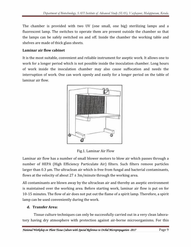
Laminar air flow cabinet
It is the most suitable, convenient and reliable instrument for aseptic work. It allows one to work for a longer period which is not possible inside the inoculation chamber. Long hours of work inside the inoculation chamber may also cause suffocation and needs the interruption of work. One can work openly and easily for a longer period on the table of laminar air flow.
Fig.1. Laminar Air Flow
Laminar air flow has a number of small blower motors to blow air which passes through a number of HEPA (High Efficiency Particulate Air) filters. Such filters remove particles larger than 0.3 µm. The ultraclean air which is free from fungal and bacterial contaminants, flows at the velocity of about 27 ± 3m/minute through the working area.
All contaminants are blown away by the ultraclean air and thereby an aseptic environment is maintained over the working area. Before starting work, laminar air flow is put on for 10-15 minutes. The flow of air does not put out the flame of a spirit lamp. Therefore, a spirit lamp can be used conveniently during the work.
d. Transfer Area:
Tissue culture techniques can only be successfully carried out in a very clean labora-tory having dry atmosphere with protection against air-borne microorganisms. For this
Department of Biotechnology, SAFI Institute of Advanced Study (SIAS), Vazhayoor, Malappuram, Kerala.
National Workshop on Plant Tissue Culture with Special Reference to Orchid Micropropagation -2017 Page 10
purpose a sterile dust-free room/cabinet is needed for routine transfer and manipulation work. The ‘laminar air flow cabinet’ Fig. 1 is the most common accessory used for aseptic manipulations now-a-days. The cabinet may be designed with horizontal air flow or vertical air flow where the air is forced into the cabinet through a bacterial HEPA (High Efficiency Particulate Air) filter. The air flows over the working bench at a constant rate which prevents the particles (microorganisms) from settling on the bench.
Before operation in the laminar air flow cabinet, the interior of the cabinet is sterilised with the ultraviolet (UV) germicidal light and wiping the floor of cabinet with 70% alcohol. Inoculation chamber, a specially designed air tight glass chamber fitted with UV light, may also be used as transfer area.
e. Culture Room:
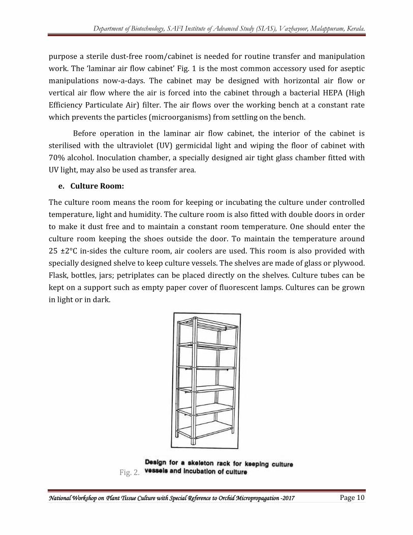
The culture room means the room for keeping or incubating the culture under controlled temperature, light and humidity. The culture room is also fitted with double doors in order to make it dust free and to maintain a constant room temperature. One should enter the culture room keeping the shoes outside the door. To maintain the temperature around 25 ±2°C in-sides the culture room, air coolers are used. This room is also provided with specially designed shelve to keep culture vessels. The shelves are made of glass or plywood. Flask, bottles, jars; petriplates can be placed directly on the shelves. Culture tubes can be kept on a support such as empty paper cover of fluorescent lamps. Cultures can be grown in light or in dark.
Fig. 2.
Department of Biotechnology, SAFI Institute of Advanced Study (SIAS), Vazhayoor, Malappuram, Kerala.
National Workshop on Plant Tissue Culture with Special Reference to Orchid Micropropagation -2017 Page 11
For light arrangement, each culture rack is provided with fluorescent lamps which are photo periodically controlled by an automatic timer. Racks covered with black curtains are suitable for dark incubation of culture. A thermometer and a hygrometer are fixed on the wall at the safety corner of the room to check temperature and relative humidity respectively.
The relative humidity of the culture room is maintained above 50%. Some small shelves are placed in the culture room for temporarily keeping the autoclaved articles and the culture vials containing the medium. The culture room should also have a shaker for suspension culture or single cell culture in moving liquid medium. The speed of revolution of the shaker can be controlled. The shaker is also provided with light. The platform of the shaker is fitted with clips for holding conical flasks (150 ml to 200 ml)
f. Data Collection Area:
The growth and development of tissues cultured in vitro are generally monitored by observing cultures at regular intervals in the culture room or incubators where they have been maintained under controlled environmental conditions.
Arrangement should be there where the observations can be done under aseptic conditions using microscope. Special facilities are required for germplasm conservation i.e., cryopreservation accessories should be there.
g. Transplantation Area:
Plants regenerated from in vitro tissue culture are transplanted to soil in pots. The potted plants are ultimately transferred to greenhouse but prior to transfer the tissue culture grown plants are allowed for acclimatization under well humid condition and controlled temperature and under controlled entry of sunlight.
Department of Biotechnology, SAFI Institute of Advanced Study (SIAS), Vazhayoor, Malappuram, Kerala.
National Workshop on Plant Tissue Culture with Special Reference to Orchid Micropropagation -2017 Page 12
6. LABORATORY REQUIREMENTS FOR PLANT TISSUE CULTURE
Plant tissue culture techniques are essential to many types of academic inquiry, as well as to many applied aspects of plant science. In the past, plant tissue culture techniques have been used in academic investigations of totipotency and the roles of hormones in Cytodifferentiation and Organogenesis. Currently, tissue-cultured plants that have been genetically engineered provide insight into plant molecular biology and gene regulation. Plant tissue culture techniques are also central to innovative areas of applied plant science, including plant biotechnology and agriculture. For example, select plants can be cloned and cultured as suspended cells from which plant products can be harvested. In addition, the management of genetically engineered cells to form transgenic whole plants requires tissue culture procedures; tissue culture methods are also required for the formation of somatic haploid embryos from which homozygous plants can be generated. Thus, tissue culture techniques have been, and still are, prominent in academic and applied plant science.
General Organization
Localize each portion of the tissue culture procedure in a specified place in the laboratory. An assembly-line arrangement of work areas (such as media preparation, glassware washing, sterilization, microscopy, and aseptic transfers) facilitates all operations and enhances cleanliness.
Glassware
Use glassware that has only been used for tissue culture and not other experiments. Toxic metal ions absorbed on glassware can be especially troublesome. Wash glassware with laboratory detergent, then rinse several times with tap water and, finally, rinse with purified water.
High-purity Water
Use only high-purity water in tissue culture procedures. Double glass distilled water or demonized water from an ion-exchanger are acceptable. Water should not be stored but used immediately. Regular maintenance and monitoring of water purification equipment are necessary. Purified water for tissue culture can also be purchased.
Plant Material
Plants used in tissue culture need to be healthy and actively growing. Stressed plants, particularly water-stressed plants, usually do not grow as tissue cultures. Insect and disease-free greenhouse plants are rendered aseptic more readily, so contamination rate is lower when these plants are used in tissue culture procedures. Seeds that can be easily
Department of Biotechnology, SAFI Institute of Advanced Study (SIAS), Vazhayoor, Malappuram, Kerala.
National Workshop on Plant Tissue Culture with Special Reference to Orchid Micropropagation -2017 Page 13
surface sterilized usually produce contamination-free plants that can be grown under clean greenhouse conditions for later experimental use.
Aseptic Technique
The essence of the aseptic technique is the exclusion of invading microorganisms during experimental procedures. If sterile tissues are available, then the exclusion of microorganisms is accomplished by using sterile instruments and culture media concurrently with standard bacteriological transfer procedures to avoid extraneous contamination. Media and apparatus are rendered sterile by autoclaving at 15 lbs/inch2 (121°C) for 15 minutes. The use of disposable sterile plastic ware reduces the need for some autoclaving. Alternative sterilization techniques such as filter sterilization must be employed for heat-labile substances like cytokinins. Aseptic transfers can be made on the laboratory bench top by using standard bacteriological techniques (i.e., flaming instruments prior to use and flaming the opening of receiving vessels prior to transfer). Aseptic transfers are more easily performed in a transfer chamber such as a laminar flow hood, which is also preferably equipped with a Bunsen burner. If experimental tissues are not aseptic, then surface sterilization procedures specific to the tissues are employed. Common sterilants are ethyl alcohol and/or Clorox with an added surfactant. The Concentration of sterilants and exposure time are determined empirically.
The following items are also essential in the Tissue culture room
- Different types of glasswares - Different kinds of balances - Required chemicals - Hot plates and Stirrer - Water bath - pH meter - Autoclave and Hot air oven - Microwave oven - Vortex, Shaker - Centrifuge - Refrigerator and Freezer - Storage cabinet (Dust-free)
Hot Air Oven: It is necessary for drying the washed glass goods. For this purpose, a number of enameled trays of different sizes are required for keeping wet glass goods inside the oven. Refrigerator:
Department of Biotechnology, SAFI Institute of Advanced Study (SIAS), Vazhayoor, Malappuram, Kerala.
National Workshop on Plant Tissue Culture with Special Reference to Orchid Micropropagation -2017 Page 14
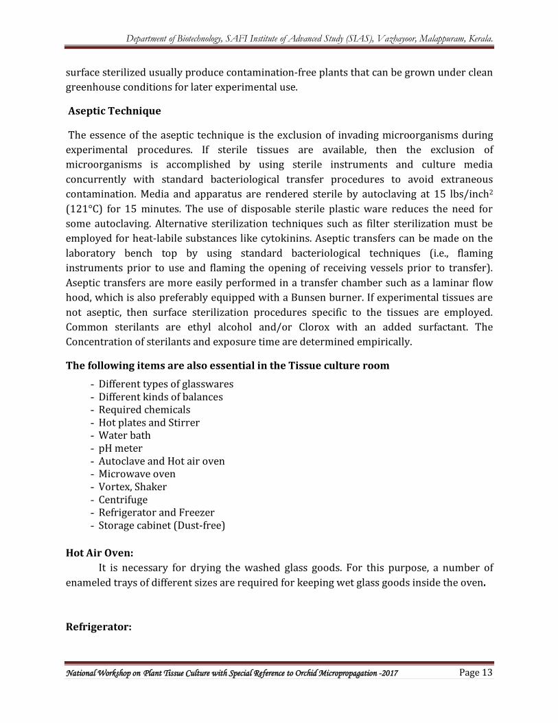
It is essential for storing various thermo- labile chemicals like vitamins, hormones, amino acids, casein-hydrolysate, yeast extract, coconut milk, etc. Stock solutions of salts are also kept to prevent contamination. Distillation Plant: A single distillation and a double distillation water plant are indispensible. Two big plastic containers are required for storing the distilled water. Weighing Balance: Three types of weighing balances viz. pan balance; chemical balance and electric balance are required for weighing chemicals, sugars, agar- agar and others. pH Meter: It is necessary for the measurement and adjustment of pH of the nutrient medium
Fig.3. Digital pH meter
Vacuum Pump:
It is required for filtering liquid media, sugar solution etc. through filter apparatus using air suction.
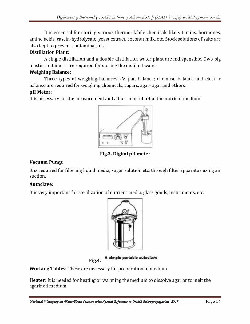
Autoclave: It is very important for sterilization of nutrient media, glass goods, instruments, etc.
Fig.4. Working Tables: These are necessary for preparation of medium Heater: It is needed for heating or warming the medium to dissolve agar or to melt the agarified medium.
Department of Biotechnology, SAFI Institute of Advanced Study (SIAS), Vazhayoor, Malappuram, Kerala.
National Workshop on Plant Tissue Culture with Special Reference to Orchid Micropropagation -2017 Page 15
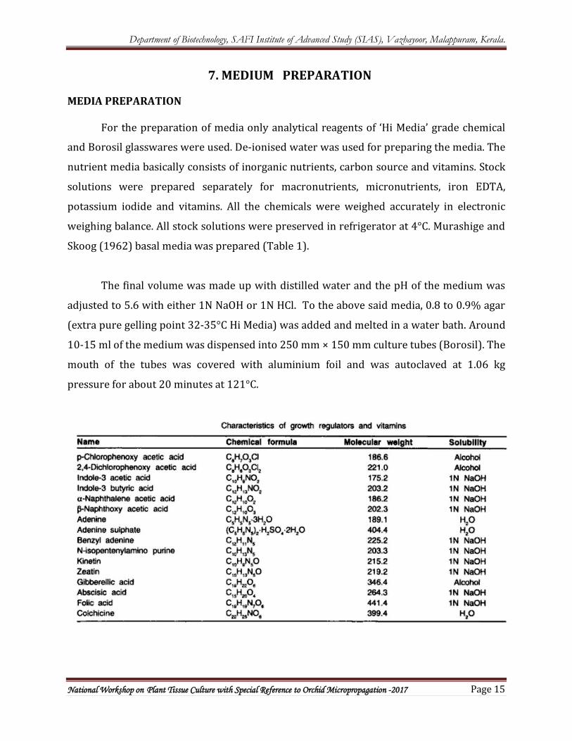
7. MEDIUM PREPARATION
MEDIA PREPARATION
For the preparation of media only analytical reagents of ‘Hi Media’ grade chemical
and Borosil glasswares were used. De-ionised water was used for preparing the media. The
nutrient media basically consists of inorganic nutrients, carbon source and vitamins. Stock
solutions were prepared separately for macronutrients, micronutrients, iron EDTA,
potassium iodide and vitamins. All the chemicals were weighed accurately in electronic
weighing balance. All stock solutions were preserved in refrigerator at 4°C. Murashige and
Skoog (1962) basal media was prepared (Table 1).
The final volume was made up with distilled water and the pH of the medium was
adjusted to 5.6 with either 1N NaOH or 1N HCl. To the above said media, 0.8 to 0.9% agar
(extra pure gelling point 32-35°C Hi Media) was added and melted in a water bath. Around
10-15 ml of the medium was dispensed into 250 mm × 150 mm culture tubes (Borosil). The
mouth of the tubes was covered with aluminium foil and was autoclaved at 1.06 kg
pressure for about 20 minutes at 121°C.
Department of Biotechnology, SAFI Institute of Advanced Study (SIAS), Vazhayoor, Malappuram, Kerala.
National Workshop on Plant Tissue Culture with Special Reference to Orchid Micropropagation -2017 Page 16
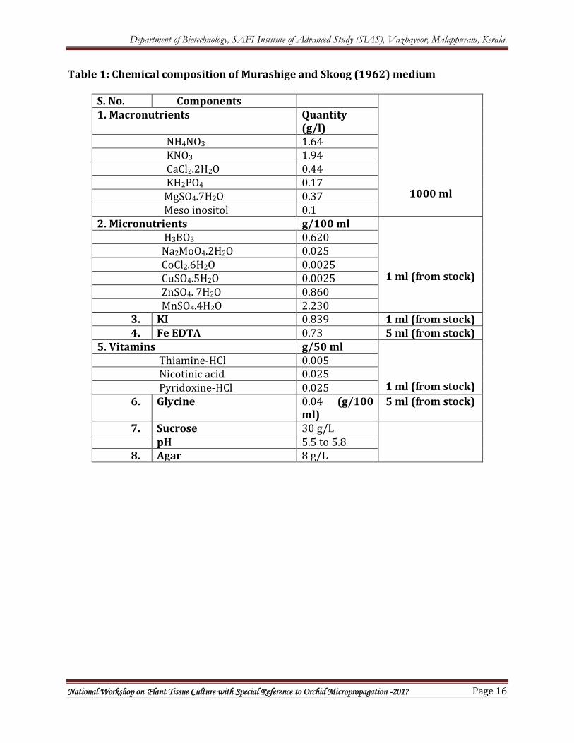
Table 1: Chemical composition of Murashige and Skoog (1962) medium
S. No. Components
1000 ml
1. Macronutrients Quantity (g/l)
NH4NO3 1.64 KNO3 1.94 CaCl2.2H2O 0.44 KH2PO4 0.17 MgSO4.7H2O 0.37 Meso inositol 0.1 2. Micronutrients g/100 ml
1 ml (from stock)
H3BO3 0.620 Na2MoO4.2H2O 0.025 CoCl2.6H2O 0.0025 CuSO4.5H2O 0.0025 ZnSO4. 7H2O 0.860 MnSO4.4H2O 2.230
3. KI 0.839 1 ml (from stock) 4. Fe EDTA 0.73 5 ml (from stock)
5. Vitamins g/50 ml 1 ml (from stock)
Thiamine-HCl 0.005 Nicotinic acid 0.025 Pyridoxine-HCl 0.025
6. Glycine
0.04 (g/100 ml)
5 ml (from stock)
7. Sucrose 30 g/L pH 5.5 to 5.8 8. Agar 8 g/L
Department of Biotechnology, SAFI Institute of Advanced Study (SIAS), Vazhayoor, Malappuram, Kerala.
National Workshop on Plant Tissue Culture with Special Reference to Orchid Micropropagation -2017 Page 17
8. MEDIUM STERILIZATION
STERILIZATION METHODS
Sterilization is defined as the maintenance of aseptic environment for successful experimentation. To maintain sterile environment all culture vessels, media and instruments used in handling tissues as well as the explant itself must be sterilized. All operations are carried out in biosafety hood / laminar air flow cabinet as UV light penetration helps in elimination of pathogenic organisms.
Dry sterilization
The sterilization of glassware and metallic instruments can be carried out in dry heat for 3 hr. at 160-180°C. Moist heat sterilization can be done at 121°C, 15 psi for 15 minutes duration. Glass wares, metal instruments like forceps, scalpels surgical blades etc., should be wrapped by aluminium foil and autoclaved followed by heating in an oven. Domestic pressure cookers are very useful in sterilizing a small amount of media.
Steam / Wet sterilization
Nutrient media are sterilized by using either autoclave or pressure cookers. For the glass containers with a capacity of 20-40 ml of nutrient media autoclave with 121°C, 15 lbs pressure and 15- 20 minutes conditions are used. An autoclave has a normal temperature range of 115-130 °C. Proper sterilization relies on time, pressure, temperature and volume of the object to be sterilized. The uses of an autoclave are speed, easiness to handle and destruction of viruses and microbes, while the demerits are change in pH by 0.2 -0.5 units components becoming isolated and occurrence of chemical reactions, resulting in a loss of activity of media constituents.
Filter Sterilization
Vitamins, amino acids, growth regulators and toxins are heat labile and get destroyed during autoclaving with the other nutrients compounds. Those compounds can be sterilized by filtration through a sieve or filter membranes of 0.45 to 0.22 μm. Other membrane filters (Sartorius, Labgene, Millipore etc.,) and related equipment are available for sterilization of different volumes of the liquid in the range of 1 – 200 ml. Most filters are of cellulose acetate, cellulose nitrate and /or nitrocellulose are available. During filter sterilization, all the particles, microbes and viral particles which are bigger than the pore diameter of the filter used are eliminated.
Department of Biotechnology, SAFI Institute of Advanced Study (SIAS), Vazhayoor, Malappuram, Kerala.
National Workshop on Plant Tissue Culture with Special Reference to Orchid Micropropagation -2017 Page 18
Procedure for Aseptic Transfer
Switch on the UV lamp for half an hour and close the door, afterwards switch off the light, open the door and switch on the air flow. The person who is working should spray 70% ethanol on his hands and rub with cotton and be sterile. Rub on all sides of the laminar flow cabinet with 70% ethanol. Use head cap, nasal covering in order to avoid contamination by air. It is always advisable to keep a sterile towel soaked with 70% ethanol in the working area.
The glassware may be tubes, petridishes, bottles with prepared and autoclaved media within are carefully transferred to the laminar flow cabinet by washing the surface of the glassware with 70% ethanol so that we can avoid transmission of surface contaminants. Set up all the glassware aside and then switch on the Bunsen burner / spirit lamp and flame sterilize the filter papers and be ready the explant sterilization.
Explant sterilization
Explants (e.g: nodal segment, axillary bud, shoot tip, hairy root, anther, ovary etc.,) taken from the main plant are thoroughly washed in tap water then rinsed in Teepol or Tween 20 for three times then wash in distilled and autoclaved distilled water thrice. Now the explants are rinsed in any one of the following chemical sterilants viz.,
1. Mercuric Chloride 0.01-1% for 2-5 min
2. Sodium hypochlorite 1-1.4% for 5-30 min
3. Hydrogen peroxide 10-12% for 5-15 min
4. Calcium hypochlorite 4-10% for 5-30 min
5. Bromine water 1-2 % for 2-10 min
6. Silver nitrate 1% for 5- 30 min
7. Antibiotics 4-50 mg/l for 30-60 min
Explants after treatment with sterilants, must be thoroughly rinsed with several changes of sterile distilled water because retention of such noxious chemicals will seriously affect the establishment of culture.
Department of Biotechnology, SAFI Institute of Advanced Study (SIAS), Vazhayoor, Malappuram, Kerala.
National Workshop on Plant Tissue Culture with Special Reference to Orchid Micropropagation -2017 Page 19
9. CULTURE ROOM
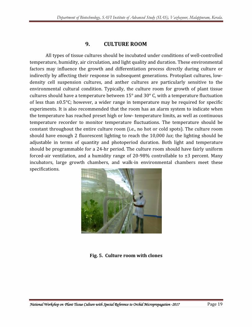
All types of tissue cultures should be incubated under conditions of well-controlled temperature, humidity, air circulation, and light quality and duration. These environmental factors may influence the growth and differentiation process directly during culture or indirectly by affecting their response in subsequent generations. Protoplast cultures, low-density cell suspension cultures, and anther cultures are particularly sensitive to the environmental cultural condition. Typically, the culture room for growth of plant tissue cultures should have a temperature between 15° and 30° C, with a temperature fluctuation of less than ±0.5°C; however, a wider range in temperature may be required for specific experiments. It is also recommended that the room has an alarm system to indicate when the temperature has reached preset high or low- temperature limits, as well as continuous temperature recorder to monitor temperature fluctuations. The temperature should be constant throughout the entire culture room (i.e., no hot or cold spots). The culture room should have enough 2 fluorescent lighting to reach the 10,000 lux; the lighting should be adjustable in terms of quantity and photoperiod duration. Both light and temperature should be programmable for a 24-hr period. The culture room should have fairly uniform forced-air ventilation, and a humidity range of 20-98% controllable to ±3 percent. Many incubators, large growth chambers, and walk-in environmental chambers meet these specifications.
Fig. 5. Culture room with clones
Department of Biotechnology, SAFI Institute of Advanced Study (SIAS), Vazhayoor, Malappuram, Kerala.
National Workshop on Plant Tissue Culture with Special Reference to Orchid Micropropagation -2017 Page 20
10. TISSUE CULTURE METHOD
The whole plant can be regenerated virtually from any plant part (referred to explant) or cells. Steps
Plant tissue culture techniques involve the following steps:
a) Preparation and selection of suitable nutrient media.
b) Selection of explants such as shoot tip.
c) Surface sterilization of the explants by disinfectants
E.g. sodium or calcium hypochlorite solution 0.3–0.6% followed by washing the explants with sterile distilled water. d) Inoculation or Transfer of the explants onto the suitable pre-sterilized nutrient medium in culture vessels under sterile conditions (using laminar flow hood).
e) Incubation or growing the cultures in the growth chamber or plant tissue culture room at optimum physical conditions of light (16 hours of photoperiod), diurnal illumination, temperature (25+/- 20˚C) and relative humidity (50% - 60%).
f) Regeneration of plants from cultured plant tissues.
g) Hardening: It is the gradual exposure of plantlets for acclimatization to environmental conditions.
h) Transfer of plants to the field conditions following the acclimatization/ hardening of the regenerated plants.
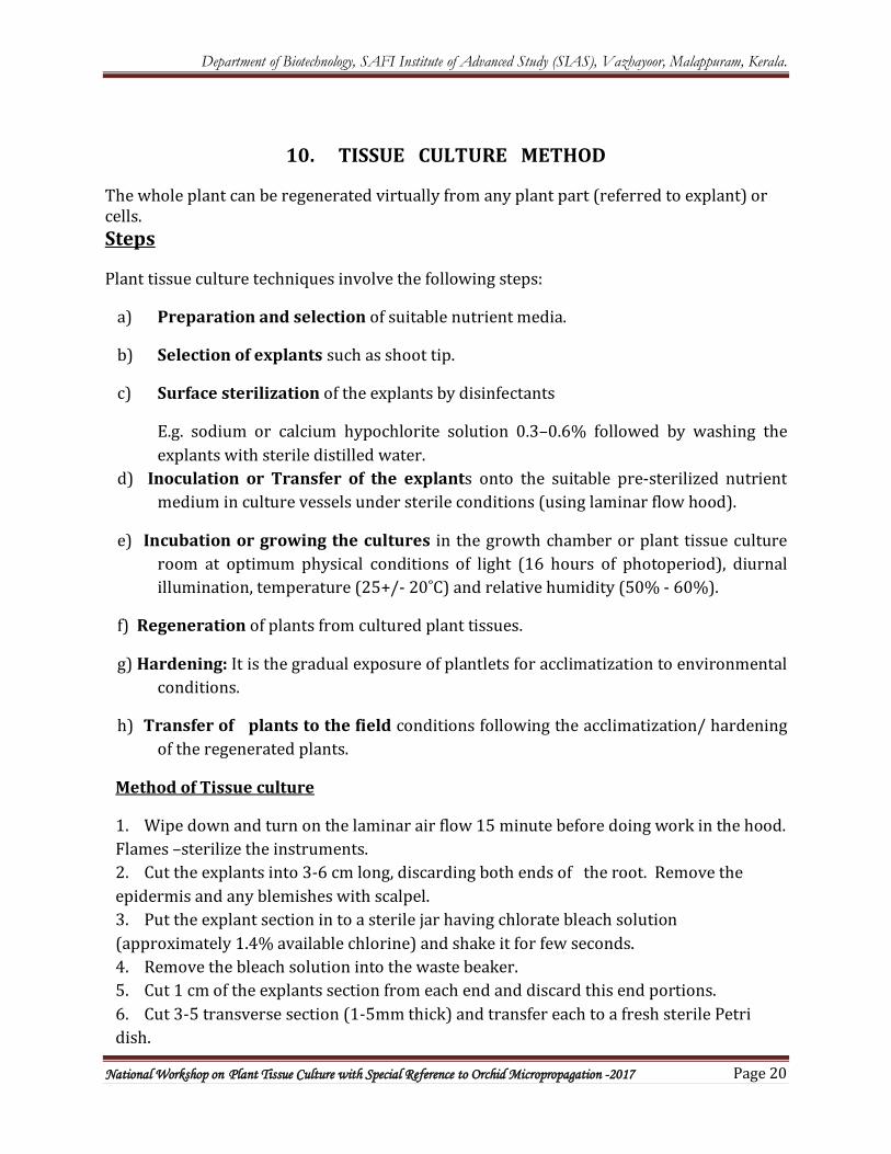
Method of Tissue culture
1. Wipe down and turn on the laminar air flow 15 minute before doing work in the hood. Flames –sterilize the instruments. 2. Cut the explants into 3-6 cm long, discarding both ends of the root. Remove the epidermis and any blemishes with scalpel. 3. Put the explant section in to a sterile jar having chlorate bleach solution (approximately 1.4% available chlorine) and shake it for few seconds. 4. Remove the bleach solution into the waste beaker. 5. Cut 1 cm of the explants section from each end and discard this end portions. 6. Cut 3-5 transverse section (1-5mm thick) and transfer each to a fresh sterile Petri dish.
Department of Biotechnology, SAFI Institute of Advanced Study (SIAS), Vazhayoor, Malappuram, Kerala.
National Workshop on Plant Tissue Culture with Special Reference to Orchid Micropropagation -2017 Page 21
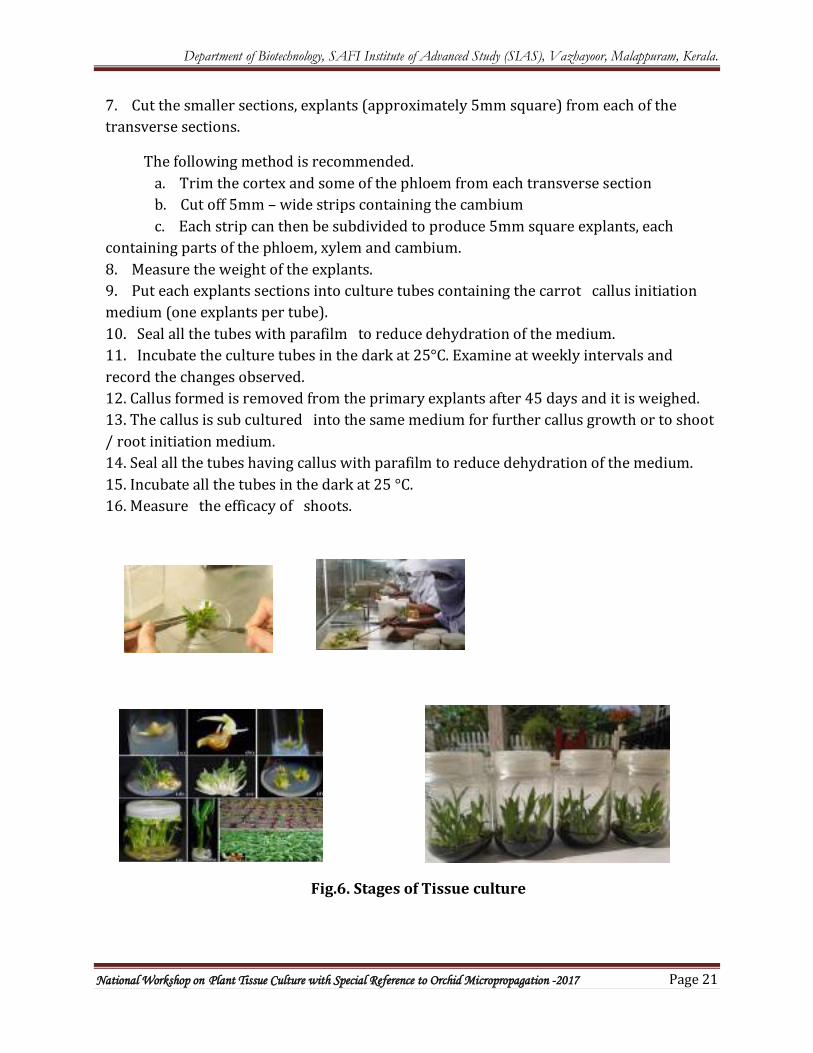
7. Cut the smaller sections, explants (approximately 5mm square) from each of the transverse sections.
The following method is recommended. a. Trim the cortex and some of the phloem from each transverse section b. Cut off 5mm – wide strips containing the cambium c. Each strip can then be subdivided to produce 5mm square explants, each containing parts of the phloem, xylem and cambium. 8. Measure the weight of the explants. 9. Put each explants sections into culture tubes containing the carrot callus initiation medium (one explants per tube). 10. Seal all the tubes with parafilm to reduce dehydration of the medium. 11. Incubate the culture tubes in the dark at 25°C. Examine at weekly intervals and record the changes observed. 12. Callus formed is removed from the primary explants after 45 days and it is weighed. 13. The callus is sub cultured into the same medium for further callus growth or to shoot / root initiation medium. 14. Seal all the tubes having callus with parafilm to reduce dehydration of the medium. 15. Incubate all the tubes in the dark at 25 °C. 16. Measure the efficacy of shoots.
Fig.6. Stages of Tissue culture
Department of Biotechnology, SAFI Institute of Advanced Study (SIAS), Vazhayoor, Malappuram, Kerala.
National Workshop on Plant Tissue Culture with Special Reference to Orchid Micropropagation -2017 Page 22
PROPAGATION OF CULTURED CLONES
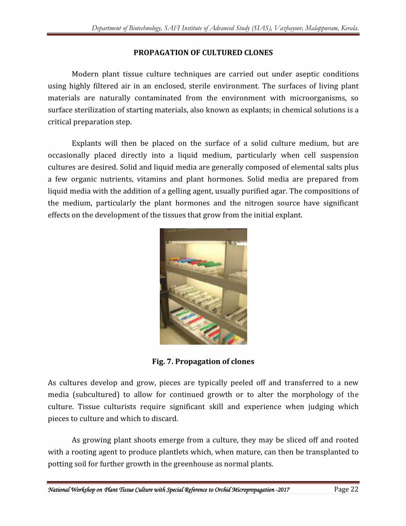
Modern plant tissue culture techniques are carried out under aseptic conditions using highly filtered air in an enclosed, sterile environment. The surfaces of living plant materials are naturally contaminated from the environment with microorganisms, so surface sterilization of starting materials, also known as explants; in chemical solutions is a critical preparation step.
Explants will then be placed on the surface of a solid culture medium, but are occasionally placed directly into a liquid medium, particularly when cell suspension cultures are desired. Solid and liquid media are generally composed of elemental salts plus a few organic nutrients, vitamins and plant hormones. Solid media are prepared from liquid media with the addition of a gelling agent, usually purified agar. The compositions of the medium, particularly the plant hormones and the nitrogen source have significant effects on the development of the tissues that grow from the initial explant.
Fig. 7. Propagation of clones
As cultures develop and grow, pieces are typically peeled off and transferred to a new media (subcultured) to allow for continued growth or to alter the morphology of the culture. Tissue culturists require significant skill and experience when judging which pieces to culture and which to discard.
As growing plant shoots emerge from a culture, they may be sliced off and rooted with a rooting agent to produce plantlets which, when mature, can then be transplanted to potting soil for further growth in the greenhouse as normal plants.
Department of Biotechnology, SAFI Institute of Advanced Study (SIAS), Vazhayoor, Malappuram, Kerala.
National Workshop on Plant Tissue Culture with Special Reference to Orchid Micropropagation -2017 Page 23
11. MICROPROPAGATION OF ORCHID-
Introduction
Orchidaceae is a highly successful family, with representatives capable of occupying
almost every ecological situation, apart from marine environments and habitats characterized by
extreme cold throughout the year. Orchids exhibit an incredible range of diversity in size, shape
and colour of their flowers and as a result, these plants have great ornamental value. Around 10%
(3000) of the world’s total orchid species are believed to be endangered in their native habitats.
Orchids are subject to high levels of threat, through both natural and anthropogenic causes. The
greatest threat to orchid diversity is habitat loss. Clearance of natural vegetation for ranching,
monocrop agriculture, mining, logging, burning and urban development, has decimated many
orchid species. Habitat destruction triggers the loss of pollinators, other plants and fungi on
which the orchids mostly depend for their survival. Micropropagation is an excellent tool for
conservation of orchids.
Micropropagation
The freshly collected capsules were surface sterilized in sodium hypochlorite solution
(NaClO, 0.6 % w/v) or Mercuric Chloride (0.1 %), then rinse thrice with sterile distilled water,
dipped in 70 % (v/v) ethanol for few seconds and flame it. Seeds from the surface sterilized
capsules can be extracted by splitting the capsule longitudinally with a sharp sterilized surgical
blade. The seeds are then spread as thin film in the test tube containing 10 ml of solid culture
medium.
Medium for orchids
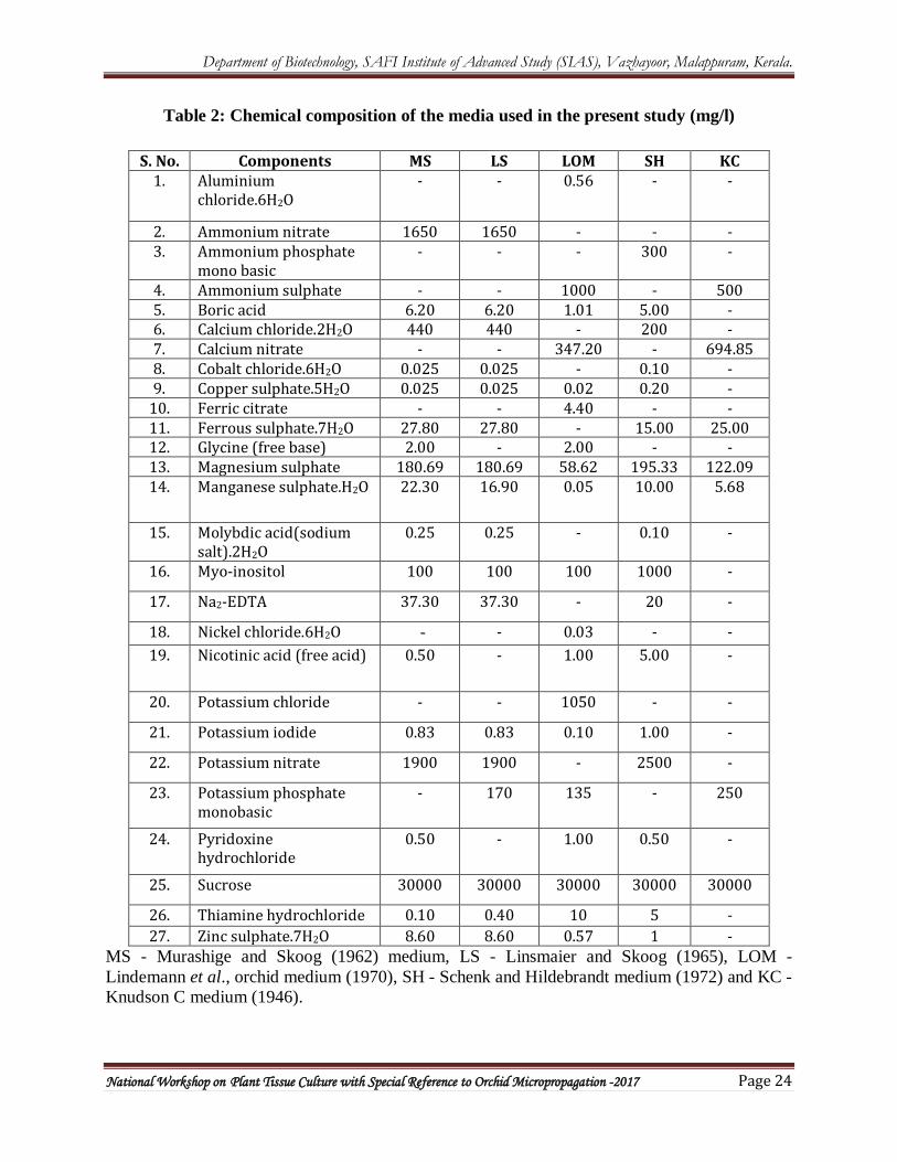
Different type of orchid medium and normal plant tissue culture medium is available
(Table 2).
Department of Biotechnology, SAFI Institute of Advanced Study (SIAS), Vazhayoor, Malappuram, Kerala.
National Workshop on Plant Tissue Culture with Special Reference to Orchid Micropropagation -2017 Page 24
Table 2: Chemical composition of the media used in the present study (mg/l)
S. No. Components MS LS LOM SH KC 1. Aluminium
chloride.6H2O - - 0.56 - -
2. Ammonium nitrate 1650 1650 - - - 3. Ammonium phosphate
mono basic - - - 300 -
4. Ammonium sulphate - - 1000 - 500 5. Boric acid 6.20 6.20 1.01 5.00 - 6. Calcium chloride.2H2O 440 440 - 200 - 7. Calcium nitrate - - 347.20 - 694.85 8. Cobalt chloride.6H2O 0.025 0.025 - 0.10 - 9. Copper sulphate.5H2O 0.025 0.025 0.02 0.20 -
10. Ferric citrate - - 4.40 - - 11. Ferrous sulphate.7H2O 27.80 27.80 - 15.00 25.00 12. Glycine (free base) 2.00 - 2.00 - - 13. Magnesium sulphate 180.69 180.69 58.62 195.33 122.09 14. Manganese sulphate.H2O 22.30 16.90 0.05 10.00 5.68
15. Molybdic acid(sodium salt).2H2O
0.25 0.25 - 0.10 -
16. Myo-inositol 100 100 100 1000 -
17. Na2-EDTA 37.30 37.30 - 20 -
18. Nickel chloride.6H2O - - 0.03 - - 19. Nicotinic acid (free acid) 0.50 - 1.00 5.00 -
20. Potassium chloride - - 1050 - -
21. Potassium iodide 0.83 0.83 0.10 1.00 -
22. Potassium nitrate 1900 1900 - 2500 -
23. Potassium phosphate monobasic
- 170 135 - 250
24. Pyridoxine hydrochloride
0.50 - 1.00 0.50 -
25. Sucrose 30000 30000 30000 30000 30000
26. Thiamine hydrochloride 0.10 0.40 10 5 - 27. Zinc sulphate.7H2O 8.60 8.60 0.57 1 -
MS - Murashige and Skoog (1962) medium, LS - Linsmaier and Skoog (1965), LOM - Lindemann et al., orchid medium (1970), SH - Schenk and Hildebrandt medium (1972) and KC - Knudson C medium (1946).