plasmapheresis why when and how
TRANSCRIPT

DR AYMAN SEDDIK , M.sc, MD
ASS.PROF.NEPHROLOGIST AIN SHAMS UNIVERSITY
NEPHROLOGY CONSULTANT
DUBAI HEALTH AUTHORITY
DR AYMAN SEDDIK , PLASMAPHERESIS WHY , WHEN AND HOW

Outline Introduction to aphaeresis
therapies
Why ….. Plasmaphaeresis hypothesis
When ……. Therapeutic indications
How …… technical aspects
DR AYMAN SEDDIK , PLASMAPHERESIS WHY , WHEN AND HOW

History
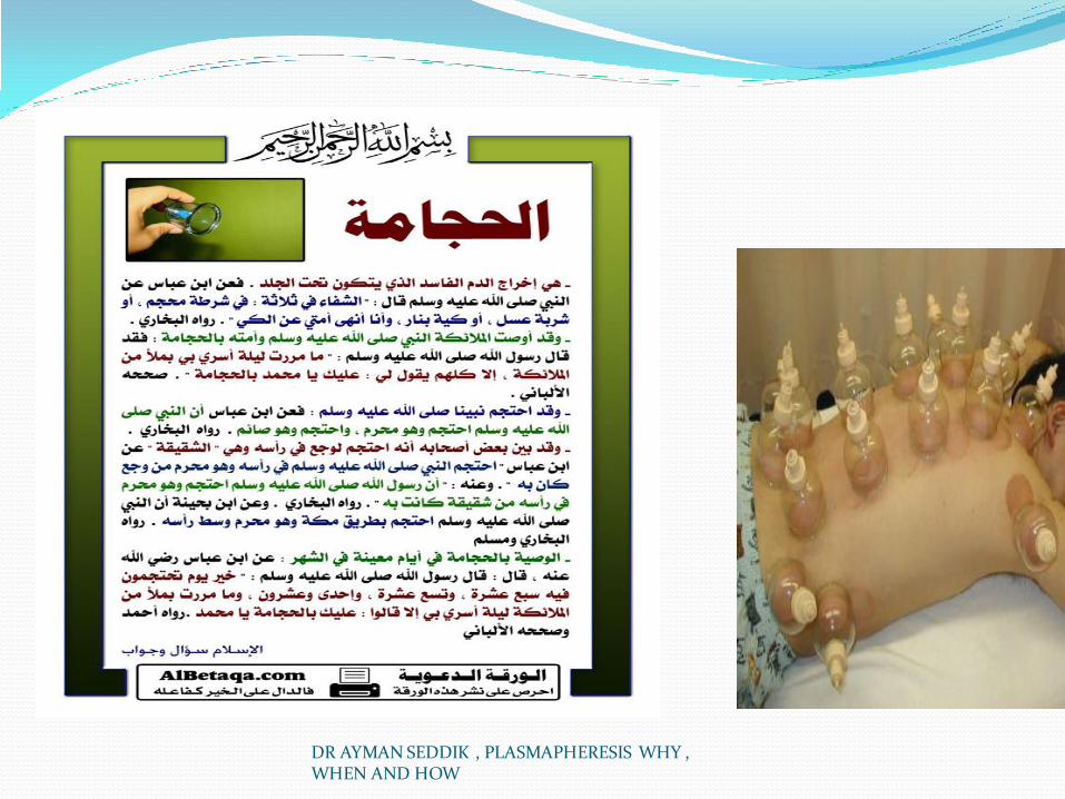
The term plasmapheresis is derived partly from the Greek word apheresis, which means “taking away” or removal. It is unclear when the notion of therapeutic removal of blood components first originated, but it was flourishing even before Hippocrates in the fifth century bc .
Bloodletting to remove evil “humors” was a commonplace medical practice, partly because of the lack of understanding of disease processes and the paucity of effective therapies
DR AYMAN SEDDIK , PLASMAPHERESIS WHY , WHEN AND HOW

DR AYMAN SEDDIK , PLASMAPHERESIS WHY , WHEN AND HOW

DR AYMAN SEDDIK , PLASMAPHERESIS WHY , WHEN AND HOW

DR AYMAN SEDDIK , PLASMAPHERESIS WHY , WHEN AND HOW

Effectiveness of TPE depends on:
Volume of plasma removed relative to total plasma volume
Distribution of substance to be removed Between intra and extravascular compartments
Speed at which the substance equilibrates between compartments
Rate at which substance is synthesized

Normal Immunoglobulins One plasma volume exchange:
IgG drops to 34% of baseline
IgA drops to 39% of baseline
IgM drops to 31% of baseline
Varying reports as to time to recovery of Ig
Ranges from 3 days to 5 weeks to full recovery
Variation due to different methods of calculating recovery, some patients on immunosuppressive medications

Metabolic Characteristics of Plasma Proteins Protein Concentration
in plasma
(mg/mL)
% intravascular Change in
catabolism with
decrease conc.
Molecular
weight
(kDa)
IgG 12.1 45 Decrease 150
IgA 2.6 42 Constant 160
IgM 0.9 76 Constant 950
IgD 02.6.02 75 Increase 175
IgE 0.0001 41 Increase 190
Albumin 42 40 Decrease 66
Fibrinogen 2-4 80 Constant 340
C3 1.5 53 240
A2
macroglobulin
100 constant 820

DR AYMAN SEDDIK , PLASMAPHERESIS WHY , WHEN AND HOW

DR AYMAN SEDDIK , PLASMAPHERESIS WHY , WHEN AND HOW

DR AYMAN SEDDIK , PLASMAPHERESIS WHY , WHEN AND HOW
DR AYMAN SEDDIK , PLASMAPHERESIS WHY , WHEN AND HOW

DR AYMAN SEDDIK , PLASMAPHERESIS WHY , WHEN AND HOW

DR AYMAN SEDDIK , PLASMAPHERESIS WHY , WHEN AND HOW

DR AYMAN SEDDIK , PLASMAPHERESIS WHY , WHEN AND HOW

DR AYMAN SEDDIK , PLASMAPHERESIS WHY , WHEN AND HOW

DR AYMAN SEDDIK , PLASMAPHERESIS WHY , WHEN AND HOW
DR AYMAN SEDDIK , PLASMAPHERESIS WHY , WHEN AND HOW

DR AYMAN SEDDIK , PLASMAPHERESIS WHY , WHEN AND HOW

DR AYMAN SEDDIK , PLASMAPHERESIS WHY , WHEN AND HOW

DR AYMAN SEDDIK , PLASMAPHERESIS WHY , WHEN AND HOW

DR AYMAN SEDDIK , PLASMAPHERESIS WHY , WHEN AND HOW

DR AYMAN SEDDIK , PLASMAPHERESIS WHY , WHEN AND HOW

DR AYMAN SEDDIK , PLASMAPHERESIS WHY , WHEN AND HOW

DR AYMAN SEDDIK , PLASMAPHERESIS WHY , WHEN AND HOW

Spectrum of Blood Purification
Bun 28,Urea 60
Vitamin B12 1,355
Vancomycin 1,468
2-microglobulin 11.600
Albumin 69,000
IgG 180,000
IgM 900,000
LDL-cholesterol 1,300,000
Cell
HD HF
Plasma
exchange
Double filtration
plasmapheresis
Cytapheresis

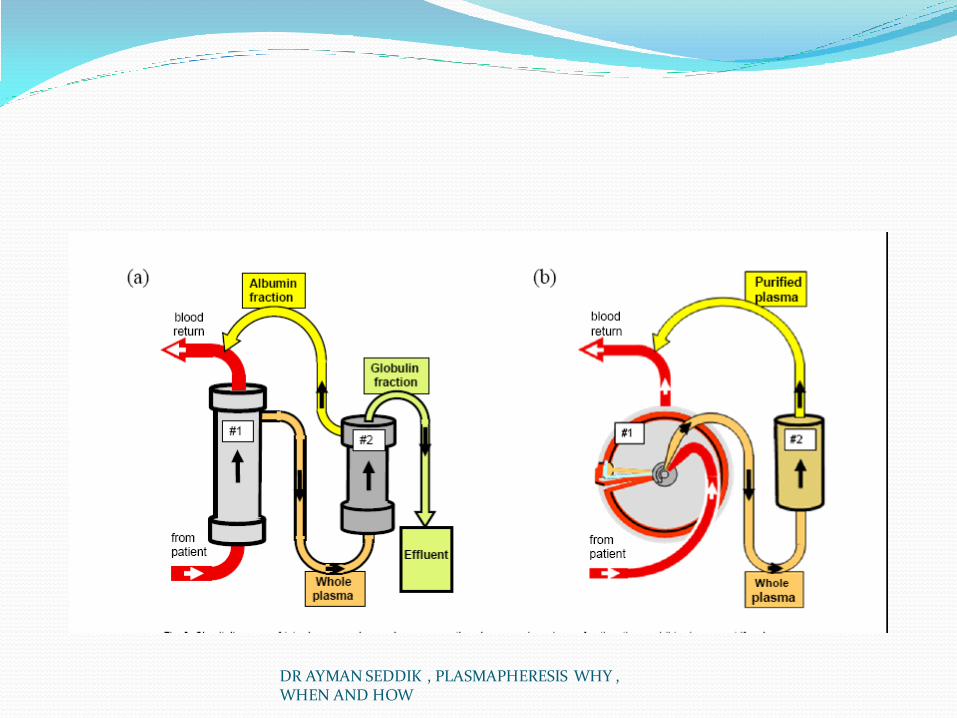
MECHANISMS OF DOUBLE FILTRATION PLASMAPHERESIS
Plasma
Plasma separator
Blood cells
Plasma filter
IgG, Immune complex, Lipoprotein, etc.

PLASMA LEVELS OF IGG BEFORE AND AFTER PLASMAPHERESIS

DR AYMAN SEDDIK , PLASMAPHERESIS WHY , WHEN AND HOW

DR AYMAN SEDDIK , PLASMAPHERESIS WHY , WHEN AND HOW

DR AYMAN SEDDIK , PLASMAPHERESIS WHY , WHEN AND HOW
DR AYMAN SEDDIK , PLASMAPHERESIS WHY , WHEN AND HOW

DR AYMAN SEDDIK , PLASMAPHERESIS WHY , WHEN AND HOW

DR AYMAN SEDDIK , PLASMAPHERESIS WHY , WHEN AND HOW

DR AYMAN SEDDIK , PLASMAPHERESIS WHY , WHEN AND HOW

DR AYMAN SEDDIK , PLASMAPHERESIS WHY , WHEN AND HOW

DR AYMAN SEDDIK , PLASMAPHERESIS WHY , WHEN AND HOW

DR AYMAN SEDDIK , PLASMAPHERESIS WHY , WHEN AND HOW

DR AYMAN SEDDIK , PLASMAPHERESIS WHY , WHEN AND HOW

DR AYMAN SEDDIK , PLASMAPHERESIS WHY , WHEN AND HOW

DR AYMAN SEDDIK , PLASMAPHERESIS WHY , WHEN AND HOW

DR AYMAN SEDDIK , PLASMAPHERESIS WHY , WHEN AND HOW

DR AYMAN SEDDIK , PLASMAPHERESIS WHY , WHEN AND HOW

DR AYMAN SEDDIK , PLASMAPHERESIS WHY , WHEN AND HOW
DR AYMAN SEDDIK , PLASMAPHERESIS WHY , WHEN AND HOW

DR AYMAN SEDDIK , PLASMAPHERESIS WHY , WHEN AND HOW

DR AYMAN SEDDIK , PLASMAPHERESIS WHY , WHEN AND HOW

DR AYMAN SEDDIK , PLASMAPHERESIS WHY , WHEN AND HOW
DR AYMAN SEDDIK , PLASMAPHERESIS WHY , WHEN AND HOW

DR AYMAN SEDDIK , PLASMAPHERESIS WHY , WHEN AND HOW

DR AYMAN SEDDIK , PLASMAPHERESIS WHY , WHEN AND HOW

DR AYMAN SEDDIK , PLASMAPHERESIS WHY , WHEN AND HOW

DR AYMAN SEDDIK , PLASMAPHERESIS WHY , WHEN AND HOW
DR AYMAN SEDDIK , PLASMAPHERESIS WHY , WHEN AND HOW

DR AYMAN SEDDIK , PLASMAPHERESIS WHY , WHEN AND HOW

DR AYMAN SEDDIK , PLASMAPHERESIS WHY , WHEN AND HOW

DR AYMAN SEDDIK , PLASMAPHERESIS WHY , WHEN AND HOW

DR AYMAN SEDDIK , PLASMAPHERESIS WHY , WHEN AND HOW

Complications
DR AYMAN SEDDIK , PLASMAPHERESIS WHY , WHEN AND HOW

DR AYMAN SEDDIK , PLASMAPHERESIS WHY , WHEN AND HOW

DR AYMAN SEDDIK , PLASMAPHERESIS WHY , WHEN AND HOW

DR AYMAN SEDDIK , PLASMAPHERESIS WHY , WHEN AND HOW

DR AYMAN SEDDIK , PLASMAPHERESIS WHY , WHEN AND HOW

DR AYMAN SEDDIK , PLASMAPHERESIS WHY , WHEN AND HOW

DR AYMAN SEDDIK , PLASMAPHERESIS WHY , WHEN AND HOW

DR AYMAN SEDDIK , PLASMAPHERESIS WHY , WHEN AND HOW

DR AYMAN SEDDIK , PLASMAPHERESIS WHY , WHEN AND HOW

DR AYMAN SEDDIK , PLASMAPHERESIS WHY , WHEN AND HOW

DR AYMAN SEDDIK , PLASMAPHERESIS WHY , WHEN AND HOW

DR AYMAN SEDDIK , PLASMAPHERESIS WHY , WHEN AND HOW

DR AYMAN SEDDIK , PLASMAPHERESIS WHY , WHEN AND HOW
DR AYMAN SEDDIK , PLASMAPHERESIS WHY , WHEN AND HOW

DR AYMAN SEDDIK , PLASMAPHERESIS WHY , WHEN AND HOW

DR AYMAN SEDDIK , PLASMAPHERESIS WHY , WHEN AND HOW

DR AYMAN SEDDIK , PLASMAPHERESIS WHY , WHEN AND HOW

DR AYMAN SEDDIK , PLASMAPHERESIS WHY , WHEN AND HOW
DR AYMAN SEDDIK , PLASMAPHERESIS WHY , WHEN AND HOW

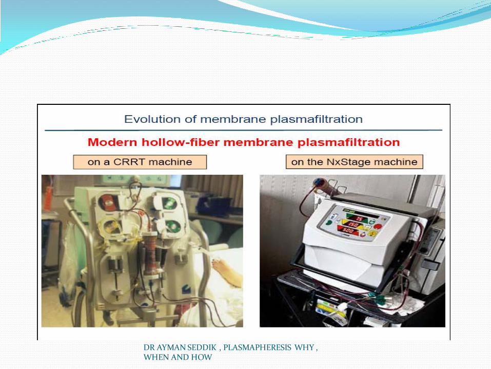
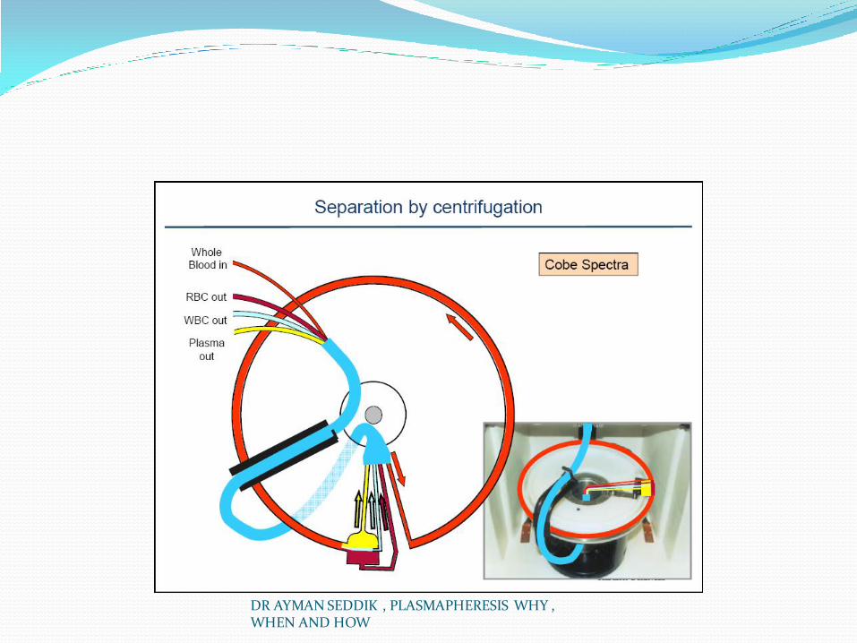
HOW ?
DR AYMAN SEDDIK , PLASMAPHERESIS WHY , WHEN AND HOW

DR AYMAN SEDDIK , PLASMAPHERESIS WHY , WHEN AND HOW

DR AYMAN SEDDIK , PLASMAPHERESIS WHY , WHEN AND HOW

DR AYMAN SEDDIK , PLASMAPHERESIS WHY , WHEN AND HOW

DR AYMAN SEDDIK , PLASMAPHERESIS WHY , WHEN AND HOW

DR AYMAN SEDDIK , PLASMAPHERESIS WHY , WHEN AND HOW

DR AYMAN SEDDIK , PLASMAPHERESIS WHY , WHEN AND HOW

DR AYMAN SEDDIK , PLASMAPHERESIS WHY , WHEN AND HOW

DR AYMAN SEDDIK , PLASMAPHERESIS WHY , WHEN AND HOW

DR AYMAN SEDDIK , PLASMAPHERESIS WHY , WHEN AND HOW

DR AYMAN SEDDIK , PLASMAPHERESIS WHY , WHEN AND HOW


DR AYMAN SEDDIK , PLASMAPHERESIS WHY , WHEN AND HOW

DR AYMAN SEDDIK , PLASMAPHERESIS WHY , WHEN AND HOW

DR AYMAN SEDDIK , PLASMAPHERESIS WHY , WHEN AND HOW

DR AYMAN SEDDIK , PLASMAPHERESIS WHY , WHEN AND HOW

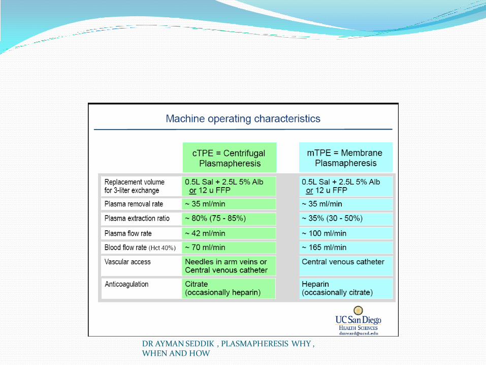
Advantages Disadvantages
Membrane
apheresis
Fast and efficient
plasmapheresis
No citrate requirements
Can be adapted for
cascade filtration
Removal of substances limited by
sieving coefficient of membrane
Unable to perform cytapheresis
Requires high blood flows, central
venous access Requires heparin anticoagulation,
limiting use in bleeding disorders
Centrifugal
devices
Capable of performing
cytapheresis
No heparin requirement
More efficient removal
of all plasma components
Expensive
Requires citrate anticoagulation
Loss of platelets
Brenner: Brenner and Rector's The Kidney, 8th ed 96

Portion of Plasma
Volumea
Exchanged (Ve/Vp)
Volume
Exchanged
(Ve, mL)
Immunoglobulin or
Other Substance
Removed (MRR, %)
0.5 1,400 39
1.0 2,800 63
1.5 4,200 78
2.0 5,600 86
2.5 7,000 92
3.0 8,400 95 aPlasma volume = 2,800 mL in a 70-kg patient, assuming
hematocrit = 45%.
Ve, volume of plasma exchanged; Vp, estimated plasma
volume; MRR, macromolecule reduction ratio.
Handbook of Dialysis 97

Why Pathologic Factors Removed by Plasmapheresis
Autoantibodies
Immune complexes
Myeloma proteins
Cryoglobulin
Complement products
ADAMTS13 (metalloproteinase)
Lipoproteins
Protein-bound toxins ADAMTS13, A member of the ADAMTS (A Disintegrin And Metalloproteinase with
Thrombospondin Motifs) family of peptidases.
DR AYMAN SEDDIK , PLASMAPHERESIS WHY , WHEN AND HOW

DR AYMAN SEDDIK , PLASMAPHERESIS WHY , WHEN AND HOW

When plasmapheresis in renal diseases Summary of Renal Diseases Treated with
Plasmapheresis DISEASE CATEGORY :
Antiglomerular basement membrane disease I Rapidly progressive glomerulonephritis II Hemolytic uremic syndrome III Thrombotic thrombocytopenia purpura I Renal transplant rejection IV Desensitization for renal transplantation II Recurrent focal segmental glomerulosclerosis III Cryoglobulinemia II Systemic lupus erythematosus III ∗ Category I, Standard primary therapy; category II, supportive therapy; category III,
when the evidence of benefit is unclear; category IV, when there is no current evidence of benefit or for research protocols.
DR AYMAN SEDDIK , PLASMAPHERESIS WHY , WHEN AND HOW

A) Use of Plasmapheresis in Renal Disease
DR AYMAN SEDDIK , PLASMAPHERESIS WHY , WHEN AND HOW

1-Anti–glomerular Basement Membrane Disease: Anti–glomerular basement membrane (anti-GBM) disease
is a disorder in which circulating antibodies are directed against the noncollagenous (NC1) domain of the α3 chain of type IV collagen, which results in rapidly progressive glomerulonephritis (RPGN). Goodpasture’s syndrome is classically defined as the triad of pulmonary hemorrhage, RPGN, and circulating anti-GBM antibodies. More than 90% of affected patients have circulating anti-GBM antibodies, the titer of which is correlated with disease activity. 6 7 Approximately 60% to 70% of patients have pulmonary disease in addition to RPGN, and in rare cases, a patient has pulmonary hemorrhage and no renal involvement
DR AYMAN SEDDIK , PLASMAPHERESIS WHY , WHEN AND HOW

ANTI-GBM ANTIBODY Goodpasture syndrome
(lung and kidney involvement)
Anti-GBM disease (only kidney involvement)
Note: 10-40% of patients may be ANCA positive.

Anti–Glomerular Basement Membrane Disease:
Before the use of current therapies, the mortality rate
exceeded 90%, and the mean survival time after diagnosis was less than 4 months. Currently, with the combination of plasmapheresis, corticosteroids, and cyclophosphamide, the mortality rate has been reduced to less than 20%. The role of plasmapheresis in anti-GBM diseases is the rapid removal of the pathogenic antibodies; cyclophosphamide and the corticosteroids are essential to prevent additional antibody synthesis and to reduce inflammation. A rapid reduction in anti-GBM antibody levels is necessary in view of the speed of glomerular damage, and this cannot be achieved by drug therapy alone.
DR AYMAN SEDDIK , PLASMAPHERESIS WHY , WHEN AND HOW

Anti–glomerular Basement Membrane Disease: Plasmapheresis was first used for the treatment of
anti-GBM disease in 1975, 8 and numerous uncontrolled studies and series published since the mid-1980s have suggested the beneficial effect of plasmapheresis on overall survival and renal preservation rates. Some of the major studies 9 10 11 12 13 14
15 are summarized in Table 67-3 , and although none were prospective randomized trials, the use of plasmapheresis is now considered standard therapy.
DR AYMAN SEDDIK , PLASMAPHERESIS WHY , WHEN AND HOW

KIDNEY TRANSPLANT AND RECURRENCE POST ANTI GBM DISEASE Most patients with anti-GBM disease who undergo
kidney transplantation have no recurrence of the disease in the allograft, although up to 50% may show linear immunoglobulin G (IgG) staining of the glomerular basement membrane. 18 The delay of kidney transplantation for 12 months after the disappearance of anti-GBM antibodies and the degree of immunosuppression necessary to maintain a functioning renal allograft are thought to be the main reasons why recurrences are very rare.
DR AYMAN SEDDIK , PLASMAPHERESIS WHY , WHEN AND HOW

2-rapidly Progressive Glomerulonephritis RPGN is characterized by rapid deterioration in renal function occurring over a
period ranging from a few days to a few weeks. Untreated RPGN usually leads to end-stage renal disease. RPGN is characterized by severe inflammation and necrosis of most glomeruli and, frequently, by fibrocellular crescents (crescentic glomerulonephritis). There are three major subgroups of RPGN: (1) anti-GBM disease and Goodpasture’s syndrome (discussed previously); (2) immune complex–mediated processes in which immune deposition occurs, usually as a result of autoimmune diseases such as systemic lupus erythematosus, postinfectious processes, mixed cryoglobulinemia, and immunoglobulin A nephropathy; and (3) pauci-immune diseases that are most often (in about 80% of patients) associated with anti–neutrophil cytoplasmic antibody (ANCA), including necrotizing granulomatous vasculitis (formerly known as Wegener’s granulomatosis), or microscopic polyarteritis. A therapeutic role for plasmapheresis in anti-GBM disease is discussed previously in this chapter, and its role in immune complex mediated processes is still uncertain (see later discussion).
DR AYMAN SEDDIK , PLASMAPHERESIS WHY , WHEN AND HOW

Crescentic Glomerulonephritis
I: Anti-GBM type (3%)
II: Immune-complex type (45%)
III: Pauci-immune type(50%)

For pauci-immune ANCA-associated diseases
(necrotizing granulomatous vasculitis and microscopic polyarteritis), plasmapheresis was used initially because the renal pathologic processes of these disorders were similar to those of Goodpasture disease; in fact, some patients have both anti-GBM antibodies and ANCA. Plasmapheresis was first used for the treatment of RPGN associated with necrotizing granulomatous vasculitis in 1977; the combination of plasmapheresis, oral prednisolone, and cyclophosphamide was associated with rapid renal recovery
DR AYMAN SEDDIK , PLASMAPHERESIS WHY , WHEN AND HOW

PAUCI-IMMUNE Wegener’s granulomatosis (WG)
Microscopic polyangiitis (MPA)
Renal-limited necrotizing crescentic glomerulonephritis (NCGN)
Churg-Strauss syndrome
Note: 80-90% of patients are ANCA positive.

However, several studies through the 1990s did not demonstrate an additional benefit for the use of plasmapheresis in the treatment of ANCA-associated diseases. For example, the Hammersmith Hospital reported a controlled trial of plasmapheresis in focal necrotizing glomerulonephritis with 48 patients randomly assigned to receive conventional treatment with oral steroids and cyclophosphamide, followed by azathioprine, with or without intensive plasmapheresis (at least five exchanges in the first 7 days). There was no benefit for patients with moderate or severe renal disease who were not dialysis dependent at presentation. 21 However, the results of this study were the first to suggest that some patients who were dialysis dependent might be able to discontinue dialysis after treatments that included plasmapheresis (10 of 17 patients receiving plasmapheresis versus 3 of 8 patients not receiving plasmapheresis).
DR AYMAN SEDDIK , PLASMAPHERESIS WHY , WHEN AND HOW

The Canadian Apheresis Study Group randomly assigned 32 patients with RPGN to receive intravenous methylprednisolone, followed by oral prednisolone and azathioprine, with or without plasmapheresis (10 exchanges in the first 16 days). Again, no benefit of plasmapheresis was demonstrated in the non–dialysis-dependent patients; however, a nonsignificant trend in benefit was observed in the dialysis-dependent patients: of four patients receiving plasmapheresis, three were able to discontinue dialysis, in comparison with only two of seven control subjects. 22 In 62 patients with Churg-Strauss syndrome or polyarteritis nodosa, patients who received plasmapheresis in addition to cyclophosphamide and steroids exhibited no additional benefit. 23
More recently, in a prospective, multicenter study, Zauner and associates 24 randomly assigned 39 patients with RPGN to receive immunosuppressive therapy alone or immunosuppressive therapy and plasmapheresis; they found that plasmapheresis had no significant effect on renal or patient survival, independently of age, sex, or serum creatinine level at the time of diagnosis
DR AYMAN SEDDIK , PLASMAPHERESIS WHY , WHEN AND HOW

However, other studies have shown that plasmapheresis treatment may improve prognosis in patients with ANCA-associated glomerulonephritis. Frasca and colleagues 25 restrospectively analyzed data from 26 patients with acute renal failure caused by ANCA-associated vasculitis. They reported that the patients who received immunosuppressive treatment plus plasmapheresis experienced a more favorable outcome than did patients who received immunosuppressive treatment alone. Results from the multicenter European Vasculitis Study Group have also been reported. 26 27 In this randomized, controlled clinical trial, plasma exchange was compared with intravenous methylprednisolone in ANCA-associated vasculitis in patients with severe renal involvement (creatinine level >500 μmol/L, or >5.7 mg/dL). All patients received oral cyclophosphamide for 3 months, followed by azathioprine. Treatment with plasmapheresis was associated with lower incidence of dialysis dependence at 12 months; these results, together with those of previous studies, provide strong support for additional therapy with plasmapheresis in patients with severe ANCA-associated glomerulonephritis and advanced renal failure
DR AYMAN SEDDIK , PLASMAPHERESIS WHY , WHEN AND HOW

However, other studies have shown that plasmapheresis treatment may improve prognosis in patients with ANCA-associated glomerulonephritis. Frasca and colleagues 25 restrospectively analyzed data from 26 patients with acute renal failure caused by ANCA-associated vasculitis. They reported that the patients who received immunosuppressive treatment plus plasmapheresis experienced a more favorable outcome than did patients who received immunosuppressive treatment alone. Results from the multicenter European Vasculitis Study Group have also been reported
DR AYMAN SEDDIK , PLASMAPHERESIS WHY , WHEN AND HOW

In conclusion, data now support plasmapheresis as having a beneficial role in the treatment of patients with ANCA-associated vasculitis and severe kidney failure at presentation. In patients with ANCA and anti-GBM disease, as well as in any patient with diffuse pulmonary alveolar hemorrhage, plasmapheresis is beneficial for the recovery and reduction of risk progression to dialysis. 30 31 32
Therefore, plasmapheresis is, at present, the best complement to immunosuppressive therapy for patients with advanced kidney disease from these mechanisms of injury. The usefulness of plasmapheresis for less severe kidney disease, however, remains unresolved.
DR AYMAN SEDDIK , PLASMAPHERESIS WHY , WHEN AND HOW

Finally, there is little evidence for the use of plasmapheresis in other causes of RPGN, although there is one report of benefit in children with RPGN from Henoch-Schönlein purpura. 34 Results of one study 35 suggest that plasmapheresis may be beneficial in kidney transplant recipients with recurrent Henoch-Schönlein purpura nephritis, but no prospective studies or protocols have yielded results indicating the optimal therapeutic regimen in this group of patients.
DR AYMAN SEDDIK , PLASMAPHERESIS WHY , WHEN AND HOW

RANDOMIZED TRIAL OF PLASMA EXCHANGE OR HIGH-DOSAGE METHYLPREDNISOLONE AS ADJUNCTIVE THERAPY FOR SEVERE RENAL VASCULITIS
David R.W. Jayne. J Am Soc Nephrol 18: 2180–2188, 2007
MEPEX

3-Lupus Nephritis Acute and chronic kidney diseases are common and
potentially serious complications of systemic lupus erythematosus. Traditionally, lupus nephritis was treated with corticosteroids, azathioprine, and intravenous cyclophosphamide, but safer and more effective therapies have been sought. More recent studies have shown that mycophenolate mofetil 36 is an alternative to intravenous cyclophosphamide in patients with active proliferative disease; rituximab 37 has also been considered as alternative in the treatment of proliferative lupus nephritis, but data suggest that rituximab is not effective in the treatment of refractory disease
DR AYMAN SEDDIK , PLASMAPHERESIS WHY , WHEN AND HOW

The use of plasmapheresis for patients with proliferative lupus nephritis was first reported in the 1970s, but it was not until the Lupus Nephritis Collaborative Study Group in 1992 undertook a randomized study to systematically examine the safety and efficacy of plasmapheresis. The Lupus Nephritis Collaborative Study Group 38 was a large, randomized, controlled multicenter trial comparing a standard-therapy regimen of prednisone and cyclophosphamide with a regimen of standard therapy plus plasmapheresis in patients with severe lupus nephritis.
DR AYMAN SEDDIK , PLASMAPHERESIS WHY , WHEN AND HOW

Forty-six patients were randomly assigned to receive standard therapy, and 40 were randomly assigned to receive plasmapheresis. Histologic categories included lupus nephritis types III, IV, and V. Plasmapheresis was carried out three times per week for 4 weeks, and drug therapy was standardized. The mean follow-up period was 136 weeks. Although patients treated with plasmapheresis experienced more rapid reduction of antibodies to double-stranded DNA and cryoglobulins, the addition of plasmapheresis did not improve clinical outcomes
DR AYMAN SEDDIK , PLASMAPHERESIS WHY , WHEN AND HOW

Of the 46 patients who received standard therapy, 8 (17%) developed kidney failure and 6 (13%) died; in comparison, of the 40 patients who received plasmapheresis, 10 (25%) developed kidney failure and 8 (20%) died. Results were similar in magnitude and direction after an extended follow-up of 277 weeks. Another small trial confirmed these findings: Wallace and colleagues 39 randomly assigned nine patients to receive either 6 months of intravenous cyclophosphamide and prednisone and nine to receive plasmapheresis before each infusion of cyclophosphamide. In each group, two patients developed end-stage renal disease, and three patients achieved renal remission at 24 months. Together, the results of these studies show that addition of plasmapheresis to conventional treatment for lupus nephritis does not improve the prognosis of lupus nephritis, despite more rapid reduction in circulating autoantibodies
DR AYMAN SEDDIK , PLASMAPHERESIS WHY , WHEN AND HOW

The main side effects were herpes zoster, and four women developed irreversible amenorrhea. Danieli and associates 41
compared two groups of patients with proliferative lupus nephritis at 4 years of follow-up. The first group (12 patients) received synchronized therapy with plasmapheresis and cyclophosphamide, whereas the second group (16 patients) received intermittent cycles of cyclophosphamide; at the end of the follow-up period, the patients who received synchronized therapy achieved remission faster than did the other group, but their renal outcomes were not superior at long-term follow-up analysis. Yamaji and colleagues 42 reported a retrospective analysis of 38 patients in which they found that synchronized therapy with plasmapheresis and cyclophosphamide might be superior to plasmapheresis or cyclophosphamide alone in achieving complete remission of lupus nephritis and in minimizing the risk of relapse.
DR AYMAN SEDDIK , PLASMAPHERESIS WHY , WHEN AND HOW
Thus, the available published evidence does not support the addition of plasmapheresis to immunosuppressive therapy for lupus nephritis. As a result, the American Society for Apheresis considered plasmapheresis as a category IV (no evidence) therapy for lupus. 43 Although plasmapheresis does not demonstrate clear benefits in severe lupus nephritis, immunoadsorption with protein A, protein C1q, or dextran sulfate cellulose columns was shown in small case series to be useful in treating refractory lupus nephritis
DR AYMAN SEDDIK , PLASMAPHERESIS WHY , WHEN AND HOW

3-Mixed Cryoglobulinemia Cryoglobulinemia is the presence of serum proteins that
precipitate at temperatures below 37°C and redissolve on rewarming. More than 80% of patients with mixed cryoglobulinemia (in which the cryoglobulin contains both a polyclonal IgG [which may either act as an antigen or be directed against an antigen] and a monoclonal IgM rheumatoid factor directed against the IgG) are infected by hepatitis C virus, and cryoglobulinemia can often be found in patients with membranoproliferative glomerulonephritis related to this virus. The glomerular injury is the consequence of glomerular deposition of immune complexes, and the renal manifestations range from isolated proteinuria to overt nephritic or nephrotic syndrome, with variable progression toward end-stage renal disease.
DR AYMAN SEDDIK , PLASMAPHERESIS WHY , WHEN AND HOW
The use of plasmapheresis for cryoglobulinemia has not been studied in randomized, controlled trials; however, the notion that plasmapheresis may remove pathogenic cryoglobulins is rational, and numerous anecdotal case reports and uncontrolled studies have demonstrated that plasmapheresis may benefit patients with severe active disease manifested by progressive kidney failure, severe or malignant hypertension, purpura, and advanced neuropathy. 46 47 In the treatment of severe acute flares of cryoglobulinemia with glomerulonephritis or vasculitis, one approach is combination antiviral therapy with peginterferon and ribavirin for 48 weeks, plus corticosteroids and cyclophosphamide as needed to control severe symptoms. In the most severe cases, the addition of plasmapheresis (exchanges of 3 L of plasma three to four times per week for 2 to 3 weeks) can be helpful. In uncontrolled studies with more than five patients, plasmapheresis induced rapid reduction in the cryocrit, improved kidney function in 55% to 87% of patients, and improved survival (≈25% mortality rate) in comparison to historical data (≈55% mortality rate). 48
DR AYMAN SEDDIK , PLASMAPHERESIS WHY , WHEN AND HOW

Because of the unique characteristics of cryoglobulins, the plasmapheresis technique has been modified to enhance their removal. Cryofiltration cools the plasma in an extracorporeal circuit, allowing for more efficient removal of the pathogenic proteins. However, this technique is most efficiently performed by a continuous process that requires a specialized machine designed for this purpose. An alternative protocol is a two-step procedure in which the patient’s own plasma can be reinfused after incubation in the cold to cause the abnormal proteins to precipitate out. 49
DR AYMAN SEDDIK , PLASMAPHERESIS WHY , WHEN AND HOW

4-kidney Failure Associated With Multiple Myeloma And Other Hematologic Disorders
Renal disease is a common finding in multiple myeloma; in 20% to 50% of affected patients, the plasma creatinine concentration exceeds 1.5 mg/dL (133 μmol/L). Kidney function can be impaired by a variety of factors, including precipitation of myeloma light chains within renal tubules (Bence-Jones proteins) that can lead to direct tubular toxicity. Other factors frequently implicated in myeloma associated kidney failure include hypercalcemia, hyperuricemia, amyloidosis, hyperviscosity, infections, and chemotherapeutic agents. 50 Plasmapheresis could be of benefit in preventing tubular damage by the removal of nephrotoxic Bence-Jones proteins.
DR AYMAN SEDDIK , PLASMAPHERESIS WHY , WHEN AND HOW

DR AYMAN SEDDIK , PLASMAPHERESIS WHY , WHEN AND HOW

Paraproteins Removal of paraproteins (ie myeloma) is 50% of
predicted Some cases can have greater removal than predicted (see
last 2 reasons)
Due to: Increase in plasma volume (up to 1.5x greater, especially
if IgG >40g/L)
Some myeloma patients have higher proportion of IgG in intravascular space (56-85%)
As remove paraprotein in TPE, plasma volume progressively decreases
An early study of 29 patients with multiple myeloma and acute kidney injury included 24 patients on dialysis and an additional 5 with creatinine concentrations higher than 5 mg/dL. The patients were randomly assigned to one of two groups: 15 patients received plasmapheresis plus standard therapy, and 14 patients received standard therapy alone. Of the 15 patients who received plasmapheresis, 13 patients recovered renal function (creatinine concentration < 2.5 mg/dL), in contrast to only 2 of the 14 receiving standard therapy. 51 However, in a study of 21 patients who were randomly assigned to receive either plasmapheresis plus chemotherapy or chemotherapy alone, Johnson and colleagues 52 reported no difference in patient survival or in recovery of kidney function. The mortality rate at 6 months was 20% in each group, which increased to 60% to 80% at 12 months. In the largest study to date, 97 patients with multiple myeloma and acute kidney injury were randomly assigned to receive either conventional therapy alone or conventional therapy plus five to seven plasma exchanges (5% human serum albumin) of 50 mL per kilogram of body weight for 10 days. The primary endpoint (death, dialysis, or glomerular filtration rate <30 mL/min) occurred in 33 (56.9%) of 58 patients who received plasmapheresis and in 27 (69.2%) of 39 control subjects. 53
DR AYMAN SEDDIK , PLASMAPHERESIS WHY , WHEN AND HOW

Together, the results of these studies leave unresolved the role of plasmapheresis in the management of cast nephropathy. Questions remain about subgroups of patients who may benefit; in general, these results suggest that caution be used when plasmapheresis is considered for patients with acute kidney injury in association with multiple myeloma.
DR AYMAN SEDDIK , PLASMAPHERESIS WHY , WHEN AND HOW

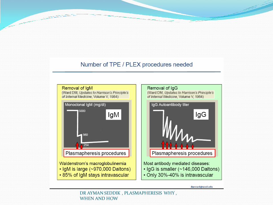
Waldenström’s macroglobulinemia is a B cell disorder resulting from the accumulation of clonally related immunoglobulin M (IgM)–secreting lymphoplasmacytic cells. The morbidity associated with Waldenström’s macroglobulinemia is typically mediated by tissue infiltration by neoplastic cells and by the physicochemical and immunologic properties of the monoclonal IgM. In affected patients with symptomatic hyperviscosity, cryoglobulinemia, or moderate to severe cytopenia, the burden of plasma paraproteins should be reduced rapidly. In these circumstances, plasmapheresis can be initially performed. Typically, two to three sessions of plasmapheresis are necessary to reduce serum IgM levels by 30% to 60%. Treatment should be initiated as soon as possible with a regimen that includes bortezomib, dexamethasone, and rituximab to achieve more rapid disease control.
DR AYMAN SEDDIK , PLASMAPHERESIS WHY , WHEN AND HOW

Plasmapheresis has been widely used in hematologic and oncologic diseases; however, only the following disorders are considered category I (standard primary therapy) by the American Society for Apheresis: (1) leukocytosis and thrombocytosis (cytapheresis), (2) thrombotic thrombocytopenic purpura (TTP; discussed next section), (3) posttransfusion purpura (plasmapheresis), (4) sickle cell disease (red blood cell exchange), (5) ABO-incompatible bone marrow transplantation (red blood cell removal from the marrow, plasmapheresis in the recipient to eliminate ABO antibodies is considered category II [supportive therapy]), (6) hyperviscosity in monoclonal gammopathies, and (7) cutaneous T cell lymphoma (photopheresis).
DR AYMAN SEDDIK , PLASMAPHERESIS WHY , WHEN AND HOW

5-Thrombotic Thrombocytopenic Purpura and Hemolytic Uremic Syndrome TTP and hemolytic uremic syndrome (HUS) share a spectrum of
abnormalities in numerous organ systems and are characterized by the presence of thrombocytopenia and microangiopathic hemolytic anemia.
In HUS, the prominent features are hemolytic anemia, thrombocytopenia, and advanced acute or chronic kidney disease. The finding of neurologic symptoms with fever and perhaps less severe kidney failure is classically considered TTP.
However, these designations are artificial, and both syndromes are characterized with pathologic changes of endothelial injury and platelet microthrombi. With two exceptions, the causes for these disorders remain unknown and can be viewed as complications of drug therapy (mitomycin, cyclosporine, ticlopidine), autoimmune disorders (systemic lupus erythematosus, anti–phospholipid antibody syndrome), and pregnancy.
DR AYMAN SEDDIK , PLASMAPHERESIS WHY , WHEN AND HOW

Sadler J E Blood 2008;112:11-18
©2008 by American Society of Hematology
pathogenesis
TTP has been shown to be associated with a severe (<5%) deficiency of plasma ADAMTS13

One well-defined cause of HUS is the syndrome associated with hemorrhagic diarrhea caused by Escherichia coli O157:H7. In this disease, the enterotoxin induces colonic vascular injury, which leads to systemic absorption and activation of numerous pathways and results in endothelial cell damage over several days. Platelet microthrombi are particularly prominent in the glomerular capillaries and often cause severe renal failure. The disease is often self-limited in children, and a role for plasmapheresis is not clear. In adults and in patients with severe or persistent disease, plasmapheresis is often used. In the largest uncontrolled trial for plasmapheresis in E. coli –associated HUS, 22 patients in Scotland were confirmed to have E. coli O157:H7 infection.
DR AYMAN SEDDIK , PLASMAPHERESIS WHY , WHEN AND HOW

Plasmapheresis could be performed in only 16 patients, of whom 5 (31%) died; of 6 patients who did not receive plasmapheresis, 5 (83%) died of the disease. There is also evidence from a single report about 60 patients that plasmapheresis improves outcomes with ticlopidine-associated HUS-TTP (mortality rates, 50% of control subjects vs. 24% of plasmapheresis recipients 59 ). However, there is no evidence for a beneficial role of plasmapheresis in patients with HUS-TTP secondary to cancer chemotherapy, calcineurin inhibitors, or bone marrow transplantation.
DR AYMAN SEDDIK , PLASMAPHERESIS WHY , WHEN AND HOW

The mechanism of some causes of TTP are now partially understood and reveal why plasmapheresis with plasma exchange is beneficial. Genetic studies of congenital TTP led to the identification of defects in the metalloproteinase named von Willebrand factor (vWF)–cleaving protease (A Disintegrin-like And Metalloprotease with Thrombospondin type 1 repeats [ADAMTS13]).
DR AYMAN SEDDIK , PLASMAPHERESIS WHY , WHEN AND HOW

TTP can result from the accumulation of ultra-large vWF. Multimers of VWf normally accumulate on the endothelial cell membrane and are rapidly cleaved into normal-sized multimers by the ADAMTS13 protease. In some patients, ADAMTS13 deficiency leads to accumulation of ultra-large VWf multimers, resulting in platelet microthrombus formation and subsequent microangiopathic hemolytic anemia. An inhibitory autoantibody to the ADAMTS13 metalloproteinase has been found at varying titers among a high percentage of patients with the idiopathic form of this disease
DR AYMAN SEDDIK , PLASMAPHERESIS WHY , WHEN AND HOW

By removing autoantibodies to ADAMTS13 and replacing with normal plasma (containing ADAMTS13 activity), plasmapheresis can reverse the TTP syndrome caused by ADAMTS13 deficiency. However, ADAMTS13 deficiency may be necessary but is not sufficient to account for many cases of TTP. Furthermore, enzyme activity is significantly reduced in numerous other conditions, including infection, cancer, cirrhosis, uremia, systemic lupus erythematosus, and disseminated intravascular coagulation.
DR AYMAN SEDDIK , PLASMAPHERESIS WHY , WHEN AND HOW
Before the introduction of plasma infusion and plasmapheresis, the disease rapidly progressed and was almost uniformly fatal (90% mortality rate). 64 In 1977 it was discovered that infusion of fresh-frozen plasma or plasmapheresis with fresh-frozen plasma replacement could reverse the course of disease. 65 66 The efficacy of plasma exchange in the treatment of TTP-HUS in adults was demonstrated in two trials that included 210 patients. 67 68 Plasma exchange with fresh-frozen plasma was more effective than plasma infusion alone. At 6 months, the remission rate was 78% versus 31%, respectively, and the survival rates with these two procedures were 78% versus 50%, respectively. Patients treated with plasma exchange received approximately three times as much plasma as those treated with plasma infusion alone (whereby the amount of plasma administration was limited by the risk of volume overload). Therefore, it is possible that the benefit observed with plasma exchange may have resulted from infusion of more plasma rather than from the removal of a toxic substance.
DR AYMAN SEDDIK , PLASMAPHERESIS WHY , WHEN AND HOW

The optimal duration of plasmapheresis treatment for HUS-TTP is not known, but it is performed daily until the platelet count has risen to nearly normal and evidence for hemolysis (schistocytes, elevation of lactose dehydrogenase levels) has resolved. 60 A wide range of exchanges (3 to 145) have been reported; on average, 7 to 16 daily exchanges are necessary to induce remission. 60 64 67 68 The American Association of Blood Banks recommends daily plasmapheresis until the platelet count exceeds 150,000/μL for 2 to 3 days, and the American Society for Apheresis recommends daily plasmapheresis until the platelet count is above 100,000/μL and lactose dehydrogenase level is nearly normal. 69 When present, neurologic symptoms rapidly improve, and the serum lactose dehydrogenase level tends to improve over the first 1 to 3 days. The platelet count may not rise for several days, and improvements in renal function often take longer. Patients requiring dialysis at presentation may be able to recover enough function to discontinue dialysis, but many patients have residual chronic kidney disease.
DR AYMAN SEDDIK , PLASMAPHERESIS WHY , WHEN AND HOW

When a normal platelet count has been achieved, plasma exchange is gradually tapered by increasing the interval between treatments. Many patients (one third to one half) abruptly develop recurrent thrombocytopenia and increased evidence of hemolysis when daily plasma exchanges are tapered or stopped. Some of these patients may benefit from the addition of prednisone or other immunosuppressive therapy (cyclosporine, rituximab), although few data validate any benefits of these agents.
DR AYMAN SEDDIK , PLASMAPHERESIS WHY , WHEN AND HOW

6-Recurrent Focal Segmental Glomerulosclerosis Focal segmental glomerulosclerosis (FSGS) is a
common cause of end-stage renal failure, and recurrent primary FSGS occurs at a rate of 20% to 30% in kidney transplant recipients. The risk of relapse is particularly high (80% to 90%) in such patients with a prior history of allograft loss resulting from recurrent FSGS. Additional factors associated with an increased risk of recurrence are rapid progression to end-stage renal disease, mesangial hypercellularity, and younger age
DR AYMAN SEDDIK , PLASMAPHERESIS WHY , WHEN AND HOW

The mechanisms of recurrent FSGS and early detection of proteinuria after kidney transplantation is unclear, but the early reappearance of proteinuria suggests that a circulating factor that alters glomerular permeability and cannot be eliminated by dialysis may be present
DR AYMAN SEDDIK , PLASMAPHERESIS WHY , WHEN AND HOW
Removal of a circulating factor by immunoadsorption or plasma exchange may account for the remission of the disease in some patients. 72 The potential circulating factor may be a nonimmunoglobulin protein with a molecular weight of less than 100 kDa, although there are discrepancies on the characteristics of this permeability factor
DR AYMAN SEDDIK , PLASMAPHERESIS WHY , WHEN AND HOW

An alternative hypothesis is that nephrotic patients lack one or more factors necessary for the maintenance of normal glomerular permeability, and a factor in normal serum (i.e., clusterin) may be lost or diminished. 73 74 75 However, at this time the mechanisms of recurrent proteinuria and FSGS remain unresolved.
DR AYMAN SEDDIK , PLASMAPHERESIS WHY , WHEN AND HOW

The treatments currently available for recurrent FSGS are immunosuppressive drugs (cyclophosphamide and methylprednisolone), plasmapheresis, and, according to some reports, rituximab. Zimmerman 76 first reported on a 38-year-old patient with recurrent FSGS who was successfully treated with plasmapheresis. Cochat and colleagues 77 studied three patients with recurrent FSGS in a prospective uncontrolled trial in which early plasmapheresis was used in combination with methylprednisolone pulses and cyclophosphamide over a 2-month period. All three patients achieved remission within 12 to 24 days, which suggests that plasma exchange instituted early in the course of recurrent nephrotic syndrome may be beneficial in patients with FSGS. Artero and associates 78 treated nine patients within 1 week of the onset of proteinuria; seven had a mean reduction in protein excretion from 11.5 to 0.8 g/day, and these remissions were sustained for up to 27 months.
DR AYMAN SEDDIK , PLASMAPHERESIS WHY , WHEN AND HOW
In recurrent FSGS after kidney transplantation, beneficial results have been reported in children treated with plasmapheresis and cyclophosphamide. In a study of 11 children with recurrent FSGS after transplantation, nine were treated with plasmapheresis (6 to 10 times over 15 to 24 days), and in seven, remission persisted after a follow-up of 32 months. 79 Likewise, in Cheong and colleagues’ 80
report of six children with recurrent FSGS, treatment with plasmapheresis plus cyclophosphamide resulted in complete or partial remissions in all the patients
DR AYMAN SEDDIK , PLASMAPHERESIS WHY , WHEN AND HOW

With regard to adult patients, controlled trials are lacking, but early plasmapheresis is also recommended. Deegens and associates 81 analyzed data from 23 patients with FSGS and renal transplants, of whom 13 were treated with plasmapheresis and 10 were historical controls. After a median follow-up of 3.5 years, 2 (15%) patients who had been treated with plasmapheresis had lost their allografts, in comparison with all 10 controls. In the patients with recurrent proteinuria, FSGS recurred within 4 weeks after transplantation (77%), and plasmapheresis was initiated within 14 days of recurrence (85%). In most studies, researchers reported a remission rate between 70% and 80%, but 33% of patients experienced relapse after the end of the treatment. 82 Nevertheless, retrospective evaluation of patients managed without plasmapheresis indicates that early rates of graft failure were as high as 80%; therefore, plasmapheresis is indicated as initial therapy for recurrent FSGS.
DR AYMAN SEDDIK , PLASMAPHERESIS WHY , WHEN AND HOW

Some reports have suggested that for individuals at high risk, preemptive treatment with plasmapheresis in the pretransplantation or perioperative period may alter or even prevent disease recurrence. In Ohta and associates’ 83 report, 15 patients received preoperative plasmapheresis, and FSGS recurred in 5 (33%), whereas of 6 who did not receive preoperative plasmapheresis, 4 (66%) developed recurrence. Gohh and colleagues 84 reported on 10 patients at high risk for FSGS recurrence because of rapid progression to renal failure ( n = 4) or prior posttransplantation recurrence of FSGS ( n = 6). Patients underwent a course of eight plasmapheresis treatments in the perioperative period. Seven patients, including all 4 with first grafts and 3 of 6 with prior recurrence, were free of recurrence at follow-up (238 to 1258 days), and the final serum creatinine concentration in 8 patients with functioning kidneys averaged 1.53 mg/dL. Therefore, the use of preoperative and prophylactic postoperative plasmapheresis appears promising in patients at high risk, but controlled multicenter trials are warranted to delineate the optimal preventive approach.
DR AYMAN SEDDIK , PLASMAPHERESIS WHY , WHEN AND HOW

7-Kidney Transplantation Plasmapheresis has been used in different clinical
scenarios involving kidney transplantation. These include ABO blood group–incompatible transplants, positive T cell cross-match, acute humoral rejection, and FSGS in the transplant
DR AYMAN SEDDIK , PLASMAPHERESIS WHY , WHEN AND HOW

ABO-Incompatible Kidney Transplantation The ABO blood group antigen system was discovered on
red blood cells by K. Landsteiner in 1901; these antigens are expressed throughout the body, and in the kidney they are found in the distal tubules, collecting tubules, and vascular endothelium of peritubular and glomerular capillaries.
The ABO antibodies (isoagglutinins) are produced in the first years of life by sensitization to environmental substances such as food, bacteria, and viruses and are usually of the IgM type.
These antibodies against ABO antigens generally preclude kidney transplantation across ABO barriers and are the key mediators of antibody-mediated rejection.
DR AYMAN SEDDIK , PLASMAPHERESIS WHY , WHEN AND HOW
In the early days of kidney transplantation, the results with ABO-incompatible organs were disappointing. In 1981, Slapak and colleagues 89 described a patient with blood group O who inadvertently received a mismatched kidney from a donor of blood group A; 2 days after transplantation, the patient experienced acute rejection that was treated successfully with plasmapheresis.
DR AYMAN SEDDIK , PLASMAPHERESIS WHY , WHEN AND HOW

Twenty months after transplantation, the patient had normal kidney function. From 1982 to 1989, Squifflet and associates 90 performed 39 ABO-incompatible kidney transplantations and were the first group to attempt kidney transplantation with ABO incompatibilities. The protocol to prepare the living donor recipient included two to five plasmapheresis sessions, pretransplantation immunosuppressive therapy, and splenectomy to remove antibodies; graft survival rates were better among the patients younger than 15 years (89% at 5 years) than among those older than 15 years (77% at 5 years).
DR AYMAN SEDDIK , PLASMAPHERESIS WHY , WHEN AND HOW

In Japan, ABO-incompatible renal transplantation flourished in the 1990s, and the outcomes to date have been excellent. Takahashi and colleagues 91 reported the outcomes of 441 ABO-incompatible kidney transplantations performed at 55 centers across Japan from 1989 to 2001. The rates of graft survival were 84% in the first year and 59% at 9 years of follow-up; these rates of survival were not statistically significant in comparison with historic recipients of ABO-compatible living donor organs. The therapy used to prepare the recipients consisted of four components: (1) extracorporeal immunomodulation to remove AB antibodies before the transplantation, (2) use of immunosuppressive drugs, (3) splenectomy, and (4) anticoagulation therapy. Plasmapheresis and immunoadsorption were the two techniques performed to remove the AB antibodies, and the goal with either technique was to decrease pretransplantation serum AB titers by 8- to 16-fold. Antibody removal was usually not performed after transplantation.
DR AYMAN SEDDIK , PLASMAPHERESIS WHY , WHEN AND HOW

The Japanese literature has emphasized the need for splenectomy at the time of transplantation, but the Johns Hopkins group has established a preconditioning protocol of plasmapheresis, cytomegalovirus hyperimmune globulin (CMVIg), and anti-CD20 (rituximab) to enable the success of ABO-incompatible renal transplantation without splenectomy. The treatment protocol requires four to five preoperative sessions of plasmapheresis to remove anti-A and anti-B antibodies, and each session is followed by the administration of CMVIg. After achieving pretransplantation A- and B-antibody titers of less than 1:16
DR AYMAN SEDDIK , PLASMAPHERESIS WHY , WHEN AND HOW

a single dose of rituximab is given 1 or 2 days before transplantation. Thereafter, immunosuppression therapy with tacrolimus and mycophenolate mofetil is initiated, followed by steroids and daclizumab after transplantation. Postoperative treatment included another three sessions of plasmapheresis and CMVIg administration on days 1, 3, and 5. The 5-year graft survival rate for a cohort of 60 consecutive patients was of 88.7%.
DR AYMAN SEDDIK , PLASMAPHERESIS WHY , WHEN AND HOW

Plasmapheresis has been used with methylprednisolone for the treatment of acute antibody-mediated rejection in patients with ABO-incompatible renal transplants. In five patients with acute antibody-mediated rejection after they received ABO-incompatible kidney transplants, Gloor and colleagues 94 95 treated with plasmapheresis and steroids, and three patients demonstrated improvements in renal function
DR AYMAN SEDDIK , PLASMAPHERESIS WHY , WHEN AND HOW

Positive T Cell Cross-Match High sensitization to HLA indicates positive T cell
cross-matches with multiple potential donors. The degree of sensitization is quantified as the percentage of the donor pool with which the serum of the patient had positive T cell cross-matches: the panel reactive antibody status. The patients whose panel reactive antibodies are persistently higher than 50% are generally considered “highly sensitized”
DR AYMAN SEDDIK , PLASMAPHERESIS WHY , WHEN AND HOW

Kidney International(2011) 79, 583 – 586.

Therapeutic strategies antibody-mediated rejection

primary sensitization results from exposure to foreign HLA antigens through transplantation, transfusion, or pregnancy, although infection and other conditions can also alter sensitization status. Patients with preformed antibodies against HLA antigens have a lower probability of receiving a matched kidney from a deceased or living donor. Furthermore, presensitized recipients experience less favorable outcomes after deceased-donor kidney transplantation and are at increased risk for hyperacute or acute antibody-mediated rejection and graft loss. Successful transplantation in these patients requires a protocol of desensitization to a specific donor in order to reduce the risk of hyperacute rejection and immediate graft loss
DR AYMAN SEDDIK , PLASMAPHERESIS WHY , WHEN AND HOW

The general approach in protocols to reduce HLA antibodies involves the use of high-dose IgG and plasmapheresis. Plasmapheresis is performed to remove anti-HLA antibodies and is followed by infusion of low doses of IgG during hemodialysis. The rationale is that low-dose IgG has beneficial immunomodulating effects. Concurrently with plasmapheresis initiation, patients are treated with tacrolimus, mycophenolate mofetil, steroids, and antimicrobial prophylaxis. Plasmapheresis is continued thrice weekly until the T cell cross-match is negative, and transplantation usually takes place within 24 hours. Plasmapheresis and low-dose IgG are usually repeated several times during the first 2 weeks after transplantation to remove any rebounding antibody.
DR AYMAN SEDDIK , PLASMAPHERESIS WHY , WHEN AND HOW

Plasmapheresis-based protocols are usually not suitable for highly sensitized patients awaiting deceased-donor transplantation because the availability of suitable organs is unpredictable and plasmapheresis is both difficult and very expensive to continue indefinitely; if plasmapheresis is stopped, anti-HLA antibody titers rebound
DR AYMAN SEDDIK , PLASMAPHERESIS WHY , WHEN AND HOW

Acute Humoral Rejection Acute humoral rejection is characterized by severe allograft
dysfunction in association with the presence of circulating donor-specific antibodies. Very poor outcomes are observed with acute humoral rejection, and treatment with pulse steroids and antilymphocyte therapy is often ineffective. 100
Removal of the donor-specific antibodies with plasmapheresis has been successful when this treatment is combined with tacrolimus and mofetil mycophenolate. 101 It is now proposed that the combination of plasmapheresis and IVIG may lead to short-term recovery from acute antibody-mediated rejection in more than 80% of cases
DR AYMAN SEDDIK , PLASMAPHERESIS WHY , WHEN AND HOW

b)Use of Plasmapheresis in NONRenal Disease
DR AYMAN SEDDIK , PLASMAPHERESIS WHY , WHEN AND HOW

Plasmapheresis and Nonrenal Disease According to several national registries, TTP, myasthenia gravis,
chronic inflammatory demyelinating polyneuropathy, Waldenström’s macroglobulinemia, and Guillain-Barré syndrome are the most frequent indications for plasmapheresis, and results of randomized controlled trials have been indicative of benefits for patients with these disorders. 104 There are now nearly 100 rational indications for plasmapheresis, and the American Society for Apheresis published an exhaustive review of the experimental data supporting the different indications for plasmapheresis. 56 In many clinical settings, the nephrologist is asked to initiate plasmapheresis. Therefore, it is essential that nephrologists be generally familiar with the literature supporting the use of plasmapheresis for these conditions.
DR AYMAN SEDDIK , PLASMAPHERESIS WHY , WHEN AND HOW

Plasma exchange is a well-established therapeutic procedure commonly used in many neurologic disorders of autoimmune origin. It is thought that the beneficial effects of plasmapheresis occur through the removal of inflammatory mediators, including autoantibodies, complement components, and cytokines. Guillain-Barré syndrome, myasthenia gravis, chronic inflammatory demyelinating polyneuropathy, and demyelinating polyneuropathy with IgG/immunoglobulin A are considered category I indications by the American Society for Apheresis
DR AYMAN SEDDIK , PLASMAPHERESIS WHY , WHEN AND HOW

Guillain-Barré syndrome Guillain-Barré syndrome develops shortly after an
infection, most commonly caused by Campylobacter jejuni. A large number of diverse antibodies against different glycolipids, including GM1, GD1a, and GQ1b, have been describedPlasma Exchange in Guillain-Barré Syndrome 108
established the optimal numbers of plasmapheresis sessions in the treatment of this disease: two for patients with mild disability and four for patients with moderate and severe disability. Plasmapheresis is considered as efficacious as IVIG therapy, and combined treatment of plasmapheresis and IVIG does not seem to yield additional benefit.
DR AYMAN SEDDIK , PLASMAPHERESIS WHY , WHEN AND HOW

Chronic inflammatory demyelinating polyneuropathy (CIDP) is a common and potentially treatable disease with an
estimated prevalence of about 1 to 2 per 100,000 adults. Symmetric weakness in both proximal and distal muscles that progressively increases for more than 2 months is the pivotal symptom in the diagnosis of this disease. CIDP is associated with impaired sensation, absence or diminishment of tendon reflexes, an elevated protein level in cerebrospinal fluid, demyelinating nerve findings in conduction studies, and signs of demyelination in nerve biopsy specimens.
DR AYMAN SEDDIK , PLASMAPHERESIS WHY , WHEN AND HOW

The presence of autoantibodies against various proteins and glycolipids of the peripheral nerve in samples of serum and cerebrospinal fluid from patients with CIDP may provide a rationale for the therapeutic use of plasmapheresis. The treatments most widely used for CIDP consist of IVIG, plasmapheresis, and corticosteroids. According to published data, there appears to be no difference in efficacy among these three main therapies
DR AYMAN SEDDIK , PLASMAPHERESIS WHY , WHEN AND HOW

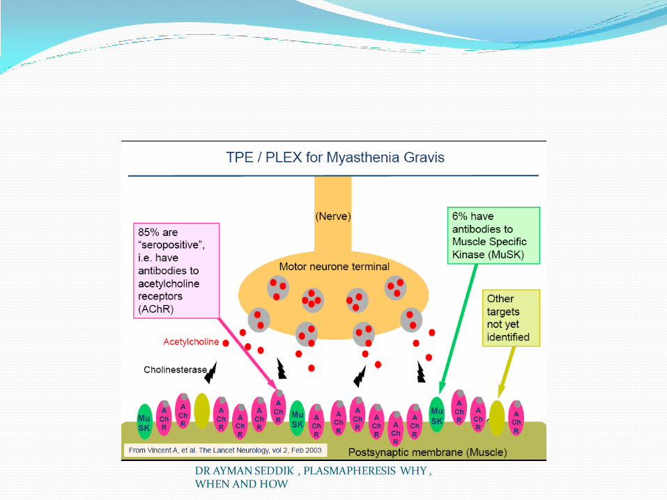
Myasthenia gravis is an autoimmune-mediated disorder of the neuromuscular
junction, clinically characterized by fluctuating muscle weakness and fatigability. The most common variant of the disease is mediated by circulating autoantibodies against the nicotinic acetylcholine receptor (AChR). Mechanisms responsible for loss of functional nicotinic AChR that compromise neuromuscular transmission include the degradation of the receptor, complement-mediated lysis of the receptor, and interference with neurotransmitter binding. In subgroups of patients negative for nicotinic AChR antibody, antibodies against the receptor tyrosine kinase can be detected.
DR AYMAN SEDDIK , PLASMAPHERESIS WHY , WHEN AND HOW

The treatment of myasthenia gravis includes thymectomy, acetylcholine esterase inhibitors, corticosteroids, immunosuppressive agents, plasmapheresis, and IVIG. It is presumed that by eliminating circulating nicotinic AChR antibodies and other humoral factors, plasmapheresis accounts for the observed beneficial effects. Indications for plasmapheresis include situations that necessitate rapid clinical improvement, such as myasthenic crisis, impending crisis, and preoperative stabilization; patients in whom long-term control of symptoms is suboptimal with other forms of therapy also benefit from plasmapheresis. On occasion, patients require long-term outpatient exchange in order to achieve adequate control of myasthenia gravis symptoms. Treatment consists of four to six exchanges, each removing 3 to 5 L of plasma, performed daily or every other day. The duration of maximal improvement is 2 to 3 weeks in 65% of cases, and any degree of improvement lasts less than 3 months in 68% of cases. Sometimes patients have a more prolonged response.
DR AYMAN SEDDIK , PLASMAPHERESIS WHY , WHEN AND HOW

Catastrophic antiphospholipid syndrome (CAPS) is a rapidly progressive and life-threatening disease
that results in thromboses in multiple organs in the presence of antiphospholipid antibodies. Rapid-onset thromboses in multiple organs and extensive involvement of small and medium-sized vessels in atypical locations are the general characteristics of CAPS
DR AYMAN SEDDIK , PLASMAPHERESIS WHY , WHEN AND HOW

Treatment with anticoagulation, corticosteroids, and plasmapheresis or IVIG can be initiated. Plasmapheresis can remove pathologic antiphospholipid antibodies, as well as cytokines, tumor necrosis factor-α, and complement products. Although plasmapheresis improves outcomes in patients with CAPS, most reports of such patients have specified plasmapheresis with fresh-frozen plasma as the replacement fluid. Fresh-frozen plasma contains natural anticoagulants (such as antithrombin III and protein C), as well as clotting factors, so it is unknown whether plasmapheresis per se or the fresh-frozen plasma replacement provides the benefits to patients with CAPS. No randomized controlled studies of plasmapheresis use in this condition are currently under way.
DR AYMAN SEDDIK , PLASMAPHERESIS WHY , WHEN AND HOW

familial hypercholesterolemia the successful use of plasmapheresis was first described in
1975. Subsequent reports showed that long-term, repetitive procedures had a beneficial effect on aortic and coronary atherosclerosis and significantly prolonged survival in comparison with untreated siblings with homozygous familial hypercholesterolemia. However, although plasmapheresis is still used in some centers to treat severe hypercholesterolemia, low-density lipoprotein (LDL) apheresis is now accepted as the treatment of choice for patients with homozygous familial hypercholesterolemia and for heterozygotes with cardiovascular disease refractory to lipid-lowering drug therapy.
DR AYMAN SEDDIK , PLASMAPHERESIS WHY , WHEN AND HOW

REMOVAL OF TOXINS Plasmapheresis has also been used to remove toxins, depending
on the effective clearance, plasma protein binding, and volume of distribution of the toxic substance. Plasmapheresis is used to treat mushroom intoxication by Amanita phalloides, but some reports suggest that forced diuresis is the treatment of choice. 120
There is controversy about the beneficial effect of plasmapheresis in the treatment of life-threatening intoxications with tricyclic antidepressants, benzodiazepines, quinine, and phenytoin. Other drugs such as L-thyroxine, verapamil, diltiazem, carbamazepine, and theophylline, as well as heavy metals, are removed effectively by plasmapheresis, but the overall change in total body toxin level is usually not clinically significant. Because of the lack of controlled studies, it is difficult to make recommendations for the treatment of poisonings and overdoses
DR AYMAN SEDDIK , PLASMAPHERESIS WHY , WHEN AND HOW

Pregnancy and Plasmapheresis Plasmapheresis can be performed safely during pregnancy,
and introduction of plasmapheresis during pregnancy for diseases necessitating that procedure has improved maternal and fetal survival rates.
Plasmapheresis has been safely carried out in patients with myasthenic crisis, Guillain-Barré syndrome, anti-GBM disease, acute fatty liver of pregnancy, and TTP. Until the effectiveness of plasmapheresis was recognized, the rate of mortality from TTP was 95%; in cases of pregnancy-related TTP, maternal survival was rare, and the fetal mortality rate approached 80%. Since 1990, numerous reports have revealed the efficacy of plasma exchange, and TTP has become a curable disease, with a response rate of about 80%, with minimal or no sequelae DR AYMAN SEDDIK , PLASMAPHERESIS WHY ,
WHEN AND HOW

RISK Plasmapheresis can result in premature delivery
because hormones crucial in maintaining pregnancy are removed. Other complications can result from hypovolemic reaction, allergy, transitory cardiac arrhythmias, nausea, and impaired vision. During the exchanges, hypotension must be carefully monitored and corrected, and in the second or third trimester, it is preferable to place the patient on her left side to avoid compression of the inferior vena cava by the gravid uterus
DR AYMAN SEDDIK , PLASMAPHERESIS WHY , WHEN AND HOW

DR AYMAN SEDDIK , PLASMAPHERESIS WHY , WHEN AND HOW

For most conditions, the aim of this procedure is the removal of pathologic autoantibodies or toxins, and the initial treatment goal is to exchange 1 to 1.5 times the plasma volume per plasmapheresis procedure. This lowers plasma macromolecule levels by 60% to 75%, respectively.
DR AYMAN SEDDIK , PLASMAPHERESIS WHY , WHEN AND HOW

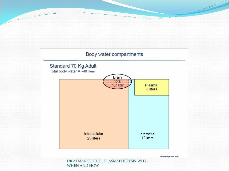
Estimated plasma volume(in liters)=
0.07×weight(in
kg)×(1−hematocrit)
DR AYMAN SEDDIK , PLASMAPHERESIS WHY , WHEN AND HOW

For removal of components restricted predominantly to the plasma space, the use of higher exchange volumes requires significantly longer procedure times and yields no additional clinical benefit. The ultimate clinical success of the procedure depends on both the abundance of the abnormal protein in plasma and its rate of production. Unless the removal of the protein by plasmapheresis is combined with additional therapies (usually immunosuppressive or cytotoxic) to eliminate or reduce the source of the abnormal protein or proteins, the procedure is unlikely to provide clinical benefit. The time needed to suppress abnormal protein production can be several weeks; that is why plasmapheresis protocols often require daily apheresis (or near daily) for prolonged times.
DR AYMAN SEDDIK , PLASMAPHERESIS WHY , WHEN AND HOW
DR AYMAN SEDDIK , PLASMAPHERESIS WHY , WHEN AND HOW

COMLICATIONS Complications involving vascular access Hematomas Pneumothorax Catheter infections Complications involving replacement fluids Anaphylactoid reactions to fresh-frozen plasma Coagulopathies Transmission of viral infections Hypocalcemia Hypokalemia Other complications Hypotension Dyspnea Thrombocytopenia Removal of erythropoietin and drugs bound to plasma proteins
DR AYMAN SEDDIK , PLASMAPHERESIS WHY , WHEN AND HOW

REGISTERY PROVED COMPLICATIONS The Swedish Therapeutic Apheresis Registry reported
on more than 14,000 procedures from 1996 to 1999; adverse events occurred in 4.2% of procedures; no fatalities were reported; and 1% of all the apheresis procedures had to be interrupted because of an adverse event. The most common adverse effects reported were paresthesias (0.52%), hypotension (0.5%), urticaria (0.34%), shivering, and nausea. This events were most frequent in patients with Goodpasture’s syndrome (12.5%), patients with TTP/HUS (10.5%), and patients with Guillain-Barré syndrome (11.0%).
DR AYMAN SEDDIK , PLASMAPHERESIS WHY , WHEN AND HOW
In another report of 17,940 procedures performed on 3583 patients, 144 adverse events occurred in 3.9% of all procedures. The following adverse reactions were documented: reactions related to citrate toxicity (3%), vasovagal reactions and hypotension (0.5%), vascular access–related complications (0.15%), reactions related to fresh-frozen plasma (0.12%), hepatitis B from fresh-frozen plasma (0.06%), arrhythmias (0.01%), hemolysis caused by inappropriate dilution of 25% albumin (0.01%), and one death (from underlying disease) during a plasmapheresis procedure (0.006%). No significant bleeding complications were observed. Patients receiving fresh-frozen plasma had significantly higher rates of adverse reactions than did patients receiving other exchange fluids.
DR AYMAN SEDDIK , PLASMAPHERESIS WHY , WHEN AND HOW

HYPOCALCEMIA One of the most frequent complications of plasmapheresis is hypocalcemia,
related to citrate infusion as anticoagulant for the extracorporeal system or to the fresh-frozen plasma administered as a replacement fluid. 145 Citrate binds to free calcium to form soluble calcium citrate, thereby lowering the free (but not the total) serum calcium concentration. Hypocalcemia is manifested by perioral and distal extremity paresthesias. Symptoms can be prevented and reduced by administration of either intravenous or oral calcium if the plasmapheresis session lasts longer than an hour. Either the administration of oral calcium carbonate or the addition of calcium gluconate to the return fluid is a useful maneuver to prevent hypocalcemia. 146 The incidence of hypocalcemic symptoms is lowered with the prophylactic administration of calcium; in one study, without calcium prophylaxis, the incidence of symptoms was 9.1% (six in 66 treatments), whereas with calcium prophylaxis, the incidence was reduced to 1% (six in 633 treatments). Marques and Huang 147
reported an incidence of hypocalcemia of 3% when calcium gluconate is infused in 5% albumin.
DR AYMAN SEDDIK , PLASMAPHERESIS WHY , WHEN AND HOW

DR AYMAN SEDDIK , PLASMAPHERESIS WHY , WHEN AND HOW

Electrolytes Potassium decrease (minimal)(0.25meq/L with
albumin and up to 0.7meq/L with FFP
No change in sodium and glucose
Bicarbonate decrease 6meq/L and chloride increase 4meq/L with albumin and this reverses with FFP (more citrate in FFP)

METABOLIC ALKALOSIS Another complication of citrate administration is the
development of metabolic alkalosis, but critical levels of bicarbonate higher than 35 mEq are rarely seen. Risk factors are use of fresh-frozen plasma and the presence of concurrent renal failure (i.e., TTP), because the excess of citrate generates bicarbonate, the excretion of which is limited by the renal failure. Replacement regimens involving saline and albumin solutions can result in a 25% reduction in the plasma potassium concentration in the postapheresis period, and this can be minimized by adding 4 mEq of potassium per liter to the replacement solution. Hypokalemia is also a consequence of metabolic alkalosis.
DR AYMAN SEDDIK , PLASMAPHERESIS WHY , WHEN AND HOW

HYPOTENSION Plasmapheresis can lead to a reduction in blood pressure,
usually as a result of a decrease in intravascular volume. Because the volume of extracorporeal whole blood is greater with intermittent centrifugation techniques, hypotension episodes are more common than with continuous modalities. Hypotension can also occur in response to complement-mediated reactions to the membrane filter or as a sensitivity to the ethylene oxide that is used to sterilize the membrane. Fresh-frozen plasma is also associated with anaphylactoid reactions that, in rare cases, result in death. Reactions to fresh-frozen plasma are most often characterized by fever, rigors, urticaria, wheezing, and hypotension.
DR AYMAN SEDDIK , PLASMAPHERESIS WHY , WHEN AND HOW
PULMONAARY EDEMA The development of dyspnea suggests that pulmonary
edema is present as a result of fluid overload; noncardiogenic edema can occur in rare instances as a component of anaphylactic reactions. Another cause of acute-onset dyspnea is the presence of massive pulmonary emboli that have been reported to develop when the reinfused blood components are not adequately anticoagulated
DR AYMAN SEDDIK , PLASMAPHERESIS WHY , WHEN AND HOW

COAGULOPATHY Plasma exchange with albumin replacement produces a
predictable decrease in clotting factors that may predispose to bleeding ( Table 67-5 ). A single plasma volume exchange increases the prothrombin time by 30% and the partial thromboplastin by 100%; these changes revert toward normal within several hours, but with repeated plasmapheresis sessions, these abnormalities can persist. In reported studies, the most significant change is in the fibrinogen levels. Keller and associates 148 reported that fibrinogen levels were lowered to 25% of levels before apheresis and recovered to baseline levels after 2 to 3 days. Therefore, 3 to 4 units of fresh-frozen plasma should be substituted as the replacement fluid each week or sooner in patients at risk for bleeding
DR AYMAN SEDDIK , PLASMAPHERESIS WHY , WHEN AND HOW

DR AYMAN SEDDIK , PLASMAPHERESIS WHY , WHEN AND HOW

Coagulant Proteins Fibrinogen:
Decrease to 25% of pretreatment with single exchange of 1 PV
Decrease to 10-30% of pretreatment with consecutive daily 1 PV exchange
recover to 100% of pretreatment levels by 2-3 days

Coagulant Proteins Prothrombin: Decreased to 30% of baseline Factor VII & factor VIII: Decreased to 45-50% of baseline Factor IX: Decreased to 60% of baseline Factor V, X, XI: Decrease to 38% of baseline Antithrombin: Activity to 40%, Ag to 70%

Thrombocytopenia Thrombocytopenia is also a consequence of plasma
removal; removal of larger volumes is associated with greater platelet loss, and the mean reduction in platelets after a plasmapheresis procedure ranges from 9.4% to 52.6%. Clinical bleeding associated with plasmapheresis is rarely reported, and when plasmapheresis-related hemorrhage is present, it is more likely to be a consequence of thrombocytopenia or inadequate heparin neutralization
DR AYMAN SEDDIK , PLASMAPHERESIS WHY , WHEN AND HOW

IMMUNEDEFICIENCY STATE Removal of immunoglobulins and complement could
result in an immunodeficient state. However, in a randomized, controlled trial of plasmapheresis in patients with lupus nephritis, TTP, or multiple myeloma, patients receiving plasmapheresis were not more prone to infection than were the other patients. 151 Nevertheless, repeated apheresis treatments with albumin replacement deplete the patient’s reserve of immunoglobulins for several weeks. If an infection occurs, a single infusion of IVIG (400 mg/kg) restores the plasma immunoglobulin concentration toward normal.
DR AYMAN SEDDIK , PLASMAPHERESIS WHY , WHEN AND HOW

BLOOD TRANSMITTED DISEASES Although estimates for the risk of viral
transmission by the use of fresh-frozen plasma are low, the large volumes from multiple donors increase the risk in patients receiving long-term plasmapheresis therapy. Use of large-volume plasma units collected from a single donor and the use of hepatitis B vaccine may reduce the risk of virally transmitted infections.
DR AYMAN SEDDIK , PLASMAPHERESIS WHY , WHEN AND HOW

DRUG REMOVAL Substantial drug removal by plasmapheresis occurs with
drugs that are highly protein bound and therefore primarily limited to the vascular space. Of the drugs used to treat renal diseases, prednisone is not substantially
removed, whereas cyclophosphamide and azathioprine are removed to some extent. This
potential problem can be circumvented by administering the drug after a plasma exchange treatment
ALWAYS GIVE YOUR PULSE CYCLOPHOSPHAMIDE POST PLASMAPHERESIS SESSION NOT BEFORE
DR AYMAN SEDDIK , PLASMAPHERESIS WHY , WHEN AND HOW

DR AYMAN SEDDIK , PLASMAPHERESIS WHY , WHEN AND HOW

Drug Removal Can remove:
ASA, tobramycin, dilantin, vancomycin, propranolol
May reduce plasma levels of enzymes that metabolize drugs
May reduce plasma levels of proteins that bind and transport drugs
Depends on distribution of drug between intra/extravascular space, half life of drug in circulation, timing of administration of drug, protein bound status, not lipid or tissue bound
1% of prednisone removed IVIG mainly removed as remains intravascularly Ideally give medications after exchange
ACE INHIBITORS Flushing, hypotension, abdominal cramping, and other
gastrointestinal symptoms have been reported during plasmapheresis in patients receiving angiotensin converting enzyme (ACE) inhibitors. In one report of 299 consecutive patients undergoing plasmapheresis, these atypical symptoms occurred in all 14 patients who received an ACE inhibitor, in contrast to only 7% of those not treated with this medication. 152
The administration of an ACE inhibitor may prolong the half-life of bradykinin, which enables patients to attain a clinically significant concentration in the plasma; therefore,
it is recommended that ACE inhibitors be withheld 24 hours before plasmapheresis
DR AYMAN SEDDIK , PLASMAPHERESIS WHY , WHEN AND HOW

CONCLUSION The use of plasmapheresis to treat a variety of kidney
diseases has expanded significantly since the 1990s. In some cases, the rationale and benefit are supported by data from clinical studies, but in many cases, the benefits are not well established. Nevertheless, the rationale of removing plasma containing pathogenic antibodies is now well established. Additional studies are needed to determine the potential benefits for plasmapheresis in these other conditions
DR AYMAN SEDDIK , PLASMAPHERESIS WHY , WHEN AND HOW

Conclusion In the early days, the utility of plasmapheresis was
judged on the basis of anecdotal or uncontrolled studies; more recently, the number of clinical indications for plasmapheresis has been growing. However, the number of clinical conditions that have been rigorously studied with prospective and randomized controlled trials remains small, and decisions for the implementation of plasmapheresis (an invasive and potentially dangerous procedure) are still based on results of anecdotal and uncontrolled studies in many circumstances
DR AYMAN SEDDIK , PLASMAPHERESIS WHY , WHEN AND HOW

refferences Buhaescu I., Covic A., and Levy J.: Systemic vasculitis: still a challenging disease. Am J Kidney Dis 2005; 46:
pp. 173-185 De Lind van Wijngaarden R.A., et al: Clinical and histologic determinants of renal outcome in ANCA-
associated vasculitis: a prospective analysis of 100 patients with severe renal involvement. J Am Soc Nephrol 2006; 17: pp. 2264-2274
Jayne D.R., et al: Randomized trial of plasma exchange or high dosage methylprednisolone as adjunctive therapy for severe renal vasculitis. J Am Soc Nephrol 2007; 18: pp. 2180-2188
Bosch X., et al: Treatment of antineutrophil cytoplasmic antibody–associated vasculitis. JAMA 2007; 298: pp. 655-669
Hattori M., et al: Plasmapheresis as the sole therapy for rapidly progressive Henoch-Schönlein purpura nephritis in children. Am J Kidney Dis 1999; 33: pp. 427-433
Walsh M., et al: Rituximab in the treatment of anti–neutrophil cytoplasm antibody associated vasculitis and systemic lupus erythematosus: past, present and future. Kidney Int 2007; 72: pp. 676-682
Ferri C., et al: Mixed cryoglobulinemia: demographic, clinical, and serologic features and survival in 231 patients. Semin Arthritis Rheum 2004; 33: pp. 355-374
Dominguez J.H., and Sha E.: Apheresis in cryoglobulinemia complicating hepatitis C and in other renal diseases. Ther Apher 2002; 6: pp. 69-76
Plasmapheresis Madore F.: Technical aspects and indications. Crit Care Clin 2002; 18: pp. 375-392 Szczepiorkowski Z.M., et al: Guidelines on the use of therapeutic apheresis in clinical practice—evidence-based approach from
the Apheresis Applications Committee of the American Society for Apheresis. J Clin Apher 2007; 22: pp. 106-175 Brenner anr rector 9th edition 2012
DR AYMAN SEDDIK , PLASMAPHERESIS WHY , WHEN AND HOW

DR AYMAN SEDDIK , PLASMAPHERESIS WHY , WHEN AND HOW