plasmid insertion mutagenesis gene fusionwith mini …jb.asm.org/content/158/2/488.full.pdfplasmid...
TRANSCRIPT
JOURNAL OF BACTERIOLOGY, May 1984, p. 488-4950021-9193/84/050488-08$02.00/0Copyright © 1984, American Society for Microbiology
Vol. 158, No. 2
Plasmid Insertion Mutagenesis and lac Gene Fusion with Mini-MuBacteriophage Transposons
BEATRIZ A. CASTILHO,' PAUL OLFSON,2 AND MALCOLM J. CASADABANl.3*
Committee on Genetics,1 Department of Microbiology,2 and Department ofBiophysics and Theoretical Biology,3 TheUniversity of Chicago, Chicago, Illinois 60637
Received 8 August 1983/Accepted 13 February 1984
Small bacteriophage Mu transposable elements containing the lac operon structural genes wereconstructed to facilitate the isolation and use of Mu insertions and lac gene fusions. These mini-Muelements have selectable genes for either ampicillin or kanamycin resistance and can be used to form bothtranscriptional and translational lac gene fusions. Some of the mini-Mu-lac elements constructed are deletedfor the Mu A and B transposition genes and form stable insertions that cannot undergo transposition unlesscomplemented for these functions. A procedure was developed for selecting mini-Mu insertions specificallyinto plasmids, including commonly used high-copy-number cloning vectors such as pBR322. Mu itisertionsin pBR322 were found to be distributed around the plasmid, but insertions in certain regions occurred more
frequently than in others.
Several properties of bacteriophage Mu make it veryuseful for genetic manipulations (8, 23, 31, 55). It is atemperate phage which replicates by a process that involvesDNA transposition. Transposition to new sites can be in-duced at a high rate by lytic growth, or it can be repressed inthe lysogenic state. In addition, Mu has a headful mechanismfor packaging its DNA so that adjacent DNA sequences, upto a total of approximately 38 kilobase pairs (kb) includingthe Mu genome, can be taken up into a viral particle andcarried to a new cell upon infection (10). New DNA se-quences can be incorporated between the ends of the Mugenome and carried, within the limits of DNA packaging,continually as a part of the phage to new cells and genomiclocations. Segments of the Mu genome can be deletedwithout affecting phage growth, and even larger segmentscan be removed or altered without affecting intracellulartransposition. If removed, both of these functions can becomplemented by a nondefective helper Mu prophage aslong as the DNA sequences at each Mu end are maintained(54).For forming gene fusions, gene segments can be incorpo-
rated in the Mu genome close enough to the right end thattranscription and translation can proceed from sequencesoutside of Mu across a small right-end segment into theincorporated sequence. The Mu dllac phage (16) (Fig. 1) hasthe lacZYA operon such that transcription from a promoternear the Mu insertion can proceed into the Mu dI phage toexpress the lac genes. The Mu dlllac phage (14a) has asimilar structure but allows both transcription and transla-tion (of the resulting transcript) to proceed to express the lacgenes with the formation of enzymatically active hybrid ,B-galactosidase (lacZ) proteins that have their amino-terminalamino acids derived from sequences outside of Mu. Thesetypes of gene fusions are useful in the study of generegulation (2) and protein structure (28, 35, 43, 44).Here we describe the construction of a series of mini-Mu-
lac gene fusion elements that have several advantages overthe previously described Mu dlI and Mu dII301 lac genefusion elements. These mini-Mu elements have an additional
* Corresponding author.
selectable drug resistance gene (Kmr). Some are missing theMu transposition genes but can be complemented for thesefunctions to form insertions that are genetically stable andincapable of subsequent transposition. The small size ofthese elements also permits the use of the Mu transductionprocess to select Mu insertions, including insertions in high-copy-number plasmids.
MATERIALS AND METHODSStrains are listed in Table 1. Bacterial growth conditions
and genetic manipulation procedures including conjugation(42), standard DNA cloning procedures (39), and methodsfor handling bacteriophage Mu (9, 13, 16) have been de-scribed previously. Antibiotics and the concentrations usedwere ampicillin (25 ,ug/ml), kanamycin (20 pug/ml), spectino-mycin (40 ,4g/ml), streptomycin (100 ,ug/ml), and tetracyclinehydrochloride (10 ,ug/ml). All Mu elements used containedthe cts62 temperature-sensitive repressor mutation so thatlytic growth could be induced from the lysogenic state. Mulysogenic strains were grown at 30°C or below unless other-wise stated. Since Mu lysates can be unstable, Mu phagewere stored as lysogens and lysates were freshly prepared byheat induction.
RESULTSOverview of the genetic manipulations. An additional selec-
table drug resistance marker for kanamycin resistance wasfirst introduced into the original Apr Mu dlI and Mu dII301lac fusion phage. The Mu dII Kmr Apr recombinant phagewas then inserted into the pSC101 plasmid in which deletionsof internal restriction fragments were made by DNA cloningtechnology to form mini-Mu dIl prophage. Some of thesewere moved to the chromosome along with a complementingMu cts prophage to form strains which are convenient forinducing lysates. Mini-Mu dl transcription fusion-formingphage were constructed by recombination of Mu dlI withmini-Mu dII phage. Next, the mini-Mu phage were tested forgrowth, transduction, and lac fusion formation.
Procedures for localizing mini-Mu insertions into plasmidswere then developed. Recombination defective recA- ver-
488
on June 19, 2018 by guesthttp://jb.asm
.org/D
ownloaded from
MINI-Mu-lac GENE FUSION 489
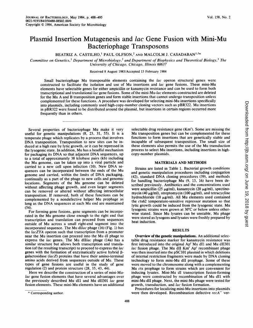
MudI (Ap Il)
X Ec ner A B APrA Y Z trp x Px
MudJi (Ap (gl)
X c ner AB APrA Y Z 'X Px
FIG. 1. Mu-/ac gene fusion phage. Indicated are the original37.2-kb Mu dlI (Ap trp'CBA'lac'ZYA) lac transcription (16) andthe 35.6-kb Mu d1I301 (Ap lac'ZYA) lac transcription lacZ transla-tion (14a) fusion-forming phage. These phage are defective (d) forMu phage growth but are proficient for intracellular transposition.They differ only to the right of the lac sequences. Mu dl has asegment from the trp operon from the W209 trp-lac fusion which isprobably missing the first two codons of lacZ and which probablyuses the translation initiation site from or within the trpA gene (M.Berman, personal communication). Mu dll has a 116-base-pairsegment from the Mu right end joined to the eighth codon of lacZ,with a BamHI site formed at the joint. Transcription and translationfrom outside of Mu on the right can proceed across this small Musegment into the lacZ gene to form enzymatically active hybridproteins. The relevant early Mu repressor gene c and the transposi-tion-replication genes A and B are indicated on the left side of Mu.An unselected and irrelevant IS121 insertion sequence whichtransposed from the E. coli chromosome was found to be present inthe middle of the original Mu dlI phage (45) and is retained in MudII301 (Fig. 2). The phage are drawn as insertions into a hypotheti-cal gene X with promoter Px which initiates transcription (arrows)toward the lac sequences to encode proteins indicated by thesquiggly lines below.
sions of the mini-Mu lysogens were made to minimize theformation of multimeric forms of plasmids. Insertions intopBR322 were examined in detail.Kmr derivatives of Mu-lac fusion phage. A Kmr-selectable
marker was introduced into Mu dlI (Apr) and Mu dII301(Apr) by recombination with the Mu pf7701 (Kmr) phage(Table 1). Recombinants were obtained by infecting theMAL113 and POL3 lysogens of Mu dlI and Mu dII301 with a
lysate of Mu pf7701. Kmr Apr transductants were selectedand scored for the presence of recombinants. About half ofthe transductants were recombinants as judged by theirfailure to release plaque-forming phage. The Mu dl and MudlI phage are defective for phage growth because they aremissing essential Mu genes, whereas Mu pf7701 is notdefective for growth. Recombinants between the two wouldalso be defective. The other half of the transductants pre-sumably became Kmr by a new insertion of Mu pf7701 in thechromosome. The recombinant nature of these Mu dI7701-1(strain BACI) and Mu dII7701-301 (strain P03) elements wasverified by the cotransduction of the Apr and Kmr markersby lysates prepared from these strains after introduction byconjugation of a complementing Mu cts prophage present onthe F' episome from strain EC601.9. The physical structureof Mu dII7701-301 is discussed below and shown in Fig. 2.
Insertion of Mu dII7701-301 into pSC101. Mu dII7701-301was inserted into the intermediate-copy-number plasmidpSC101 (21) by first incorporating it into an F' ar-a episomeand then by using the process of plasmid mobilization toselect Mu dlI insertions into pSC101, as has been describedfor the isolation of Mu cts insertions into pSC101 (19).The F' ara from strain EFO was introduced by conjuga-
tion, with selection for Ara' Kmr, into strain P03, which hasa Mu dII7701-301 insertion in the ara genes (as an ara-lacfusion). Recombinant F' ara episomes, which had picked upthe ara::Mu dII7701-301 insertion by homologous recombi-nation, were selected by mating with strain M8820S. Kmrand Spcr exconjugants were picked and scored for Leu (leiais also carried on the episome), Apr, Strs, and the ability totransfer the episome again, along with all its markers, to ther-psL strain M8820 Mu cts. Expression of/ac was also shownto be still induced by L-arabinose, as indicated by theformation of red Lact colonies on lactose MacConkey agarwhen L-arabinose was added as inducer at 0.1%.The pSC101 plasmid was introduced into one of these F'
ara::Mu dII7701-301 strains (named P0401) by transforma-tion. These cells were grown at 37°C to induce Mu transposi-tion which would occasionally mediate the joining of thepSC101 plasmid to the F'::Mu dlI episome by a cointegratetransposition structure. Such a cointegrate structure couldthen be transferred by conjugation to strain M8820 andselected as Tc", Kmr, and Strr cells on lactose MacConkeyindicator agar, with 0.1% L-arabinose as inducer. About halfof the exconjugants formed white colonies, implying that theF' ara::Mu dII7701-31 episome had not been established andhad presumably been separated from the cointegrate pSC101by homologous recombination within the duplicated Musequences. These white colonies were also Leu- and thusdid not have the episome. Plasmid DNA (as pPO1669)prepared from three of these strains had structures expectedfor Mu dII7701-301 insertions in pSC101, as judged by 0.7%agarose gel electrophoresis after digestion with the enzymesBamHI, BgII, EcoRI, and HindlIl (Fig. 2).
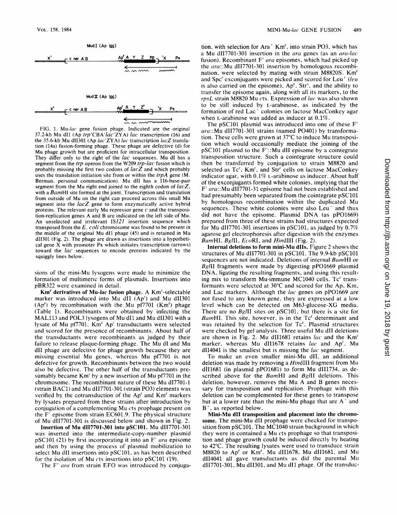
Internal deletions to form mini-Mu dlls. Figure 2 shows thestructures of Mu dII7701-301 in pSC101. The 9.9-kb pSC101sequences are not indicated. Deletions of internal BamHI orBglII fragments were made by digesting pPO1669 plasmidDNA, ligating the resulting fragments, and using this result-ing mix to transform Mu-immune MC1040 cells. Tcr trans-formants were selected at 30°C and scored for the Ap, Km,and Lac markers. Although the lac genes on pPO1669 arenot fused to any known gene, they are expressed at a lowlevel which can be detected on M63-glucose-XG media.There are no BglII sites on pSC101, but there is a site forBa(mHI. This site, however, is in the Tcr determinant andwas retained by the selection for Tcr. Plasmid structureswere checked by gel analysis. Three useful Mu dlI deletionsare shown in Fig. 2. Mu dII1681 retains lac and the Km"marker, whereas Mu dII1678 retains lac and Apr. MudII4041 is the smallest but is missing the lac segment.To make an even smaller mini-Mu dll, an additional
deletion was made by removing a HindIll fragment from MudII1681 (in plasmid pPO1681) to form Mu dII1734, as de-scribed above for the BamnHI and BglII deletions. Thisdeletion, however, removes the Mu A and B genes neces-sary for transposition and replication. Prophage with thisdeletion can be complemented for these genes to transposebut at a lower rate than the mini-Mu phage that are A' andB as reported below.Mini-Mu dlI transposition and placement into the chromo-
some. The mini-Mu dII prophage were checked for transpo-sition from pSC101. The MC1040 strain background in whichthey were in contained a Mu cts prophage so that transposi-tion and phage growth could be induced directly by heatingto 42°C. The resulting lysates were used to transduce strainM8820 to Apr or Kmr. Mu dII1678, Mu dII1681, and MudII4041 all gave transductants as did the parental MudII7701-301, Mu dII301, and Mu dlI phage. Of the transduc-
VOL. 158, 1984
on June 19, 2018 by guesthttp://jb.asm
.org/D
ownloaded from
490 CASTILHO, OLFSON, AND CASADABAN
TABLE 1. Strains, phage, and plasmids
Strain, phage, and Description or genotype Source or referenceplasmidStrainBAC1BAC101CT152D7053D7053.3EFO
EC601.9
M8820M8820MuM882OMuctsM8820TRM8820TRMuctsM88205SM8820SMuctsMAL103MAL113MAL315
MALII1681MALII1681TRMALII1734MALII1734TRMC1000MC1040MC4100P011678POII1678P011681POI1681TRPOI11681POII1681TRP011734POI11734P03POL3P0401
MC4100 with araB::Mu d17701-1M8820TRMucts with Mu dII4041F- gal lac rpsL Mu pf7701Hfr Cav thr araDI39 trp metD7053 with ara::(Mu cts)3F'KLF-1 thr+ araC+O+l+B+A+D+ leu+ F- araD139 A(araCOIBA-
leu)7697F'ts114 lacI::Mu cts62 lacP+O+Z+ Y+A+ F-A(lacIPOZYA-argF)U169 melB? glp?
F-, araD,139 A(ara-leu)7697 A(proAB-argF-lacIPOZYA)XIII rpsLM8820 with MuM8820 with Mu ctsM8820 with recA56 srl::TnJO (Tcr)M8820TR with Mu ctsM8820 with spcA rpsLM8820S with Mu ctsF- Mu dlI ara::(Mu cts)3 A(proAB-argF-1acIPOZYA) XIII rpsLMC4100 with araB::Mu dl (an ara-lac fusion)Mu dII301 malK or T::Mu cts araD139 A(ara-leu)7697 A(proAB-argF-lacIPOZYA)XIII rpsL, unknown auxotroph
MAL315 with Mu dII1681 (Kmr) in place of Mu dII301MALII1681 with recA56 srl::TnlO (Tcr)MAL315 with Mu dII1734 (Kmr) in place of Mu dII301MAL111734 with recA56 srl::TnlO (Tcr)F- araD- A(ara-leu)7697 A(lacIPOZY-A?) X74 galU galK rpsLF- araD139 ara::(Mu cts)3 A(lac)X74 galU galK rpsLF- araD139 A(lacIPOZYA-argF)U169 rpsL thiP011681 with Mu d11678 (Apr) in place of Mu dI1681P0111681 with Mu dII1678 (Apr) in place of Mu dII1681MAL103 with Mu d11681 (Km) in place of Mu dIlP011681 with recA56 srl::TnlO (Tcr)M8820 with Mu dII1681 ara::(Mu cts)3 araD- leu+ lac+ pro+P0111681 with recA56 srl::TnJO (Tcr)MAL103 with Mu d11734 (Kmr) in place of Mu dlI (Apr)MC1040(Mu cts) with Mu dII1734 lac+MC4100 with araB or A::Mu dII7701-301MC4100 with araB or A::Mu dII301 (an ara-lac fusion)M8820S Mu cts with F'KLF-1 thr+ araB or A::Mu dII7701-301 leu+
MAL113 x Mu pf7701InfectionM. Howe12Lysogeny14
13a
12"LysogenyLysogenyP1 transductionLysogenyP1 transductionLysogeny161614a
x Mu dII1681P1 transductionx Mu dII1734P1 transduction13D7053.3 x MC100013ax Mu d11678x Mu d11678x Mu d11681P1 transductionMu dII1681 lysogen x D7053.3P1 transductionx Mu d11734(D7053.3 x Mu dII1734) x MC1040POL3 x Mu pf7701Lysogeny(EFO x P03) x M8820SMucts
Mu c+Mu cts62Mucts62::Tn5 (Kmr) (AIS50 right) A(445-3) a plaque-forming Kmrphage
Mu cts62::IS121 d(Apr trp'B+A'-AW209-lac'ZYA) a defective lactranscription fusion phage
Mu cts62::IS121 d(Apr lac'ZYA) a defective lac transcription-lacZtranslation fusion phage
Mu dlI with Kmr of Mu pf7701Mu dII301 with Kmr of Mu pf7701
Mu d17701-1 with A(Kmr) BglIIMu dII7701-301 with A(Kmr) BglIIMu d17701-1 with A(Apr) BamHIMu d17701-301 with A(Apr) BamHIMu d11681 with A(Mu A B) HindIllMu dII1681 with A(Mu A B) HindlllMu dII7701-301 with A(Apr lac) BamHI
Tcr Apr replicon ColElTcr replicon pSC101pSC101::Mu dII7701-301 (Apr Kmr lac)pSC101::MudII1678 (Apr lac)pSC101::Mu dII1681 (Kmr lac)pSC101::Mu dII1734 (Kmr lac)pSC101::Mu dII4041 (Kmr)
A. BukhariM. HoweM. Howe, CT152
13; MAL103 (Fig. 1)
14a; MAL315 (Fig. 1)
BAC1P03 (Fig.2)
P011678pPO1678 (Fig. 2)P011681pPO1681 (Fig. 2)P011734pPO1734 (Fig. 2)pPO4041 (Fig. 2)
5221P0401 with pSC101 X MC1040pP01669 deletion (Fig. 2)pP01669 deletion (Fig. 2)pP01669 deletion (Fig. 2)pP01669 deletion (Fig. 2)
a Deletions of argF are not Arg- because E. coli K-12 has an isozyme gene argI.
PhageMuMu ctsMu pf7701
Mu dlI
Mu dII301
Mu d17701-1Mu dII7701-
301Mu d11678Mu dII1678Mu d11681Mu dII1681Mu d11734Mu dII1734Mu dII4041
PlasmidspBR322pSClOlpPO1669pPO1678pPO1681pPO1734pPO4041
J. BACTERIOL.
on June 19, 2018 by guesthttp://jb.asm
.org/D
ownloaded from
MINI-Mu-lac GENE FUSION 491
MudI 1681
MudI 4041
udll 1678
MudI 1734
B=BOMHIb=BgllH=Hin dM
FIG. 2. Mini Mu-lac's. Shown is the genetof Mu dII7701-301 on the pPO1669 plasmnid wthis phage into pSC101 (Tcr). The IS121 insert1) was retained as determined by its characterisites. This restriction map is correlated withsequences of Mu (1, 35, 46), Mu dlI (16, 4.pf7701 (C. Thompson and M. Howe, unpublis(3, 49), Tn3 (Apr) (30), and lac (7, 15, 17, 34)the phenotypes of the deletions isolated. Un(fragments deleted by restriction digestion, ligEtion to select the clones pPO1678, pPOpPO4041. The resulting plasmids, deletions,designated with the same numbers.
tants, 99% were Tcs and approximatelylactose MacConkey media, implying thano longer connected to the Tcr pSC101and apparently inserted into different lo(mosome. The Mu dII1734 lysate, howevfewer Kmr transductants, but the sam
complementing Mu cts prophage per ml. (dIIl734 Kmr transductants were also Tcoriginal pSC101 plasmid with the Mu dlfrequently being transduced without trandII1734 to the chromosome, as would beA- B- phage.To propagate the mini-Mu dlI phage
plasmid, Mu dII1678 and Mu dII1681M8820 were associated with a complemphage by mating them with the Hfr Muwith selection for Leu+, Kmr or Apr, ancSelection for Leu+ ensured that the Muadjacent ara genes of D7053.3 was crosL
deletion strain M8820. This formed strPOII1681. A similar strain, BAC101, wasin a different way, by infecting strain Mthe Mu dII4041 lysate and selecting fowhich the Mu dII4041 had inserted intoviolation of the Mu cts immunity. (It wviolation occurred more frequently whelried out at the partially inducing temper;than at repressing temperatures below 3A similar strain with Mu dII1734 was n
to ensure that the Mu dII1734 was in thdII1734 on pPO1734 was introduced iD7053.3 by transformation, partially indtion by growth at 37°C, and mated w
lysogen strain MC1040, with selection
SIZE DRUG Since Hfr strains only transfer DNA from the chromosome,(Kb) RESISTANCE and not from the pSC101 plasmid, it was expected that the
35.6 Km, Ap Kmr Mu dII1734 would be conjugally transferred only if ithad transposed to the chromosome. Indeed, all the exconju-gants tested (as POI11734) were Tcs and had no detectableplasmid DNA in extracts run on cesium chloride-ethidiumbromide gradients.
14.2 Km The frequency of Mu transduction and lac fusion forma-tion was checked for all these strains which no longer had
75 Km the pSC101 plasmid. All except for P0111734, with the MuA- B- HindIII deletion phage Mu dII1734, gave the same
22.4 Ap titers as the original Mu dII301 lysogen strain MAL315: 2 x10⁹ to 6 x 10⁹ plaques per ml and 5 x 10⁵ to 50 x 105 Kmr or
9.7 Km Apr transductants per ml, with approximately 1% of thetransductants being Lac' for the type II phage. The 1734phage gave the same plaque titers, but only 104 Kmr trans-ductants per ml, and only 10-4 of these were Lac'. Themechanism by which the 1734 phage gives rise to anylysogens at all is discussed below.
ic and restriction map Construction of mini-Mu dI phage by recombination withthich is an insertion of Mini-Mu dlI. The 1678, 1681, and 1734 deletion derivativesstic HindIII and BgIII of Mu dII7701-301 shown in Fig. 2 were transferred to thethe maps and known Mu dlI transcription fusion phage by homologous recom-5), Mu dlI (14a), Mu bination by Mu and lac homology. Strain MAL103, contain-shed data), TnS (Kmr) ing the Apr Mu dIl and the complementing Mu cts pro-and is consistent with phages, was infected at 30°C with lysates of the Kmr Muderlined are the DNA dII1681 and Mu dII1734 phage from strains P0111681 andation, and transforma- POII1734, with selection for Kmr. Recombinants were1681, pPO1734, and scored by their loss of the Apr marker, and the strains wereand Mu d phage are named P011681 and P011734. Similarly, Mu d11678 (Apr)
recombinants were selected in a second step by infecting theMu dI1681 (Kmr) recombinant strain P011681 with a Mu
1% were Lac' on dII1678 lysate with selection for Apr and scoring for Kms. InLt the Mu dIls were these crosses, approximately half of the selected coloniesplasmid sequences were recombinants, with the others presumably being newcations in the chro- insertions in violation of immunity, as was seen in the earlierer, yielded 100-fold cross between Mu pf7701 (Kmr) and POL3 (Mu dII301, Apr).ie 109 PFU of the Alternative strains MALII1681 and MAL111734 and MuDver 90% of the Mu dII1681 and Mu dII1734, and a complementing Mu cts, werer, implying that the made by recombination with the Apr Mu dII301 lysogenI1734 insertion was MAL315 with selection for Kmr and scoring for Aps. Thissposition of the Mu established a set of mini-Mu dl and Mu dlI lysogens whichexpected for a Mu were isogenic with the original Mu dlI and Mu dII301
lysogenic strains MAL103 and MAL315, which are usefulwithout the pSC101 for careful comparisons. These strains also gave slightlylysogens of strain higher phage titers than the POII strains.Lenting Mu cts pro- Mini-Mu insertions into plasmids. Lysogeny with bacterio-cts strain D7053.3 phage Mu results in Mu insertions in different sites in the
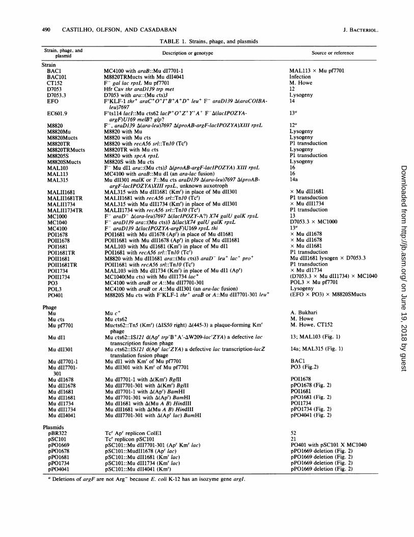
d Strr exconjugants. chromosome or in a plasmid present in a cell. To select Mucts insertion in the insertions located exclusively in a small genetic region, wesed into the ara-leu devised the mini-Mu plasmid transduction scheme outlined*ains P0111678 and in Fig. 3. This procedure can be carried out with small high-made for Mu d4041 copy-number plasmids whose DNA can be easily isolated18820TRMucts with and examined for its physical structure by gel analysis.r Kmr lysogens for Alternative localization procedures involve phage (54) orthe chromosome in plasmid transfer (19, 25, 37, 41, 55).as noticed that this To test this scheme for selecting insertions into plasmids,n infection was car- we first chose the pSC101 intermediate-copy-number plas-ature of 37°C rather mid which we knew would be viable with a Mu insertion.0°C.) This plasmid was introduced, by DNA transformation withnade in another way selection for Tcr, into strains P011678, P011681, POII1678,[e chromosome. Mu and POII1681, which have Apr and Kmr mini-Mu dl and Muinto the Hfr strain dII prophage along with a complementing Mu cts prophage.luced for transposi- All these strains yielded phage lysates which gave the,ith the F- Mu cts expected 2 x 10⁹ to 5 x 10⁹ plaques per ml and 5 x 10⁵ to 50for Kmr and Strr. x 105 Apr or Kmr transductants per ml in nonimmune M8820
VOL. 158, 1984
on June 19, 2018 by guesthttp://jb.asm
.org/D
ownloaded from
492 CASTILHO, OLFSON, AND CASADABAN
mini-Mu
I INDUCTION
mini-Mu
INFECTION OF Mu IMMUNE,fA CELLS
e HOMOLOGOUS RECOMBINATION
,p
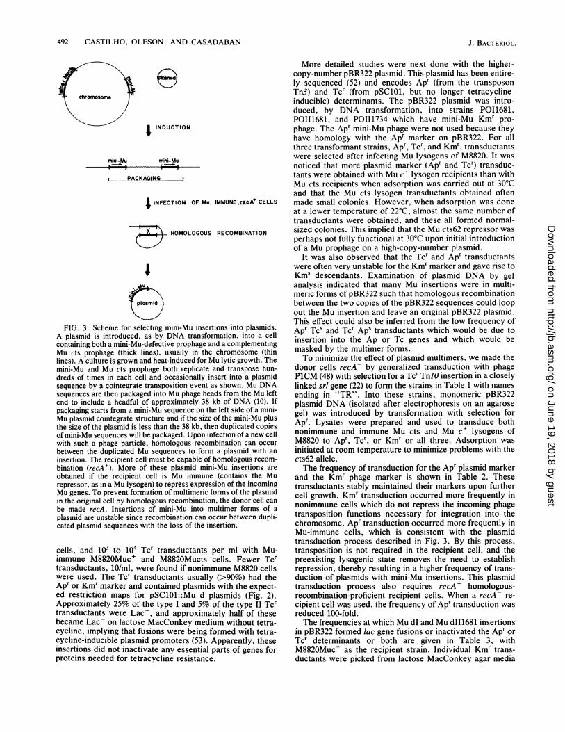
FIG. 3. Scheme for selecting mini-Mu insertions into plasmids.A plasmid is introduced, as by DNA transformation, into a cellcontaining both a mini-Mu-defective prophage and a complementingMu cts prophage (thick lines), usually in the chromosome (thinlines). A culture is grown and heat-induced for Mu lytic growth. Themini-Mu and Mu cts prophage both replicate and transpose hun-dreds of times in each cell and occasionally insert into a plasmidsequence by a cointegrate transposition event as shown. Mu DNAsequences are then packaged into Mu phage heads from the Mu leftend to include a headful of approximately 38 kb of DNA (10). Ifpackaging starts from a mini-Mu sequence on the left side of a mini-Mu plasmid cointegrate structure and if the size of the mini-Mu plusthe size of the plasmid is less than the 38 kb, then duplicated copiesof mini-Mu sequences will be packaged. Upon infection of a new cellwith such a phage particle, homologous recombination can occurbetween the duplicated Mu sequences to form a plasmid with aninsertion. The recipient cell must be capable of homologous recom-bination (recA+). More of these plasmid mini-Mu insertions areobtained if the recipient cell is Mu immune (contains the Murepressor, as in a Mu lysogen) to repress expression of the incomingMu genes. To prevent formation of multimeric forms of the plasmidin the original cell by homologous recombination, the donor cell canbe made recA. Insertions of mini-Mu into multimer forms of aplasmid are unstable since recombination can occur between dupli-cated plasmid sequences with the loss of the insertion.
cells, and 103 to 104 Tcr transductants per ml with Mu-immune M8820Muc+ and M882OMucts cells. Fewer Tcrtransductants, 10/ml, were found if nonimmune M8820 cellswere used. The Tcr transductants usually (>90%) had theApr or Kmr marker and contained plasmids with the expect-ed restriction maps for pSC101::Mu d plasmids (Fig. 2).Approximately 25% of the type I and 5% of the type II Tcrtransductants were Lac+, and approximately half of thesebecame Lac- on lactose MacConkey medium without tetra-cycline, implying that fusions were being formed with tetra-cycline-inducible plasmid promoters (53). Apparently, theseinsertions did not inactivate any essential parts of genes forproteins needed for tetracycline resistance.
More detailed studies were next done with the higher-copy-number pBR322 plasmid. This plasmid has been entire-ly sequenced (52) and encodes Apr (from the transposonTn3) and Tcr (from pSC101, but no longer tetracycline-inducible) determinants. The pBR322 plasmid was intro-duced, by DNA transformation, into strains P011681,POI11681, and POII1734 which have mini-Mu Kmr pro-phage. The Apr mini-Mu phage were not used because theyhave homology with the Apr marker on pBR322. For allthree transformant strains, Apr, Tcr, and Kmr, transductantswere selected after infecting Mu lysogens of M8820. It wasnoticed that more plasmid marker (Apr and Tcr) transduc-tants were obtained with Mu c+ lysogen recipients than withMu cts recipients when adsorption was carried out at 30°Cand that the Mu cts lysogen transductants obtained oftenmade small colonies. However, when adsorption was doneat a lower temperature of 22°C, almost the same number oftransductants were obtained, and these all formed normal-sized colonies. This implied that the Mu cts62 repressor wasperhaps not fully functional at 30°C upon initial introductionof a Mu prophage on a high-copy-number plasmid.
It was also observed that the Tcr and Apr transductantswere often very unstable for the Kmr marker and gave rise toKms descendants. Examination of plasmid DNA by gelanalysis indicated that many Mu insertions were in multi-meric forms of pBR322 such that homologous recombinationbetween the two copies of the pBR322 sequences could loopout the Mu insertion and leave an original pBR322 plasmid.This effect could also be inferred from the low frequency ofApr Tcs and Tcr Aps transductants which would be due toinsertion into the Ap or Tc genes and which would bemasked by the multimer forms.To minimize the effect of plasmid multimers, we made the
donor cells recA- by generalized transduction with phageP1CM (48) with selection for a Tcr TnJO insertion in a closelylinked srl gene (22) to form the strains in Table 1 with namesending in "TR". Into these strains, monomeric pBR322plasmid DNA (isolated after electrophoresis on an agarosegel) was introduced by transformation with selection forApr. Lysates were prepared and used to transduce bothnonimmune and immune Mu cts and Mu c+ lysogens ofM8820 to Apr, Tcr, or Kmr or all three. Adsorption wasinitiated at room temperature to minimize problems with thects62 allele.The frequency of transduction for the Apr plasmid marker
and the Kmr phage marker is shown in Table 2. Thesetransductants stably maintained their markers upon furthercell growth. Kmr transduction occurred more frequently innonimmune cells which do not repress the incoming phagetransposition functions necessary for integration into thechromosome. Apr transduction occurred more frequently inMu-immune cells, which is consistent with the plasmidtransduction process described in Fig. 3. By this process,transposition is not required in the recipient cell, and thepreexisting lysogenic state removes the need to establishrepression, thereby resulting in a higher frequency of trans-duction of plasmids with mini-Mu insertions. This plasmidtransduction process also requires recA + homologous-recombination-proficient recipient cells. When a recA- re-cipient cell was used, the frequency of Apr transduction wasreduced 100-fold.The frequencies at which Mu dI and Mu dII1681 insertions
in pBR322 formed lac gene fusions or inactivated the Apr orTcr determinants or both are given in Table 3, withM8820Muc+ as the recipient strain. Individual Kmr trans-ductants were picked from lactose MacConkey agar media
J. BACTERIOL.
on June 19, 2018 by guesthttp://jb.asm
.org/D
ownloaded from

MINI-Mu-lac GENE FUSION 493
TABLE 2. Mini-Mu duction of pBR322
Mini-Mu's in Recipient strains (transductants/ml)donors with Selectionmonomeric M8820 M882OMucts M8820Muc+pBR322
Kmr 1 X 10⁵ 7 x 10³ 1 X 10³Mu dI1681 Apr 3 x 10' 4 x 10³ 2 x 10³
Kmr Apr 4 x 10' 4 x 10³ 1 X 10³Kmr 1 x 10⁷ 7 x 10⁵ 2 x 10⁵
Mu dII1681 Apr 1 x 10² 4 x 10⁵ 4 x 10⁵Kmr Apr 3 x 100 2 x 10⁵ 1 x 10⁵
and checked for Apr and Tcr. The 3% of the Kmr transduc-tants examined which were both Ap5 and Tcs are notincluded in these data since for all cases examined these didnot contain any plasmid DNA and presumably were due torare Mu transposition or recombination in the Mu-immunecells.We noted that the frequency of Mu d insertions in the Apr
or Tcr determinants (Table 3) was approximately sixfoldlower than would be expected from the size of these regionson pBR322 (52). The frequency of insertions that are Lac' inthe Ap and Tc resistance determinants is 69 and 41%,respectively, for Mu dI1681, and 12 and 7%, respectively,for Mu dII1681, in comparison to an expected almost 50%(two possible orientations) for Mu dl transcription fusionsand 16.7% (two orientations and three phases) for Mu dII in-frame translation fusions. These variations from expecta-tions may be due to nonrandomness of Mu insertion (seebelow) or to lethality of some insertions. The Apr determi-nant codes for a periplasmic protein P-lactamase and the Tcrdeterminant codes for membrane-bound proteins, and hy-brid protein P-galactosidase gene fusions to these types ofgenes have previously been reported (28) to often be lethal.
Plasmid DNAs from several insertions of each type shownin Table 3 were isolated and examined by gel analysis afterdigestion with several restriction enzymes. All were found tohave appropriate structures for Mu d insertions into pBR322.No detectable deletions or rearrangements were found,except that for occasional DNA preparations, some faintrestriction digestion-generated bands could be seen on gelswhich may have been due to Mu-induced rearrangements ina small fraction of the plasmids. The positions and orienta-tions of mini-Mu insertions in pBR322 were mapped andfound to be distributed around the plasmid, although notnecessarily at random (see below). Insertions that formedlac fusions were mapped in the Apr and Tcr determinants, inthe rop gene (18), and in unknown genetic regions (51, 52) at1.4 and 3.15 kb from the EcoRI site (in the clockwisedirection). Insertions of the 1734 phage were also examinedand found to have structures expected for simple insertions.Mu d insertions into pBR322 were also shown not to have
any detectable defects for Mu growth or transduction. Mucts lysogens containing mini-Mu insertions in pBR322 wereinduced and the lysates used to transduce M8820 cells. TheKmr transductants obtained had new insertions, as judgedby the various levels of lac expression observed on lactoseMacConkey medium and by the loss of the plasmid markerin most cases. However, the titers of Mu d (Kmr) transduc-tion and of the complementing Mu cts phage were reducedapproximately 10- and 103-fold, respectively, as comparedwith the original phage lysates. This we suspect is due tointerference by the high number of copies of the defectiveMu d phage on the pBR322 plasmid which could competi-tively inhibit the growth of the unit copy complementing Mu
cts phage in the chromosome. When Mu d insertions inpBR322 were moved to the chromosome (as Kmr transduc-tants of M8820) and induced (after the introduction of a Mucts-complementing prophage from a cross with D7053.3),normal transduction frequencies and phage titers were ob-served.
DISCUSSION
We described the construction of small Mu-/ac gene fusionelements that can be used in vivo to form either transcrip-tional or transcriptional-translational lac gene fusions. Fu-sions were formed with the ampicillin (,B-lactamase) andtetracycline resistance determinants and the rop gene onpBR322 and with tetracycline-inducible genes on pSC101.The latter fusions were selected by their regulation of lacexpression and not by an insertion-inactivation phenotypesuch as sensitivity to tetracycline.
Insertions of these mini-Mu elements can be localized tosequences on particular plasmids, including the high-copy-number plasmids commonly used for DNA cloning. Inser-tions have been obtained in plasmids with several types ofreplicons and copy numbers, including pSC101 and ColEltype plasmids (such as pBR322) as described here, and Ffactor- and plSa- (as pACYC184) (20) derived plasmids. Notype of plasmid was found that could not tolerate insertionsof these mini-Mu elements. The only known limitation to theplasmid insertion procedure described here (Fig. 3) is thesize of the plasmid, since bacteriophage Mu can packageonly approximately 38 kb of DNA (10). For larger plasmids,other procedures, such as conjugation (19, 25, 37, 41, 55),can be used to select insertions specifically in the plasmid.As few as 116 base pairs from the Mu right end and 1,006
base pairs from the left end were retained in these mini-phages (Fig. 2), which are still competent for intracellulartransposition and growth as a phage when complemented bya wild-type Mu phage. Most of the DNA sequences on thesephage have been determined previously (3, 7, 33, 34, 46, 49).These mini-Mu phage are deleted for most of the Mu genes,including the lys (lysis) and kil (host cell killing) genes whichmay affect cell viability when present in a high-copy-numberplasmid. Insertions of these elements are stabie in ColEl-derived plasmids, in contrast to some but not all reports forfull-length Mu insertions. Inselburg (32) and others (asdiscussed in reference 50) have reported the failure to obtainMu insertions in ColEl-derived plasmids, whereas Maynard-Smith et al. (40) and Liebart et al. (38) have reported theisolation of such insertions with Mu c+ and Mu cts. The firstof the positive reports (40) have presented only three inser-tions, two of which have deletions of plasmid sequences nearthe insertions, and the other one (38) does not characterizethe insertions.
lac gene fusions formed by Mu-lac transposition can begenetically unstable in the presence of the Mu transposition
TABLE 3. Frequency of lac+, Aps, and Tcs insertions in pBR322% of inserts with following
Phage Phenotype phenotypes:Apr Tc' Apr Tc' Ap5 Tc'
Mu dI1681 Lac' 43.0 1.7 2.7Lac- 49.0 2.4 1.2
Mu dII1681 Lac' 5.1 0.5 0.3Lac- 85.3 6.7 2.1
VOL. 158, 1984
on June 19, 2018 by guesthttp://jb.asm
.org/D
ownloaded from
494 CASTILHO, OLFSON, AND CASADABAN
genes since the Mu-lac can transpose to new sites or maketransposition-generated terminal deletions. This can particu-larly interfere with selections for mutants that have alteredgene expression. To circumvent this problem, fusions can bemade with a transposition-defective phage such as the Mud1734 phage described here. Alternatively, fusions madewith a transposition-proficient phage can be stabilized byremoving the transposition genes or a Mu end by mutation orby recombination with a defective phage such as Mu d1734or A (lac Mu) (13, 36). Mu insertions in plasmids can also bestabilized by removing appropriate restriction fragments.The scheme outlined in Fig. 3 can be used to isolate
insertions of the transposition gene-deleted phage Mu d1734,since there is complementation in the donor cell and thetransposase gene is not needed in the recipient cell. We havealso found that Mu d1734 insertions into the chromosomecan be formed after infection with a lysate, even though noMu transposition genes were expected to be present in therecipient. This transposition was not due to transient com-plementation by a coinfecting Mu cts phage, since lysogenswere obtained linearly with dilutions of the phage lysate.Recent preliminary experiments (P. Olfson, S. Suh, B.Castilho, and M. Casadaban, unpublished data) indicate thatthis transposition does depend on a Mu A gene being carriedinto the recipient cell, since the number of transductants isgreatly reduced when the 1734 phage is grown with a Mu Aamber helper phage in an amber-suppressing cell and theresulting lysate is used to infect a nonsuppressing cell.Perhaps the Mu transposition genes are occasionally carriedon the flanking DNA packaged from the chromosome whenthere is a nearby insertion of a helper Mu prophage in thedonor cell.The process depicted in Fig. 3 for localization of mini-Mu
insertions into plasmids can be used to obtain hundreds ofinsertions in a small-scale experiment which should beenough to saturate all possible Mu insertion sites in a smallplasmid. This brings up the question ofjust how random Muinsertion sites are. Most insertions that have been studied ina defined region, such as in a particular gene, have beenshown to be in different sites, although a few, as determinedby recombination tests, may have occurred in precisely thesame location (11, 24, 26, 47). Precise sequence mapping ofthese insertions have not been reported. Insertions of othertransposons in limited regions have been shown upon carefulanalysis to have hot-spot sites with various specificities (4, 5,27, 29, 56, 57). In further experiments (unpublished data), weused the procedure outlined in Fig. 3 to prepare pools ofmini-Mu insertions in a shortened version of the pBR322plasmid and found that many insertions do indeed occur in afew specific hot-spot regions, with particular sites withineach region being used at different frequencies.Mu insertions in plasmids can be used as a convenient
source of Mu DNA for further Mu genetic or biochemicalanalysis (19). For example, Mu dII4041 insertions inpBR322-derived plasmids have been used to isolate deletionswhich define the functional ends of bacteriophage Mu. Sofar, the essential sequences on the right end of Mu have beenlocalized to begin between 93 and 68 base pairs from that end(unpublished data).Mu insertion plasmid DNA can also be used to conve-
niently introduce Mu into other species. Mu dII1681 inser-tions in pBR322 have been used as a source of DNA totransform the gram-negative bacterium Agrobacterium tu-mefaciens, in which Mu-lac has been found to transpose andform lac fusions (F. Richaud and M. Casadaban, unpub-lished data). More conventional methods for Mu transfer to
other gram-negative bacterial species have been carried outwith Mu dII1681 insertions into permissive conjugativeplasmids for transfer between Escherichia coli and A. tume-faciens (F. Richaud and M. Casadaban, unpublished data)and Pseudomonas aeruginosa (J. Shapiro, personal commu-nication). Mu dII1681 has also been introduced into thelower eucaryotic yeast Saccharomyces cerevisiae with in-sertions in E. coli-S. cerevisiae shuttle vector plasmids (6),but no evidence for transposition was found by using theformation of P-galactosidase gene fusions as an assay fortransposition (M. Ditto and M. Casadaban, unpublisheddata).
ACKNOWLEDGMENTSWe thank Ni. O'Connor and M. Malamy for information about the
IS121 insertion in Mu dIl, and M. Howe for Mu pf7701.This work was supported by Public Health Service grant GM
29067 fronm the National Institutes of Health. B.A.C. was supportedby grants from CNPq and FAPESP (Brazil), P.O. was supported byPublic Health Service grant CA 09273 from the National Institutes ofHealth, and M.J.C. was supported by Public Health Service grantAl 00468 from the National Institutes of Health.
LITERATURE CITED1. Allet, B., F. Blattner, M. Howe, M. Magazin, D. Moore, K.
O'Day, D. Schultz, and J. Schumm. 1977. Genetic and physicalmap of bacteriophage Mu, p. 745-746. In A. I. Bukhari, J. A.Shapiro, and S. L. Adhya (ed.), DNA insertion elements,plasmids and episomes. Cold Spring Harbor Laboratory, ColdSpring Harbor, N.Y.
2. Bassford, P., J. Beckwith, M. Berman, E. Brickman, M. Casada-ban, L. Guarante, I. Saint-Girons, A. Sarthy, M. Schwartz, andT. Silhavy. 1978. Genetic fusions of the lac operon: a newapproach to the study of biological processes, p. 245-261. InJ. H. Miller and W. S. Reznikoff (ed.), The operon. Cold SpringHarbor Laboratory, Cold Spring Harbor, N.Y.
3. Beck, E., G. Lugwig, E. A. Auerswald, B. Reiss, and H. Schaller.1982. Nucleotide sequence and exact localization of the neomy-cin phosphotransferase gene from transposon Tn5. Gene19:327-336.
4. Berg, D. E., A. Weiss, and L. Crossland. 1980. Polarity of TnSinsertion mutations in Escherichia coli. J. Bacteriol. 142:439-446.
5. Bossi, L., and M. S. Campi. 1981. DNA sequences at the sites ofthree insertions of the transposable element Tn5 in the histidineoperon of Salmonella. Mol. Gen. Genet. 183:406-408.
6. Botstein, D., C. Falco, S. Stewart, M. Brennan, S. Scherer, D.Stinchcomb, K. Struhl, and R. Davis. 1979. Sterile host yeasts: aeukaryotic system of biological containment for recombinantDNA experiments. Gene 8:17-24.
7. Buchel, D. B., B. Gronenborn, and B. Muller-Hill. 1980. Se-quence of the lactose permease gene. Nature (London) 283:541-545.
8. Bukhari, A. I. 1976. Bacteriophage Mu as a transpositionelement. Annu. Rev. Genet. 10:389-411.
9. Bukhari, A. I., and E. Ljungquist. 1977. Bacteriophage Mu:methods for cultivation and use, p. 749-756. In A. I. Bukhari,J. A. Shapiro, and S. L. Adhya (ed.), DNA insertion elements,plasmids and episomes. Cold Spring Harbor Laboratory, ColdSpring Harbor, N.Y.
10. Bukhari, A. I., and A. L. Taylor. 1975. Influence of insertions onpackaging of host sequences covalently linked to bacteriophageMu DNA. Proc. Natl. Acad. Sci. U.S.A. 72:4399-4403.
11. Bukhari, A. I., and D. Zipser. 1972. Random insertion of Mu-1DNA within a single gene. Nature (London) New Biol. 236:240-243.
12. Casadaban, M. 1975. Fusion of the Escherichia coli lac genes tothe ara promoter: a general technique using bacteriophage Mu-1insertions. Proc. Natl. Acad. Sci. U.S.A. 72:809-813.
13. Casadaban, M. 1976. Transposition and fusion of the lac genesto selected promoters in E. coli using bacteriophage lambda andMu. J. Mol. Biol. 104:541-555.
J. BACTERIOL.
on June 19, 2018 by guesthttp://jb.asm
.org/D
ownloaded from
MINI-Mu-lac GENE FUSION 495
14. Casadaban, M. 1976. Regulation of the regulatory gene for thearabinose pathway, arcaC. J. Mol. Biol. 104:557-566.
14a.Casadaban, M., and J. Chou. 1984. In Oivo formation of hybridP-galactosidase gene fusions in one step with a new transpos-able Mu-lac transducing phage. Proc. Natl. Acad. Sci. U.S.A.81:535-539.
15. Casadaban, M., J. Chou, and S. N. Cohen. 1980. In vitro genefusions that join an enzymatically active 3-galactosidase seg-ment to amino-terminal fragments of exogenous proteins: Esch-erichia coli plasmid vectors for the detection and cloning oftranslational initiation signals. J. Bacteriol. 143:971-980.
16. Casadaban, M., and S. N. Cohen. 1979. Lactose genes fused toexogenous promoters in one step using a Mu-/ac bacteriophage:in vivo probe for transcriptional control sequences. Proc. Natl.Acad. Sci. U.S.A. 76:4530-4533.
17. Casadaban, M., and S. N. Cohen. 1980. Analysis of gene controlsignals by DNA fusion and cloning in Escherichia coli. J. Mol.Biol. 138:179-207.
18. Cesareni, G., M. A. Muesing, and B. Polisky. 1982. Control ofColEl DNA replication: the rop gene product negatively affectstranscription from the replicon primer promoter. Proc. Natl.Acad. Sci. U.S.A. 79:6313-6317.
19. Chaconas, G., F. J. deBruijn, M. Casadaban, J. R. Lupski, T. J.Kwok, R. M. Harshey, M. S. DuBow, and A. I. Bukhari. 1981. Invitro and in i'iw'o manipulations of bacteriophage Mu DNA:cloning of Mu ends and construction of mini-Mu's carryingselectable markers. Gene 13:37-46.
20. Chang, A. C. Y., and S. N. Cohen. 1978. Construction andcharacterization of amplifiable multicopy DNA cloning vehiclesderived from the P1SA cryptic miniplasmid. J. Bacteriol.134:1141-1156.
21. Cohen, S. N., and A. C. Y. Chang. 1977. Revised interpretationof the origin of the pSC101 plasmid. J. Bacteriol. 132:734-737.
22. Csonka, L. N., and A. J. Clark. 1979. Deletions generated by thetransposon TnlO in the srl recA region of the Escherichia coli K-12 chromosome. Genetics 93:321-343.
23. Csonka, L. N., M. M. Howe, J. L. Ingraham, L. S. Pierson III,and C. L. Turnbough, Jr. 1981. Infection of Sal/nonella typhi-murium with coliphage Mu dl(Ap' lac): construction of pvr:lacgene fusions. J. Bacteriol. 145:299-305.
24. Daniell, E., R. Roberts, and J. Abelson. 1972. Mutation in thelactose operon caused by bacteriophage Mu. J. Mol. Biol. 69:1-8.
25. Dixon, R., R. R. Eady, G. Espin, S. Hill, M. laccarino, D. Kahn,and M. Merrick. 1980. Analysis of regulation of K/ebsiellapneumoniae nitrogen fixation (nif) gene cluster with gene fu-sions. Nature (London) 286:128-132.
26. Emr, S. D., and T. Silhavy. 1980. Mutations affecting localiza-tion of an Escherichia coli outer membrane protein, the bacte-riophage A receptor. J. Mol. Biol. 141:63-90.
27. Galas, D. J., M. P. Calos, and J. H. Miller. 1980. Sequenceanalysis of Tn9 insertions in the lacZ gene. J. Mol. Biol. 144:19-41.
28. Hall, M. N., and T. J. Silhavy. 1981. Genetic analysis of themajor outer membrane proteins of Escherichia coli. Annu. Rev.Genet. 15:91-142.
29. Hailing, S. M., and N. Kleckner. 1981. A symmetrical sixbasepair target site sequence determines TnlO insertion speci-ficity. Cell 28:155-163.
30. Heifron, F., B. J. McCarthy, H. Ohtsubo, and E. Ohtsubo. 1979.DNA sequence analysis of the transposon Tn3: three genes andthree sites involved in transposition of Tn3. Cell 18:1153-1163.
31. Howe, M. M., and E. G. Bade. 1975. Molecular biology ofbacteriophage Mu. Science 190:624-632.
32. Inselburg, J. 1974. Isolation and characterization of mutants ofcolicin plasmids El and E2 after Mu bacteriophage infection. J.Bacteriol. 119:469-477.
33. Kahmann, R., and D. Kamp. 1979. Nucleotide sequences of theattachment sites of bacteriophage Mu DNA. Nature (London)280:247-250.
34. Kalmins, A., K. Otto, U. Ruther, and B. Muller-Hill. 1983.Sequence of the lacZ gene of Escherichia coli. EMBO J. 2:593-
597.35. Kania, J., and B. Muller-Hill. 1977. Construction, isolation and
implications of repressor-,B-galactosidase: 3-galactosidase hy-brid molecules. Eur. J. Biochem. 79:381-383.
36. Komeda, Y., and T. lino. 1979. Regulation of expression of theflagellin gene (hag) in Escherichia coli K-12: analysis of hag-lacgene fusions. J. Bacteriol. 139:721-729.
37. Lee, J.-H., L. Heffernan, and G. Wilcox. 1980. Isolation of alta-lac gene fusions in Salmonella tvphimurium LT2 by usingtransducing bacteriophage Mu d(Apr lac). J. Bacteriol.143:1325-1331.
38. Liebart, J. C., P. Ghelardini, and L. Paolozzi. 1982. Conserva-tive integration of bacteriophage Mu DNA into pBR322 plas-mid. Proc. Natl. Acad. Sci. U.S.A. 79:4362-4366.
39. Maniatis, T., E. Fritsch, and J. Sambrook. 1982. Molecularcloning. Cold Spring Harbor Laboratory. Cold Spring Harbor,N.Y.
40. Maynard-Smith, S., D. Leach, A. Coelho, J. Carey, and N.Symonds. 1980. The isolation and characteristics of plasmidsderived from the insertion of Mup Apl into pML2: theirbehavior during transposition. Plasmid 4:34-50.
41. Mendoza, D., D. Clark, and J. E. Cronan, Jr. 1981. One-stepgene amplification by Mu-mediated transposition of E. coligenes to a multicopy plasmid. Gene 15:27-32.
42. Miller, J. H. 1972. Experiments in molecular genetics. ColdSpring Harbor Laboratory, Cold Spring Harbor, N.Y.
43. Muller-Hill, B., G. Heidecker, and J. Kania. 1976. Repressor-galactosidase chimaeras, p. 167-179. In R. Markham and R. W.Horne (ed.), Structure-function relationships of proteins.North-Holland Publishing Co., New York.
44. Muller-Hill, B., and J. Kania. Lac repressor can be fused to -galactosidase. 1974. Nature (London) 249:561-562.
45. O'Connor, M. B., and M. H. Malamy. 1983. A new insertionsequence, IS121, is found on the Mu dll(Ap lac) bacteriophageand the Escherichia coli K-12 chromosome. J. Bacteriol.156:669-679.
46. Priess, H., D. Kamp, R. Kahmann, B. Brauer, and H. Delius.1982. Nucleotide sequence of the immunity region of bacterio-phage Mu. Mol. Gen. Genet. 186:315-321.
47. Raibaud, O., M. Roa, C. Braun-Breton, and M. Schwartz. 1979.Structure of the malB region in Escherichia coli K-12. 1. Geneticmap of the ma/K-/amB operon. Mol. Gen. Genet. 174:241-248.
48. Rosner, J. 1972. Formation, induction and curing of bacterio-phage P1 lysogens. Virology 48:679-689.
49. Rothstein, S. J., R. A. Jorgensen, K. Postle, and W. S. Reznikoff.1980. The inverted repeats of TnS are functionally different. Cell19:795-805.
50. Schumann, W., and E. G. Bade. 1979. In vitro constructedplasmids containing both ends of bacteriophage Mu DNAexpress phage functions. Mol. Gen. Genet. 169:97-105.
51. Stuber, D., and H. Bujard. 1981. Organization of transcriptionalsignals in plasmids pBR322 and pACYC184. Proc. Natl. Acad.Sci. U S.A. 78:167-171.
52. Sutcliffe, J. G. 1979. Complete nucleotide sequence of theEscherichia coli plasmid pBR322. Cold Spring Harbor Symp.Quant. Biol. 43:77-90.
53. Tait, R. C., and H. W. Boyer. 1978. On the nature of thetetracycline resistance controlled by the plasmid pSC101. Cell13:73-81.
54. Toussaint, A., M. Faelen, and A. Resibois. 1981. Chromosomalarrangements induced by mini-Mu and mini-D108. Mini-reviewand new data. Gene 14:115-119.
55. Toussaint, A., and A. Resibois. 1983. Phage Mu: transposition asa life-style, p. 105-158. In J. Shapiro (ed.), Mobile geneticelements. Academic Press, Inc., New York.
56. Tu, C.-P. D., and S. N. Cohen. 1980. Translocation specificity ofthe Tn3 element: characterization of sites of multiple insertions.Cell 19:151-160.
57. Weisberg, R. A., and A. Landy. 1983. Site-specific recombina-tion in phage lambda, p. 211-250. In R. W. Hendrix, J. W.Roberts, F. W. Stahl, and R. A. Weisberg (ed.), Lambda II.Cold Spring Harbor Laboratory, Cold Spring Harbor, N.Y.
VOL. 158, 1984
on June 19, 2018 by guesthttp://jb.asm
.org/D
ownloaded from