please cite this article in press as: carman et al ... · total duration of diapedesis and the...
TRANSCRIPT
Immunity
Article
Transcellular Diapedesis Is Initiatedby Invasive PodosomesChristopher V. Carman,1,5 Peter T. Sage,1,5 Tracey E. Sciuto,2 Miguel A. de la Fuente,3 Raif S. Geha,3
Hans D. Ochs,4 Harold F. Dvorak,2 Ann M. Dvorak,2 and Timothy A. Springer1,*1The CBR Institute for Biomedical Research, Department of Pathology, Harvard Medical School, Boston, MA 02115, USA2Department of Pathology, Beth Israel Deaconess Medical Center, Harvard Medical School, Boston, MA 02215, USA3Division of Immunology, Children’s Hospital, Boston, MA 02115, USA4Department of Pediatrics, University of Washington School of Medicine, Seattle, WA 98195, USA5Present address: Department of Medicine, Beth Israel Deaconess Medical Center, Harvard Medical School, Boston,MA 02215, USA*Correspondence: [email protected] 10.1016/j.immuni.2007.04.015
SUMMARY
Diapedesis is critical for immune system func-tion and inflammatory responses. This occursby migration of blood leukocytes either directlythrough individual microvascular endothelialcells (the ‘‘transcellular’’ route) or betweenthem (the ‘‘paracellular’’ route). Mechanismsfor transcellular pore formation in endotheliumremain unknown. Here we demonstrate thatlymphocytes used podosomes and extended‘‘invasive podosomes’’ to palpate the surfaceof, and ultimately form transcellular poresthrough, the endothelium. In lymphocytes,these structures were dependent on Src kinaseand the actin regulatory protein WASP; inhibi-tion of podosome formation selectively blockedthe transcellular route of diapedesis. In endo-thelium, membrane fusion events dependenton the SNARE-containing membrane fusioncomplex and intracellular calciumwere requiredfor efficient transcellular pore formation inresponse to podosomes. These findingsprovide insights into basic mechanisms forleukocyte trafficking and the functions ofpodosomes.
INTRODUCTION
Whereas functions for most cells of the body are coupledto their organization in tissues, functions for blood leuko-cytes are uniquely centered on their ability to efficientlycross tissue barriers, i.e., invasiveness. In order to conductimmune surveillance, respond to infection, and undergodifferentiation, leukocytes must traffic throughout thebody, moving in and out of the bone marrow, lymphoidorgans, and inflamed tissues (von Andrian and Mackay,2000). A central component to this process is the repeatedcrossing of vascular and lymphatic endothelial barriers,i.e., diapedesis, as leukocytes leave and re-enter the circu-
lation (von Andrian and Mackay, 2000). Fundamentalaspects of the mechanisms for this barrier-crossing func-tion remain unknown.Diapedesis occurs in vivo either directly through individ-
ual endothelial cells (the transcellular route) or betweenthem at interendothelial cell junctions (the paracellularroute) (Feng et al., 1998; Greenwood et al., 1994; Lossin-sky and Shivers, 2004; Marchesi, 1961; Marchesi andGowans, 1963). In both cases, the process begins withthe accumulation of leukocytes on the luminal surface ofthe endothelium through a well-characterized sequenceof rolling, activation, and firm adhesion events (Springer,1994). Subsequent integrin-dependent lateral migrationis an essential next step that seems to allow leukocytesto search out sites permissive for endothelial barrierpenetration (Schenkel et al., 2004). The final and criticalsteps of identifying such sites and then formally breachingthe endothelium are incompletely understood.Interendothelial cell junctions provide a defined and
intuitive site for paracellular diapedesis, and mechanisticmodels for this process have been proposed (Muller,2003). In contrast, neither an intuitive locus nor an obviousmechanism for transcellular diapedesis exists. Whateverthe mechanism, transcellular diapedesis requires, atminimum, local displacement of endothelial cytoplasmicorganelles and fusion of the apical and basal plasmamembranes. Formally, this process could be driven bythe leukocyte, by the endothelium, or by contributionsfrom both.On the part of the leukocyte, actin-dependent protru-
sive structures might be able to both displace endothelialorganelles and facilitate membrane fusion by forcingapical and basal membranes into close opposition. Candi-date protrusive structures include the well-characterizedfilopodia, lamellipodia, and related pseudopodia (Ridleyet al., 2003), along with more recently discovered protru-sive organelles termed podosomes (‘‘foot-protrusions’’)and invadopodia (Buccione et al., 2004; Linder andAepfelbacher, 2003; Yamaguchi et al., 2005b).Podosomes and invadopodia are micron-scale cylindri-
cal protrusions formed on the ventral aspect of adherentcells with the defining architecture of a peripheral ring ofb2 or b3 integrin, talin, and vinculin, and a central core of
Immunity 26, 1–14, June 2007 ª2007 Elsevier Inc. 1
Please cite this article in press as: Carman et al., Transcellular Diapedesis Is Initiated by Invasive Podosomes, Immunity (2007),doi:10.1016/j.immuni.2007.04.015
F-actin (Buccione et al., 2004; Linder and Aepfelbacher,2003). These protrusive structures share a strong depen-dence on Src kinase and the cytoskeletal regulatorWiskott-Aldrich syndrome (WAS) protein (WASP) orN-WASP in nonhematopoietic cells (Calle et al., 2004;Linder and Aepfelbacher, 2003). Distinction betweenpodosomes and invadopodia are seen in their size (withsubstantially greater protrusion depths observed in inva-dopodia), dynamics, and apparent functions (Buccioneet al., 2004; Yamaguchi et al., 2005b). Podosomes havebeen described in a wide range of migratory cell types,most notably leukocytes, where they form dynamic clus-tered arrays or ‘‘rosettes’’ (Buccione et al., 2004; Evanset al., 2003; Linder and Aepfelbacher, 2003). However,their function is unclear, though generally linked to adhe-sion and migration. In contrast, invadopodia, which havebeen described only in transformed cell types, havea more defined function in burrowing across matrixbarriers, thereby conferring invasiveness to metastatictumor cells (Buccione et al., 2004; Yamaguchi et al.,2005a).
An active role for the endothelium during diapedesis hasbeen widely demonstrated through, for example, leuko-cyte-triggered calcium and Rho signaling (Adamsonet al., 1999; Carman and Springer, 2004; Etienne-Manne-ville et al., 2000; Huang et al., 1993; Muller, 2003). We andothers have previously hypothesized that such intraendo-thelial signals could potentially trigger membrane fusionevents that facilitate transcellular pore formation (Carmanand Springer, 2004; Feng et al., 2002; Lossinsky andShivers, 2004; Olah and Glick, 1985).
Here we demonstrate that leukocytes dynamicallyinserted numerous podosomes and extended ‘‘invasivepodosomes,’’ the latter of which exhibit similarities toinvadopodia, into the endothelial surface both in vitroand in vivo. This serves to probe for sites permissive totranscellular pore formation and to trigger endothelialfusion activity that supports this process. This previouslyunrecognized palpation behavior was critical for transcel-lular diapedesis and may also play broader roles in leuko-cyte path finding during trafficking.
RESULTS
Transcellular Diapedesis Is Quantitatively Importantin Microvascular EndotheliumGiven that the vast majority of leukocyte traffic in vivooccurs through the microvasculature (von Andrian andMackay, 2000), in this study we investigated migrationon human dermal (HDMVEC) and lung (HLMVEC) micro-vascular endothelial monolayers. By using our previouslyestablished fixed-cell imaging approach (Carman andSpringer, 2004) with IL-2-cultured lymphocytes, wedemonstrate that approximately one-third of diapedesisevents occurred via the transcellular route on tumornecrosis factor (TNF)-a-activated HDMVEC and HLMVEC(Figure 1A). We also examined and found comparableresults for dermal lymphatic endothelium (Figure 1A). Bycontrast, approximately 10% of lymphocytes transmi-
grated via the transcellular route on HUVEC (humanumbilical vein endothelial cells), a phenotypically distinctmacrovascular endothelium (Figure 1A; Carman andSpringer, 2004). Dynamic live-cell imaging experimentsshowed that the kinetic parameters of para- and transcel-lular diapedesis are largely indistinguishable (Figure 1B).This confirmed the validity of using a fixed-cell, single-time-point analysis for quantitative comparison of para-and transcellular diapedesis.We attempted to selectively block paracellular diapede-
sis. Paracellular diapedesis is predicted to be uniquelydependent on molecules enriched at the endothelialjunctions including platelet-endothelial cellular adhesionmolecule-1 (PECAM-1), junctional adhesion molecule-1(JAM-1), and CD99 (Muller, 2003). Thus, we employeda widely used PECAM-1 function-blocking antibody(HEC7) (Muller, 2003). To our surprise, we found thatalthough HEC7 inhibited diapedesis, it affected bothpara- and transcellular routes comparably (Figure 1C).Immunofluorescence imaging of PECAM-1 (Figure 1D)and JAM-1 (Figure S1 in the Supplemental Data availablewith this article online) showed that these molecules weresimilarly enriched around both trans- and paracellulardiapedesis events in previously described ‘‘transmigra-tory cups’’ (Barreiro et al., 2002; Carman and Springer,2004). Though the role of PECAM-1 in lymphocytediapedesis in vivo remains unclear, these data suggestthat PECAM-1 and JAM-1may not be specific for paracel-lular transmigration.
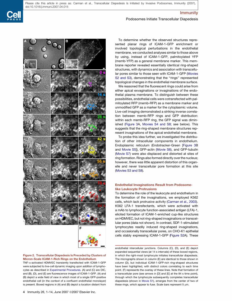
Clusters of Dynamic, Micron-Scale Invaginationsinto the Endothelial Surface PrecedeTranscellular DiapedesisLive-cell imaging experiments, combining fluorescenceand DIC (differential interference contrast) modalities,were used to dissect mechanisms for transcellulardiapedesis. To monitor the endothelial cell-surfacedynamics, we imaged lymphocyte migration on micro-vascular endothelial cells transfected with intercellularadhesion molecule (ICAM)-1 fused to GFP (ICAM-1-GFP). Lymphocytes migrated laterally over the endothe-lium and underwent both trans- and paracellular diapede-sis. Strikingly, clusters of micron-scale fluorescent rings(!0.2–1 mm in diameter) formed intermittently undermost spread and laterally migrating lymphocytes (Figure 2andMovie S1). The initial appearance of a single or severalrings was typically followed by the rapid (within !30 s)appearance of clusters containing 10–30 rings (coveringan area of !100 mm2) as lymphocytes spread (Figure 2).For laterallymigrating lymphocytes, rings formed predom-inantly just behind the leading edge (see Movie S3) butalso formed under the trailing edges and various lateralextensions (see below and Movie S9). Individual ringswere highly dynamic and appeared, disappeared, andchanged size and shape on the time scale of tens of sec-onds (Figure 2).Most importantly, formation of transcellular pores at the
initiation of transcellular diapedesis was essentiallyalways preceded by the appearance of rings (Figure 2
Immunity
Podosomes Initiate Transcellular Diapedesis
2 Immunity 26, 1–14, June 2007 ª2007 Elsevier Inc.
Please cite this article in press as: Carman et al., Transcellular Diapedesis Is Initiated by Invasive Podosomes, Immunity (2007),doi:10.1016/j.immuni.2007.04.015

and Movie S1). An analysis of 58 transcellular diapedesisevents from representative videos demonstrated thatas few as 4 and as many as 216 rings (average ± SEM =62.1 ± 7.1) were seen prior to the formation of eachtranscellular pore. In most cases, the initial pores wid-ened rapidly (expanding their diameter an average
rate ± SEM = 2.6 ± 0.2 mm/min; n = 28) to a maximumof !6 mm, as lymphocyte diapedesis proceeded, and thenconstricted and resealed (Movie S1). These observationssuggested a potential functional relationship betweenthe ring-shaped structures and the initiation of transcellu-lar diapedesis.
Figure 1. Transcellular Diapedesis on Microvascular Endothelium(A) IL-2-cultured lymphocytes were incubated with TNF-a-activated endothelial monolayers for 10 min followed by fixation, staining, and scoring as
described in Experimental Procedures. Endothelial cells included HDMVEC and HLMVEC from Cambrex, as well as HDMVEC (HDMVEC*) and
lymphatic ECs (Lymph*) selectively isolated from fresh neonatal foreskins. y axis represents the percentage of the total diapedesis that was trans-
cellular. Values are mean ± SEM for at least three separate experiments.
(B) Videos from live-cell imaging of lymphocyte diapedesis on HDMVEC and HLMVEC (described in Figure 2, below) were analyzed to determine the
total duration of diapedesis and the amount of time lymphocytes resided at a location before initiating diapedesis (dwell time) for transcellular (black
bars) or paracellular (gray bars) events. Data for the HLMVECs (L) and HDMVECs (D) were analyzed both separately and together (T). Values are the
mean ± SEM for at least seven measurements.
(C) Lymphocytes were incubated with activated HLMVEC as in (A) above, in the absence (gray bars) and presence (black bars) of PECAM-1 function-
blocking mAb HEC7 (40 mg/ml). Graph depicts total diapedesis, as well as the separate the para- and transcellular diapedesis components, each as
a percentage of total cells. Values are the mean ± SEM of three separate experiments. 40 or 120 mg/ml isotype control antibody had no effect on
diapedesis (not shown).
(D) Lymphocyteswere incubatedwithactivatedHLMVEC for 10min followedbyfixationandstainingwith fluorescent antibodies to integrinb2, ICAM-1,
and PECAM-1 and subjected to confocal microscopy. Images are Z-stack projections of representative transcellular (left) and paracellular (right)
diapedesis events. Note that under one of the two lymphocytes on the left, two transcellular pores have formed. Arrows indicate the PECAM-1-
enriched interendothelial junctions. Scale bar represents 5 mm.
Immunity 26, 1–14, June 2007 ª2007 Elsevier Inc. 3
Please cite this article in press as: Carman et al., Transcellular Diapedesis Is Initiated by Invasive Podosomes, Immunity (2007),doi:10.1016/j.immuni.2007.04.015
Immunity
Podosomes Initiate Transcellular Diapedesis
To determine whether the observed structures repre-sented planar rings of ICAM-1-GFP enrichment orinvolved topological perturbations in the endothelialmembrane, we conducted analyses similar to those aboveby using, instead of ICAM-1-GFP, palmitoylated YFP(memb-YFP) as a general membrane marker. This mem-brane reporter revealed essentially identical ring-shapedstructures, with dynamics and association with transcellu-lar pores similar to those seen with ICAM-1-GFP (MoviesS2 and S3), demonstrating that the ‘‘rings’’ representedtopological changes in the endothelial membrane surface.We reasoned that the fluorescent rings could arise from
either apical exvaginations or invaginations of the endo-thelial plasma membrane. To distinguish between thesepossibilities, endothelial cells were cotransfectedwith pal-mitoylated RFP (memb-RFP) as a membrane marker andunmodified GFP as a marker for the cytoplasmic volume.Live-cell imaging demonstrated a striking inverse correla-tion between memb-RFP rings and GFP distribution:within each memb-RFP ring, the GFP signal was dimin-ished (Figure 3A, Movies S4 and S8; see below). Thissuggests that the ring-shaped membrane structures rep-resent invaginations of the apical endothelial membrane.To probe this idea further, we investigated the distribu-
tion of other intracellular components in endothelium.Endoplasmic reticulum (Endotracker-Green [Figure 3Band Movie S5]), GFP-actin (Movie S6), and GFP-tubulin(Movie S7) were also displaced and distorted at sites ofring formation. Rings also formeddirectly over the nucleus;however, there was little apparent distortion of this organ-elle and never transcellular pore formation at this site(Movies S3 and S8).
Endothelial Invaginations Result from Podosome-like Leukocyte ProtrusionsTo determine the role of the leukocyte and endothelium inthe formation of the invaginations, we employed K562cells, which lack protrusive activity (Carman et al., 2003).K562 LFA-1 transfectants, which were activated witha mAb to lymphocyte function-associated antigen (LFA)-1,elicited formation of ICAM-1-enriched cup-like structuresonHDMVEC, but not ring-shaped invaginations or transcel-lular pores (data not shown). In contrast, SDF-1-stimulatedlymphocytes readily induced ring-shaped invaginations,and occasionally transcellular pores, on CHO-K1 epithelialcells stably expressing ICAM-1-GFP (Figure S2A). These
Figure 2. Transcellular Diapedesis Is Preceded by Clusters ofMicron-Scale ICAM-1-Rich Rings on the EndotheliumTNF-a-activated HDMVEC transiently transfected with ICAM-1-GFP
were subjected to live-cell dynamic imaging upon addition of lympho-
cytes as described in Experimental Procedures. (A) and (C) are DIC,
and (B), (D), and (E) are fluorescence images of ICAM-1-GFP. (A) and
(B) depict a wide field of view in which most of a single GFP-positive
endothelial cell (in the context of a confluent endothelial monolayer)
is present. Boxed regions in (A) and (B) depict a location distant from
endothelial intercellular junctions. Columns (C), (D), and (E) depict
expanded sequential views (at 14 s intervals) of these boxed regions,
in which the right-most lymphocyte initiates transcellular diapedesis.
The micrographs shown in column (E) are identical to those shown in
column (D), but individual ICAM-1-GFP-rich ring-shaped structures
have been highlighted, with distinct colors correlating to each time
point. (F) represents the overlay of these lines. Note that formation of
a transcellular pore (see arrows in [D] and [E] at the 84 s time point),
through which the lymphocyte subsequently completes transcellular
diapedesis (shown in Movie S1), emerges from the center of two of
these rings, which appear to fuse. Scale bars represent 5 mm.
4 Immunity 26, 1–14, June 2007 ª2007 Elsevier Inc.
Please cite this article in press as: Carman et al., Transcellular Diapedesis Is Initiated by Invasive Podosomes, Immunity (2007),doi:10.1016/j.immuni.2007.04.015
Immunity
Podosomes Initiate Transcellular Diapedesis

data suggest a critical proactive role for the lymphocyte information of the endothelial invaginations.
Given the size, dynamics, and clustered patterns of theendothelial cell invaginations, we hypothesized that theymight be formed in response to podosome protrusionsgenerated by leukocytes. After a brief incubation oflymphocytes with endothelium, samples were fixed andstained for ICAM-1 and F-actin, along with lymphocyteb2-integrin (aLb2; LFA-1) or talin-1. High-resolution confo-
cal imaging revealed the existence of clusters of ventralleukocyte protrusions, each !0.2–1 mm in diameter, withLFA-1 (Figures 4A and 4B)- and talin-1 (Figure 4C)-richouter rings and F-actin-rich inner cores (Figures 4A–4C).These features support the designation of the lymphocyteprotrusions as podosomes. We term the correspondingICAM-1-rich invaginations on the endothelial surface‘‘podoprints.’’ Podoprints were of slightly greater diameterthan their cognate podosomes (Figures 4A and 4B).
Figure 3. ‘‘Ring Structures’’ Represent Endothelial Cell-Surface InvaginationsTNF-a-activated HLMVEC were subjected to live-cell dynamic imaging upon addition of lymphocytes.
(A) HLMVEC were transiently cotransfected with memb-RFP and unmodified GFP. Panels depict DIC, memb-RFP, GFP, and merge of memb-RFP
and GFP. Upper panels show a time point shortly after lymphocytes have settled on the endothelium, but before formation of memb-RFP ring struc-
tures (relative time = 0 s). Lower panels show a time point after lymphocyte spreading and formation of rings (relative time 170 s). Note that for each
memb-RFP ring, a circular region of diminished GFP signal is formed. See corresponding Movie S4.
(B) HDMVEC were transiently transfected with memb-RFP and prestained with ER-tracker green. Panels depict DIC, memb-RFP, ER-tracker green,
and Merge of memb-RFP and ER-tracker green. Upper panels show a time point before (relative time = 0 s) and lower panels show a time point after
(relative time 460 s) the formation of memb-RFP ring structures. Note that memb-RFP ring structures seem to form within the individual reticula of the
ER often distorting and expanding these structures. These features can be more readily appreciated in the corresponding Movie S5. Scale bars
represent 5 mm.
Immunity 26, 1–14, June 2007 ª2007 Elsevier Inc. 5
Please cite this article in press as: Carman et al., Transcellular Diapedesis Is Initiated by Invasive Podosomes, Immunity (2007),doi:10.1016/j.immuni.2007.04.015
Immunity
Podosomes Initiate Transcellular Diapedesis

We further examined lymphocyte actin dynamics duringpodosome and podoprint formation in live cells (Figure 4Dand Movie S9). GFP-actin expressed in transiently trans-fected primary human lymphocytes formed dynamicpuncta similar to those typically observed for podosomes(Evans et al., 2003) that were spatially and temporallycorrelated with the formation of podoprints on the endo-thelium (Figure 4D and Movie S9).In addition to the IL-2-cultured effector type lympho-
cytes used throughout this study, we also examinedIL-15-cultured memory type (Weninger et al., 2001) lym-phocytes. These formed podoprints, podosomes, andtranscellular pores through TNF-a-activated endotheliumwith roughly similar efficiency as IL-2-cultured lympho-cytes (data not shown). Alternatively, freshly isolatedperipheral blood lymphocytes (PBLs) underwent littleadhesion, migration, or diapedesis in this inflamed endo-thelial model, which correlated with infrequent initiationof podosomes (<5% of PBLs) or transcellular pores(<1% of PBLs) (data not shown) and the predominanceof naive lymphocytes, which fail to emigrate at sites ofinflammation in vivo (Weninger et al., 2001).Podosomes have not previously been observed in
lymphocytes but have beenwidely observed in leukocytesof myeloid lineage (Buccione et al., 2004; Linder andAepfelbacher, 2003). Primary human monocytes, a repre-sentative myeloid lineage cell, induced podoprints thatwere essentially identical to those induced by lympho-cytes (Figure S2B).
Lymphocyte Podosomes Extend and Invadeinto the Endothelial CytoplasmTransmission electron microscopy (TEM) was used tofurther investigate the lymphocyte-protrusive structuresand their relation to transcellular diapedesis. Consistentwith fluorescence imagingdata, TEM revealed lymphocytes
Figure 4. Endothelial Invaginations Result from Podosome-like Protrusive Structures(A–C) Confocal imaging of lymphocyte-endothelial interactions.
Lymphocytes were incubated with TNF-a-activated HDMVEC for
5 min followed by fixation, staining for F-actin (blue), leukocyte b2
integrin (red), endothelial ICAM-1 (green in [A] and [B]), or talin-1 (green
in [C]), and confocal microscopy described in Experimental Proce-
dures. Images are representative Z-stack projections of confocal sec-
tions near the plane of the leukocyte-endothelial interaction interface.
(A) A merged image of b2 integrin, F-actin, and ICAM-1 is shown in
which two adjacent lymphocytes, one spread and somewhat dumb-
bell-shaped (lower left) and one rounded (upper right), adhere to the
surface of the endothelium.
(B) An expanded view of the boxed region in (A) is shown as both
separate and merged fluorescent channels.
(C) In a separate experiment, samples were stained for b2 integrin,
F-actin, and talin-1.
(D) Live-cell imaging of lymphocyte actin. Lymphocytes transiently
expressing GFP-actin were incubated with activated HDMVEC
expressing memb-RFP and subjected to live-cell imaging. Left and
right panels depict a lymphocyte at time points before (relative time
point = 0 s) and after (relative time point = 266 s), respectively, endo-
thelial podoprint formation. Memb-RFP, GFP-actin, and merged
images are as indicated. Arrows indicate the appearance of podo-
some-like (Evans et al., 2003) actin puncta in lymphocytes that are
centered within the memb-RFP rings of the endothelial podoprints.
Note the distinctly green areas (actin) evident at the center of each
ring. See corresponding Movie S8. Scale bars represent 5 mm.
6 Immunity 26, 1–14, June 2007 ª2007 Elsevier Inc.
Please cite this article in press as: Carman et al., Transcellular Diapedesis Is Initiated by Invasive Podosomes, Immunity (2007),doi:10.1016/j.immuni.2007.04.015
Immunity
Podosomes Initiate Transcellular Diapedesis

inserting numerous ventral protrusions into opposed en-dothelial cells (Figure 5). Endothelial invaginations werenot observed in the absence of lymphocyte protrusions.In many cases, the lymphocyte protrusions and endothe-lial cell invaginations extended deeply into the endothelialcytoplasm (Figure 5A, see ‘‘I’’). These often extended toa depth in which the apical endothelial plasma membranewas brought into close proximity to the basal aspectof the endothelium (Figure 5B, see ‘‘I1–I3’’). We alsoobserved lymphocytes that had formally breached theendothelium and were at various stages of transcellularmigration at sites distant from intact interendothelial celljunctions (Figure S3). In addition, podosomes were seenthat pressed against the endothelial cell nucleus; how-ever, these were invariably shallow and never provideda pathway for lymphocyte transmigration (Figures 5Cand 5D).
For 143 lymphocyte protrusions from randomlyselected sections, dimensions were determined in they axis normal to the endothelial monolayer and in thexz plane of the monolayer. The average width alongthe x axis was 344 ± 13 nm. For several podosomes, wealso estimated the width in the z axis by determining theminimal number of 90 nm-thick serial sections that con-tained the entire podosome. The average z:x ratio was1.2 ± 0.06 (n = 8), demonstrating that these structureswere basically symmetrical in agreement with the confocalanalysis above. The depth of protrusions into the endothe-lium in the y axis was 482 ± 29 nm (n = 143). Thus, thesestructures were consistent with podosomes (width anddepth both !200–500 nm [Buccione et al., 2004; Linderand Aepfelbacher, 2003]).
However, diameters ranged from 94 to 1046 nm anddepths ranged from 81 to 2145 nm. Thus, many of the pro-trusions exceeded typical size criteria for podosomes andexhibited widths and, particularly, depths that were moreconsistent with invadopodia (Figures 5A and 5B, see ‘‘I’’and ‘‘I1–I3,’’ respectively; Buccione et al., 2004; Linderand Aepfelbacher, 2003). We term these extended protru-sions ‘‘invasive podosomes.’’ Importantly, the sizes werecontinuously distributed between the extremes, ratherthan bimodally (Figure S4), consistent with a continuumof related protrusive structures rather than the coexis-tence of categorically distinct ones.
To assess the physiologic relevance of these structures,we examined leukocyte-endothelial interactions in twodistinct settings in vivo. TEM of inflamed vessels ina guinea pig model of dermatitis demonstrated both lym-phocytes (Figure 5E) and basophils (Figure 5F) bearingmultiple ventral podosomes and invasive podosomes(essentially identical to those seen in vitro) that protrudedinto endothelial invaginations. Similar observations weremade for murine mononuclear cells bound to tumor-associated vessels (Figure S5). These results demonstratethat the podosomes and invasive podosomes form in vivoand in the context of shear flow. We further confirmed thislatter point by assessing lymphocyte podoprint formationon HLMVECs in the presence of 2 dyne/cm2 laminar shearflow in vitro. In this setting, the overall efficiency and
dynamics of podosome, podoprint, and transcellularpore formation were similar to those seen under staticconditions (data not shown).
Podosomes Are Required for Efficient TranscellularPore FormationThe regulatory protein WASP has been shown to be criti-cal for podosome formation (Calle et al., 2004; Linder andAepfelbacher, 2003). To investigate the role of WASP inour system, we obtained lymphocytes from WAS andXLT (X-linked thrombocytopenia) patients bearing theWASP-destabilizing mutations W64R or P58A, respec-tively. Immunoblotting demonstrated that WASP expres-sion was either undetectable (W64R) or !10% (P58A)the amount of control lymphocytes (Figure 6A). Live-cellimaging with these lymphocytes demonstrated thatspreading and lateral migration on endothelium was gen-erally normal (Movie S10). Indeed, lateral migration veloc-ities for W64R (4.9 ± 0.3 mm/min, n = 69) and P58A (5.1 ±0.5 mm/min, n = 21) lymphocytes were indistinguishablefrom control (4.6 ± 0.4 mm/min, n = 28). In contrast, how-ever, podoprint induction (a readout for podosome forma-tion) was reduced to 25% of control (Figure 6B and MovieS10). In addition, dynamic imaging suggested a selectivereduction in transcellular diapedesis (Movie S10). Thisphenotype was quantified through fixed-cell imagingexperiments (Figures 6C and 6D). Whereas paracellulardiapedesis was similar to control (Figure 6C), transcellulardiapedesis was reduced by more than 80% in WASP-deficient lymphocytes (Figure 6D).Podosomes are reported to depend strongly on Src
kinase activity (Buccione et al., 2004; Linder andAepfelbacher, 2003). Thus, we pretreated lymphocyteswith Src inhibitor PP2 or vehicle (DMSO, control) andconducted dynamic and fixed-cell analysis similar tothose described above for WASP-deficient cells. Live-cell imaging demonstrated that the PP2-treated lym-phocytes spread, polarized, and migrated laterally onendothelium with velocities (4.6 ± 0.5 mm/min, n = 14)similar to control (5.2 ± 0.5 mm/min, n = 24). However,the Src-inhibited lymphocytes displayed significantlyreduced podoprint formation (p = 0.0005) (Figure 6E)that correlated with a selective decrease in transcellulardiapedesis (Figures 6F and 6G). Together, the results withWASP-deficient and Src-inhibited lymphocytes demon-strate the importance of leukocyte podosomes in transcel-lular pore formation.
Regulated Membrane Fusion in EndothelialCells Is Required for Efficient TranscellularPore FormationDuring both in vitro and in vivo ultrastructure analysis, wenoted an enrichment of endothelial vesicles and VVOs(vesiculo-vacuolar organelles) (Feng et al., 2002) immedi-ately adjacent to sites of podosome protrusion (Figure 5and Figure S5). Quantitative analysis revealed a !4-foldincrease in total vesicle density in cultured endotheliumwithin 500 nm of podosomes, compared to endothe-lium without attached lymphocytes or with attached
Immunity 26, 1–14, June 2007 ª2007 Elsevier Inc. 7
Please cite this article in press as: Carman et al., Transcellular Diapedesis Is Initiated by Invasive Podosomes, Immunity (2007),doi:10.1016/j.immuni.2007.04.015
Immunity
Podosomes Initiate Transcellular Diapedesis

8 Immunity 26, 1–14, June 2007 ª2007 Elsevier Inc.
Please cite this article in press as: Carman et al., Transcellular Diapedesis Is Initiated by Invasive Podosomes, Immunity (2007),doi:10.1016/j.immuni.2007.04.015
Immunity
Podosomes Initiate Transcellular Diapedesis

lymphocytes but lacking podosomes (Figure 7A). More-over, the density of vesicles and VVOs that were fusedor docked to the endothelial cell plasma membrane wasalso increased by !4-fold at these locations (Figure 7A).These data suggest that lymphocyte podosomes triggerlocal recruitment and fusion of endothelial vesicles withthe plasma membrane.
We sought to identify and monitor these vesicles vialive-cell fluorescence imaging during podoprint formation.However, markers for caveolae (cav1-GFP) (Movie S11),endosomes (pACGFP-Endo) (not shown), and lysosomes(LAMP1-GFP; Lyso-Tracker Green) (not shown) were notenriched adjacent to podoprints, as quantified by Pear-son’s correlation analysis of the distribution of thesemarkers relative to memb-RFP (Figure 7B). In contrast,we found that two important fusogenic proteins in endo-thelium, the SNAREs VAMP2 (p < 0.0001) and VAMP3(p < 0.0001), became significantly enriched in podoprintsand early-stage transcellular pores, exhibiting largeincreases in Pearson’s coefficients (Figures 7B and 7Cand Movie S12).
To investigate whether fusion activity is important fortranscellular pore formation, we blocked function of theNSF-, SNAP-, and SNARE-complex in endothelium eitherby chelating intracellular calcium with BAPTA-AM or byinactivating NSF (NEM-sensitive factor) with N-ethylmaleimide (NEM). Through live-cell imaging, we observedthat whereas podoprints were essentially normal, trans-cellular pore formation was reduced in each of thesesettings (not shown). This was quantified by fixed-cellimaging experiments; these showed significant reduc-tions in transcellular pore formation of 87% (p < 0.0001)and 72% (p = 0.0004) by BAPTA and NEM treatments,respectively (Figure 7E). Paracellular diapedesis wasonlymodestly reduced (by!10%) by BAPTA in this settingand was actually increased by NEM (Figure 7D), correlat-ing with partial junction disruption effect by NEM (notshown).
DISCUSSION
Crossing endothelial barriers is a critical requirement forleukocyte trafficking and immune system function. Our re-sults here, coupled with recent in vitro (Carman andSpringer, 2004; Cinamon et al., 2004; Millan et al., 2006;Nieminen et al., 2006; Yang et al., 2005) and previousin vivo (Feng et al., 1998;Greenwood et al., 1994; Lossinskyand Shivers, 2004; Marchesi and Gowans, 1963; Wolburget al., 2005) investigations, demonstrate that transcellulardiapedesis is a quantitatively important contributor tooverall physiologic leukocyte trafficking. Lossinsky andShivers (2004) have proposed that diapedesis occursthrough the ‘‘path of least resistance,’’ an idea that was re-cently supported by in vivo studies (Wang et al., 2006). Fortranscellular diapedesis in particular, the means by whichleukocytes identify sites permissive to transcellular poreformation have remained mysterious. The dynamic podo-some- and invasive podosome-mediated protrusive be-havior uncovered in the current study provides a mecha-nism for leukocytes to both discover and exploit suchpathways.Through combined fluorescence and ultrastructure im-
aging studies, we discovered highly dynamic leukocyteprotrusions that form against the endothelial surface.Based on their architecture, dynamics, and WASP andSrc dependence, these protrusions were determined torepresent podosomes and extended ‘‘invasive podo-somes.’’ These were formed by lymphocytes, monocytes,and basophils and observed both in vitro and several set-tings in vivo. Most importantly, transcellular diapedesis,but not paracellular diapedesis or lateral migration, wasfound to be highly dependent on podosomes and invasivepodosomes.The dozens of podosome protrusions that form rapidly
during lateral migration seem to provide an efficientmeans for leukocytes to probe or palpate the local surfaceresistance of the endothelium (Figure S6). In response to
Figure 5. In Vitro and In Vivo Ultrastructure of Lymphocyte Podosomes and Invasive Podosomes(A–D) Podosomes in vitro. Lymphocytes migrating on TNF-a-activated HDMVEC were fixed after a 5 min coincubation and processed for transmis-
sion EM as described in Experimental Procedures.
(A) A lymphocyte extends three shallow (!0.3 mm) podosomes (‘‘P1–P3’’) and one deep (!1.5 mm) invasive podosome (‘‘I’’) into endothelial invagi-
nations. Inset is an expanded view of podosome ‘‘P2.’’
(B) A lymphocyte extends three shallow (!0.2–0.5 mm) podosomes (‘‘P1–P3’’) and three deep (!1.5 mm) invasive podosomes (‘‘I1–I3’’) that span to
nearly the basal surface.
(C and D) Shallow lymphocyte podosomes ‘‘P1–P6’’ that have formed directly over the endothelial cell nucleus. ‘‘P1’’ shows the apical plasma
membrane nearly in contact with the nuclear envelope and ‘‘P6’’ shows it mildly indenting, but not invaginating, the nucleus.
(E and F) Podosomes in vivo. A guinea pig model of dermatitis was prepared and processed for TEM as described in Experimental Procedures. The
representative lymphocyte (E) and basophil (F) are from an extensive analysis, including four different experiments and examination of at least 100
distinct lymphocytes and at least 100 distinct basophils. More than half of all both lymphocytes and basophils exhibited at least one podosome in any
given section.
(E) Lymphocyte podosomes (‘‘P1–P3;’’ !0.2–0.5 mm) and an invasive podosome (‘‘I;’’ !0.8 mm) project into the endothelial surface of an inflamed
vessel. Note that ‘‘I’’ has traversed the entire depth of the endothelial cell and placed the luminal and abluminal membranes into extremely close
apposition.
(F) Multiple basophil podosomes (‘‘P1–P8;’’ !0.2–0.5 mm) protrude into the endothelial surface.
In all panels, leukocytes are indicated by a 5%opacity red overlay. In all panels, arrows indicate endothelial vesicles and VVOs, both apparently free in
the cytoplasm and fused or docked to the plasmamembrane, enriched near leukocyte protrusions. Scale bars represent 500 nm in (A)–(C), (E), and (F)
and 100 nm in insert in (A) and (D).
Immunity 26, 1–14, June 2007 ª2007 Elsevier Inc. 9
Please cite this article in press as: Carman et al., Transcellular Diapedesis Is Initiated by Invasive Podosomes, Immunity (2007),doi:10.1016/j.immuni.2007.04.015
Immunity
Podosomes Initiate Transcellular Diapedesis

podosomes, endothelial cell-surface invaginations, i.e.,podoprints, were formed, which displaced and distortedthe cytoplasm, actin filaments, microtubules, and endo-plasmic reticulum to varying degrees. Shallow podo-somes and podoprints were also frequently observed toencounter the endothelial nucleus, but withmodest distor-tion and never transcellular pore formation at these sites(see model, Figure S6). We presume that the nuclearenvelope and its underlying lamina provide too greata resistance for deeper penetration.
The finding of podoprints over nuclei suggests thatleukocytes do not know a priori that they cannot diape-dese directly through the nucleus, but must discovera route through trial and error. Even in nonnuclear areas,the vast majority of podosomes were inserted less than600 nm into the endothelium (!half of the total average en-dothelial cell thickness) and were rapidly retracted after!10–30 s without transcellular pore formation (Figure S6).This suggests that endothelial locations permissive fordeeper protrusion and transcellular diapedesis are limit-ing. We anticipate that the efficiency of transcellular dia-
pedesis is governed by the sum local resistances providedby endothelial organelles including the plasmamembrane,nucleus, cytoskeleton, and endoplasmic reticulum. Wepropose that podosome-mediated palpation during lateralmigration provides leukocytes with a stochastic mecha-nism to efficiently identify locations of relatively low totalendothelial resistance (Figure S6). When such locationsare encountered, podosomes are able to progressivelyextend (becoming invasive podosomes) all the way tothe basal endothelial surface, resulting in transcellularpore formation (Figure S6).Our studies here demonstrate a distinct proactive role
for the endothelium in locally modulating permissivenessto leukocyte protrusions. We found that both plasmamembrane-fused vesicles and VVOs and fusogenicproteins (i.e., the SNAREs VAMP2 and 3) were substan-tially enriched at endothelial sites of podosome protru-sion. Furthermore, the efficiency of transcellular poreformation was attenuated by endothelial treatments (i.e.,BAPTA and NEM) that inhibit SNARE fusogenic activity.We envision that podosomes induce endothelial signals
Figure 6. Podosomes and Transcellular Diapedesis Are Dependent on Lymphocyte Src and WASP Activity(A) Lysates from IL-2-cultured control and WASP-deficient (P58A and W64R) lymphocytes were immunoblotted for WASP and actin as described
(Jin et al., 2004).
(B) Podosome formation by WASP-deficient lymphocytes. Control, P58A, and W64R lymphocytes were incubated with TNF-a-activated HLMVEC
transiently transfected with memb-YFP and subjected to live-cell dynamic imaging. As a readout for podosome formation, podoprint indices were
quantified as described in Experimental Procedures. Values are mean ± SEM of measurements obtained from at least 40 lymphocytes. See corre-
sponding Movie S10.
(C and D) Para- and transcellular diapedesis inWAS lymphocytes. Control, P58A, andW64R lymphocytes were incubated with activated HLMVEC for
5 min and then fixed, stained, and scored for para- and transcellular diapedesis.
(C) Paracellular diapedesis by P58A and W64R lymphocytes were expressed as a percentage of the control value (36.5% ± 1.7% of total cells).
(D) Transcellular diapedesis by P58A and W64R lymphocytes were expressed as a percentage of the control value (22.5% ± 1.6% of total cells).
Values are mean of normalized values ± SEM for at least three separate experiments.
(E) Podoprint indices for lymphocytes pretreated with the Src inhibitor PP2 (1 mM, 60 min) or DMSO (control) were assessed as in (B).
(F and G) Para- and transcellular diapedesis in lymphocytes pretreated with PP2 (1 mM, 60 min) or DMSO (control) was as described in (C) and (D),
respectively. Control values for para- and transcellular diapedesis were 21.5% ± 2.6% (F) and 12.9% ± 1.6% (G) of total cells, respectively.
10 Immunity 26, 1–14, June 2007 ª2007 Elsevier Inc.
Please cite this article in press as: Carman et al., Transcellular Diapedesis Is Initiated by Invasive Podosomes, Immunity (2007),doi:10.1016/j.immuni.2007.04.015
Immunity
Podosomes Initiate Transcellular Diapedesis

(such as calcium fluxes initiated through endothelial adhe-sion receptors or stretch-activated ion channels) thattrigger site-specific SNARE complex-mediated vesicleand VVO fusion. Such vesicle fusion could serve to lowerlocal surface tension, by providing additional plasma-membrane surface area and thereby allow invasive podo-somes to probe progressively deeper.
In vivo ultrastructure studies here in two distinct settingsdemonstrated physiologic relevance for both the leuko-cyte palpation behavior and the resulting endothelial ves-icle enrichment. A re-examination of the existing literatureprovides further support for this. Indeed, though referredto variously as ‘‘processes,’’ ‘‘filopodia-like structures,’’and ‘‘pseudopods,’’ ultrastructurally defined protrusionsremarkably similar to the podosomes and invasive podo-somes characterized in the current study have beendemonstrated in vivo to be formed by lymphocytes (Fujitaet al., 1991; Lossinsky and Shivers, 2004; Olah and Glick,
1985; Raine et al., 1990; Wolburg et al., 2005; Wolose-wick, 1984), monocytes (Greenwood et al., 1994), andtumor cells (De Bruyn et al., 1989) during transcellulardiapedesis across a variety of vascular and lymphaticendothelia, often in association with visible endothelialvesicle enrichment.Beyond vascular and lymphatic endothelium, the act of
palpating in order to identify the ‘‘path of least resistance’’could be important for other substrates encountered byleukocytes during trafficking. For example, it was recentlydemonstrated that neutrophil extravasation in vivo occurspreferentially near pre-existing attenuations in the base-ment membrane, suggesting that leukocytes possessmechanisms to identify weak points in the matrix (Wanget al., 2006). It has also been demonstrated that neutro-phils are capable of transcellular diapedesis through thepericytes underlying the vascular endothelium (Fenget al., 1998). Moreover, leukocytes are known to cross
Figure 7. Endothelial Calcium- and SNARE-Mediated Membrane Fusion Is Required for Efficient Transcellular Diapedesis(A) The density of both free (gray) and plasma-membrane-fused or docked (black) vesicles in HDMVEC was quantified in samples without (") or with
(+) lymphocyte incubation and without (") or with (+) podosomes, as described in Experimental Procedures. Values are mean ± SEM for at least 74
linear segments, each 500 nm long.
(B) Pearson’s correlation for the distribution of caveolin-1-GFP (cav1), LAMP1-GFP (LAMP1), pACGFP-Endo (Endo), GFP, VAMP2-GFP (VAMP2), and
VAMP3-GFP (VAMP3) relative to memb-RFP, both prior to (gray bars) and during (black bars) podoprint formation, was quantified from live-cell
imaging experiments as described in Experimental Procedures. Values are mean ± SEM for at least five measurements.
(C) DIC, memb-RFP, VAMP3-GFP, and merged memb-RFP and VAMP3-GFP images are as indicated. Upper panels show a time point before
(relative time = 0 s) and lower panels show a time point after (relative time 290 s) the formation of memb-RFP podoprints. See corresponding Movie
S12. Scale bars represent 5 mm.
(D and E) Activated HLMVEC were pretreated with BAPTA-AM (20 mM, 60 min), NEM (300 mM, 5 min), or DMSO (control) and then incubated with
lymphocytes for 5 min followed by fixation, staining, and scoring. Control values for para- and transcellular diapedesis were 29% ± 1.2% (D) and
14.9% ± 1.8% (E) of total cells, respectively.
Immunity 26, 1–14, June 2007 ª2007 Elsevier Inc. 11
Please cite this article in press as: Carman et al., Transcellular Diapedesis Is Initiated by Invasive Podosomes, Immunity (2007),doi:10.1016/j.immuni.2007.04.015
Immunity
Podosomes Initiate Transcellular Diapedesis

the intestinal epithelium by yet incompletely understoodmechanisms (Zen and Parkos, 2003). Interestingly, wefound here that lymphocytes readily formed podosomesand transcellular pores across epithelial cells, i.e., CHO-K1 expressing ICAM-1-GFP. Thus, podosome-dependentleukocyte-protrusive behavior might indeed have broadroles in leukocyte trafficking.
Our studies point to important new ideas regardingthe function of podosomes and their relation to invado-podia, a recent subject of intense investigation. Thestructures formally defined as ‘‘invadopodia’’ have beendemonstrated only in transformed cells where they func-tion to confer invasiveness, i.e., the ability to cross tissuebarriers. Functions for podosomes have remainedunclear. To our knowledge, the functional role of leuko-cyte podosomes has not previously been studied in thecontext of the endothelium, a physiologic and ‘‘soft’’substrate. Instead, the vast majority of studies have em-ployed solid glass and plastic substrates, which wouldlikely frustrate the formation of extended invasive podo-somes and mask the protrusive and invasive functions(Yamaguchi et al., 2005b) that we have described here.Moreover, assays directly implicating podosomes (andrelated signaling activities including WASP and Src) inleukocyte locomotion and lateral migration have beenperformed on glass and plastic substrates with inclusionof only a single chemoattractant. This contrasts with therobust and physiologic substrate provided by the acti-vated endothelium on which lymphocyte lateral migrationwas independent of podosomes. We found, in the phys-iologic setting of leukocyte-endothelial interaction, thedominant function of podosomes was in protrusion andinvasion, functions akin to those ascribed to tumor-cellinvadopodia.
A precursor role of podosomes relative to invadopodiahas been hypothesized (Buccione et al., 2004; Linderand Aepfelbacher, 2003; Yamaguchi et al., 2005a) butremains controversial. On the ‘‘soft’’ endothelial substrate,lymphocyte podosomes were free, at permissive loca-tions, to progressively extend to invadopodia-like depths(several microns) as they transition to ‘‘invasive podo-somes.’’ These structures served an unambiguously inva-sive function; indeed, the act of passing directly throughanother cell would seem to be the ultimate act of invasive-ness. Leukocyte podosomes have also recently beenshown to possess invadopodia-like matrix-degradingcapabilities (Buccione et al., 2004; Linder and Aepfel-bacher, 2003). These observations suggest that leuko-cytes are capable of expressing a continuum of relatedpodosomes and invadopodia-like structures.
Thus, we propose that (with the exception of osteo-clasts, which have the distinction of interacting witha physiologic solid substrate, i.e., bone [Buccione et al.,2004; Linder and Aepfelbacher, 2003]) podosomes areprobing or palpating organelles, uniquely designed for‘‘path finding,’’ rather than locomotion per se, during theprocess of tissue-barrier crossing and invasion. Tumor-cell invadopodia likely represent the pathologic subver-sion of the physiologic invasive podosome.
These findings add to our understanding of basic mech-anisms for leukocyte trafficking. Future investigation ofhow leukocyte podosomes are influenced by diversein vivo settings may provide even greater understandingof both normal and pathologic immune cell trafficking.
EXPERIMENTAL PROCEDURES
Antibodies and Reagents
See Supplemental Experimental Procedures.
Cells and Cell Culture
Studies of human blood were approved by the institutional review
board at the CBRI, Harvard Medical School. Written informed consent
was obtained from all the subjects. Animal studies were conducted
under protocols approved by the Committee on Use and Care of
Animals at BIDMC, Harvard Medical School. Preparation of mitogen-
activated, IL-2-cultured primary human lymphocytes (model effector
type lymphocytes [Weninger et al., 2001]) was as described (Carman
and Springer, 2004). Resulting cells were !97% CD3+, CD56",
!50% CD4+, and !50% CD8+. Freshly isolated human PBL and
monocytes were prepared as described (Carman et al., 2003; Carman
and Springer, 2004). IL-2-cultured lymphocytes, from patients with
WASP-destabilizing mutations (W64R or P58T) and healthy volunteers
(control), were prepared and assessed by immunoblotting as
described (Jin et al., 2004). The W64R patient presented a classic
WAS phenotype (clinical score of 4), and the P58A patient presented
a milder phenotype (clinical score of 2) with features of XLT (Jin
et al., 2004). Stably transfected K562 cells expressing LFA-1 and
CHO-K1 cells expressing ICAM-1-GFP were described (Carman
et al., 2003). HUVEC, HDMVEC, and HLMVEC were purchased from
Cambrex (East Rutherford, NJ) and cultured on fibronectin (20 mg/
ml)-coated substrates in either EGM-2 (for HUVEC) or EGM-2MV
(HDMVEC and HLMVEC) media (Cambrex). In addition, HDMVEC
(HDMVEC*) and lymphatic ECs (Lymph*) were isolated from fresh
neonatal foreskins (Richard et al., 1998). EC and lymphocyte transient
transfection was by Amaxa Nucleofection according to the manufac-
turer’s instructions (Amaxa Inc., Gaithersburg, MD). pAcGFP1-Endo,
membrane-YFP, monomeric-DSRed (RFP), pEGFP-tubulin, and
pEGFP-actin plasmids were purchased from Clontech (Mountain
View, CA). Memb-RFP was generated by overlap-extension PCR to
add an N-terminal palmitoylation sequence (from membrane-YFP) to
monomeric-DSRed. Other DNA constructs were kind gifts from
T. Kirchhausen (LAMP1-GFP, CBR Institute for Biomedical Research),
M. Lisanti (Caveolin-1-GFP, Thomas Jefferson University), E. Masuda
(VAMP2-GFP, Rigel Pharmaceuticals, San Francisco, CA), and
W.S. Trimble (VAMP3-GFP, University of Toronto).
Lymphocyte Diapedesis
ECs were plated at 90% confluency on fibronectin (20 mg/ml)-coated
glass surfaces and cultured for 48–72 hr and then activated for 12 hr
with TNF-a (50 ng/ml) before use. Where indicated, ECs were also
preincubated with BAPTA-AM (20 mM, 1 hr), NEM (300 mM, 5 min), or
equal dilutions of vehicle (DMSO). In addition, for specified experi-
ments, ECs were prelabeled with either Endo- or Lyso-Tracker Green
(100 nM for 30 min at 37#C). ECs were washed five times prior to
addition of lymphocytes with Hank’s balanced salt solution supple-
mented with 20 mM HEPES (pH 7.2) and 1% human serum albumin
(Buffer A). Lymphocytes were washed and resuspended in Buffer A
and then added to EC monolayers and incubated at 37#C for the
indicated times. Where indicated, lymphocytes were preincubated
with the Src-inhibitor PP2 (1 mM, for 60 min) or an equal dilution of
vehicle (DMSO).
Fixed-Cell Imaging
Samples were fixed and stained for b2 (LFA1/7-488, -Cy3, or -Cy5),
ICAM-1 (IC1/11-488 or IC1/11-Cy3), VE-cadherin (55-7H1-488, -Cy3,
12 Immunity 26, 1–14, June 2007 ª2007 Elsevier Inc.
Please cite this article in press as: Carman et al., Transcellular Diapedesis Is Initiated by Invasive Podosomes, Immunity (2007),doi:10.1016/j.immuni.2007.04.015
Immunity
Podosomes Initiate Transcellular Diapedesis

or -Cy5), PECAM-1 (9G11-Cy3), JAM-1 (polyclonal anti-JAM-1-488),
talin-1 (polyclonal anti-talin-1 followed by goat anti-rabbit-488), and
F-actin (phalloidin-488, -546, or -647) as described (Carman and
Springer, 2004). The stages and route of leukocyte diapedesis were
determined from the relative distribution (assessed by confocal
microscopy) of ICAM-1 and LFA-1 and VE-cadherin, PECAM-1,
JAM-1 or actin fluorescence in both the x-y and z dimensions (Carman
and Springer, 2004). Diapedesis adjacent to endothelial cell-cell
junctions was scored as paracellular; diapedesis where no part of
the transmigration passage was within 1 mm of a junction was scored
as transcellular.
Live-Cell Imaging
For live-cell imaging, ECs growth on Bioptechs delta-T chambers were
maintained at 37#C in Buffer-A before and during addition of
leukocytes. DIC and epifluorescence images were acquired at
intervals of 10–15 s as indicated for durations of !20 min. Migrating
lymphocytes were determined to be above (bright edges) or below
(dark edges) the endothelium based on their diffractive properties in
DIC. Initiation of diapedesis was determined by the appearance, under
bound lymphocytes, of an endothelial region in which the fluorescent
membrane reporter was completely displaced, forming a ‘‘black
spot.’’ This readout was validated by confocal microscopy as
described (Carman and Springer, 2004). Endothelial junctions could
readily be seen by DIC. However, determination of transcellular
diapedesis in most cases was simplified by imaging individual
reporter-positive EC transfectants surrounded by reporter-negative
ECs. Unambiguous transcellular pores could be seen as dark spots
formed >3 mm away from the edge of these individual cells (the criteria
of 3 mm was used here, rather than 1 mm used above for confocal
analysis, to account for the relative decrease in resolution with this
technique). Details for analysis of live-cell imaging are provided in
Supplemental Experimental Procedures.
Light-Microscopy Image Acquisition and Processing
Wide-field DIC and epifluorescence imaging were on a Zeiss Axiovert
S200 epifluorescence microscope (Germany), with a 633 oil objective
and a Hamamatsu Orca CCD (Japan). Confocal imaging was
performed with a Biorad Radiance 2000 laser-scanning confocal
system on an Olympus BX50BWI microscope with a 1003 water-
dipping objective. For serial Z-stacks, the Z-step was between 0.1
and 0.3 mm. Pearson’s analysis was conducted with Volocity 3.6.1
software (Improvision). Other analysis was with Openlab software
(Improvision). Images were exported to Adobe Photoshop or Apple
Quicktime software for preparation of final images or videos,
respectively.
Transmission Electron Microscopy
TNF-a-activated HDMVEC grown on fibronectin-coated coverglass,
with or without lymphocyte coincubation (as indicated) were fixed
with 2.5% glutaraldehyde and 2% paraformaldehyde in 1.0 M sodium
cacodylate buffer (pH 7.4), for 2 hr, postfixed in 1.5% sym-collidine-
buffered OsO4 for 1 hr, stained en bloc with uranyl acetate, dehydrated
in a series are graded alcohols, and embedded in Eponate. The cover-
glass was then separated from the basal aspect of the endothelium by
brief treatment with liquid nitrogen (a step that causes mild disruption
to the basal plasma membrane, lowering its contrast in EM), and the
samples were then re-embedded with the monolayer parallel to the
long axis of the embedding block. Thin Eponate sections of 90 nm,
cut perpendicular to the long axis of the block (and to the plane of
the monolayer), were visualized with a Philips CM-10 electron micro-
scope at 80 kV. Preparation of Hartley guinea pigs sensitized for cuta-
neous basophil hypersensitivity reactions (Dvorak and Dvorak, 1993)
and solid MOT tumors grown in C3Heb/FeJ mice (Nagy et al., 1995)
were described. Tissue was processed for TEM as described (Feng
et al., 2002). Details for the quantitation of ultrastructure are provided
in Supplemental Experimental Procedures.
Supplemental Data
Six figures, twelve movies, and Experimental Procedures are available
at http://www.immunity.com/cgi/content/full/26/6/---/DC1/.
ACKNOWLEDGMENTS
We thank C. Peruzzi for providing isolated HDMVEC and lymphatic
endothelium. This work was supported by grants from the Arthritis
Foundation (C.V.C.) and the National Institutes of Health (T.A.S.,
H.F.D., H.D.O., and R.S.G.).
Received: December 15, 2006
Revised: March 28, 2007
Accepted: April 10, 2007
Published online: June 14, 2007
REFERENCES
Adamson, P., Etienne, S., Couraud, P.O., Calder, V., and Greenwood,
J. (1999). Lymphocyte migration through brain endothelial cell
monolayers involves signaling through endothelial ICAM-1 via a rho-
dependent pathway. J. Immunol. 162, 2964–2973.
Barreiro, O., Yanez-Mo, M., Serrador, J.M., Montoya, M.C., Vicente-
Manzanares, M., Tejedor, R., Furthmayr, H., and Sanchez-Madrid, F.
(2002). Dynamic interaction of VCAM-1 and ICAM-1 with moesin and
ezrin in a novel endothelial docking structure for adherent leukocytes.
J. Cell Biol. 157, 1233–1245.
Buccione, R., Orth, J.D., and McNiven, M.A. (2004). Foot and mouth:
podosomes, invadopodia and circular dorsal ruffles. Nat. Rev. Mol.
Cell Biol. 5, 647–657.
Calle, Y., Chou, H.C., Thrasher, A.J., and Jones, G.E. (2004). Wiskott-
Aldrich syndrome protein and the cytoskeletal dynamics of dendritic
cells. J. Pathol. 204, 460–469.
Carman, C.V., and Springer, T.A. (2004). A transmigratory cup in leuko-
cyte diapedesis both through individual vascular endothelial cells and
between them. J. Cell Biol. 167, 377–388.
Carman, C.V., Jun, C.-D., Salas, A., and Springer, T.A. (2003). Endo-
thelial cells proactively form microvilli-like membrane projections
upon ICAM-1 engagement of leukocyte LFA-11. J. Immunol. 171,
6135–6144.
Cinamon, G., Shinder, V., Shamri, R., and Alon, R. (2004). Chemoat-
tractant signals and beta 2 integrin occupancy at apical endothelial
contacts combine with shear stress signals to promote transendothe-
lial neutrophil migration. J. Immunol. 173, 7282–7291.
De Bruyn, P.P., Cho, Y., and Michelson, S. (1989). Endothelial attach-
ment and plasmalemmal apposition in the transcellular movement of
intravascular leukemic cells entering the myeloid parenchyma. Am.
J. Anat. 186, 115–126.
Dvorak, A.M., and Dvorak, H.F. (1993). Cutaneous basophil hypersen-
sitivity: a 20-year perspective, 1970-1990. In Immunopharmacology of
Mast Cells and Basophils, J.C. Foreman, ed. (London: Academic
Press), pp. 153–180.
Etienne-Manneville, S., Manneville, J.B., Adamson, P., Wilbourn, B.,
Greenwood, J., and Couraud, P.O. (2000). ICAM-1-coupled cytoskel-
etal rearrangements and transendothelial lymphocyte migration
involve intracellular calcium signaling in brain endothelial cell lines.
J. Immunol. 165, 3375–3383.
Evans, J.G., Correia, I., Krasavina, O., Watson, N., and Matsudaira, P.
(2003). Macrophage podosomes assemble at the leading lamella by
growth and fragmentation. J. Cell Biol. 161, 697–705.
Feng, D., Nagy, J.A., Pyne, K., Dvorak, H.F., and Dvorak, A.M. (1998).
Neutrophils emigrate from venules by a transendothelial cell pathway
in response to FMLP. J. Exp. Med. 187, 903–915.
Feng, D., Nagy, J.A., Dvorak, H.F., andDvorak, A.M. (2002). Ultrastruc-
tural studies define soluble macromolecular, particulate, and cellular
Immunity 26, 1–14, June 2007 ª2007 Elsevier Inc. 13
Please cite this article in press as: Carman et al., Transcellular Diapedesis Is Initiated by Invasive Podosomes, Immunity (2007),doi:10.1016/j.immuni.2007.04.015
Immunity
Podosomes Initiate Transcellular Diapedesis
transendothelial cell pathways in venules, lymphatic vessels, and
tumor-associated microvessels in man and animals. Microsc. Res.
Tech. 57, 289–326.
Fujita, S., Puri, R.K., Yu, Z.X., Travis, W.D., and Ferrans, V.J. (1991). An
ultrastructural study of in vivo interactions between lymphocytes and
endothelial cells in the pathogenesis of the vascular leak syndrome
induced by interleukin-2. Cancer 68, 2169–2174.
Greenwood, J., Howes, R., and Lightman, S. (1994). The blood-retinal
barrier in experimental autoimmune uveoretinitis. Leukocyte interac-
tions and functional damage. Lab. Invest. 70, 39–52.
Huang, A.J., Manning, J.E., Bandak, T.M., Ratau, M.C., Hanser, K.R.,
and Silverstein, S.C. (1993). Endothelial cell cytosolic free calcium reg-
ulates neutrophil migration across monolayters of endothelial cells.
J. Cell Biol. 120, 1371–1380.
Jin, Y., Mazza, C., Christie, J.R., Giliani, S., Fiorini, M., Mella, P., Gan-
dellini, F., Stewart, D.M., Zhu, Q., Nelson, D.L., et al. (2004). Mutations
of the Wiskott-Aldrich Syndrome Protein (WASP): hotspots, effect on
transcription, and translation and phenotype/genotype correlation.
Blood 104, 4010–4019.
Linder, S., and Aepfelbacher, M. (2003). Podosomes: adhesion hot-
spots of invasive cells. Trends Cell Biol. 13, 376–385.
Lossinsky, A.S., and Shivers, R.R. (2004). Structural pathways for
macromolecular and cellular transport across the blood-brain barrier
during inflammatory conditions. Histol. Histopathol. 19, 535–564.
Marchesi, V.T. (1961). The site of leucocyte emigration during inflam-
mation. Q. J. Exp. Physiol. 46, 115–133.
Marchesi, V.T., andGowans, J.L. (1963). Themigration of lymphocytes
through the endothelium of venules in lymph nodes: an electron micro-
scope study. Proc. R. Soc. Lond. B. Biol. Sci. 159, 283–290.
Millan, J., Hewlett, L., Glyn, M., Toomre, D., Clark, P., and Ridley, A.J.
(2006). Lymphocyte transcellular migration occurs through recruitment
of endothelial ICAM-1 to caveola- and F-actin-rich domains. Nat. Cell
Biol. 8, 113–123.
Muller, W.A. (2003). Leukocyte-endothelial-cell interactions in leuko-
cyte transmigration and the inflammatory response. Trends Immunol.
24, 327–334.
Nagy, J.A., Masse, E.M., Herzberg, K.T., Meyers, M.S., Yeo, K.T., Yeo,
T.K., Sioussat, T.M., and Dvorak, H.F. (1995). Pathogenesis of ascites
tumor growth: vascular permeability factor, vascular hyperpermeabil-
ity, and ascites fluid accumulation. Cancer Res. 55, 360–368.
Nieminen, M., Henttinen, T., Merinen, M., Marttila-Ichihara, F., Eriks-
son, J.E., and Jalkanen, S. (2006). Vimentin function in lymphocyte
adhesion and transcellular migration. Nat. Cell Biol. 8, 156–162.
Olah, I., and Glick, B. (1985). Lymphocyte migration through the
lymphatic sinuses of the chicken’s lymph node. Poult. Sci. 64, 159–
168.
Raine, C.S., Cannella, B., Duijvestijn, A.M., and Cross, A.H. (1990).
Homing to central nervous system vasculature by antigen-specific
lymphocytes. II. Lymphocyte/endothelial cell adhesion during the
initial stages of autoimmune demyelination. Lab. Invest. 63, 476–489.
Richard, L., Velasco, P., and Detmar, M. (1998). A simple immunomag-
netic protocol for the selective isolation and long-term culture of
human dermal microvascular endothelial cells. Exp. Cell Res. 240, 1–6.
Ridley, A.J., Schwartz, M.A., Burridge, K., Firtel, R.A., Ginsberg, M.H.,
Borisy, G., Parsons, J.T., and Horwitz, A.R. (2003). Cell migration: inte-
grating signals from front to back. Science 302, 1704–1709.
Schenkel, A.R., Mamdouh, Z., and Muller, W.A. (2004). Locomotion of
monocytes on endothelium is a critical step during extravasation. Nat.
Immunol. 5, 393–400.
Springer, T.A. (1994). Traffic signals for lymphocyte recirculation and
leukocyte emigration: the multi-step paradigm. Cell 76, 301–314.
von Andrian, U.H., andMackay, C.R. (2000). T-cell function and migra-
tion. Two sides of the same coin. N. Engl. J. Med. 343, 1020–1034.
Wang, S., Voisin, M.B., Larbi, K.Y., Dangerfield, J., Scheiermann, C.,
Tran, M., Maxwell, P.H., Sorokin, L., and Nourshargh, S. (2006). Venu-
lar basement membranes contain specific matrix protein low expres-
sion regions that act as exit points for emigrating neutrophils. J. Exp.
Med. 203, 1519–1532.
Weninger, W., Crowley, M.A., Manjunath, N., and von Andrian, U.H.
(2001). Migratory properties of naive, effector, and memory CD8(+) T
cells. J. Exp. Med. 194, 953–966.
Wolburg, H., Wolburg-Buchholz, K., and Engelhardt, B. (2005). Diape-
desis of mononuclear cells across cerebral venules during experimen-
tal autoimmune encephalomyelitis leaves tight junctions intact. Acta
Neuropathol. (Berl.) 109, 181–190.
Wolosewick, J.J. (1984). Distribution of actin in migrating leukocytes
in vivo. Cell Tissue Res. 236, 517–525.
Yamaguchi, H., Lorenz, M., Kempiak, S., Sarmiento, C., Coniglio, S.,
Symons, M., Segall, J., Eddy, R., Miki, H., Takenawa, T., and Condee-
lis, J. (2005a). Molecular mechanisms of invadopodium formation: the
role of the N-WASP-Arp2/3 complex pathway and cofilin. J. Cell Biol.
168, 441–452.
Yamaguchi, H., Wyckoff, J., and Condeelis, J. (2005b). Cell migration
in tumors. Curr. Opin. Cell Biol. 17, 559–564.
Yang, L., Froio, R.M., Sciuto, T.E., Dvorak, A.M., Alon, R., and Luscin-
skas, F.W. (2005). ICAM-1 regulates neutrophil adhesion and transcel-
lular migration of TNF-alpha-activated vascular endothelium under
flow. Blood 106, 584–592.
Zen, K., and Parkos, C.A. (2003). Leukocyte-epithelial interactions.
Curr. Opin. Cell Biol. 15, 557–564.
14 Immunity 26, 1–14, June 2007 ª2007 Elsevier Inc.
Please cite this article in press as: Carman et al., Transcellular Diapedesis Is Initiated by Invasive Podosomes, Immunity (2007),doi:10.1016/j.immuni.2007.04.015
Immunity
Podosomes Initiate Transcellular Diapedesis

Immunity, Volume 26
1
Supplemental Data
Transcellular Diapedesis Is Initiated
by Invasive Podosomes
Christopher V. Carman, Peter T. Sage, Tracey E. Sciuto, Miguel A. de la Fuente, Raif S. Geha, Hans
D. Ochs, Harold F. Dvorak, Ann M. Dvorak, and Timothy A. Springer
Supplemental Experimental Procedures
Antibodies and Reagents
Antibodies to human !L (TS2/4), "2 (CBR-LFA1/7 and CBR-LFA1/2) and ICAM-
1
(CBR-IC1/11) have been described (Carman et al., 2003). Anti-VE-cadherin
(clone 55-7H1) was from BD Pharmingen (San Diego, CA). Monoclonal anti-
PECAM-1 (clone 9G11), polyclonal anti-JAM1 and IL-2 were from R&D Systems
(Minneapolis, MN). PECAM-1 function-blocking mAb HEC7 was from RDI
(Valhalla, NY). Anti-WASP mAb B9 was from Santa Cruz (CA), while anti-actin
mAb was from Chemicon (Temecula, CA). Rabbit polyclonal anti-sera to talin-1
was a generous gift from Dr. K. Burridge (University of North Carolina). Antibody
conjugation to Cy3, Cy5 (Amersham, Piscataway, NJ) or Alexa488 (Molecular
Probes, Carlsbad, CA) bis-reactive dyes were according to manufacturer’s
instructions. BAPTA-AM and NEM were from Sigma (St. Louis, MO). ER-Tracker
Green and Lyso-Tracker Green were from Molecular Probes.
Phytohaemagglutinin (PHA) was from Remel (Lenexa, KS). PP2 was from
Calbiochem (San Diego, CA).
Culture of Model Memory Lymphocytes
Preparation of IL-15-cultured primary human lymphocytes (models for memory
type lymphocytes (Weninger et al., 2001)) was as described (Shimaoka et al.,
2006) and included 3 days of activation with PHA (1 µg/ml) followed by 3-6 days
of differentiation in the presence of IL-15 (20 ng/ml).
Analysis of Live-Cell Imaging
Lymphocyte lateral migration over the endothelium was analyzed by manually
tracing individual cell outlines in successive video frames, followed by plotting a
line connecting the centroids of these outlines. The length of the line was divided
by time interval during which it was recorded to calculate average migration
velocity. Supplemental Data

Transcellular Diapedesis Is Initiated
by Invasive Podosomes
Christopher V. Carman, Peter T. Sage, Tracey E. Sciuto, Miguel A. de la Fuente, Raif S. Geha, Hans
D. Ochs, Harold F. Dvorak, Ann M. Dvorak, and Timothy A. Springer
Supplemental Experimental Procedures
Antibodies and Reagents
Antibodies to human !L (TS2/4), "2 (CBR-LFA1/7 and CBR-LFA1/2) and ICAM-
1
(CBR-IC1/11) have been described (Carman et al., 2003). Anti-VE-cadherin
(clone 55-7H1) was from BD Pharmingen (San Diego, CA). Monoclonal anti-
PECAM-1 (clone 9G11), polyclonal anti-JAM1 and IL-2 were from R&D Systems
(Minneapolis, MN). PECAM-1 function-blocking mAb HEC7 was from RDI
(Valhalla, NY). Anti-WASP mAb B9 was from Santa Cruz (CA), while anti-actin
mAb was from Chemicon (Temecula, CA). Rabbit polyclonal anti-sera to talin-1
was a generous gift from Dr. K. Burridge (University of North Carolina). Antibody
conjugation to Cy3, Cy5 (Amersham, Piscataway, NJ) or Alexa488 (Molecular
Probes, Carlsbad, CA) bis-reactive dyes were according to manufacturer’s
instructions. BAPTA-AM and NEM were from Sigma (St. Louis, MO). ER-Tracker
Green and Lyso-Tracker Green were from Molecular Probes.
Phytohaemagglutinin (PHA) was from Remel (Lenexa, KS). PP2 was from
Calbiochem (San Diego, CA).
Culture of Model Memory Lymphocytes
Preparation of IL-15-cultured primary human lymphocytes (models for memory
type lymphocytes (Weninger et al., 2001)) was as described (Shimaoka et al.,
2006) and included 3 days of activation with PHA (1 µg/ml) followed by 3-6 days
of differentiation in the presence of IL-15 (20 ng/ml).
Analysis of Live-Cell Imaging
Lymphocyte lateral migration over the endothelium was analyzed by manually
tracing individual cell outlines in successive video frames, followed by plotting a
line connecting the centroids of these outlines. The length of the line was divided
by time interval during which it was recorded to calculate average migration
velocity.
Immunity, Volume 26
2
As a readout for podosome formation, we quantified the time-averaged
number of podoprints formed under individual spread and migrating lymphocytes
(the podoprint index). For this, podoprints formed under individual lymphocytes
were counted in sequential video frames over the interval in which the

lymphocyte was adherent and within the field of view. The total number of
podoprints was then divided by the number of frames within the interval.
To compare relative enrichment of molecules within podoprints, we
employed Pearson’s correlation analysis (a statistical test for a linear
relationship between two variables in which values of 1 and –1 reflect perfect
positive or negative correlation, respectively, and 0 indicates no correlation).
For imaging, the two variables are the fluorescence intensities of two distinct
fluorophores, which are correlated on a pixel-by-pixel basis. The signal intensity
of memb-RFP (to monitor podoprints) was compared to that of various GFP-
labeled molecules within endothelial cells. Video frames from live-cell imaging
experiments were tightly cropped around regions where clusters of podoprints
formed at some point during the duration of an imaging experiment. Sets of at
least three successive frames with podosomes present (“podosome-positive”)
and three successive frames from time points prior to podoprint formation
(“podosome-negative”) were selected. For each frame, pixel-by-pixel green and
red channel intensities were compared, using Volocity software, to generate a
Pearson’s coefficient. Similar sets of Pearson’s coefficients were obtained for at
least five distinct podosome clusters (each corresponding to a distinct
lymphocyte). The sum of all podosome-negative and all podosome-positive
coefficients (at least 15 values for each) were each averaged.
Quantitation of Ultrastructure
Since lymphocyte protrusions that had breached the endothelium completely
were extremely heterogeneous in size and shape, we limited our analysis to
those protrusions that had not yet breached the endothelium. As the protrusion
angles relative to the cross section cannot be known, we could not measure
protrusion length, per se. Instead, for consistency, we measured the depth of
each protrusion, from its base to its distal end, along the axis perpendicular to the
plane of the endothelium as defined under “Transmission Election Microscopy” in
Experimental Procedures. Endothelial thickness immediately adjacent to each
projection was also measured in a similar manner. In addition, analysis of the
overall thickness (not specifically related to site of lymphocyte protrusion) of in
vitro endothelial monolayers (as well as of guinea pig dermal venular
endothelium) was accomplished by taking the average from (at least 50)
thickness measurements made at regular intervals along the length of the
endothelium from randomly selected representative micrographs. Lymphocyte
protrusion width measurements were taken across the location that represented
50% of the total depth.
Total endothelial vesicles were counted within a depth 500 nm from the
apical endothelial surface in 500 nm horizontal intervals in micrographs without
Immunity, Volume 26
lymphocytes or with lymphocytes but lacking protrusions. In micrographs
containing protrusions measurements were made 500 nm to the left and the right

of the protrusion center. Individual vesicles were identified as being apparently
free in the cytoplasm or fused/docked with the apical plasma membrane.
Measurements were taken from no less than 74 intervals from micrographs of
randomly selected sections.
Supplemental References
Carman, C.V., and Springer, T.A. (2004). A transmigratory cup in leukocyte diapedesis
both through individual vascular endothelial cells and between them. J. Cell Biol. 167,
377–388.
Carman, C.V., Jun, C.-D., Salas, A., and Springer, T.A. (2003). Endothelial cells
proactively form microvilli-like membrane projections upon ICAM-1 engagement of
leukocyte LFA-11. J. Immunol. 171, 6135–6144.
Evans, J.G., Correia, I., Krasavina, O., Watson, N., and Matsudaira, P. (2003).
Macrophage podosomes assemble at the leading lamella by growth and fragmentation. J.
Cell Biol. 161, 697–705.
Shimaoka, M., Kim, M., Cohen, E. H., Yang, W., Astrof, N., Peer, D., Salas, A., Ferrand,
A., and Springer, T. A. (2006). AL-57, a ligand-mimetic antibody to integrin LFA-1,
reveals chemokine-induced affinity upregulation in lymphocytes. Proc Natl Acad Sci U S
A 103, 13991-13996.
Weninger, W., Crowley, M.A., Manjunath, N., and von Andrian, U.H. (2001). Migratory
properties of naive, effector, and memory CD8(+) T cells. J. Exp. Med. 194, 953–966.!