plos one, 12(9): e0184117 pokrzywa, m., pawelek, k.,...
TRANSCRIPT

http://www.diva-portal.org
This is the published version of a paper published in PLoS ONE.
Citation for the original published paper (version of record):
Pokrzywa, M., Pawelek, K., Kucia, W E., Sarbak, S., Chorell, E. et al. (2017)Effects of small-molecule amyloid modulators on a Drosophila model of Parkinson's disease.PLoS ONE, 12(9): e0184117https://doi.org/10.1371/journal.pone.0184117
Access to the published version may require subscription.
N.B. When citing this work, cite the original published paper.
Permanent link to this version:http://urn.kb.se/resolve?urn=urn:nbn:se:umu:diva-139797

RESEARCH ARTICLE
Effects of small-molecule amyloid modulators
on a Drosophila model of Parkinson’s disease
Małgorzata Pokrzywa1*, Katarzyna Pawełek1, Weronika Elżbieta Kucia1¤,
Szymon Sarbak1¤, Erik Chorell2, Fredrik Almqvist2, Pernilla Wittung-Stafshede3*
1 Airoptic Sp. z o.o., Poznań, Poland, 2 Department of Chemistry, UmeåUniversity, Umeå, Sweden,
3 Department of Biology and Biological Engineering, Chalmers University of Technology, Gothenburg,
Sweden
¤ Current address: Faculty of Physics, Adam Mickiewicz University, Poznań, Poland
* [email protected] (MP); [email protected] (PWS)
Abstract
Alpha-synuclein (aS) amyloid formation is involved in Parkinson’s disease (PD); therefore,
small molecules that target aS and affect its aggregation are of interest as future drug candi-
dates. We recently reported modified ring-fused 2-pyridones that modulate aS amyloid for-
mation in vitro. Here, we describe the effects of such molecules on behavioral parameters of
a Drosophila model of PD (i.e., flies expressing human aS), using a new approach (imple-
mented in a commercially available FlyTracker system) to quantify fly mobility. FlyTracker
allows for automated analysis of walking and climbing locomotor behavior, as it collects
large sequences of data over time in an unbiased manner. We found that the molecules per
se have no toxic or kinetic effects on normal flies. Feeding aS-expressing flies with the amy-
loid-promoting molecule FN075, remarkably, resulted in increased fly mobility at early time
points; however, this effect switched to reduced mobility at later time points, and flies had
shorter life spans than controls. In contrast, an amyloid inhibitor increased both fly kinetics
and life span. In agreement with increased aS amyloid formation, the FN075-fed flies had
less soluble aS, and in vitro aS-FN075 interactions stimulated aS amyloid formation. In addi-
tion to a new quantitative approach to probe mobility (available in FlyTracker), our results
imply that aS regulates brain activity such that initial removal (here, by FN075-triggered
assembly of aS) allows for increased fly mobility.
Introduction
Parkinson’s disease (PD) is the second most common neurological disorder and the most
common movement disorder. It is characterized by widespread degeneration of subcortical
structures of the brain, especially dopaminergic neurons in the substantia nigra. These changes
are coupled with bradykinesia (slowness in execution and decrease of amplitude and range of
movements), rigidity and tremor, resulting in difficulties in walking and abnormal gait in
patients [1]. The assembly process of the intrinsically-unstructured protein α-synuclein (aS)
has been linked to the molecular basis of PD. aS is a major component of the amyloid
PLOS ONE | https://doi.org/10.1371/journal.pone.0184117 September 1, 2017 1 / 21
a1111111111
a1111111111
a1111111111
a1111111111
a1111111111
OPENACCESS
Citation: Pokrzywa M, Pawełek K, Kucia WE,
Sarbak S, Chorell E, Almqvist F, et al. (2017)
Effects of small-molecule amyloid modulators on a
Drosophila model of Parkinson’s disease. PLoS
ONE 12(9): e0184117. https://doi.org/10.1371/
journal.pone.0184117
Editor: Marcel Daadi, Stanford University School of
Medicine, UNITED STATES
Received: December 5, 2016
Accepted: August 20, 2017
Published: September 1, 2017
Copyright: © 2017 Pokrzywa et al. This is an open
access article distributed under the terms of the
Creative Commons Attribution License, which
permits unrestricted use, distribution, and
reproduction in any medium, provided the original
author and source are credited.
Data Availability Statement: All relevant data are
within the paper and its Supporting Information
files.
Funding: This study was supported by the Knut
and Alice Wallenberg Foundation (PWS), the
Swedish Research Council (PWS, FA), the
Chalmers Foundation (PWS), Goran Gustafsson
Foundation (FA), the Swedish Foundation for
Strategic Research (FA) and the Michael J. Fox
Foundation (FA). The funders had no role in study

aggregates found in Lewy body inclusions, which are pathological hallmarks of PD, and dupli-
cations, triplications and point-mutations in the aS gene are related to familial PD cases [2, 3].
The exact function of aS is unknown, but aS is suggested to be involved in synaptic vesicle
release and trafficking, physiological regulation of enzymes and transporters, and in cell sur-
vival by controlling the neuronal apoptotic response [4, 5]. aS appears to be present in soluble
and membrane-associated forms at presynaptic nerve terminals [6–8].
aS can assemble via oligomeric intermediates to amyloid fibrils and, finally, to inclusion
bodies (Lewy bodies) under pathological conditions [9]. Although soluble aS oligomers have
been proposed to be the most toxic species in PD-related neurodegeneration [10, 11], recent
work with pre-formed aS in fibrils has demonstrated that the amyloid fibrils themselves may
be toxic and can amplify in vivo, transmit to other cells, and cross the blood-brain barrier [12–
14]. Despite the lack of a mechanistic understanding of PD, many studies have focused on
small synthetic or natural molecules that inhibit aS monomers to assemble into toxic oligomers
and/or amyloid fibrils, or that divert the aS assembly process toward non-toxic aggregates, as
an approach to counteract the disease [15, 16]. Inversely, the identification of small molecules
that promote the aggregation of aS into oligomers and amyloid fibers could be helpful as
research tools for the elucidation of early events during PD development in animal models.
Most current animal models of PD are limited to studies of later events during disease progres-
sion, as they involve the use of toxic chemicals with non-aS targets that directly kill neurons
[17–20].
FN075 is a low molecular weight peptidomimetic molecule that promotes aS amyloid for-
mation in vitro via rapid formation of soluble oligomers [21]. Small-angle X-ray scattering
(SAXS) data demonstrated that the FN075-initiated oligomers were structurally very similar
to aS oligomers formed without FN075 and, as an indication of toxicity, they readily caused
leakage of lipid vesicles in vitro [21, 22]. FN075 has a dihydro thiazolo ring-fused 2-pyridone
central fragment designed to mimic a small C-terminal peptide with an extended β-sheet con-
formation [21, 23, 24]. A small chemical modification to the FN075 central fragment can
change the properties so that the molecule becomes an inhibitor of aS aggregation [25]. We
performed several in vitro characterizations of the designed 2-pyridone compounds on differ-
ent amyloidogenic proteins, with an emphasis on aS [21, 24–26]. We recently extended our
work to mice and injected FN075 into the brains of normal mice [27]. We discovered that a
single dose of FN075, months later, promotes neuronal damage and symptoms similar to early
PD. None of these effects were found upon the injection of an aS amyloid inhibitor molecule,
or when FN075 was injected into aS knock-out mice [27]. In comparison to mice, fly models
are attractive as they have a short life cycle, very low comparative costs and allow for powerful
genetic manipulations [28]. It has been shown that several fly models recapitulate the essential
features of PD [28] upon aS (wild-type and disease mutants) over-expression [1, 29]. As evi-
denced previously, it causes selective and progressive loss of dopaminergic neurons that are
associated with the presence of aS inclusions in the form of Lewy bodies and Lewy neurites.
Global neural expression of aS is also reported to cause gradual behavioral and locomotion
defects, without having an impact on fly longevity [28, 30].
In the present study, we used three molecules with the same ring-fused 2-pyridone central
fragment: FN075 that promotes aS amyloid (described above), one inhibitor (MS400) that is
structurally very similar to FN075 but carries an amine in position C6 [25], and a control
2-pyridone molecule (C10) that has no effect on aS amyloid in vitro (S1B Fig). The effects of
these molecules together with the established drug, L-Dopa, have been studied when fed to
Drosophila flies. For this, we applied a new technology to better quantify fly activity, named
FlyTracker. This is an automated video tracking system that allows for simultaneous recording
and measurement of locomotor behavior in modular array by capturing images over several
Small-molecule effects on Drosophila model of Parkinson’s
PLOS ONE | https://doi.org/10.1371/journal.pone.0184117 September 1, 2017 2 / 21
design, data collection and analysis, decision to
publish, or preparation of the manuscript.
Competing interests: The authors have declared
that no competing interests exist.

fly tubes (S1A Fig). This system is unique in comparison to other developed fly tracking sys-
tems, where the number of tracked flies is limited to one vial [31–33]. We found that Drosoph-ila flies over-expressing human aS and fed with FN075, notably, became more active at a
younger age than the vehicle-treated flies. However, the effect was reversed with age, and the
FN075-fed flies ended up with shorter life spans than the vehicle- and inhibitor-fed aS-express-
ing flies. In vitro experiments and aS quantification in the fly brains supported the finding that
the FN075 effects were caused by aS interactions.
Materials and methods
Drosophila stocks
Expression of wild type aS (Bloomington Drosophila Stock Center BDSC, Indiana University,
stock #8146; w�; P{w+mC = UAS-Hsap\SNCA.F}5B) was achieved with a nSyb-Gal4#2–1 driver
line used previously [34], where expression of Gal4 transcriptional activator was under the reg-
ulatory control of the neuronal synaptobrevin (nSyb) gene located on the third chromosome
(kind gift of Dr. Julie Simpson; Howard Hughes Medical Institute, MD). In control experi-
ments, we used wild-type Oregon-R strain originally obtained from the BDSC #6361 and
crossed with the nSyb-GAL4 (w+; +; +/nSyb-GAL4, denoted CTRL VEH), as well as the follow-
ing genotypes: w; +; UAS-Hsap/+, denoted CTRL TG VEH and aS-expressing flies w; +;UAS-Hsap/nSyb-Gal4, denoted AS VEH. Prior to experiments, the genetic backgrounds of all
strains were equilibrated to that of the w1118 by five generations of out-crossing.
Fly rearing and drug feeding assay
Flies were kept at 60% humidity at +20˚C under a 12:12 h light:dark cycle (8 a.m. to 8 p.m.
daily) until eclosion and at +29˚C post eclosion. This temperature shift was adopted to lower
the expression of aS during development before adding the tested compounds. The crossings
were reared in bottles containing standard Drosophila food (corn meal, corn syrup solids,
yeast, water and agar). Newly eclosed female flies (10 flies per vial) were transferred into 5 ml
ventilated vials (75 x 13 mm, polystyrene tubes with archiving caps with filter, Sarstedt, Num-
brecht, Germany), containing low-melt fly food and tested compounds according to the for-
mula developed by [35] for mixing drugs in low volumes. Briefly, the food was prepared with
distilled water containing 2% (wt/vol) autoclaved yeast, 7% (vol/vol) corn syrup liquids, and
1.5% (wt/vol) agarose (composed of 1 part standard agarose to 11 parts low-melt agarose). The
food was mixed as a liquid with drugs at 37˚C. The ring-fused 2-pyridone compounds (FN075,
MS400, C10; chemical structures shown in S1B Fig) [21, 25] were dissolved in 95% ethanol
and mixed into the low-melt fly food at appropriate concentrations (final 2.5% ethanol sol-
vent). The resulting food-plus compound mixtures solidified at 30˚C into soft fly-edible gels.
L-Dopa (3,4-Dihydroxy-L-phenylalanine #D9628 Sigma-Aldrich, Saint Lois, MO) containing
food at the final concentration of 1mM was made by mixing with ascorbic acid (25mg/100ml)
and then adding into 37˚C freshly made food. Ascorbic acid was used to prevent drug oxida-
tion. For larval feeding regime, parental crosses were placed for 1 day in vials with standard
Drosophila food, containing the respective drug at appropriate concentration (prepared as
above). Larvae were allowed to feed and develop in the vials at 25˚C; thereafter, the female
progeny having both the nSyb-Gal4 driver and UAS-Hsap (w; +; UAS-Hsap/nSyb-Gal4) were
transferred to 5 ml ventilated vials (10 flies per vial) containing low-melt fly food and tested
compounds, and continued as in adult fly feeding regime. Every 2–3 days, the flies were trans-
ferred to fresh vials, and the number of dead flies was recorded throughout the lifetime of all
flies. Graphs and statistical comparisons were generated with IBM SPSS 20 Statistics (IBM
Corporation, Armonk, NY).
Small-molecule effects on Drosophila model of Parkinson’s
PLOS ONE | https://doi.org/10.1371/journal.pone.0184117 September 1, 2017 3 / 21
Locomotion tracking with FlyTracker
Fly locomotion was tracked using FlyTracker (a new system commercially available from Air-
optic Sp. z o.o., Poland; product number FT10.01.04–AAA; URL http://www.airoptic.pl/en/
products), as described below and in the Results section. Briefly, the FlyTracker apparatus con-
sists of a plastic frame, which incorporates a VGA camera (640 x 480 pixels resolution) and a
tube holder rack. The camera is fixed at a distance of 105 mm from the center of the tubes and
is connected to the computer with a USB interface (S1A Fig). Images over 10 consecutive sec-
onds from four individual vials were simultaneously acquired at 30 frames per second and
quantified using the accompanying dedicated FlyTracker Windows-based software. This soft-
ware consists of two modules: one for detection of individual flies and the other for tracking
fly movements. Measurements of the fly movements (starting 10 female flies per vial) were
recorded once per week until the time when all the flies had died or lost climbing ability. The
number of surviving flies was counted each time. The locomotor parameters computed by
the FlyTracker system are described in the Results section. Recordings from each vial were
acquired in two independent sequences, one after another, and the results were averaged; each
treatment was run in 3–10 independent vials. The fly tube holder rack was tapped down three
times before each trial to activate the locomotion. The freshly made food, containing drugs
was changed every 2–3 days at 10 a.m. The flies were allowed to feed and accommodate to the
vials in the incubator, and the readouts were carried out at 2 p.m. the same day the food was
changed in order to ensure constant conditions.
Protein aggregation in vitro
Freshly thawed sample of recombinant human wild-type aS at 1 mg/ml in buffered solution
(phosphate buffer saline pH 7.4, 100 mM NaCl, 2.5% v/v EtOH) was incubated at 37˚C with
slight agitation (100 r.p.m.), with and without the addition of 100 μM FN075.
ATR-FTIR measurements
The Attenuated Total Reflectance Fourier Transform Infrared (ATR-FTIR) spectra at room
temperature was measured on a Nicolet iS50 FTIR spectrophotometer equipped with one pass
diamond crystal ATR module (Thermo Fisher Scientific, Waltham, MA). The spectrometer
was purged with N2 to remove the contribution of atmospheric water vapor and CO2 from all
spectra. Each spectrum was an average of 100 scans at room temperature with a resolution of 2
cm-1 in the spectral range of 4000–400 cm−1. At indicated time points (0h–144h), 5 μl protein
samples were taken out of the incubator and dropped on top of the ATR crystal. Raw data cor-
responding to amide-I region (1700–1600 cm-1) were deconvoluted by using the Fourier self-
deconvolution (FSD) method. The deconvoluted spectra in the amide-I region were subse-
quently subject to Gaussian curve-fitting procedure in order to quantify the secondary struc-
ture content in aS. The water component was subtracted from each of the sample spectra. Data
analysis was performed with Omnic program (Thermo Fisher Scientific, MA) and OriginPro
2015 (OriginLab Corporation, MA), according to the manufacturers’ instructions.
Statistical data analysis
Graphs and statistical comparisons were generated with IBM SPSS 20 Statistics (IBM Corpora-
tion, Armonk, NY). Survival data were analyzed with Kaplan-Meier method, and statistical
comparisons were made with log-rank pairwise analysis. Statistical significance for locomotor
effects and ELISA tests was determined by General Linear Model multivariate analysis of vari-
ance (Multivariate GLM, also known as MANOVA), followed by Fisher’s post hoc. The mean
Small-molecule effects on Drosophila model of Parkinson’s
PLOS ONE | https://doi.org/10.1371/journal.pone.0184117 September 1, 2017 4 / 21

difference was considered to be statistically significant at the 95% confidence level. Final fig-
ures were assembled with Adobe Photoshop and Illustrator CC 2015.5 (Adobe Systems, San
Jose, CA).
aS extraction and quantification
For immunoblotting and ELISA, protein extracts were prepared according to the protocol
modified from [36]. For details, see S1 Supporting Materials and Methods.
Results
FlyTracker system
The FlyTracker system is an automated video tracking system that consists of hardware and
software (S1A Fig). The hardware is a single digital web-camera, mounted on a plastic cham-
ber at a fixed distance (105 mm) from a custom designed fly tube holder rack. The camera is
connected to a computer via a USB interface. The FlyTracker Windows-based software pro-
vides two modes of operation: a fly detector and a fly tracking mode based on algorithms
parameterizing fly trajectories from acquired images. The system allows for simultaneous
recording and measurement of locomotor behavior in modular array by capturing images
over several independent fly tubes (four vials in this study). Despite the fact that our approach
is based on a two-dimensional monitoring system, acquisition of data at 30 frames per second
facilitates individual fly trajectories to be tracked and a number of computed locomotor
parameters to be discerned. These include several quantitative measures extracted such as
mean velocity, maximum velocity, total walking duration, total walking distance (i.e., total tra-
jectory length), mean trajectory length per fly, percentage of time of fly motion, mean trajec-
tory length per episode, mean number of trajectories and some others. In addition, the
paramount feature of the FlyTracker software is an ability to compute the number of flies pres-
ent in the vials. Thus it is possible to calculate and normalize the fly parameters despite con-
stant changes in the number of flies in a vial. This normalization scheme implemented in the
FlyTracker allows for a fast and robust estimation of vital behavioral parameters, necessary for
sensitive screening of drug candidates. Our approach engages an application of small volume
tubes requiring as little as 0.5 ml fly food that may be supplemented with small molecule com-
pounds. These tubes easily accommodate 10 flies each, and special ventilation caps ensure a
constant air exchange. The reduced need for large amounts of often expensive molecules
makes the FlyTracker system a suitable method for high-throughput behavioral screening in
forward genetic, candidate drug or toxicological screens. Here, we tested the FlyTracker sys-
tem on a well-described fly model of sporadic PD to demonstrate its power as a sensitive
method for investigation of the in vivo effects of small molecules, previously reported to modu-
late aS aggregation in vitro.
Age-dependent climbing deficits in PD flies
To ensure a high and robust expression of human aS, we used a neural synaptobrevin pro-
moter (nSyb-GAL4), a type that was previously shown to yield about 60% increased aS levels
compared to the standard and broadly used elav-GAL4 neuronal promoter [1]. Protein head
extracts probed with an antibody specific to human aS confirmed the presence of aS in both
soluble and insoluble fractions (Fig 1). Signal intensity quantification allowed for estimation
of aS expression levels, showing insoluble aS amount to be roughly 1/3 of that of soluble aS
(S1C Fig; 3,4 ng soluble and 1,4 ng insoluble aS per fly head).
Small-molecule effects on Drosophila model of Parkinson’s
PLOS ONE | https://doi.org/10.1371/journal.pone.0184117 September 1, 2017 5 / 21

As evidenced previously, we also found that pan-neuronal expression of aS preserves the
motion behavior in young flies but accelerates climbing deficits normally seen later in life in
the control flies (Fig 2). This premature locomotor decline was earlier reported as being associ-
ated with intracellular accumulation of aS and the specific loss of dopaminergic neurons. Lon-
gevity, on the other hand, was shown to be insensitive to aS expression in flies [28, 30].
Quantification of fly trajectories with the FlyTracker system showed that subtle and non-
significant changes between the aS-expressing and control flies were already present on day 1
and worsened with time as measured on days 7, 16, 21, 30 and 42 (Fig 2, for statistical compari-
son see S1 Table). This is reflected by the sharp drops in the mean and maximum velocity on
day 7 in aS flies, and continuously decreasing each week until the flies became immobile
around day 42 (Fig 2A–2D). The rate of decrease in the mean velocity was markedly different
between the aS and the control flies, with drops from 5.6 mm/s down to 2.5 mm/s vs. 6mm/s
to 5 mm/s for aS vs. control flies, respectively, within the first 3 weeks. Thereafter, the speed at
which the aS flies moved leveled out, as values below 2 mm/s were not recognized as progres-
sive fly movement (Fig 2A and 2B). Therefore, we set another parameter to quantify the per-
centage of time that the flies move faster than the cut-off speed of 2.5 mm/s. This value,
described as the percentage of time that the flies are in motion (%), revealed no difference on
day 1 but was significantly reduced on day 21 for the aS vs. the control flies (Fig 2I and 2J).
A significant reduction of other quantitative locomotion descriptors for aS flies was clearly
apparent for the total walking distance and mean trajectory length (Fig 2G, 2H, 2K and 2L), as
well as the mean trajectory length per episode (Fig 2M and 2N). Also, as expected, shorter tra-
jectories and lower velocities were mirrored by a reduced duration of fly movement measured
within the experimental time frame. Sample trajectories extracted by the FlyTracker software
showing the effects of aS expression and aging on flies are presented in Fig 2O and 2P.
Small molecule effects on fly life span
As shown in S2A Fig, the selected molecules (FN075 and MS400, shown in S1B Fig) have no
toxic effects on the development of aS-expressing and control flies. Similarly, these compounds
did not affect the longevity of the control flies (S2B Fig, S2 Table). Nonsignificant changes
Fig 1. Western blot analysis of fly head protein extracts probed with antibody to human aS. The
protein extracts are divided in soluble and insoluble aS fractions prepared as described in SI. Tubulin (upper
panel), aS (lower panel). Lanes: 1. Recombinant human aS (5 ng), 2. Molecular weight marker, 3. Soluble
fraction of fly head extracted aS, 4. Insoluble fraction of fly head extracted aS.
https://doi.org/10.1371/journal.pone.0184117.g001
Small-molecule effects on Drosophila model of Parkinson’s
PLOS ONE | https://doi.org/10.1371/journal.pone.0184117 September 1, 2017 6 / 21

Fig 2. Kinetic parameters for control non-expressing UAS-aS flies (w; +; UAS-Hsap/+; CTRL) vs. aS-expressing flies
(w; +; UAS-Hsap/nSyb-Gal4; AS) measured at 1, 7, 16, 21, 30 and 42 days of fly lifetime. (A, B) Mean velocity (mm/s). (C,
D) Maximum velocity (mm/s). (E, F) Total walking duration (s). (G, H) Total walking distance (mm). (I, J) Percentage of time
that flies are in motion (%). (K, L) Mean trajectory length (mm). (M, N) Mean trajectory length per episode (mm). (O, P) Sample
tracings of fly trajectories for 1 day-young and 30-day old control and aS flies. Scattered line and bar diagrams represent the
mean values. Error bars = ± SE. * P < 0.05; ** P < 0.01; *** P <0.001.
https://doi.org/10.1371/journal.pone.0184117.g002
Small-molecule effects on Drosophila model of Parkinson’s
PLOS ONE | https://doi.org/10.1371/journal.pone.0184117 September 1, 2017 7 / 21

were noted for the mean and median lifetimes that had no apparent effect on the maximum
lifetime (S2C, S2D and S2E Fig). However, we found a reduced fly life span when feeding the
adult aS flies with FN075, which was expected because FN075 gave mice PD-like symptoms
[27], and it promoted aS amyloid formation in vitro [21]. In contrast, in accordance with being
an aS amyloid inhibitor in vitro [25], the MS400 compound increased the mean, median as
well as the maximum lifetime compared to the vehicle-treated aS and control flies (Fig 3). Both
treatments induced significant changes in the life span (log rank analysis, supplementary S3
Table). In parallel, we used the molecule C10, with the same core fragment as FN075 and
MS400 but differently substituted, as a negative control because it had no effect on aS amyloid
formation in vitro (S1B Fig). This compound, like FN075 and MS400, had no toxic effects on
the development of aS or control flies (S2A Fig), nor did it change the life span of the control
flies (S2B, S2D and S2E Fig, S2 Table). Feeding adult aS flies with C10 slightly prolonged the
maximum life span and increased the mean and median life spans (Fig 3, S3 Table).
Small molecule kinetic effects on aS flies
When assessing the kinetic effects of molecules fed to adult flies (Fig 4, S3 Fig, S4 Table), we
found that FN075 resulted in early effects with increases in mean velocity, mean trajectory
length and increased percentage of time that the flies were in motion on day 4 and 8 (Fig 4C,
4E, and 4F, respectively). MS400, on the other hand, improved the mean and maximum veloci-
ties (Fig 4A and 4B), increased total walking distance (Fig 4C) and mean trajectory length (Fig
4F), increased the percentage of time that the flies were in motion (Fig 4E) and increased the
mean trajectory length per episode up to day 29 (Fig 4H). Feeding with the control molecule
C10 had initial effects on the locomotion parameters of the aS flies, but these faded out during
the course of the trial. On day 29, however, most of the C10-related kinetic descriptors were
elevated in comparison with the vehicle-treated aS flies. We note that the detected C10 effects
on flies are in conflict with the lack of C10 modulation of aS amyloid formation in vitro. It is
thus possible there are non-amyloid-related C10 effects on aS in the flies, which need further
investigation to be explained.
In contrast, when the aS flies were treated with molecules already at the larval feeding stage,
the kinetic parameters significantly improved for both FN075 and MS400, at least up to day 21
(Fig 5, S4 Fig, S5 Table). This elevated effect was sustained for FN075 until day 30, and brought
Fig 3. Feeding effects of 2-pyridones on aS-expressing flies life span. Survival analysis is presented by Kaplan-Meier curves. (A)
Cumulative survival of control flies (w+; +; +/nSyb-GAL4; CTRL VEH, dotted black line) and aS expressing flies (w; +; UAS-Hsap/nSyb-Gal4;
AS) treated with either vehicle (AS VEH, black) or compounds: FN075 (AS FN075, magenta), MS400 (AS MS400, green line) or C10 (AS
C10, blue line) at 100 μM concentration. Bar diagrams show (B) mean, (C) median and (D) maximum lifetime. Numbers in bars represent
days of mean, median and maximum lifetime (n = 10). Error bars imply ± SE.
https://doi.org/10.1371/journal.pone.0184117.g003
Small-molecule effects on Drosophila model of Parkinson’s
PLOS ONE | https://doi.org/10.1371/journal.pone.0184117 September 1, 2017 8 / 21
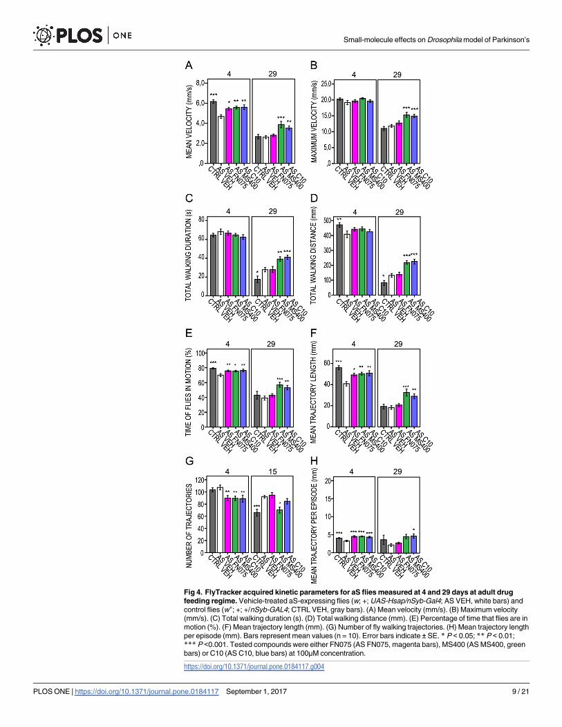
Fig 4. FlyTracker acquired kinetic parameters for aS flies measured at 4 and 29 days at adult drug
feeding regime. Vehicle-treated aS-expressing flies (w; +; UAS-Hsap/nSyb-Gal4; AS VEH, white bars) and
control flies (w+; +; +/nSyb-GAL4; CTRL VEH, gray bars). (A) Mean velocity (mm/s). (B) Maximum velocity
(mm/s). (C) Total walking duration (s). (D) Total walking distance (mm). (E) Percentage of time that flies are in
motion (%). (F) Mean trajectory length (mm). (G) Number of fly walking trajectories. (H) Mean trajectory length
per episode (mm). Bars represent mean values (n = 10). Error bars indicate ± SE. * P < 0.05; ** P < 0.01;
*** P <0.001. Tested compounds were either FN075 (AS FN075, magenta bars), MS400 (AS MS400, green
bars) or C10 (AS C10, blue bars) at 100μM concentration.
https://doi.org/10.1371/journal.pone.0184117.g004
Small-molecule effects on Drosophila model of Parkinson’s
PLOS ONE | https://doi.org/10.1371/journal.pone.0184117 September 1, 2017 9 / 21

Fig 5. FlyTracker acquired kinetic parameters for controls and aS-expressing flies measured at 1, 16, 30 days of
larval drug feeding regime. Controls: nSyb-Gal4#2–1 outcrossed with Oregon R (w+; +; +/nSyb-GAL4; CTRL VEH, dark
gray bars); non-expressing UAS–aS (w; +; UAS-Hsap/+; CTRL TG, light gray bars) and aS-expressing vehicle-treated flies
(w; +; UAS-Hsap/nSyb-Gal4; AS VEH, white bars). (A) Mean velocity (mm/s). (B) Maximum velocity (mm/s). (C) Total walking
duration (s). (D) Total walking distance (mm). (E) Percentage of time that flies are in motion (%). (F) Mean trajectory length
(mm). (G) Number of fly walking trajectories. (H) Mean trajectory length per episode (mm). Error bars indicate ± SE.
* P < 0.05; ** P < 0.01; *** P <0.001. Tested compounds were either FN075 (AS FN075, magenta bars) or MS400 (AS
MS400, green bars) or C10 (AS C10, blue bars) at 100μM concentration.
https://doi.org/10.1371/journal.pone.0184117.g005
Small-molecule effects on Drosophila model of Parkinson’s
PLOS ONE | https://doi.org/10.1371/journal.pone.0184117 September 1, 2017 10 / 21

all kinetic parameters to the level of the control non-aS expressing flies (CTRL TG). C10
showed no evident effects on aS fly locomotive behavior when fed already at the larvae stage.
To assure that feeding with molecules did not result in general (non-aS mediated) effects on
fly locomotion, we ran control experiments with aS non-expressing flies. Quantification of the
extracted fly trajectories revealed that they were similar and independent of the precise chem-
istry of the small molecule fed to the flies. Most of the kinetic parameters showed no significant
changes compared to the vehicle-treated control (S5 Fig, S6 Table). Thus, we conclude that the
specific and divergent effects found for the molecules require aS expression.
In vitro complementary analyses of FN075
To confirm the previously reported interaction between the aS and FN075 in vitro [21, 25], we
employed ATR-FTIR to probe conformational changes of purified aS (Fig 6). ATR-FTIR is a
sensitive method for measurements of protein secondary structure and can be used to study
protein aggregation in vitro [37]. The kinetics of aS amyloid formation is reflected by changes
in the secondary structure content where oligomers encompass anti-parallel β-sheet structures
and amyloid fibrils are characterized by parallel β-sheet content. FTIR analysis of aS incubated
at 37˚C with agitation was performed in the amide-I band, i.e., in the range of 1600 cm-1 to
1700 cm-1 as a function of time. Deconvolution and curve-fitting of the spectra for aS alone
and aS in the presence of FN075 are shown in Fig 6A–6F. After immediate sample preparation
(time 0 h), soluble aS (Fig 6A) showed distinguished bands in the amide-I region, which corre-
spond mostly to disordered structures, loops, turns and α-helices (1673–1646 cm-1), random
coil-like structures (major absorption at 1647 cm-1), and to β-sheet structures (1641–1612
cm-1). After 124 h of incubation, the intensity at 1647 cm-1 further increased at a disadvantage
of other structures (Fig 5C). Finally, at 144 h, an absorption peak at 1640 cm-1, typical of paral-
lel β-sheets, and thus amyloid fibrils, appeared (Fig 6E). For aS incubated with FN075, the
absorption peak characteristic of amyloid fibrils appeared already at 124 h (Fig 6D) and was
further increased at 144 h (Fig 6F). Although aS aggregation is slower here than in previous
reports [21, 25, 37], which we explain by the fact that less agitation was applied and the lack of
glass beads, importantly, the accelerating effect of FN075 remains.
The fact that FN075 interacts with aS in the flies such that aS aggregation results is indicated
from analysis of the soluble amount of aS in fly brain extracts by ELISA. As expected, if FN075
promotes aS aggregation and insolubility, we find less soluble aS in the FN075 fed flies in con-
trast to the MS400 treatment (Fig 7; for statistical analysis see S7 Table). These data are in
agreement with our previous work, which complemented the mice studies, where we analyzed
aS brain content of FN075 and MS400 fed flies by Western blot [27]. Moreover, in aS knock-
out mice, there were no effects of FN075 treatment on brain cell death, motor skills and serum
metabolite pattern at 6 months after the injection.
Testing a known compound, dopamine precursor
L-Dopa is a dopamine precursor and a common therapeutic used for PD [38, 39]. In our
study, aS-expressing flies that fed on a diet of L-Dopa (Fig 8 and S8 Table) showed an increased
mean velocity along the course of the trial (Fig 8A). This treatment improved the fly trajecto-
ries, seen as extended mean length of trajectories and decreased number of trajectories (Fig 8F
and 8H), which was inversely correlated. Although L-Dopa improved the percentage of time
that the flies were in motion already on day 4 and further corrected the fly motion index by a
factor of two on day 29 (Fig 8E), it shortened the mean, median and maximum life span of the
aS flies (Fig 8I–8L). Interestingly, there was no significant difference in the life span between
L-Dopa and FN075-treated flies (for log-rank pair-wise comparisons see S9 Table).
Small-molecule effects on Drosophila model of Parkinson’s
PLOS ONE | https://doi.org/10.1371/journal.pone.0184117 September 1, 2017 11 / 21
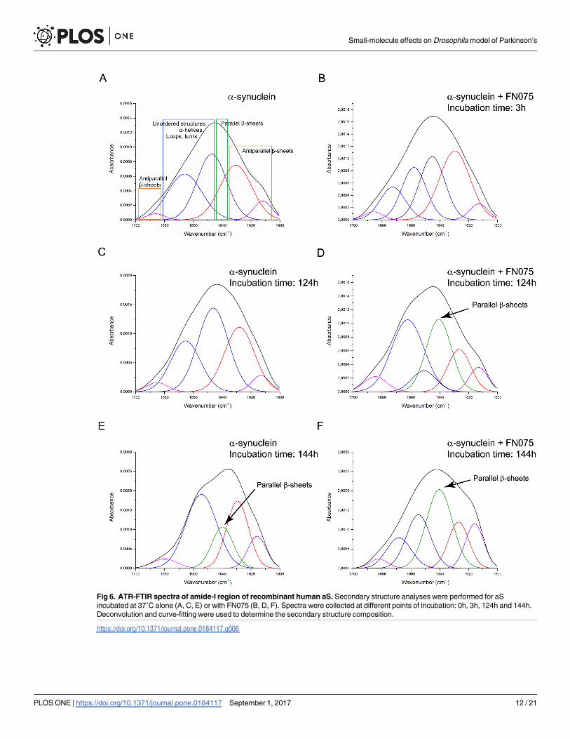
Fig 6. ATR-FTIR spectra of amide-I region of recombinant human aS. Secondary structure analyses were performed for aS
incubated at 37˚C alone (A, C, E) or with FN075 (B, D, F). Spectra were collected at different points of incubation: 0h, 3h, 124h and 144h.
Deconvolution and curve-fitting were used to determine the secondary structure composition.
https://doi.org/10.1371/journal.pone.0184117.g006
Small-molecule effects on Drosophila model of Parkinson’s
PLOS ONE | https://doi.org/10.1371/journal.pone.0184117 September 1, 2017 12 / 21

Discussion
Animal models of human diseases are helpful tools to not only evaluate the potential drug con-
cepts but also to study the underlying mechanisms of human diseases. PD is a motor neurode-
generative disease that is becoming more and more common in the population as we become
older. Although we have clues as to what proteins and reactions go awry and what the clinical
manifestations are, many fundamental aspects of PD (such as what is the trigger, order of dele-
terious events, involved biochemical pathways, normal function of aS) remain elusive, and so
far there is no cure for this disease. Therefore, new PD model and analysis systems allowing
for complementary studies of biological consequences and putative drug intervention are of
high importance.
Here, we presented a new way to probe motor skills of Drosophila flies and used this tech-
nology to assess the effects of small-molecule modulators (ring-fused 2-pyridones) of aS amy-
loid formation in vitro. The same molecules, recently tested in mice and amyloid-promoting
FN075, were found to cause signs of PD at 6 months after injection, including altered serum
metabolites, motor dysfunction in sticker-on-nose test and death of dopaminergic neurons
[27]. In contrast, in the fly model, new aspects of drug treatment were evaluated. Here, motor
function was probed differently, and the flies were monitored throughout their life span. We
Fig 7. Effects of 2-pyridones on level of soluble aS in fly brain extracts of 20 days old flies. Mean
soluble aS levels were measured with antibody specific to human aS in ELISA test. Controls: vehicle-treated
nsyb-Gal4 outcrossed with Oregon R flies (CTRL VEH) or aS expressing flies (AS VEH, white bars). Tested
compounds were FN075 (AS FN075, magenta bars) or MS400 (AS MS400, green bars) or C10 (AS C10, blue
bars) at 100μM concentration. Bars represent mean values (n = 2). Error bars indicate ± SD. * P < 0.05;
** P < 0.01; *** P <0.001. Multivariate GLM followed by Fisher’s post hoc showed P = 0.084 for AS FN075
vs AS VEH, P<0.001 for AS MS400 vs AS VEH and P = 0.002 for AS C10 vs AS VEH. For raw data see
S7 Table.
https://doi.org/10.1371/journal.pone.0184117.g007
Small-molecule effects on Drosophila model of Parkinson’s
PLOS ONE | https://doi.org/10.1371/journal.pone.0184117 September 1, 2017 13 / 21
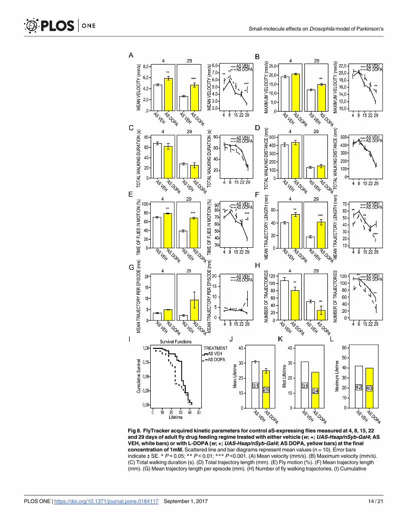
Fig 8. FlyTracker acquired kinetic parameters for control aS-expressing flies measured at 4, 8, 15, 22
and 29 days of adult fly drug feeding regime treated with either vehicle (w; +; UAS-Hsap/nSyb-Gal4; AS
VEH, white bars) or with L-DOPA (w; +; UAS-Hsap/nSyb-Gal4; AS DOPA, yellow bars) at the final
concentration of 1mM. Scattered line and bar diagrams represent mean values (n = 10). Error bars
indicate ± SE. * P < 0.05; ** P < 0.01; *** P <0.001. (A) Mean velocity (mm/s). (B) Maximum velocity (mm/s).
(C) Total walking duration (s). (D) Total trajectory length (mm). (E) Fly motion (%). (F) Mean trajectory length
(mm). (G) Mean trajectory length per episode (mm). (H) Number of fly walking trajectories. (I) Cumulative
Small-molecule effects on Drosophila model of Parkinson’s
PLOS ONE | https://doi.org/10.1371/journal.pone.0184117 September 1, 2017 14 / 21

note that loss of dopaminergic cells in the aS-expressing flies as a function of small molecules
was not tested here as previous reports have been contradictory and it is unclear if PD fly mod-
els involve the loss of dopamine-producing cells [28, 30, 40–43] or not [44–47]. The main dis-
crepancies were found due misinterpretation of the decrease in TH immunostaining, or GFP
signal, as a sign of neuronal loss without direct experimental evidence of cell death or occur-
rence of apoptotic processes in the PD fly models [44]. To avoid this uncertainty, we here
focused specifically on defects in motor behavior, which is a key feature of PD. However,
we note that future studies that reliably count TH-immunoreactive neurons as well as probe
Lewy body pathology in the fly brains as a function of small molecule treatments are desired.
In aS-expressing flies fed with FN075, we found an increase in the motor activity at a young
age (not captured in mice), followed by a reduction of activity upon aging and a shorter life
span, as compared to control and inhibitor treated flies. The fact that FN075 acted on aS in
the flies was supported by in vitro FTIR data, by the reduction of soluble levels of aS in brain
extracts of FN075-treated flies, and by the fact that the molecules had no effects on flies lacking
human aS. In analogy, we found that FN075 had no effect on the aS knock-out mice [27]. Our
finding that the initial removal of aS (via FN075-triggered aggregation) has a favorable effect
on fly neuronal activity is novel and suggests that aS moderates the neuronal activity under
normal conditions [5].
Another surprising observation we made was that larvae feeding of FN075 resulted in solely
positive effects on the flies. This may be explained by the induction of fly adaptive mechanisms
(e.g., down-regulation of target genes) upon prolonged exposure to molecules [48], but our
control experiments do not support this. Stage-specific differences between the adult- and lar-
vae-fed flies have been noted for other compounds [49] and emphasize the importance of
being aware of the fact that the experimental design may influence the outcome.
Fly models have been used to test the biological effects of many small molecules that modu-
late the aggregation of amyloidogenic proteins in vitro. For example, it was found that epicate-
chin gallate, a flavonoid antioxidant, delayed the loss of climbing ability and reduced oxidative
stress in PD flies [50]. This, together with in vitro data of aS amyloid inhibition by this flavo-
noid, suggests that this natural product may have a beneficial effect on PD patients. Interest-
ingly, in another study using a Drosophila model of Alzheimer’s disease (AD; i.e., over-
expressing the amyloid-β peptide, Aβ) it was found that curcumin reduced neurotoxicity
(reflected by increased locomotor activity and increased life span) by promoting Aβ fibrillation
such that oligomers and pre-fibrillar species were diminished [51]. Treatment with L-Dopa,
the dopamine precursor and the major drug used to mitigate the effects of dopaminergic neu-
ron loss in PD patients, was previously shown to restore locomotor deficits in aS-expressing
[1, 52] and dopamine-deficient flies [53, 54]. With respect to protein aggregation, dopamine
can protect against aS amyloid formation in vitro [55]. However, if present in the cytoplasm, it
can also make adducts with aS, and those can promote the accumulation of toxic cytoplasmic
aS proto-fibrils, which would make the dopamine neurons being selectively vulnerable [55,
56]. Such a scenario may explain why we found that flies fed with L-Dopa do better, in terms
of kinetics but still have a shorter life span than untreated aS flies. Notably, the L-Dopa results
on the aS flies parallel the FN075 results as well as long-term treatment of PD patients with
L-Dopa. In the latter case, patients initially show strong increases in their mobility, but after
survival of vehicle-treated aS flies (w; +; UAS-Hsap/nSyb-Gal4; AS VEH) and aS flies treated with L-DOPA
(w; +; UAS-Hsap/nSyb-Gal4; AS DOPA). (J) Mean, (K) median and (L) maximum lifetime. Numbers in bars
represent days of mean, median and maximum lifetime.
https://doi.org/10.1371/journal.pone.0184117.g008
Small-molecule effects on Drosophila model of Parkinson’s
PLOS ONE | https://doi.org/10.1371/journal.pone.0184117 September 1, 2017 15 / 21
long-term treatment the patients often develop L-Dopa induced dyskinesia [57]. Many future
studies are necessary to reveal the underlying biological mechanisms.
FlyTracker allows for automated analysis of walking and climbing locomotor behavior, so
called geotaxis, by collecting large sequences of data over a desired time period in an entirely
unbiased manner. An additional mechanical feature added to the holder rack enables move-
ments of fly tubes to induce the startle-induced negative geotactic response. Other approaches
based on digital imaging have been described previously but only with the main measure
extracted from the assay being height of fly negative geotaxis [58]. A somewhat similar accu-
racy of walking descriptors as with FlyTracker was described in an automatic system, but it
only tracked a small number of flies and just in an open horizontal field [1, 59]. Moreover,
there have been some reports of automated systems which track planar fly interactions that
may be useful for studies of genetic origins of fly behavior [60, 61]. In summary, the work pre-
sented demonstrates that the commercially available FlyTracker system is an excellent new
tool to study fly mobility parameters with high precision (i.e., in disease models such as the PD
flies here), requiring low amounts of test compounds.
Supporting information
S1 Fig. (A) The equipment and experimental setup of FlyTracker. 1. Fly tubes; 2. Fly tube
holder rack with a movable frame; 3. Base frame; 4. VGA camera with the USB interface fixed
at 105 mm from the center of fly tubes; 5. PC. (B) Structure of ring-fused 2-pyridones used in
this work and in vitro thioflavin T (ThT) aggregation assay for 70 μM aS alone (filled circles)
and with 100 μM FN075 (filled squares) or C10 (open circles) in 10 mM phosphate, pH 7.4
with 140 mM NaCl and 2.7 mM KCl. Experiments were performed at 37˚C with continuous
agitation using a 2 mm glass bead in each well. All samples contained 20 μM ThT and fluores-
cence was measured at 480 nm (excitation at 440 nm) in a FLUOstar Omega plate reader. (C)
Densitometric analysis of Western blots (n = 3) of fly head protein extracts probed with anti-
body specific to human aS. The protein extracts are divided in soluble and insoluble fractions
prepared as described in supplementary M&M. The aS-specific signal was normalized to its
tubulin signal and then insoluble aS level was further normalized as ratio to aS soluble level.
The diagram shows aS expression fold change in soluble and insoluble aS fractions. Bars repre-
sent mean values ± SD. Raw densitometric data presented in the table were acquired with Gel-
Doc XR+ Imager and analysed with Image Lab 5.2 software (Bio-Rad, Richmond, CA, USA).
(TIF)
S2 Fig. Compound toxicity assays. (A) Mean fraction of eclosed pupae of control (CTRL) and
aS fly lines (AS) that were fed during larval stages with either vehicle (VEH, white bars) or
tested compounds: FN075 (magenta bars), MS400 (green bars) or C10 (blue bars) at 100μM
concentration. (B) Survival analysis was analyzed by Kaplan-Meier curves. Cumulative survival
of non-expressing UAS-aS flies treated with either vehicle (CTRL TG VEH, white bars) or
compounds: FN075 (CTRL TG FN075, magenta bars), MS400 (CTRL TG MS400, greean bars)
and C10 (CTRL TG C10, blue bars) at 100μM concentration. (C) Mean, (D) median and (E)
max lifetime for control non-expressing UAS-aS flies (CTRL TG) fed with either vehicle
(VEH) or tested compounds: FN075, MS400 and C10. Numbers in bars represents mean,
median and max lifetime (days). Error bars indicate ± SE.
(TIF)
S3 Fig. FlyTracker acquired kinetic parameters for aS flies measured at 4, 8,15,22 and 29
days of adult drug feeding regime. Controls: vehicle treated nsyb-Gal4 outcrossed with Ore-
gon R (CTRL VEH, dark grey bars) or aS expressing flies fed vehicle (AS VEH, white bars) or
Small-molecule effects on Drosophila model of Parkinson’s
PLOS ONE | https://doi.org/10.1371/journal.pone.0184117 September 1, 2017 16 / 21

tested compounds: FN075 (AS FN075, magenta bars) or MS400 (AS MS400, green bars) or
C10 (AS C10, blue bars) at 100μM concentration. Error bars indicate ± SE. P values are<0,05
(�), <0,01 (��), <0,001 (���). (A) Mean velocity (mm/s). (B) Maximum mean velocity (mm/s).
(C) Total walking duration (s). (D) Total trajectory length (mm). (E) Fly motion (%). (F)
Mean trajectory length (mm). (G) Mean number of fly walking trajectories. (H) Mean trajec-
tory length per episode (mm).
(TIF)
S4 Fig. FlyTracker acquired kinetic parameters for aS flies measured at 1, 7, 16, 21, 30 and
42 days of larval drug feeding regime. Controls: nsyb-Gal4 outcrossed with Oregon R (CTRL
VEH, dark grey bars); non-expressing UAS-aS flies (CTRL TG, light grey bars). Tested com-
pounds were either FN075 (AS FN075, magenta bars) or MS400 (AS MS400, green bars) or
C10 (AS C10, blue bars) at 100μM concentration. (A) Mean velocity (mm/s). (B) Maximum
velocity (mm/s). (C) Total walking duration (s). (D) Total trajectory length (mm). (E) Fly
motion (%). (F) Mean trajectory length (mm). (G) Number of fly walking trajectories. (H)
Mean trajectory length per episode (mm). Bars represent mean values. Error bars
indicate ± SE. � P< 0,05; �� P< 0,01; ��� P<0,001.
(TIF)
S5 Fig. FlyTracker acquired kinetic parameters for non-expressing UAS-aS flies (CTRL
TG) measured at 1, 7, 16, 21, and 30 days of larval drug feeding regime. Tested compounds
were either FN075 (AS FN075, magenta bars) or MS400 (AS MS400, green bars) or C10 (AS
C10, blue bars) at 100μM concentration and vehicle (VEH, grey bars). (A) Mean velocity
(mm/s). (B) Maximum velocity (mm/s). (C) Total walking duration (s). (D) Total trajectory
length (mm). (E) Fly motion (%). (F) Mean trajectory length (mm). (G) Mean number of fly
walking trajectories. (H) Mean trajectory length per episode (mm). Bars represent mean val-
ues. Error bars indicate ± SE. � P< 0,05; �� P< 0,01; ��� P<0,001.
(TIF)
S1 Table. Kinetic parameters for aS and control flies. General Linear Model multivariate
analysis with Fisher’s post hoc test. Significant number are highlighted in red.
(PDF)
S2 Table. Pairwise comparisons and log-rank analysis of survival curves for control aS
non-expressing UAS-aS flies (CTRL TG) treated either vehicle or compounds FN075,
MS400, C10. Significant numbers are highlighted in red.
(PDF)
S3 Table. Pairwise comparisons and log-rank analysis of survival curves for aS expressing
vehicle, FN075, MS400, or C10 treated flies. Significant number are highlighted in red.
(PDF)
S4 Table. Kinetic parameters for aS flies fed vehicle or compounds FN075, MS400, C10 in
adult stages of life span. General Linear Model multivariate analysis with Fisher’s post hoc
test. Significant numbers are highlighted in red.
(PDF)
S5 Table. Kinetic parameters for aS flies fed vehicle or compounds FN075, MS400, C10
from larval stages and throughout entire life span. General Linear Model multivariate analy-
sis with Fisher’s post hoc test. Significant numbers are highlighted in red.
(PDF)
Small-molecule effects on Drosophila model of Parkinson’s
PLOS ONE | https://doi.org/10.1371/journal.pone.0184117 September 1, 2017 17 / 21

S6 Table. Kinetic parameters for control (aS non-expressing) flies fed vehicle or com-
pounds FN075, MS400, C10 from larval stages and throughout entire life span. General
Linear Model multivariate analysis with Fisher’s post hoc test. Significant numbers are
highlighted in red.
(PDF)
S7 Table. Effects of 2-pyridones on level of soluble aS in fly brain extracts of 20 days old
flies. Multiple comparisons are presented for aS flies fed compounds FN075, MS400, C10 vs.
aS flies fed vehicle (AS VEH) or vs. control aS non-expressing flies (CTRL VEH) General Lin-
ear Model multivariate analysis with Fisher’s post hoc test (upper table). Significant numbers
are highlighted in red. Raw data i.e. descriptive statistics from ELISA tests are in the lower
table.
(PDF)
S8 Table. Kinetic parameters for aS flies fed either vehicle or L-DOPA (1mM) in adult
stages. General Linear Model multivariate analysis with Fisher’s post hoc test. Significant
numbers are highlighted in red.
(PDF)
S9 Table. Pairwise comparisons and log-rank analysis of survival curves for aS expressing
flies treated either vehicle or compounds FN075, MS400, C10 and aS non-expressing flies
(CTRL VEH) vs. aS flies fed L-DOPA. Significant numbers are highlighted in red.
(PDF)
S1 Supporting Materials and Methods.
(PDF)
Acknowledgments
We would like to thank Julie H. Simpson and The Bloomington Stock Center for sharing fly
lines, AlexoTech AB for antibodies, and Jorgen Åden for his technical assistance with in vitroThT experiments.
Author Contributions
Conceptualization: Małgorzata Pokrzywa, Fredrik Almqvist, Pernilla Wittung-Stafshede.
Formal analysis: Małgorzata Pokrzywa, Katarzyna Pawełek.
Funding acquisition: Pernilla Wittung-Stafshede.
Investigation: Małgorzata Pokrzywa, Katarzyna Pawełek, Weronika Elżbieta Kucia, Szymon
Sarbak, Erik Chorell.
Methodology: Małgorzata Pokrzywa, Erik Chorell, Fredrik Almqvist, Pernilla Wittung-
Stafshede.
Project administration: Pernilla Wittung-Stafshede.
Supervision: Małgorzata Pokrzywa, Fredrik Almqvist, Pernilla Wittung-Stafshede.
Visualization: Małgorzata Pokrzywa, Katarzyna Pawełek, Weronika Elżbieta Kucia, Szymon
Sarbak, Erik Chorell, Pernilla Wittung-Stafshede.
Writing – original draft: Małgorzata Pokrzywa, Erik Chorell, Fredrik Almqvist, Pernilla Wit-
tung-Stafshede.
Small-molecule effects on Drosophila model of Parkinson’s
PLOS ONE | https://doi.org/10.1371/journal.pone.0184117 September 1, 2017 18 / 21

Writing – review & editing: Małgorzata Pokrzywa, Pernilla Wittung-Stafshede.
References
1. Chen AY, Wilburn P, Hao X, Tully T. Walking deficits and centrophobism in an alpha-synuclein fly
model of Parkinson’s disease. Genes Brain Behav. 2014; 13(8):812–20. https://doi.org/10.1111/gbb.
12172 PMID: 25113870
2. Spillantini MG, Schmidt ML, Lee VM, Trojanowski JQ, Jakes R, Goedert M. Alpha-synuclein in Lewy
bodies. Nature. 1997; 388(6645):839–40. https://doi.org/10.1038/42166 PMID: 9278044
3. Polymeropoulos MH, Lavedan C, Leroy E, Ide SE, Dehejia A, Dutra A, et al. Mutation in the alpha-synu-
clein gene identified in families with Parkinson’s disease. Science. 1997; 276(5321):2045–7. PMID:
9197268
4. Dev KK, Hofele K, Barbieri S, Buchman VL, van der Putten H. Part II: alpha-synuclein and its molecular
pathophysiological role in neurodegenerative disease. Neuropharmacology. 2003; 45(1):14–44. PMID:
12814657
5. Lassen LB, Reimer L, Ferreira N, Betzer C, Jensen PH. Protein partners of alpha-synuclein in Health
and Disease. Brain Pathol. 2016.
6. Maroteaux L, Campanelli JT, Scheller RH. Synuclein: a neuron-specific protein localized to the nucleus
and presynaptic nerve terminal. The Journal of neuroscience: the official journal of the Society for Neu-
roscience. 1988; 8(8):2804–15.
7. Eliezer D, Kutluay E, Bussell R Jr., Browne G. Conformational properties of alpha-synuclein in its free
and lipid-associated states. Journal of molecular biology. 2001; 307(4):1061–73. https://doi.org/10.
1006/jmbi.2001.4538 PMID: 11286556
8. Iwai A, Masliah E, Yoshimoto M, Ge N, Flanagan L, de Silva HA, et al. The precursor protein of non-A
beta component of Alzheimer’s disease amyloid is a presynaptic protein of the central nervous system.
Neuron. 1995; 14(2):467–75. PMID: 7857654
9. Uversky VN. Neuropathology, biochemistry, and biophysics of alpha-synuclein aggregation. Journal of
neurochemistry. 2007; 103(1):17–37. https://doi.org/10.1111/j.1471-4159.2007.04764.x PMID:
17623039
10. Xu J, Kao SY, Lee FJ, Song W, Jin LW, Yankner BA. Dopamine-dependent neurotoxicity of alpha-synu-
clein: a mechanism for selective neurodegeneration in Parkinson disease. Nature medicine. 2002; 8
(6):600–6. https://doi.org/10.1038/nm0602-600 PMID: 12042811
11. Gosavi N, Lee HJ, Lee JS, Patel S, Lee SJ. Golgi fragmentation occurs in the cells with prefibrillar
alpha-synuclein aggregates and precedes the formation of fibrillar inclusion. The Journal of biological
chemistry. 2002; 277(50):48984–92. https://doi.org/10.1074/jbc.M208194200 PMID: 12351643
12. Peelaerts W, Bousset L, Van der Perren A, Moskalyuk A, Pulizzi R, Giugliano M, et al. alpha-Synuclein
strains cause distinct synucleinopathies after local and systemic administration. Nature. 2015; 522
(7556):340–4. https://doi.org/10.1038/nature14547 PMID: 26061766
13. Luk KC, Kehm V, Carroll J, Zhang B, O’Brien P, Trojanowski JQ, et al. Pathological alpha-synuclein
transmission initiates Parkinson-like neurodegeneration in nontransgenic mice. Science. 2012; 338
(6109):949–53. https://doi.org/10.1126/science.1227157 PMID: 23161999
14. Paumier KL, Luk KC, Manfredsson FP, Kanaan NM, Lipton JW, Collier TJ, et al. Intrastriatal injection of
pre-formed mouse alpha-synuclein fibrils into rats triggers alpha-synuclein pathology and bilateral
nigrostriatal degeneration. Neurobiology of disease. 2015; 82:185–99. https://doi.org/10.1016/j.nbd.
2015.06.003 PMID: 26093169
15. Glabe CG. Structural classification of toxic amyloid oligomers. The Journal of biological chemistry.
2008; 283(44):29639–43. https://doi.org/10.1074/jbc.R800016200 PMID: 18723507
16. Braga CA, Follmer C, Palhano FL, Khattar E, Freitas MS, Romao L, et al. The anti-Parkinsonian drug
selegiline delays the nucleation phase of alpha-synuclein aggregation leading to the formation of non-
toxic species. Journal of molecular biology. 2011; 405(1):254–73.
17. Choubey V, Safiulina D, Vaarmann A, Cagalinec M, Wareski P, Kuum M, et al. Mutant A53T alpha-
synuclein induces neuronal death by increasing mitochondrial autophagy. The Journal of biological
chemistry. 2011; 286(12):10814–24. https://doi.org/10.1074/jbc.M110.132514 PMID: 21252228
18. Sterky FH, Lee S, Wibom R, Olson L, Larsson NG. Impaired mitochondrial transport and Parkin-inde-
pendent degeneration of respiratory chain-deficient dopamine neurons in vivo. Proceedings of the
National Academy of Sciences of the United States of America. 2011; 108(31):12937–42.
19. Chesselet MF, Fleming S, Mortazavi F, Meurers B. Strengths and limitations of genetic mouse models
of Parkinson’s disease. Parkinsonism & related disorders. 2008; 14 Suppl 2:S84–7.
Small-molecule effects on Drosophila model of Parkinson’s
PLOS ONE | https://doi.org/10.1371/journal.pone.0184117 September 1, 2017 19 / 21

20. Blesa J, Przedborski S. Parkinson’s disease: animal models and dopaminergic cell vulnerability. Fron-
tiers in neuroanatomy. 2014; 8:155. https://doi.org/10.3389/fnana.2014.00155 PMID: 25565980
21. Horvath I, Weise CF, Andersson EK, Chorell E, Sellstedt M, Bengtsson C, et al. Mechanisms of protein
oligomerization: inhibitor of functional amyloids templates alpha-synuclein fibrillation. Journal of the
American Chemical Society. 2012; 134(7):3439–44. https://doi.org/10.1021/ja209829m PMID:
22260746
22. Nors Perdersen M, Fodera V, Horvath I, van Maarschalkerweerd A, Norgaard Toft K, Weise C, et al.
Direct Correlation Between Ligand-Induced alpha-Synuclein Oligomers and Amyloid-like Fibril Growth.
Scientific reports. 2015; 5:10422.
23. Aberg V, Norman F, Chorell E, Westermark A, Olofsson A, Sauer-Eriksson AE, et al. Microwave-assis-
ted decarboxylation of bicyclic 2-pyridone scaffolds and identification of Abeta-peptide aggregation
inhibitors. Org Biomol Chem. 2005; 3(15):2817–23. https://doi.org/10.1039/b503294f PMID: 16032359
24. Cegelski L, Pinkner JS, Hammer ND, Cusumano CK, Hung CS, Chorell E, et al. Small-molecule inhibi-
tors target Escherichia coli amyloid biogenesis and biofilm formation. Nature chemical biology. 2009; 5
(12):913–9. https://doi.org/10.1038/nchembio.242 PMID: 19915538
25. Horvath I, Sellstedt M, Weise C, Nordvall LM, Krishna Prasad G, Olofsson A, et al. Modulation of alpha-
synuclein fibrillization by ring-fused 2-pyridones: templation and inhibition involve oligomers with differ-
ent structure. Arch Biochem Biophys. 2013; 532(2):84–90.
26. Andersson EK, Bengtsson C, Evans ML, Chorell E, Sellstedt M, Lindgren AE, et al. Modulation of curli
assembly and pellicle biofilm formation by chemical and protein chaperones. Chemistry & biology.
2013; 20(10):1245–54.
27. Chermenina M, Chorell E, Pokrzywa M, Antti H, Almqvist F, Stromberg I, et al. Single injection of small-
molecule amyloid accelerator results in cell death of nigral dopamine neurons in mice. Npj Parkinson’s
Disease. 2015; 1:15024. https://doi.org/10.1038/npjparkd.2015.24 PMID: 28725689
28. Feany MB, Bender WW. A Drosophila model of Parkinson’s disease. Nature. 2000; 404(6776):394–8.
https://doi.org/10.1038/35006074 PMID: 10746727
29. Riemensperger T, Issa AR, Pech U, Coulom H, Nguyen MV, Cassar M, et al. A single dopamine path-
way underlies progressive locomotor deficits in a Drosophila model of Parkinson disease. Cell Rep.
2013; 5(4):952–60. https://doi.org/10.1016/j.celrep.2013.10.032 PMID: 24239353
30. Auluck PK, Chan HY, Trojanowski JQ, Lee VM, Bonini NM. Chaperone suppression of alpha-synuclein
toxicity in a Drosophila model for Parkinson’s disease. Science. 2002; 295(5556):865–8. https://doi.org/
10.1126/science.1067389 PMID: 11823645
31. Kohlhoff KJ, Jahn TR, Lomas DA, Dobson CM, Crowther DC, Vendruscolo M. The iFly tracking system
for an automated locomotor and behavioural analysis of Drosophila melanogaster. Integr Biol (Camb).
2011; 3(7):755–60.
32. Grover D, Yang J, Tavare S, Tower J. Simultaneous tracking of fly movement and gene expression
using GFP. BMC Biotechnol. 2008; 8:93. https://doi.org/10.1186/1472-6750-8-93 PMID: 19087237
33. Fry SN, Sayaman R, Dickinson MH. The aerodynamics of free-flight maneuvers in Drosophila. Science.
2003; 300(5618):495–8. https://doi.org/10.1126/science.1081944 PMID: 12702878
34. Iakovleva I, Begum A, Pokrzywa M, Walfridsson M, Sauer-Eriksson AE, Olofsson A. The flavonoid
luteolin, but not luteolin-7-O-glucoside, prevents a transthyretin mediated toxic response. PLoS One.
2015; 10(5):e0128222. https://doi.org/10.1371/journal.pone.0128222 PMID: 26020516
35. Markstein M, Dettorre S, Cho J, Neumuller RA, Craig-Muller S, Perrimon N. Systematic screen of che-
motherapeutics in Drosophila stem cell tumors. Proc Natl Acad Sci U S A. 2014; 111(12):4530–5.
https://doi.org/10.1073/pnas.1401160111 PMID: 24616500
36. Auluck PK, Meulener MC, Bonini NM. Mechanisms of Suppression of {alpha}-Synuclein Neurotoxicity
by Geldanamycin in Drosophila. J Biol Chem. 2005; 280(4):2873–8. https://doi.org/10.1074/jbc.
M412106200 PMID: 15556931
37. Sarroukh R, Goormaghtigh E, Ruysschaert JM, Raussens V. ATR-FTIR: a "rejuvenated" tool to investi-
gate amyloid proteins. Biochimica et biophysica acta. 2013; 1828(10):2328–38. https://doi.org/10.1016/
j.bbamem.2013.04.012 PMID: 23746423
38. Huot P, Johnston TH, Koprich JB, Fox SH, Brotchie JM. The pharmacology of L-DOPA-induced dyski-
nesia in Parkinson’s disease. Pharmacol Rev. 2013; 65(1):171–222. https://doi.org/10.1124/pr.111.
005678 PMID: 23319549
39. Ehringer H, Hornykiewicz O. [Distribution of noradrenaline and dopamine (3-hydroxytyramine) in the
human brain and their behavior in diseases of the extrapyramidal system]. Klin Wochenschr. 1960;
38:1236–9. PMID: 13726012
40. Trinh K, Moore K, Wes PD, Muchowski PJ, Dey J, Andrews L, et al. Induction of the Phase II Detoxifica-
tion Pathway Suppresses Neuron Loss in Drosophila Models of Parkinson’s Disease. The Journal of
Small-molecule effects on Drosophila model of Parkinson’s
PLOS ONE | https://doi.org/10.1371/journal.pone.0184117 September 1, 2017 20 / 21

Neuroscience. 2008; 28(2):465–72. https://doi.org/10.1523/JNEUROSCI.4778-07.2008 PMID:
18184789
41. Cooper AA, Gitler AD, Cashikar A, Haynes CM, Hill KJ, Bhullar B, et al. α-Synuclein Blocks ER-Golgi
Traffic and Rab1 Rescues Neuron Loss in Parkinson’s Models. Science. 2006; 313(5785):324–8.
42. Chen L, Feany MB. [alpha]-Synuclein phosphorylation controls neurotoxicity and inclusion formation in
a Drosophila model of Parkinson disease. Nat Neurosci. 2005; 8(5):657–63. https://doi.org/10.1038/
nn1443 PMID: 15834418
43. Barone MC, Sykiotis GP, Bohmann D. Genetic activation of Nrf2 signaling is sufficient to ameliorate
neurodegenerative phenotypes in a Drosophila model of Parkinson’s disease. Disease Models & Mech-
anisms. 2011; 4(5):701–7.
44. Navarro JA, Hessner S, Yenisetti SC, Bayersdorfer F, Zhang L, Voigt A, et al. Analysis of dopaminergic
neuronal dysfunction in genetic and toxin-induced models of Parkinson’s disease in Drosophila. Journal
of neurochemistry. 2014; 131(3):369–82. https://doi.org/10.1111/jnc.12818 PMID: 25040725
45. Whitworth AJ, Wes PD, Pallanck LJ. Drosophila models pioneer a new approach to drug discovery for
Parkinson’s disease. Drug Discovery Today. 2006; 11(3):119–26.
46. White K, Humphrey D, Hirth F. The Dopaminergic System in the Aging Brain of Drosophila. Frontiers in
neuroscience. 2010; 4(205).
47. Pesah Y, Burgess H, Middlebrooks B, Ronningen K, Prosser J, Tirunagaru V, et al. Whole-mount analy-
sis reveals normal numbers of dopaminergic neurons following misexpression of α-Synuclein in Dro-
sophila. genesis. 2005; 41(4):154–9. https://doi.org/10.1002/gene.20106 PMID: 15789427
48. Pandey UB, Nichols CD. Human disease models in Drosophila melanogaster and the role of the fly in
therapeutic drug discovery. Pharmacol Rev. 2011; 63(2):411–36. https://doi.org/10.1124/pr.110.
003293 PMID: 21415126
49. Soh JW, Marowsky N, Nichols TJ, Rahman AM, Miah T, Sarao P, et al. Curcumin is an early-acting
stage-specific inducer of extended functional longevity in Drosophila. Exp Gerontol. 2013; 48(2):229–
39. https://doi.org/10.1016/j.exger.2012.09.007 PMID: 23063786
50. Siddique YH, Jyoti S, Naz F. Effect of epicatechin gallate dietary supplementation on transgenic Dro-
sophila model of Parkinson’s disease. J Diet Suppl. 2014; 11(2):121–30. https://doi.org/10.3109/
19390211.2013.859207 PMID: 24670116
51. Caesar I, Jonson M, Nilsson KP, Thor S, Hammarstrom P. Curcumin promotes A-beta fibrillation and
reduces neurotoxicity in transgenic Drosophila. PloS one. 2012; 7(2):e31424. https://doi.org/10.1371/
journal.pone.0031424 PMID: 22348084
52. Pendleton RG, Parvez F, Sayed M, Hillman R. Effects of Pharmacological Agents upon a Transgenic
Model of Parkinson’s Disease in Drosophila melanogaster. Journal of Pharmacology and Experimental
Therapeutics. 2002; 300(1):91–6. PMID: 11752102
53. Cichewicz K, Garren EJ, Adiele C, Aso Y, Wang Z, Wu M, et al. A new brain dopamine-deficient Dro-
sophila and its pharmacological and genetic rescue. Genes, Brain and Behavior. 2017; 16(3):394–403.
54. Riemensperger T, Isabel G, Coulom H, Neuser K, Seugnet L, Kume K, et al. Behavioral consequences
of dopamine deficiency in the Drosophila central nervous system. Proceedings of the National Academy
of Sciences. 2011; 108(2):834–9.
55. Conway KA, Rochet JC, Bieganski RM, Lansbury PT Jr. Kinetic stabilization of the alpha-synuclein pro-
tofibril by a dopamine-alpha-synuclein adduct. Science. 2001; 294(5545):1346–9. https://doi.org/10.
1126/science.1063522 PMID: 11701929
56. Li H-T, Lin D-H, Luo X-Y, Zhang F, Ji L-N, Du H-N, et al. Inhibition of α-synuclein fibrillization by dopa-
mine analogs via reaction with the amino groups of α-synuclein. FEBS Journal. 2005; 272(14):3661–
72. https://doi.org/10.1111/j.1742-4658.2005.04792.x PMID: 16008565
57. Jenner P. Molecular mechanisms of L-DOPA-induced dyskinesia. Nat Rev Neurosci. 2008; 9(9):665–
77. https://doi.org/10.1038/nrn2471 PMID: 18714325
58. Gargano JW, Martin I, Bhandari P, Grotewiel MS. Rapid iterative negative geotaxis (RING): a new
method for assessing age-related locomotor decline in Drosophila. Exp Gerontol. 2005; 40(5):386–95.
https://doi.org/10.1016/j.exger.2005.02.005 PMID: 15919590
59. Martin JR. A portrait of locomotor behaviour in Drosophila determined by a video-tracking paradigm.
Behav Processes. 2004; 67(2):207–19. https://doi.org/10.1016/j.beproc.2004.04.003 PMID: 15240058
60. Eyjolfsdottir E, Branson S, Burgos-Artizzu X, Hoopfer E, Schor J, Anderson D, et al. Detecting Social
Actions of Fruit Flies. COMPUTER VISION—ECCV 2014, PT II. 2015;8690:772–87.
61. Branson K, Robie AA, Bender J, Perona P, Dickinson MH. High-throughput ethomics in large groups of
Drosophila. Nature methods. 2009; 6(6):451–7. https://doi.org/10.1038/nmeth.1328 PMID: 19412169
Small-molecule effects on Drosophila model of Parkinson’s
PLOS ONE | https://doi.org/10.1371/journal.pone.0184117 September 1, 2017 21 / 21