postoperative complications of laparoscopic surgery · central international journal of clinical...
TRANSCRIPT

Central International Journal of Clinical Anesthesiology
Cite this article: Beleña JM, Nuñez M (2014) Postoperative Complications of Laparoscopic Surgery. Int J Clin Anesthesiol 2(3): 1034.
*Corresponding authorJosé M Beleña, Department of Anesthesiology and Critical Care, Sureste University Hospital, Ronda del Sur 10, Arganda del Rey, 28500 Madrid, Spain, Tel: 34-918394184; Email:
Submitted: 23 March 2014
Accepted: 29 March 2014
Published: 06 July 2014
ISSN: 2333-6641
Copyright© 2014 Beleña et al.
OPEN ACCESS
Review Article
Postoperative Complications of Laparoscopic SurgeryJosé M Beleña1* and Mónica Nuñez2
1Department of Anesthesiology and Critical Care, Sureste University Hospital, Spain2Department of Anesthesiology and Critical Care, Ramón y Cajal University Hospital, Spain
IntroductIon
Intestinal perforation
This is the second most common cause of mortality of laparoscopic surgery. Its incidence varies between 0.1% and 0.3% of cases. Approximately one third of these injuries occur during the access into the abdomen, but it may also occurs during removal of instrumental, dissection of structures or electrocautery burns.
One of the problems related to this complication is intraoperative difficulty to diagnose it. Most of lesions (70%) are diagnosed in the postoperative period and may have already evolved into a severe peritonitis. Depending on the time of occurrence can be classified as [1]: Early: occur in the first 48 hours after surgery.
Keywords•Laparoscopic•Postoperative
Abstract
Laparoscopic surgery is a very common and widely established technique. Benefits include decreased postoperative pain, improved patient satisfaction (including cosmetic results), reduced hospital stays and fewer postoperative complications compared with open techniques. The range of surgical techniques is increasing in complexity and about the kind of patients undergoing these procedures (pluripathological patients, associating co-morbidity). Number of emergency operations performed laparoscopically has been increased as well.
Complications of laparoscopic surgery are mainly divided into three groups: complications derived from pneumoperitoneum, complications caused by the operative procedure and postoperative complications.
Apart from the alterations caused by the pneumoperitoneum (raised intra-abdominal pressure and physiological effects especially within cardiovascular and respiratory systems), which have significant effects on the patient, especially if they are elderly or have associated morbidity, it may cause some complications such as severe hypercarbia, cardio-pulmonary compromise, air embolism or gas migration (subcutaneous emphysema, pneumomediastinum and pneumothorax.
Complications of the operative procedure can be grouped into two categories: complications of access and complications of technique.
Complications of access or trocar entry include: hollow or solid viscus perforation, abdominal wall or major vessel injury, incisional hernia and peritoneal tumor cell implantation.
In case of complications derived from the surgical technique, we can include: hemorrhage, vascular injury, retroperitoneal hematoma, bile leak, bile duct injury, bile peritonitis (with or without a bile duct injury). Postoperative complications include: intestinal perforation, bile leak, retroperitoneal hematoma, pancreatitis, subhepatic abscess and postoperative air embolism. This review discusses the complications that can occur in the postoperative period.
Delayed: appear 48 hours after the intervention. Produced in many cases by a secondary local inflammatory mechanism after laparoscopic technique.
Perforations are also classified according to the location of the lesion in the gastrointestinal tract or laparoscopic instrument type used. The presence of adhesions or history of previous laparotomies increase risk for perforation. With respect to location, the small bowel injuries are the most common (58%). In laparoscopic cholecystectomy, duodenal injury is the most common lesion. Next in frequency are colon lesions (32%) and finally gastric are rare and account for only 7% of cases. Regarding the material used at the beginning of the intervention, Veress needle and blind trocars puncture used at the beginning of the procedure are those that most frequently cause intestinal perforations.
Central
Beleña et al. (2014)Email:
Int J Clin Anesthesiol 2(3): 1034 (2014) 2/5
During the intervention, thermal injury is the most common adverse event. In some cases, these electrical burns are no identified immediately and its symptoms can appear several days after when complete necrosis of the intestinal wall occurs. If it is identified and repaired during surgery, morbidity is low, but if peritonitis is evolved, mortality increases.
Diagnosis of intestinal perforation is often difficult, being masked by factors such as postoperative pain, use of analgesic drugs or the use of antibiotics. Persistent abdominal pain, sepsis data or signs of peritoneal irritation scans, are suggestive of this complication. In many cases the peritoneal irritation is absent. One theory supports the unusual presentation of intestinal perforation in laparoscopic surgery compared to open surgery, based on the lower postoperative immune and metabolic response in the first case. When there is a clinical suspicion, an abdominal CT that allows us to identify intestinal perforation must be performed [2].
Most of the lesions (80%) are treated by conversion to laparotomy for intestinal repair. Conservative management would be based primarily on percutaneous drainage of abscesses and supportive measures such as the use of parenteral nutrition and antibiotics. Mortality, although it is a rare complication in some published studies it is around 3-6%, especially in those patients progressing to a state of sepsis and multiorgan failure. Moreover, if intestinal injury is associated with vascular injury, mortality increases. Therefore, prevention measures are essential [3].
Bile leak
Laparoscopic cholecystectomy is the standard surgical treatment in most patients with gallstones. Its benefits have been clearly demonstrated with respect to the laparotomy and this laparoscopic technique is probably the world´s most widely performed. However, the increase in the number of laparoscopic procedures has also increased the number of complications, with an incidence greater than in the case of open surgery. The incidence of lesions in the biliary tract in laparoscopic surgery is 0.3%, whereas in open surgery is around 0.2%. In patients with severe chronic cholecystitis or scleroatrophic gallbladder, the risk is higher. There are also studies showing an increased risk of perioperative complications in prolonged procedures. If laparoscopic cholecystectomy lasts more than two hours the risk is four times greater than if the procedure takes 30 to 60 minutes [1].
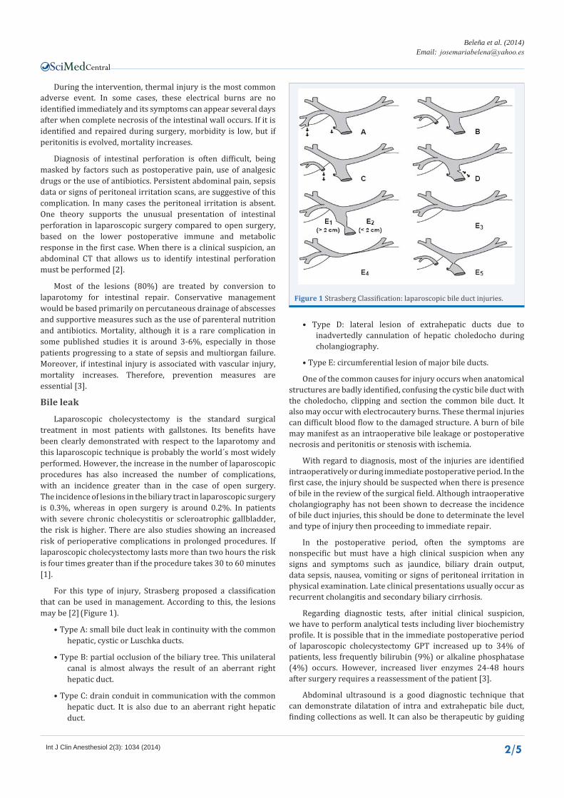
For this type of injury, Strasberg proposed a classification that can be used in management. According to this, the lesions may be [2] (Figure 1).
• Type A: small bile duct leak in continuity with the common hepatic, cystic or Luschka ducts.
• Type B: partial occlusion of the biliary tree. This unilateral canal is almost always the result of an aberrant right hepatic duct.
• Type C: drain conduit in communication with the common hepatic duct. It is also due to an aberrant right hepatic duct.
• Type D: lateral lesion of extrahepatic ducts due to inadvertedly cannulation of hepatic choledocho during cholangiography.
• Type E: circumferential lesion of major bile ducts.
One of the common causes for injury occurs when anatomical structures are badly identified, confusing the cystic bile duct with the choledocho, clipping and section the common bile duct. It also may occur with electrocautery burns. These thermal injuries can difficult blood flow to the damaged structure. A burn of bile may manifest as an intraoperative bile leakage or postoperative necrosis and peritonitis or stenosis with ischemia.
With regard to diagnosis, most of the injuries are identified intraoperatively or during immediate postoperative period. In the first case, the injury should be suspected when there is presence of bile in the review of the surgical field. Although intraoperative cholangiography has not been shown to decrease the incidence of bile duct injuries, this should be done to determinate the level and type of injury then proceeding to immediate repair.
In the postoperative period, often the symptoms are nonspecific but must have a high clinical suspicion when any signs and symptoms such as jaundice, biliary drain output, data sepsis, nausea, vomiting or signs of peritoneal irritation in physical examination. Late clinical presentations usually occur as recurrent cholangitis and secondary biliary cirrhosis.
Regarding diagnostic tests, after initial clinical suspicion, we have to perform analytical tests including liver biochemistry profile. It is possible that in the immediate postoperative period of laparoscopic cholecystectomy GPT increased up to 34% of patients, less frequently bilirubin (9%) or alkaline phosphatase (4%) occurs. However, increased liver enzymes 24-48 hours after surgery requires a reassessment of the patient [3].
Abdominal ultrasound is a good diagnostic technique that can demonstrate dilatation of intra and extrahepatic bile duct, finding collections as well. It can also be therapeutic by guiding
Figure 1 Strasberg Classification: laparoscopic bile duct injuries.

Central
Beleña et al. (2014)Email:
Int J Clin Anesthesiol 2(3): 1034 (2014) 3/5
percutaneous treatments. It is important to note that the absence of sonographic dilatation does not exclude the diagnosis of bile duct injury. Although after cholecystectomy, collections may appear in the gallbladder fossa in 10-14% of patients and the presence of liquid out of this location should not be considered a normal postoperative finding.
Abdominal CT also may show dilated intra and extrahepatic bile duct, collections, parenchyma and liver pedicle. Transhepatic cholangiography (PTC), is an invasive but fast and safe method with a low complication rate, which is able to visualize the entire biliary tree in 98% of patients with dilated bile ducts and up to 75 % of those without dilatation. Since the introduction of endoscopic retrograde cholangiopancreatography (ERCP) and magnetic resonance cholangiopancreatography, indications of PTC have decreased [4].
ERCP is the diagnostic method of choice when the presence of a biliary fistula is suspected; in addition it is a therapeutic option. Magnetic resonance cholangiopancreatography is a method with high sensitivity and specificity. Its advantages include low risks, non-invasive method, requires no contrast and fast realization.
Treatment can be performed intraoperatively or depending on early or late diagnosis. Best treatment option is prevention, which can be achieved with adequate knowledge of the anatomy and the possible variables and conducting a careful surgical technique.
Immediate repair will be carried out in the first intervention. We consider an early reoperation when it is carried out in the next few days and late reoperation when it is done from the 6th day on. During the first procedure, conversion to open surgery is in most cases the best treatment option.
In case of biliary fistula diagnosed in the immediate postoperative period, management is conservative in many cases because most close spontaneously. Some studies propose ERCP stent placement or premature papillotomy in order to increase bile flow and accelerate the closure of the fistula [5].
If diagnosis is delayed, treatment will be based on patient resuscitation and treatment of sepsis including drainage of collections. Nutritional support should be maintained until the final repair. A low albumin level have been associated with a poor prognosis and is therefore important to correct the nutritional deficit with enteral nutrition. Lack of nutrition can create a dysfunction of intestinal barrier with an increased risk of endotoxemia. Drainage of collections is usually performed percutaneously, however in the presence of an extensive biliary peritonitis or intraabdominal contamination, surgical scrub and drain placement is necessary. Late diagnosis of the biliary injury may produce systemic inflammatory response syndrome, developing a multiple organ failure secondary bile peritonitis. The presence of bile peritonitis has proven to be a risk factor for poor prognosis, liver fibrosis can appear after 6-12 months of injury if the management is not adequate [5].
With regard to the use of endoscopic techniques for repairing minor injuries in the bile duct, it is important to consider etiology and location of the damaged structure, the experience of endoscopy team, the cost of the procedure and the possibility
of short and long term monitoring. In many cases it can be an adjuvant treatment to surgery. A prerequisite for an injury to be treated by percutaneous and/or endoscopic is the continuity condition of biliary tree.
retroperitoneal hematoma
Injury of a large retroperitoneal vessel is a serious complication in laparoscopic approach. Its incidence is around 0.1%. Up to 75 % of cases, usually occurs after insertion of the Veress needle or trocar [6].
Early diagnosis of vascular injury is essential, considering that delay is an important factor to increase postoperative morbidity and mortality. However, it is often a delay in diagnosis because the retroperitoneal bleeding vascular lesion is not visible in the field. It is produced is a bulge of retroperitoneum, elevating intestine and producing that pneumoperitoneum is insufficient despite being correct [7].
In immediate postoperative period patient show tendency to hypotension and severe anemia. Clinical and analytical alterations should make us think about the diagnosis.
If retroperitoneal hematoma diagnosed intraoperatively, in most cases needs conversion to laparotomy. In case of postoperative diagnosis, patients require urgent surgical reintervention [8].
Subhepatic abscess
Incidence of postoperative infectious complications in laparoscopic surgery is low, unless it occurs secondary to other complications (intestinal perforation, bile leak, etc).
Hollow viscus injuries are more frequent in patients with adhesions and previous interventions or inflammatory processes. Routine use of nasogastric tube during surgery and urinary catheterization decrease the risk of this type of injury and facilitate viewing of the surgical field. Repair can be performed laparoscopically in case of intraoperative diagnosis, but in most cases required conversion to open surgery. The absence of intraoperative diagnosis of the injury, lead to immediate postoperative peritonitis.
During laparoscopic cholecystectomy may occur accidental perforation of the gallbladder (15-40 % of cases), bile and gallbladder stones output during the procedure. Output of gallbladder stones to the peritoneal cavity occurs in up to two thirds of patients. Should make every effort to recover laparoscopically gallstones dropped into the peritoneal cavity, which can form subhepatic adhesions and abscesses by lefting them in the abdominal cavity (0.1-2.9% of laparoscopic cholecystectomies). Formation of subhepatic abscess is more common in cholecystectomy for acute cholecystitis, in case of bile infection or calcium bilirubinate gallstones. Symptoms usually appear in the early postoperative period, being unusual late manifestation [9].
Conducting a successful laparoscopic technique, recovery gallstones and extensive washing in case of gallbladder drilling, added to the experience of the surgeon are essential to avoid a laparotomy and possible complications caused by gallstones left.

Central
Beleña et al. (2014)Email:
Int J Clin Anesthesiol 2(3): 1034 (2014) 4/5
Intra-abdominal abscess or wound infection may also occur during dissection or surgical removal of surgical specimen by extending bacteria inside the abdomen or in the access area. Diagnosis of septic complications following abdominal surgery can be difficult, in this sense postoperative pain, presence of paralytic ileus and hemodynamic or ventilatory support may mask the signs of an acute abdomen. In addition, much of the data of infection such as hyperthermia and leukocytosis, are also part of the normal response to postoperative stress, being masked clinical profile. Postoperatively, the presence of persistent sinus tachycardia (> 120 bpm), respiratory dysfunction with inability to extubate the patient, fever maintained, tendency to hypotension, hyperglycemia or the appearance of a paralytic ileus after the 7th postoperative day should make us suspect source of intra-abdominal sepsis. Early diagnosis and treatment are essential because the patient can progress to multiorgan failure with high mortality [10].
Suspecting infection it is important to culture specimens (blood, fluid drains, etc). Empiric broad- spectrum antibiotic therapy has to be initiated. In most cultures is isolated E.Coli, Enterococcus and Klebsiella pneumoniae. In addition to appropriate antibiotic therapy is essential in these patients to identify the location of the septic focus and proceed to drain it.
In terms of imaging test, CT is the most sensitive test, although ultrasound is more specific for detecting abandoned gallstones. The ultrasound image of the abscess is usually visualized as a hyperechoic lesion with acoustic shadowing. The CT is usually seen as a hypodense mass with peripheral ring of contrast enhancement. A single or subhepatic subphrenic abscess can be drained through an extraperitoneal or after the height of the 11th rib subcostal approach, enabling open the peritoneal cavity without exposing the contents of the abscess drainage. Sometimes the definitive treatment is surgical approach.
Postoperative air embolism
Gas Embolism (GE) is a rare complication (15 interventions/100,000/year) but with a high mortality (70-90%). It is produced by the passage of CO2 to the venous system and then through the right ventricle to the pulmonary circulation. The gas can also pass to arterial blood circulation in any organ causing ischemia. There are two conditions that are required in order to produce GE: the first one is a direct communication between the gas source and the vascular system, the second one is a favorable pressure gradient of gas inlet to circulation. These conditions occur during laparoscopic surgery at different times of the procedure. Considering the time of onset of symptoms the GE is classified as early (initial), during the procedure and after deflating. The first two are intraoperative complications and were treated in another part of the chapter.
GE after deflation is less frequent and difficult to explain. Experimental studies suggest that CO2 could be “trapped” in the splanchnic vessels in a high concentration, favored by the gradient of CO2 during the procedure. When abruptly release the neumoperitoneum, CO2 may form small bubbles in the circulation in a similar manner to what occurs with the nitrogen in the sudden decompression of divers. In this case the symptoms may appear in the immediate postoperative period or deferred in general in relation to active and passive mobilization of the patient [11].
Clinical is related to the amount of gas that enters the circulation and with the organ affected by the ischemia. Most GE are subclinical. However, if it is clinical, differential diagnosis with symptomatic pulmonary thromboembolism, ACVA or myocardial infarction is difficult. Crepitus in the neck vessels is uncommon but diagnostic.
Diagnosis is usually suspected in patients with acute onset of clinical support in times of risk procedure. Hemodynamic and respiratory monitoring usually helps us for diagnosis. Control of expiratory pCO2 (ETCO2) by capnography is useful since the passage of CO2 into the bloodstream, producing an initial increase in pCO2 followed by a sharp drop from pulmonary embolism. It is followed by hypoxia and fall in O2 saturation with hypotension sharp decrease in cardiac output. The absorption of CO2 produced mixed, respiratory and metabolic acidosis.
Other diagnostic procedures such as echocardiography, CT scan or pulmonary arteriogram is made on the basis of clinical status.
Most effective treatment is prevention: careful puncture gas, repeated aspiration, initial injection of gas at low flow and work with the least intra-abdominal pressure. Use of other less soluble gases like Argon also increases the risk of GE [12].
After initial treatment begins, supportive measures with vasopressors and mechanical ventilation with high oxygen concentration are necessary. In patients with a large gas bubble obstructing the right ventricular outflow, Trendelenburg position and left lateral position can reposition the air at the tip of the right ventricle allowing the pulmonary circulation (Durant maneuver). Gas extraction is also recommended by a central venous catheter in the event that the patient already had channeled. The channeling of central venous access for resuscitation is often difficult. Closed cardiac massage is an option and that chest compression could break a big bubble in other small to pass the pulmonary circulation. Hyperbaric oxygen urgent therapy is usually inaccessible in most hospitals, but is the only one that has shown a reduction in mortality.
reFerenceS1. Csendes A, Navarrete C, Burdiles P, Yarmuch J. Treatment of common
bile duct injuries during laparoscopic cholecystectomy: endoscopic and surgical management. World J Surg. 2001; 25: 1346-1351.
2. Krähenbühl L, Sclabas G, Wente MN, Schäfer M, Schlumpf R, Büchler MW. Incidence, risk factors, and prevention of biliary tract injuries during laparoscopic cholecystectomy in Switzerland. World J Surg. 2001; 25: 1325-1330.
3. Strasberg SM, Hertl M, Soper NJ. An analysis of the problem of biliary injury during laparoscopic cholecystectomy. J Am Coll Surg. 1995; 180: 101-125.
4. Gazzaniga GM, Filauro M, Mori L. Surgical treatment of iatrogenic lesions of the proximal common bile duct. World J Surg. 2001; 25: 1254-1259.
5. Connor S, Garden OJ. Bile duct injury in the era of laparoscopic cholecystectomy. Br J Surg. 2006; 93: 158-168.
6. Saville LE, Woods MS. Laparoscopy and major retroperitoneal vascular injuries (MRVI). Surg Endosc. 1995; 9: 1096-1100.
7. Guloglu R, Dilege S, Aksoy M, Alimoglu O, Yavuz N, Mihmanli M. Major

Central
Beleña et al. (2014)Email:
Int J Clin Anesthesiol 2(3): 1034 (2014) 5/5
Beleña JM, Nuñez M (2014) Postoperative Complications of Laparoscopic Surgery. Int J Clin Anesthesiol 2(3): 1034.
Cite this article
retroperitoneal vascular injuries during laparoscopic cholecystectomy and appendectomy. J Laparoendosc Adv Surg Tech A. 2004; 14: 73-76.
8. Alcázar MT, Ornaque I, Delgado MA, Martí C, Gil A, Montero A. [Abdominal aortic injury as a complication of laparoscopic cholecystectomy]. Rev Esp Anestesiol Reanim. 2004; 51: 452-455.
9. Joshi GP. Complications of laparoscopy. Anesthesiol Clin North America. 2001; 19: 89-105.
10. Kirshtein B, Domchik S, Mizrahi S, Lantsberg L. Laparoscopic diagnosis
and treatment of postoperative complications. Am J Surg. 2009; 197: 19-23.
11. Cunningham AJ. Anesthetic implications of laparoscopic surgery. Yale J Biol Med. 1998; 71: 551-578.
12. Kirchhoff P, Dincler S, Buchmann P. A multivariate analysis of potential risk factors for intra- and postoperative complications in 1316 elective laparoscopic colorectal procedures. Ann Surg. 2008; 248: 259-265.