practical histopathology in mouse models of human...
TRANSCRIPT

PRACTICAL HISTOPATHOLOGY IN MOUSE
MODELS OF HUMAN DISEASE:
GUIDES TO PHENOTYPING THE
GENETICALLY ALTERED MOUSE
http://mousepheno.ucsd.edu/
https://www.ncbi.nlm.nih.gov/pmc/articles/PMC3693904/

1. Approval to conduct experiments on animals, following ethical guidelines
2. Use of necropsy facilities in designated scientific buildings
3. Transport of cages appropriately, using covered boxes
4. Transport of and disposal of carcasses, using approved methods
5. Consultation with veterinary personnel for non-routine procedures
6. Consultation with personnel in ACP’s BSB laboratory for evaluation of blood and chemistry parameters
7. Consultation with Histology core personnel prior to mouse necropsy

10/19/17 3
Finish serum chemistry analyses before proceeding to histology

10/19/17 4
Finish hematology analyses before proceeding to histology

When tissues are removed from the body,
different preservation methods will help
ensure optimal evaluation in order to
determine the significance of the pathologic
changes induced by disease

An example of different ways to process tissues

1. NEURAL
10. SPLEEN 11. TONSIL 12. THYMUS 13. LYMPH NODES 14. BONE MARROW
15. KIDNEY 16. BLADDER 17. TESTIS 18. PROSTATE 19. UTERUS 20. OVARY 21. BREAST 22. PLACENTA
29. THYROID/ Parathyroid30. ADRENAL31. PITUITARY ---Eyes---Sinuses
24. SKIN25. SKELETAL MUSCLE 26. SMOOTH MUSCLE, ADIPOSE
27. CARTILAGE28. BONE
TUMORS
Assess for metastasis
4. LIVER 5. PANCREAS 6. SALIVARY GLAND 7. STOMACH 8. SMALL INTESTINE 9. COLON
2. HEART /Blood vessels 3. LUNGS
The various tissues and organs that are examined using microscopy
10/19/17 7

Human Mouse
Red blood cell life span
120 days 43 days
White blood cells Mostly neutrophils Mostly lymphocytes
Spleen Abundant megakaryocytes
Markers CD markers Different names
Examples of Human Mouse Differences: in blood counts

A few of many differences
Human Mouse
Brain Gyri/sulci Lissencephalic brain
Tonsil Yes No
Lungs 3 right lobes 2 left lobes
Many right lobes 1 left lobe
Stomach Glandular Squamous + glandular
Colon Proximal/distal difference
Cecum Merges large
Appendix Yes No 10/19/17 11

A few of many differences
Human Mouse
Liver Many lobes
Kidney glomeruli Gender difference
Seminal vesicles Prominent
Uterus Bi-cornuate
Ovary Several follicles develop
Placenta Distinct Different
Brown fat Not prominent Prominent
Adrenals Gender difference
Salivary glands
3 separate sites
3 grouped together, with gender difference
10/19/17 12

Mouse Lungs collapse on opening the thorax--Un-inflated lungs cannot be examined accurately by microscopy
Identify the trachea (shiny cartilagenous rings)
And insert a blunt needle
And INFLATE the lungs with
OCT:PBS 1:1 to
FREEZE for use as frozen sections in immunohistochemistry
Or
Inflate with fixative and and transfer to 70% alcohol for processing, embedding and paraffin sectioning

Separate out each of the mouse lung lobes and embed flat in order to identify abnormalities

OCT infiltrated lung prior to freezing Frozen section Good morphology
non-OCT infiltrated lung, Frozen section, poor morphology
Examples of mouse lung sections Well inflated not inflated

It is important to determine whether the spleen undergoes Fixation (flat between sponges)
Or
Whether it is cryo-protected for correct freezing for immunohistochemistry
Or
just placed in the freezer for extracts
Mouse organs especially spleen are small and delicate and have to be handled carefully
10/19/17 17

All organs need cryoprotection before freezing, for microscopic
examination by frozen sections, to prevent freeze artefact
10/19/17 18

10/19/17 20
Frozen tissue: Using specific freezing protocols Snap-freeze tissue which is then stored at minus 80 Use the cryo microtome or cryostat To do frozen sections
Fixed Tissue: in 10 volumes of fixative for 24 hours and then transfer to 70% alcohol For processing and embedding into paraffin wax for storage at room temperature To cut paraffin sections
Tissues that are removed from the body have to be processed correctly for histology

Freeze for protein, lipid, sugar, DNA/RNA etc.extracts
Isolate cells for culture
Freeze for use in immunohistochemistry
Process for EM
Immerse this sections in appropriate fixative to Process into paraffin blocks
Processing of tissue :
-Fix thin slices in correct fixative -Dehydrate in graded alcohols -Infiltrate with xylene -Infiltrate with hot paraffin wax -Make blocks for sections -Store at room temperature -Deparaffinize sections by -removing wax in xylene, - rehydrate in decreasing concentrations of alcohol to water
Dry ice in 2-methyl butane
OCT surrounds fresh tissue in plastic mold
Frozen tissue or Fixed Paraffin-embedded tissue
can then be sectioned for histology into 3--30 micron sections
Frozen for histology
10/19/17 21

Materials needed for flash freezing tissue for histology
10/19/17 22

Video of freezing technique
www.mousepheno.ucsd.edu http://mousepheno.ucsd.edu/movies/freezing.MOV

IMMUNOHISTOCHEMISTRY ASSAYS best on frozen sections but paraffin sections may also be used
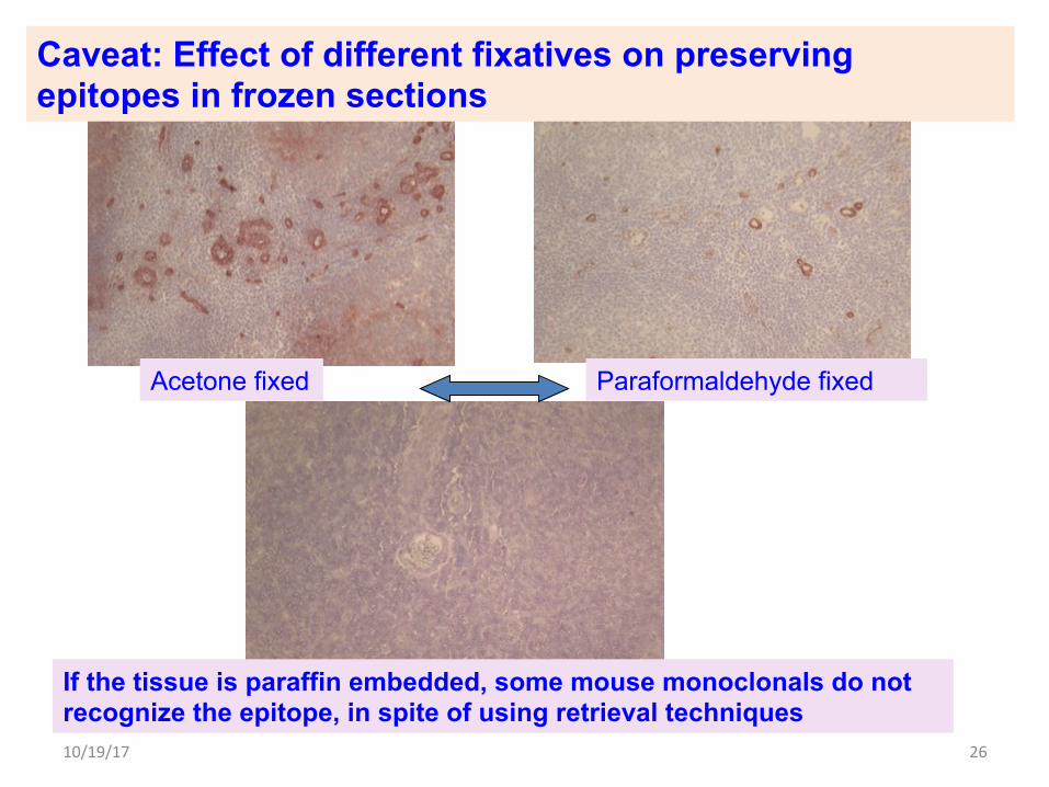
Caveat: Effect of different fixatives on preserving epitopes in frozen sections
If the tissue is paraffin embedded, some mouse monoclonals do not recognize the epitope, in spite of using retrieval techniques
Acetone fixed Paraformaldehyde fixed
10/19/17 26

FIXATIVES
• Fix Thin slices of tissue, or inflated lungs, or tissue in sponges
• use 4% freshly made paraformaldehyde for 24 hours before immersion in 70% alcohol to submit to histotech
• Or 10% buffered formalin for 24 hours before immersion in 70% alcohol to submit to histotech
• Or Bouin’s solution--has picric acid (yellow), acetic acid and formalin--fixes fast, makes tissues hard if left in it for more than 6 hours, many antibodies do not detect epitopes after Bouin’s fixation
• Or Zinc containing fixatives, preserve epitopes for immunostaining

Zinc formalin fixation requires special processing
Bouin’s fixative is quick but it hardens tissues, if fixed for too long, so move specimens to 70% alcohol in 6 hours.
10% Neutral buffered formalin
Four commonly used Fixatives for tissue processing in histology
4% paraformaldehyde is made fresh in the fume hood before use
Use 10 volumes of fixative for each samples, overnight in labeled cassettes, before transfer to 70% alcohol for processing embedding and sectioning, staining for microscopic analysis

Make 4% paraformaldehyde in the chemical hood with heating and with NaOH and PBS, cool and freeze in aliquots
Add 4 g paraformaldehyde
Wear a mask and be careful while weighing it out, it disperses easily
To 50 ml of water
Heat to 65 degrees
Add 6 ul of 10M NaOH
Solution will clear
Filter via Whatman paper
Add 10 ml of 10X PBS
Make up volume to 100 ml
Store at 4 degrees for upto 24 hours
Or freeze in aliquots
In Chemical Fume hood

Use simply labeled cassettes, using indelible pencil, to fix thin slices of organs or rolls of intestine, in 10 volumes of fixative, for less than 24 hours, before transferring to 70% alcohol, for processing into paraffin blocks.
Do not use a “Sharpie “ to label cassettes.
Use Sponges in casettes for to flatten certain organs such as: Spleen,Thymus, Pancreas, adipose tissue, skin, small organs such as adrenals, ovaries, lymph nodes
to orient them FLAT for good sections

Materials that are needed to use to freeze tissue for histology
If the animal has been perfusion fixed --the organs have to SINK (Descend to bottom of tube) in 30% sucrose/PBS
Before blotting well to remove extra sucrose, to freeze in OCT for histology examination
If you need to FREEZE FIXED tissue for histology:


Bones have to be de-calcified after fixation
Decalcification solutions: HCl; Formalin+ HCl; EDTA only- for slow decalcification for IHC

Importance of Orientation of tissues :
Coronal sections
Sagittal sections
Transverse sections

10/19/17 37
Correct orientation to gain the most information during histopathologic examination, an example of a section of mouse embryo

HEMATOXYLIN AND EOSIN STAINS
HISTO: HISTOLOGY SECTIONS FOR VIEWING UNDER THE MICROSCOPE, using BRIGHTFIELD illumination
Always review sections using the basic hematoxylin and eosin (H&E) stain
before proceeding to perform an immunohistochemical assay
in order to check out the morphology of the tissue and to determine
that what you are looking for is present in the section to be immunostained
and that the section has no other abnormalities
H&E=hematoxylinandeosin.
Hematoxylincolorsnucleiblue
Eosincolorsthecytoplasmpink

10/19/17 39
This is a photo of an unstained section on a slide, which needs histochemical stains to help with identification of the tissue

An example of a section of Mouse lung frozen section stained only with hematoxylin

An example of a section of Mouse lung stained with Hematoxylin and Eosin to demonstrate morphology

H&E is standard to assess morphology
A Trichrome stain to demosntrate collagen (blue)
PAS (periodic Acid Schiff) for carbohydrates—here demonstrated fuschia colored mucin in goblet cells of colon epithelium
Examples of Different Histochemical stains to demonstrate different components in a section of Human Colon

Reticulin stain to highlight supporting support Normal mouse liver Liver with invading cancer

Oil Red O stain of FROZEN section of mouse liver showing moderate amounts and large amounts
of fatty accumulation in hepatocytes Control: adipose tissue

Commonly used “Blue” stains in histochemistry: --hematoxylin: for nuclei --Trichrome: for collagen and for scarring/fibrosis --Alcian Blue: for mucin and for cartilage --Nissl: for nuclei in neurons --Luxol Fast Blue (LFB): for myelin

Commonly used “Red” stains in histochemistry: --Eosin: stains cytoplasm and depending in the tissue type, can vary in intensity --PAS: Periodic Acid Schiff—for carbohydrate containing material, in mucin and basement membranes --Alizarin Red: stains bone --Safranin O: stains cartilage --Oil Red O—to identify lipid containing cells , has to be done on frozen sections

Luxol Fast Blue (LFB) for myelin

Commonly used “Black “ stains in histochemistry: --PTAH: Phosphotungstic Acid Hematoxylin, to identify striations in skeletal muscle and also for collections of abnormal fibrin in clotting disorders Elastic: to identify elastic fibers Reticulin: to identify reticulin supporting fibers

Human Skeletal muscle with PhosphoTuncsticAcidHematoxylin PTAH stain to demonstrate striations

Human aorta: H&E and Elastic stain This is a large vessel with abundant elastic fibers to contribute strength

Silver stain to demonstrate reticulin supporting tissue

EPITHELIUM is the term given to the cells that
-cover the exterior surface of the body,
-lines both the internal closed cavities of the body,
-lines body tubes that communicate with the exterior
(alimentary, respiratory, genitourinary)
-comprise the various organs (liver)
Epithelium can be
-impervious (epidermis or bladder) ,
-secretory (stomach),
-absorptive (intestines),
-be a transport system(trachea),
-receive sensory stimuli (taste buds of the tongue)

Epithelium can be impervious (epidermis or bladder) Stratified Squamous epithelium—stacked up like plates
Squamous epithelium function helps with shear forces that are encountered as in
-Skin, with anuclear keratin layer
-Esophagus(No keratin layer)
-Cervix (no keratin layer)
-External ear canal
-Anus

Epithelium can be secretory (stomach), absorptive (intestines),
Columnar epithelium—is so termed because the cells are arranged like columns
Epithelial lining of intestine
The height of the cell is greater than the width

Cuboidal epithelium Ovary with developing follicles
Primordial follicles lined by flat squamous epithelium
Primary follicles lined by cuboidal epithelium
Stromal fibroblasts

Pseudo-stratified Columnar epithelium
Human trachea Mouse trachea

Transitional epithelium of bladder (mouse)

Junctional zone epithelium: Where epithelium of one kind changes naturally to another
Mouse Stomach: half is squamous epithelium
Mouse cervix: external is squamous epithelium

Junctional zone epithelium: Where epithelium of one kind changes naturally to another
HUMAN STOMACH Mouse Stomach: half is squamous epithelium

Normal breast ducts and alveoli have an Inner layer of cuboidal epithelial cells (keratin+) and an Outer layer of myoepithelial cells (smooth muscle actin)
Keratin is a marker for most epithelial cells

Vimentin is a marker for stromal fibroblasts and blood vessels

1. Frozen sections are useful for Immunohistochemistry. T/F
2. Frozen sections are useful for Morphologic examination. T/F
3. Paraffin sections are made after fixation. T/F
4. Paraffin sections can be used for immunohistochemistry T/F
5. Length of fixation affect the ability to detect antigens in paraffin sections. T/F
6. Bone has to be fixed and decalcified for two-three days before processing into paraffin blocks, for sectioning and staining. T/F

1. Your genetically altered mouse died last night.
You want to know the cause of death.
Should you fix and look paraffin sections
or plan to sacrifice littermate controls and gene altered animals
at specified time points, harvest organs,
fix and examine paraffin sections?
2. The animal has been perfused with PBS and then with fixative.
Can the tissue from various organs be now frozen
and sectioned for immunohistochemistry?