primary spinal tumors management of painful metastatic and · patients with metastatic spinal...
TRANSCRIPT

Received 01/22/2017 Review began 03/03/2017 Review ended 03/21/2017 Published 03/24/2017
© Copyright 2017Hariri et al. This is an open accessarticle distributed under the terms ofthe Creative Commons AttributionLicense CC-BY 3.0., which permitsunrestricted use, distribution, andreproduction in any medium, providedthe original author and source arecredited.
Minimally Invasive Surgical Techniques forManagement of Painful Metastatic andPrimary Spinal TumorsOmid R. Hariri , Ariel Takayanagi , Dan E. Miulli , Javed Siddiqi , Frank Vrionis
1. Department of Neurosurgery, Kaiser Permanente-Orange County, ANAHEIM, USA 2. Neurosurgery,Riverside University Health System Medical Center, Moreno Valley, USA 3. Neurosurgery, DesertRegional Medical Center, Palm Springs, USA 4. Neurological Surgery, Marcus Neuroscience Institute,Boca Raton, USA
Corresponding author: Omid R. Hariri, [email protected] Disclosures can be found in Additional Information at the end of the article
AbstractPatients with metastatic spinal disease are affected by disabling pain. The treatment of spinalmetastases is focused on pain reduction and improvement in quality of life. Until recently,many patients with metastatic spinal disease did not qualify as surgical candidates due to therisks of surgery and length of recovery period. However, recent advances in minimally invasivesurgery such as kyphoplasty and vertebroplasty allow patients to safely undergo surgery forpain relief with a short recovery period.
The studies reviewed here suggest that vertebral augmentation is successful in reducing painand disability scores in patients with painful metastases and multiple myeloma and are a safemodality to provide lasting pain relief. As the use of kyphoplasty and vertebroplasty fortreatment of vertebral metastases is becoming more common, new combinations of cementaugmentation with other techniques such as percutaneous pedicle screws and radiofrequencyablation are being explored. The implementation of kyphoplasty and vertebroplasty, inconjunction with other minimally invasive surgical techniques as well as nonsurgicalmodalities, may lead to the best palliative management of cancer patients with spinalmetastases and help them ultimately achieve a better quality of life.
Categories: Radiation Oncology, Neurosurgery, OncologyKeywords: kyphoplasty, vertebroplasty, myeloma, percutaneous, spine, tumors, palliative, metastases,minimally invasive, augmentation
Introduction And BackgroundIntroductionPain from the invasion of cancer into the spinal column is a key detriment to the quality of lifeof cancer patients. Treatment is focused on pain reduction and quality of life rather than curingthe disease process. Multimodal and multidisciplinary treatment is necessary in these patientsand includes radiation, medical management, and a variety of operative measures.
While medical treatment may be effective in managing pain for some patients, mechanical paindue to pathological fractures may require surgical intervention. Traditionally, neurologicalsymptoms have been treated by decompression of the spinal cord with a laminectomy.However, many patients with spinal tumors are at a higher risk of complications with open
1 2 2 3 4
Open Access ReviewArticle DOI: 10.7759/cureus.1114
How to cite this articleHariri O R, Takayanagi A, Miulli D E, et al. (March 24, 2017) Minimally Invasive Surgical Techniques forManagement of Painful Metastatic and Primary Spinal Tumors. Cureus 9(3): e1114. DOI10.7759/cureus.1114
surgery [1]. Recent advances in percutaneous procedures such as kyphoplasty andvertebroplasty have created options for patients who were previously not considered surgicalcandidates.
Here we provide a brief review of the pathophysiology and clinical symptoms of vertebraltumors along with diagnostic and nonsurgical treatment strategies. We then reviewpercutaneous surgical treatment with an emphasis on the efficacy and safety of vertebroplastyand kyphoplasty.
BackgroundThe metastatic neoplasms that most commonly present with spinal cord compression are breast(in 22% of patients with breast cancer), lung (15% of patients with lung cancer), and prostate(10% of patients with prostate cancer) [2]. Secondary tumors of the spine most frequently occurin the thoracic spine (70%), followed by the lumbar spine (20%), and finally the cervical spine(10%) [3].
Several theories exist regarding the pathogenesis of vertebral metastases. The 1940 Batsonstudy on cadavers suggested cancer cells are pushed into a valveless venous plexus from thechest and pelvis during periods of increased intra-abdominal pressure. The low-pressurevenous system and repeated reversal of flow likely allow for cancer cells to be lodged in thevertebral bodies [4]. A more recent study by Arguello, et al. suggests the tumors alternativelymetastasize through arterial circulation rather than through venous routes and use the bonemarrow as a “soil for proliferation” [5]. Tumors can also invade locally into the vertebral bodies[6] from retroperitoneal lymph node, lung (Pancoast tumors), thyroid, or muscle (sarcomas)metastases.
While metastases are the most common form of cancer in the spine, multiple myeloma is themost common primary tumor to invade the vertebrae. While multiple myeloma is a “primary”neoplasm in the sense that it begins in the bone marrow, it is derived from plasma cells ratherthan osteocytes like many of the other bony primary cancers.
Multiple myeloma patients are especially at risk for pathological fractures due to thecombination of local invasion of malignant myeloid cells and the cytokine-mediated activationof osteoclasts that causes an increase in bone resorption [7]. In these patients, 80% ofpathologic vertebral fractures occur from T6-L4, and 50% from T11-L1 [8]. Involvement of thecervical spine is uncommon [9].
ReviewClinical presentationThe most common symptom at the time of diagnosis is severe pain, which usually precedes theonset of neurologic dysfunction by a median time of seven weeks [10-12]. The main types ofpain experienced in these patients are local pain and axial pain.
Local pain occurs at the site of the metastasis, is severe and progressive in quality [13], and isdescribed by patients as “gnawing” or “aching” [14]. This pain classically presents nocturnallysecondary to its exacerbation by supine positioning, is relieved by standing up, and can often beelicited by percussion over the affected region [10]. Local pain is caused by inflammation andstretching of the periosteum where nociceptors are located [14]. Because back pain caused byother conditions (e.g., degenerative joint disease) typically occurs in the cervical and lumbarregions, patients with thoracic pain should be treated with a high index of suspicion for spinalmetastases [10].
2017 Hariri et al. Cureus 9(3): e1114. DOI 10.7759/cureus.1114 2 of 18
Mechanical or axial pain typically appears later than local pain and can occur secondary tovertebral instability and compression of the spinal cord. This pain is described as sharp andstabbing [10], is exacerbated by axial loading [15], and usually persists despite pain medication[6]. Regardless of the location, mechanical back pain reported by a patient diagnosed withcancer should be assumed to be due to spinal metastases until proven otherwise [1]. Axial paincan be treated at an early stage with surgical techniques such as vertebroplasty and kyphoplastyas described later.
After pain, the second most common symptom of metastatic spinal disease is neurologicdysfunction secondary to spinal cord compression. Sixty percent to 85% of patients havecorticospinal dysfunction, presenting with upper motor neuron disease. Loss of sensationoccurs along with (or soon after) the onset of weakness [11]. Patients commonly present withdifficulty in walking due to hip flexor weakness, and soon afterward complain of changes insensation. Autonomic dysfunction can also occur as a late symptom, usually presenting asurinary incontinence [11].
Similarly, multiple myeloma patients experience pain secondary to invasion of the periosteumwhich causes local pain and weakening of the vertebral body. The multiplicity of pain sites dueto the diffuse involvement of numerous vertebrae is the rule with multiple myeloma.
Radiographic findingsX-ray is typically the first imaging modality used for patients with vertebral metastases. Plainanteroposterior and lateral radiographs may reveal asymmetry with areas of radiolucency oropacity, depending on the type of lesion present. The classic “winking owl sign” can be seen onanteroposterior plain films due to a missing pedicle, but requires significant bone destructionto be visible. When an abnormality is present on an x-ray, computed tomography (CT) isrecommended to visualize the bone abnormalities at higher resolutions [15].
While x-ray and CT are important in assessing bone involvement, magnetic resonance imaging(MRI) with gadolinium enhancement is the gold standard imaging modality for suspectedspinal metastases. MRI has a high sensitivity for tumors using sagittal T1 or short T1 inversionrecovery. T2 sagittal and axial T1 or T2 are useful in detecting soft tissue involvement anddetermining the degree of spinal cord compression [12]. Moreover, focal vertebral lesions inmultiple myeloma present with hypointense on T1 and hyperintense on T2 [8].
Nonsurgical pain managementThere are several nonsurgical methods for managing spinal pain in cancer patients. The 1997World Health Organization (WHO) pain ladder remains the standard for the medicalmanagement of pain. Patients are categorized as having mild to moderate or moderate tosevere pain and are treated with the corresponding medications. Mild pain is managed withnonopioid analgesics such as nonsteroidal anti-inflammatory drugs (NSAIDs) andacetaminophen, while moderate pain is managed with weak opioids such as dihydrocodone ortramadol. NSAIDs may be added at any level on the analgesic ladder. Oral morphine is the drugof choice for treating the severe pain experienced by patients with metastatic spinal lesions [16-17]. Patients should receive continuous dosing with the administration of extra doses forbreakthrough pain [16].
Treatment of pain following the WHO guidelines is successful in reducing pain by 70% to 80%[18]. Local nerve blocks, antidepressants, anticonvulsants [19], and intrathecal analgesia [12]can be used as adjuvant treatment depending on whether the pain is local, radicular, or axial.
Corticosteroids can also reduce pain in patients with spinal metastases by inhibiting
2017 Hariri et al. Cureus 9(3): e1114. DOI 10.7759/cureus.1114 3 of 18
prostaglandin synthesis and decreasing vascular permeability. Pain is abated by reducingedema in the surrounding tissue, which prevents the compression of pain-producing structures[12]. Dexamethasone is commonly used because of its longer half-life, once-a-day-dosing, andhigher potency [18].
Radiotherapy can also be used to decrease pain from spinal metastases, especially inconjunction with surgery. Radiotherapy has become the standard of treatment of patients withmultiple metastatic lesions who are not surgical candidates due to poor prognosis and severecomorbidities. However, recent advances in percutaneous surgery indicate the benefits of theseprocedures may outweigh the risks [19].
In a prospective study of metastatic cancer pain treated with radiation therapy, 49% of patientsexperienced complete pain relief while 51% had a >50% reduction in pain [20]. While high-doseradiation can provide pain relief, it increases the risk of vertebral compression fractures due toosteonecrosis [19]. Fracture risk must be weighed against the efficacy of pain relief whendetermining stereotactic radiation treatment regimens. In selected cases, biopsy, kyphoplasty,and radiofrequency ablation can be performed prior to radiosurgery. In addition to dosage, thefrequency of radiation also has an effect on treatment outcomes. Although both split-courseand short-course regimens have similar efficacy, short-term therapy is more practical andrequires fewer patient visits [21].
Management of axial pain is similar for multiple myeloma in that the WHO guidelines are usedto manage pain medically. The most commonly used treatment of multiple myeloma ischemotherapy, while the definitive treatment requires autologous hematopoietic stem celltransplant [22].
Surgical techniquesMedical treatments may help significantly with local pain; however, they do not addressmechanical spinal pain caused by vertebral collapse and deformity [6]. Surgical treatments, onthe other hand, have been shown to be highly effective in treating axial pain [23] with fewadverse effects. Vertebroplasty and kyphoplasty offer minimally invasive surgical options forpatients who are unlikely to tolerate more extensive invasive procedures [19].
Kyphoplasty and vertebroplasty are both effective in relieving axial pain, but no consensus hasbeen made on when to implement one over the other [24]. While the type of procedure is oftendetermined by the surgeon and institutional preference, it may be valuable to develop a morestandardized treatment algorithm in the future. Figures 1-2 describe the two procedures.
2017 Hariri et al. Cureus 9(3): e1114. DOI 10.7759/cureus.1114 4 of 18

FIGURE 1: Basic steps of vertebroplasty technique.1A) Needle is inserted percutaneously through the pedicles and into the vertebral body. 1B)polymethylmethacrylate (PMMA) cement is then injected into the vertebral body to relievemechanical stress and restore height.
FIGURE 2: Basic steps of kyphoplasty.The needle is inserted percutaneously, and then a balloon is inserted (2A) and inflated (2B) tocreate a cavity inside the vertebral body and to restore height. Cement is then injected into thecavity (2C).
Vertebroplasty and kyphoplasty have traditionally been performed in the lumbar and lowerthoracic spine, using a transpedicular approach as shown in Figure 2. Intraoperative x-rays of alumbar kyphoplasty are shown in Figure 3. Although kyphoplasty and vertebroplasty may bemore difficult in the cervical spine due to the anatomy of the cervical vertebrae and theproximity of the cervical vasculature, these techniques have been successfully applied to thecervical spine. A meta-analysis of six studies included 120 patients who underwentvertebroplasty or kyphoplasty for metastases to the cervical spine and showed significantreductions in mean pain scores from 7.6 ± 0.9 preoperatively to 1.9 ±.8 (p = .0006) at the final
2017 Hariri et al. Cureus 9(3): e1114. DOI 10.7759/cureus.1114 5 of 18
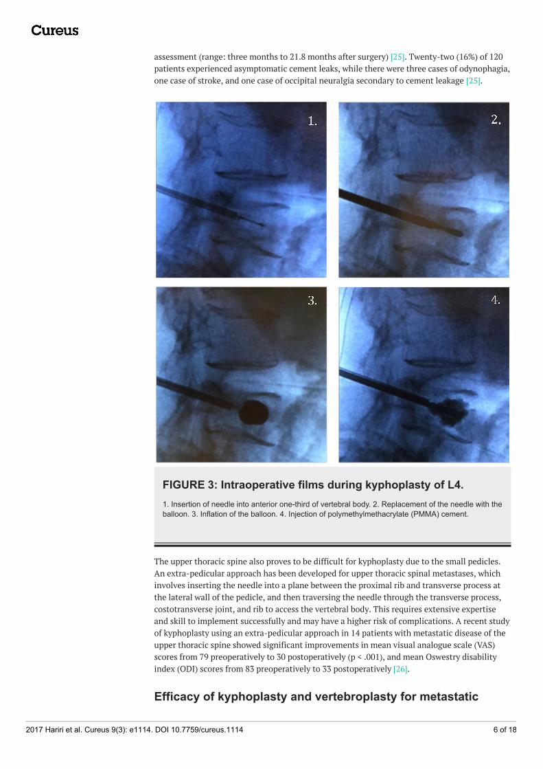
assessment (range: three months to 21.8 months after surgery) [25]. Twenty-two (16%) of 120patients experienced asymptomatic cement leaks, while there were three cases of odynophagia,one case of stroke, and one case of occipital neuralgia secondary to cement leakage [25].
FIGURE 3: Intraoperative films during kyphoplasty of L4.1. Insertion of needle into anterior one-third of vertebral body. 2. Replacement of the needle with theballoon. 3. Inflation of the balloon. 4. Injection of polymethylmethacrylate (PMMA) cement.
The upper thoracic spine also proves to be difficult for kyphoplasty due to the small pedicles.An extra-pedicular approach has been developed for upper thoracic spinal metastases, whichinvolves inserting the needle into a plane between the proximal rib and transverse process atthe lateral wall of the pedicle, and then traversing the needle through the transverse process,costotransverse joint, and rib to access the vertebral body. This requires extensive expertiseand skill to implement successfully and may have a higher risk of complications. A recent studyof kyphoplasty using an extra-pedicular approach in 14 patients with metastatic disease of theupper thoracic spine showed significant improvements in mean visual analogue scale (VAS)scores from 79 preoperatively to 30 postoperatively (p < .001), and mean Oswestry disabilityindex (ODI) scores from 83 preoperatively to 33 postoperatively [26].
Efficacy of kyphoplasty and vertebroplasty for metastatic
2017 Hariri et al. Cureus 9(3): e1114. DOI 10.7759/cureus.1114 6 of 18

neoplasmsFourney, et al. [24] studied the efficacy and safety of vertebroplasty and kyphoplasty in 56patients with either spinal metastases or spinal multiple myeloma. Eighty-four percent of thesepatients reported complete or significant pain relief, with statistically significantimprovements in median preoperative and postoperative VAS scores. A summary of the efficacyof kyphoplasty and vertebroplasty in pain reduction and disability scores is demonstrated inTable 1. All significant results in this paper correspond to a 95% confidence interval.Significant pain reduction was maintained at one, three, and six months for patients whounderwent kyphoplasty or vertebroplasty. Significant pain reduction was additionallymaintained at one year for kyphoplasty but not for vertebroplasty. No significant difference inpain reduction between kyphoplasty and vertebroplasty was noted at six months. There werealso no complications related to the procedure in any of the patients in the trial [24]. This wasthe first large study to assess the safety and benefits of kyphoplasty and vertebroplasty forpainful spinal neoplasms. The high percentage of patients with significant pain relief and lackof significant complications suggest that both kyphoplasty and vertebroplasty can provideimmediate, safe, and lasting pain relief in these patients.
StudyMedian
Pt Age
#
Pts
Procedure
(# cases)
Pathology
(#cases)
Pain Relief:
improvement
in scores
postop
Karnofsky
Performance Status
Improvement
in Disability
Scores after
KP/VP
Summary
Berenson 2011
[27]: Randomized
Controlled
Multicenter Trial
KP: 64.8,
Control:63134
KP(68) vs.
medical
management
(61)
Multiple
Myeloma: KP
(22),
nonsurgical
(27)
†Difference in
reduction of
NRS score
between KP
and medical
management.
KP > medical.
†Improvement in KP
group compared to
nonsurgical group
(mean improvement):
15.3 points (95% CI,
13.5 to 17.1; p <
.0001)
†RDQ
treatment
effect after
KP: -8.4
points (95%
CI, -7.6 to -
9.2; p <
.0001)
Sig. reductions in mean
pain scores (NRS), KPS,
RMQ for KP at one month,
but not in nonsurgical
group.Metastatic
Cancer: KP
(46), medical
(34)
1 week: -3.5
(95% CI, -3.8
to -3.2; p <
.0001)
1 mo.: -3.3
(95% CI, -3.6
to -3.0; p <
.0001) l (95%
CI, p < .0001).
Fourney 2003
[24]: Retrospective
Review
64 56
KP (34), VP
(15), KP+VP
(7)
Metastatic
cancer (35),
Multiple
Myeloma (21)
†Improvement
in VAS:
NA NA
Sig. reductions in VAS in
KP patients with MM and
spinal metastases
compared to baseline, with
significance maintained at
6 mo. for VP, and at one
year for KP.
immediately
postop: BKP,
VP (p < .05).
6 mo.: KP, VP
(p < .05)
12 mo.:KP (p <
.05)
2017 Hariri et al. Cureus 9(3): e1114. DOI 10.7759/cureus.1114 7 of 18
Markmiller
2015 [28]:
longitudinal
prospective case
series
68.7 115 Kyphoplasty
Metastatic
Cancer (92),
Multiple
Myeloma (23)
†Median VAS
after KP:†Median:
†Improvement
in mean ODI
after KP:
Sig. improvements in
median KPS, mean ODI,
and median VAS with KP
through 12 mo.
Postop: -4.0
(95% CI -5.0 to
-3.0; p <
.0001)
Postop: 15 (95% CI;
p < .0001)
Postop: -49.6
(95% CI, -
56.4 to -42.1
p < .0001)
12 mo.: -3.5
(95% CI -5.0 to
-3.0 ( p <
.0001)
12 mo: 10 (95% CI; p
< .0001)
12 mo.: -48.4
(95% CI, -
56.4 to -42.1
p < .0001)
McDonald
2009 [29]:
retrospective
review
66.2 67 VP (67)Multiple
Myeloma (67)
†median
improvement
in VAS after
VP:
NA
†Improvement
in median
RDQ scores
after VP:
Sig. improvements in
median VAS, RDQ in
patients with MM after VP
through 12 mo.
At rest: -2.7
(95% CI, -3.7
to 1.7; p <
.0001 at one
week, < .03 at
one year)
one week: -11
(95% CI -14.3
to -7.7; p <
.0001)
With activity: -
5.3 (95% CI, -
6.4 to -4.2 p <
.0001 at one
week, p < .001
at one year)
one year: (p <
.001)
Papanastassiou
2014 [31]:
retrospective
comparative
study
61.6 69
KP:
unilateral
versus
bilateral (69)
Multiple
Myeloma (69)
†Change in
mean VAS in
unilateral and
bilateral KP,
respectively
from baselineNA NA
Sig. improvement in mean
VAS scores in patients with
MM after both unilateral
and bilateral kyphoplasty,
with no difference in pain
reduction between the two
techniques.
Postop: -5.4, -
5.5(95% CI,
p<.0005>
3 mo.:
p<.0005>
Pflugmacher 2006
†Change in
mean VAS:
†Improvement
in mean ODI:
Sig. improvements in mean
Preop: 71.5%
(39-89%)
3 mo. postop:
2017 Hariri et al. Cureus 9(3): e1114. DOI 10.7759/cureus.1114 8 of 18

[30]: Longitudinal
prospective case
series
62.4 20 KP(20)Multiple
Myeloma (20)Postop: -6
(95% CI; p <
.05)
NA27.5%
improvement
27.5% (95%
CI, 11 to 41%;
p < .05)
VAS, ODI with KP in
patients with multiple
myeloma through one
year.
12 mo.: -5.1
(95% CI; p <
.05)
12 mo.:(95%
CI, 13 to 52%;
p < .05)
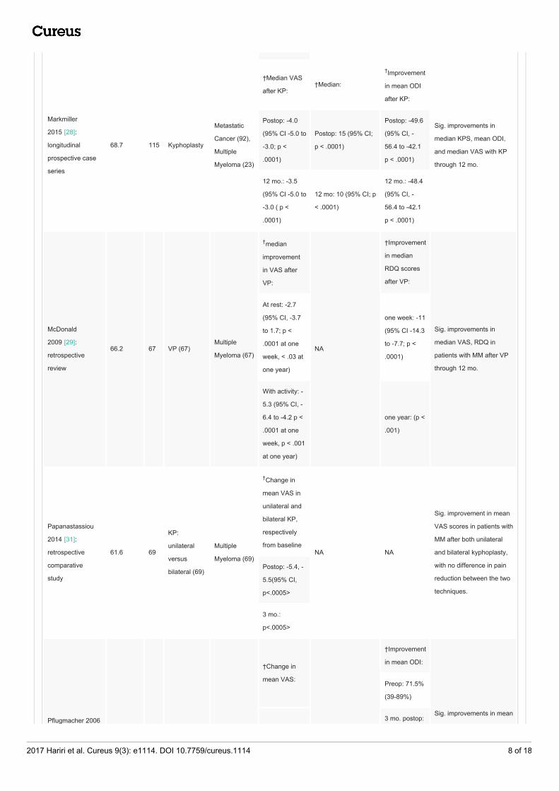
TABLE 1: Scores for pain, disability, and physical function in patients before and afterkyphoplasty and vertebroplasty.†Statistically significant.
Abbreviations: Pt, patient; KP, kyphoplasty; VP, vertebroplasty; NRS, pain numeric rating score; ODI, Oswestry disability index; VAS,visual analogue scale; RDQ, Rolland Disability Questionnaire; KPS, Karnofsky performance score; Sig., significant; MM, multiplemyeloma; mo, month; NA, not applicable; Postop, postoperatively; Preop, preoperatively; CI, confidence interval.
The Cancer Patient Fracture Evaluation (CAFÉ) trial by Berenson, et al. [27] was the firstrandomized controlled multicenter study that compared the treatment outcomes ofkyphoplasty and nonsurgical treatment in patients with painful vertebral fractures due tometastases. One hundred thirty-four patients were enrolled.
Assessment after one month showed statistically significant improvements in physicalfunction, back pain, and quality of life in the kyphoplasty group; the effects were notstatistically significant for the nonsurgical treatment group. The mean Roland Morris disabilityquestionnaire (RDQ) treatment effect for kyphoplasty was statistically significant (Table 1).Mean Karnofsky performance scores (KPS) also increased significantly, showing animprovement in functional impairment status that was not seen in the nonsurgical treatmentgroup. Kyphoplasty patients experienced an improvement in quality of life both physically andmentally, as demonstrated by significant improvements in mean Short Form Health Survey (SF-36) Physical Component Summary scores and SF-36 Mental Component Summary scores at onemonth. Reduction in back pain was assessed using the numeric pain rating scale (NRS), with asignificant difference in reduction between kyphoplasty and nonsurgical groups at one weekand one month. Thirty-eight patients in the nonsurgical group elected to receive kyphoplastywhen given the option after one month [27].
This trial was the first randomized controlled trial to assess the efficacy of kyphoplasty inpatients with painful spinal metastases. Although randomization was only sustained for follow-up at one month, results suggest that kyphoplasty is an effective palliative care strategy forpatients with painful spinal metastases, improving quality of life, functional status, and backpain.
Markmiller [28] recently published a longitudinal prospective case series of 115 patients whoreceived kyphoplasty for confirmed spinal metastases or multiple myeloma. At discharge afterkyphoplasty, a statistically significant decrease in median VAS scores was seen compared topreoperative scores; this remained significant at one year (Table 1). Similarly, median KPSscores improved significantly from preoperative scores, and improvement in function was stillfound to be significant at one year. Mean ODI scores showed significant improvementscompared to baseline, at discharge, and at one year. These results are consistent with the
2017 Hariri et al. Cureus 9(3): e1114. DOI 10.7759/cureus.1114 9 of 18

aforementioned studies, and show potential for lasting pain relief and improved functionalstatus.
Efficacy of kyphoplasty and vertebroplasty for multiplemyelomaRecently, studies on percutaneous vertebral augmentation for multiple myeloma haveemerged. While both metastatic tumors and multiple myeloma invade the vertebral bodies, thepathophysiology of multiple myeloma is drastically different, as previously described. It isimportant to investigate whether patients with multiple myeloma can benefit from kyphoplastyand vertebroplasty with as much success. McDonald, et al. [29] measured quantitativeoutcomes of pain, mobility, and function in 67 patients with multiple myeloma who underwentvertebroplasty. Median RDQ scores and VAS scores at rest and with activity improvedsignificantly (Table 1). Both RDQ and VAS scores remained significant at one week, one month,six months, and one year [29]. Similar findings were reported in a smaller study of 20 patientswith multiple myeloma who underwent kyphoplasty, with statistically significant reduction ofmean VAS and ODI scores. Improvement in both VAS and ODI scores remained significant atthe one-year follow-up [30]. These results suggest that vertebroplasty can improve pain,mobility, and function in multiple myeloma patients as it does in patients with metastatic spinedisease.
In a study comparing unilateral versus bilateral kyphoplasty in multiple myeloma patients,patients who received either form of kyphoplasty experienced improvement in pain scores. VASscores improved from baseline by 30% (p < .0005, paired samples t-test). There was, however,no significant difference in reduction of mean VAS scores between unilateral and bilateralvertebroplasty. The results of this uniform cancer population refute previous notions thatbilateral kyphoplasty is more effective in pain resolution than a unilateral approach. Theauthors recommend the use of unilateral kyphoplasty whenever feasible to reduce operationtime and risks of surgery, especially because patients with multiple myeloma often requirecement augmentation on multiple levels [31].
There is no consensus in the literature on the maximum number of adjacent levels that can betreated in one session. A study compared the biomechanics of multilevelpolymethylmethacrylate (PMMA) vertebral augmentation in cadavers [32]. Zero, one, two, andthree levels were augmented and stiffness and strength were analyzed using a univariateanalysis of variance. Stiffness (p = .0009 multilevel segments, p < .0004) and strength (p < .001)depended on bone marrow density but did not significantly differ among the groups with zero,one, two, or three levels treated. This study suggests that the strength of vertebral bodies is notnegatively affected by an increased number of adjacent levels treated with kyphoplasty orvertebroplasty. Although some sources suggest that no more than two levels should beaugmented due to possible PMMA toxicity, many experts in the field [33] have recommendedtreating up to three levels.
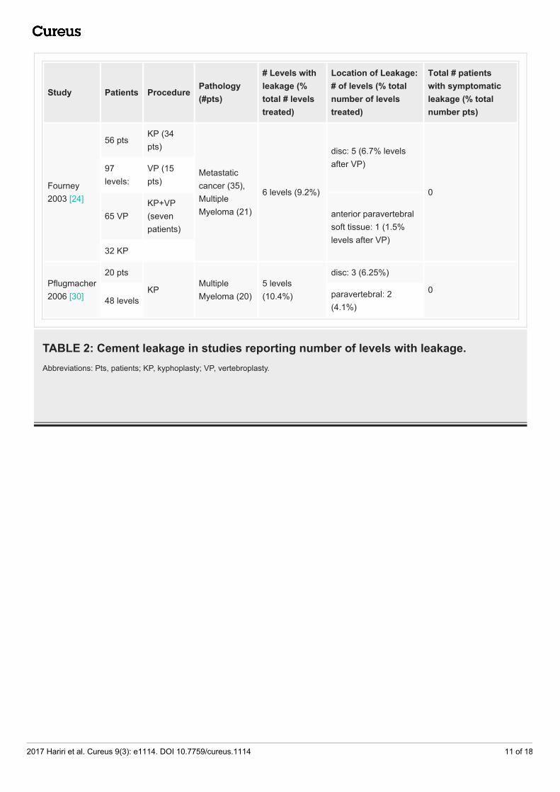
Safety of vertebroplasty and kyphoplasty in metastatic spinaldiseaseThe main concern with kyphoplasty and vertebroplasty is the potential for PMMA cementleakage. Possible but seldom seen complications secondary to cement leakage include spinalcord compression, radiculopathy, and pulmonary embolism. One-third of patients inMarkmiller, et al. experienced cement leakage; however, only three (2.6%) were symptomaticwith resolution after three months [28]. In Fourney, et al., six of 65 patients who underwentkyphoplasty or vertebroplasty for metastases showed leakage of cement on imaging, but nonewere symptomatic [24]. Leakage rates are compared in Tables 2-3.
2017 Hariri et al. Cureus 9(3): e1114. DOI 10.7759/cureus.1114 10 of 18
Study Patients ProcedurePathology(#pts)
# Levels withleakage (%total # levelstreated)
Location of Leakage:# of levels (% totalnumber of levelstreated)
Total # patientswith symptomaticleakage (% totalnumber pts)
Fourney2003 [24]
56 ptsKP (34pts)
Metastaticcancer (35),MultipleMyeloma (21)
6 levels (9.2%)
disc: 5 (6.7% levelsafter VP)
0
97levels:
VP (15pts)
65 VPKP+VP(sevenpatients)
anterior paravertebralsoft tissue: 1 (1.5%levels after VP)
32 KP
Pflugmacher2006 [30]
20 pts
KPMultipleMyeloma (20)
5 levels(10.4%)
disc: 3 (6.25%)
048 levels
paravertebral: 2(4.1%)
TABLE 2: Cement leakage in studies reporting number of levels with leakage.Abbreviations: Pts, patients; KP, kyphoplasty; VP, vertebroplasty.
2017 Hariri et al. Cureus 9(3): e1114. DOI 10.7759/cureus.1114 11 of 18

Study Patients ProcedurePathology (#cases)
# Patientswithleakage
Location ofLeakage: #patients (%patients)
Total patients withsymptomatic leakage (%total number patients)
Markmiller 2015[28]
115patients
KP
MetastaticCancer (92),MultipleMyeloma (23)
40 patients(34.8% of115patients)
disc: 17 (14.8%)
3 (2.6%)*
disc-paravertebral: 2(1.7%)
medullary canal:8 (7%)
paravertebral: 9(7.8%)
vascular: 4(3.5%)
McDonald 2009[29]
67patients
KPMultipleMyeloma (67)
13 patients(19% ofpatients)
disc: 6 (9%)
0
paravertebral 4(6%)
embolus toepidural vein: 3(4%)
Papanastassiou2014 [31]
69patients,105levels
KPMultipleMyeloma (69)
five patients(7%patients)
disc, spinalcanal
0
TABLE 3: Cement leakage in studies reporting number of patients with leakage.*Three patients with symptomatic leakage. All three patients experienced radiculopathy with no weakness and had complete resolutionof symptoms at six months. Two patients with leakage into medullary canal, one patient with paravertebral leakage.
Abbreviations: KP, kyphoplasty.
Although symptomatic cement extrusion is uncommon, there are several strategies to preventleakage. Fourney, et al. suggest that their low leak rate may be due to dedicated time forsufficient thickening of the cement before injecting into the vertebral body. Another strategy isto use smaller amounts of PMMA cement [24].
Safety of vertebroplasty and kyphoplasty in patients withmultiple myelomaIn McDonald, et al. [29], 13 of 67 patients (19%) who underwent vertebroplasty experienced theinjection of cement into areas outside of the vertebral body, but all were asymptomatic [29].
2017 Hariri et al. Cureus 9(3): e1114. DOI 10.7759/cureus.1114 12 of 18
The lack of symptomatic complications from the procedure suggests vertebroplasty is a safetreatment modality for multiple myeloma patients in addition to patients with spinalmetastases (Tables 2-3). Similar recommendations were made in a study of kyphoplasty inpatients with spinal metastases and multiple myeloma. Because patients with multiplemyeloma of the spine experienced increased cement leakage rates due to softer integrity of thebone, the authors suggested waiting for cement to become more viscous to prevent leakage[30].
Efficacy of minimally invasive options: percutaneous pediclescrews combined with vertebral augmentationIn patients with involvement of the pedicle and posterior elements, cement augmentation maynot be adequate treatment. Moreover, open resection, decompression, and fusion can bemorbid secondary to steep recovery in late-stage cancer patients. In patients who requireadditional stabilization but cannot tolerate open surgery, percutaneous pedicle screw fixationmay be considered. In a study by Chi, et al. in 2013 [34], 16 patients with pathologic fracturessecondary to spinal metastases underwent pedicle screw fixation using fluoroscopic guidance.In 14 of 16 patients, vertebroplasty was performed following screw fixation. Pain significantlydecreased postoperatively as measured by the numeric pain rating scale (p < .01). Patients alsohad a significant improvement in kyphotic angle (p < .01). Similar to kyphoplasty orvertebroplasty alone, the percutaneous treatment of instability allows patients to recoverquickly and start or return to radiotherapy or chemotherapy shortly after surgery. Thecombination of percutaneous pedicle screw fixation and vertebral augmentation is perhaps anoption in patients with significant neoplastic destabilization who will not tolerate moreextensive and open surgical approaches.
Zairi, et al. [35] studied 10 patients with spinal metastases who experienced neurologicalcompromise and underwent minimally invasive transpedicular vertebrectomy with spinal corddecompression and subsequent percutaneous stabilization. Eight of 10 patients improved atleast one Frankel grade, and all patients experienced a reduction in pain using the VAS score.Patients received radiotherapy or chemotherapy. This approach allows for tumor controlbecause radiation and chemotherapy can be initiated almost immediately after surgery withoutconcern of wound dehiscence.
One concern of pedicle fixation is the risk of screw pullout due to the poor bone quality and lackof strong purchase of the pedicle screws in the bone. Moussazadeh, et al. [36] combined twoprocedures to address this issue. Forty-four patients with spinal instability due to vertebralmetastatic tumors underwent percutaneous short-segment pedicle screw fixation with cementaugmentation at the affected levels. Transpedicular cement augmentation was followed byscrew placement into the cement. Subsequently, kyphoplasty was done at the level of thefracture, and rods were secured to the pedicle screws. Twenty-nine of 44 patients reportedcomplete resolution of symptoms, 13 reported mild pain, and two reported moderate pain aftersurgical intervention, with a significant decrease of pain on the Serlin scale (p < .001). Therewere few complications, with one adjacent-level fracture, and one asymptomatic screw pullout.The cement augmentation in the levels with screw fixation allowed the pedicle screws to befixed more securely, while kyphoplasty at the level of the fracture created better anteriorstabilization and prevented future kyphotic deformity [36]. The combination of percutaneouspedicle screw fixation and vertebral augmentation is perhaps an option in patients withsignificant neoplastic destabilization who will not tolerate more extensive and open surgicalapproaches.
Gu, et al. [37] demonstrated the benefits of minimally invasive pedicle screw fixation andpercutaneous vertebroplasty followed by neurologic decompression and partial tumor resectionusing a mini-posterior-midline approach. In the study, 18 patients with spinal cord
2017 Hariri et al. Cureus 9(3): e1114. DOI 10.7759/cureus.1114 13 of 18

compression due to vertebral tumors were treated. One year after the operation, the medianVAS scores decreased significantly from nine preoperatively to three (p < .001). Four patientspresented with complete loss of motor function (ASIA scale B), and 14 presented withincomplete motor paralysis (ASIA scale C or D). All patients experienced improvement inparaplegia postoperatively, and 13 of 18 improved to ASIA scale E by the one-year follow-up.
Experimental use of chemotherapeutic agent-eluting acryliccement in vertebroplastyExperimental use of PMMA cement mixed with chemotherapeutic agents is currently beingstudied. Rosa, et al. [38] showed that methotrexate, cisplatin, and doxorubicin retain biologiceffects on in-vitro breast cancer cells when mixed with acrylic cement. However, the amount ofdrug released decreased to values near zero by 15 days. Maccauro, et al. [39] showed thatmethotrexate does not weaken the compressive properties of acrylic cement. Possibleadvantages of adding chemotherapeutic agents to PMMA for vertebroplasty and kyphoplastyinclude the drug is less likely to be eliminated systemically before reaching its target cellssecondary to its local release. Although chemotherapeutic agents seem to be able to retain anti-cancer properties and are unlikely to affect the strength of acrylic cement, thepharmacodynamics of the timing and dosage of the drug must be studied further.
Radiofrequency ablation and cement augmentationRecently, the application of radiofrequency ablation (RFA) has been used in tumors that areunresectable and not responsive to radiation therapy. The affected vertebral body ispercutaneously accessed, and a high-frequency alternating current is sent to cause thermally-induced necrosis of the tumor. The pathophysiology of pain relief caused by RFA is unclear. Oneexplanation is that it reduces pain by destroying nociceptors as well as reducing the tumorburden [40]. Gevargez, et al. [41] showed that 41 patients treated with RFA for vertebralmetastases experienced a significant reduction in VAS scores within six weeks (p = .001) andcontinued to be significant at six months (p = 0.002).
RFA treatment of tumors within the vertebral body may cause instability if done alone. In acadaveric study comparing RFA and vertebroplasty with RFA alone, RFA was shown to decreasemechanical stability and increase the risk of subsequent burst fractures [42]. Combining RFAand vertebroplasty provides a way to decrease tumor burden and retain or augment thesupportive structure of the vertebral body. Wallace, et al. [40] performed 72 RFA treatmentswith subsequent vertebroplasty in patients with spinal tumors. Patients treated with thiscombination of techniques experienced a significant decrease in VAS pain scores at one weekand four weeks (median 3.25, 2.75, respectively; p < .0001). Like vertebroplasty or kyphoplastyalone, RFA can result in almost immediate pain relief with a short recovery period [40]. Zheng,et al. [43] showed similar results using RFA combined with kyphoplasty. Thus, RFA may be asafe technique to offer to patients in combination with vertebral augmentation, especially forpatients with a large tumor burden. However, further comparative studies are indicated tomeasure pain reduction in patients treated with percutaneous cement augmentation aloneversus RFA.
Separation surgery and spinal laser interstitial thermal therapySeparation surgery, as described by Bilsky and Smith [44], is a technique in which a portion ofan epidural tumor adjacent to the dura is resected, and instrumentation is done forstabilization without further tumor resection. This technique can be used in patients withradioresistant tumors who would likely benefit from stereotactic radiosurgery. The goal is todecompress the spinal cord and create a safe margin between the thecal sac and the tumor forhigh-dose radiation therapy while preventing direct damage to the spinal cord. Moreover, it
2017 Hariri et al. Cureus 9(3): e1114. DOI 10.7759/cureus.1114 14 of 18

prevents the more aggressive and invasive approaches required when attempting a gross totalresection. Therefore, patients proceed to radiation therapy and other adjunct therapy withoutsignificant delay [45].
Tatsui, et al. [46] recently described an innovative technique called spinal laser interstitialthermotherapy (SLITT). For select patients, this is an alternative to separation surgery in whichthe tumor adjacent to the dura is ablated to create a space between the spinal cord and thetumor in preparation for high-dose radiation therapy [46]. A probe is inserted into the epiduraltumor percutaneously under CT-based image guidance and advanced to a safe distance from thethecal sac. The tumor is thermally ablated under real-time thermal MRI monitoring to protectthe spinal cord from thermal injury [45].
In another study, Tatsui, et al. [47] reported 19 patients with metastatic epidural spinal cordcompression who received SLITT experienced a significant decrease in VAS pain scores from4.72 to 2.56 after one month, which remained significant at three months postoperatively (p =.43 and p = .21, respectively) [47]. All patients were able to undergo subsequent stereotacticradiosurgery (SRS), with a median time from laser ablation to SRS of three days. With oneexception, no patients experienced adverse events due to the procedure. The median thicknessof the epidural tumor decreased significantly from 8.0 mm preoperatively to 6.4 mm, twomonths postoperatively (p = .012) [47].
The study demonstrates that SLITT can be performed safely to relieve epidural spinal cordcompression with low morbidity in select patients, namely those with radioresistant tumors,and allows for the prompt resumption of adjunct therapy. Like other minimally invasivetechniques, this offers a viable alternative for patients who are not candidates for open surgery.
In addition, SLITT can be combined with percutaneous instrumentation in patients who requireimmediate stabilization [48]. A pilot study of eight patients with epidural spinal cordcompression underwent SLITT combined with percutaneous spinal stabilization with a mediantime to radiation therapy or chemotherapy of five days [48]. Tatsui, et al. showed that thermalMRI guidance for laser ablation has high accuracy and can be used effectively to visualize thespinal cord even when severe spinal cord compression is present [49].
ConclusionsPatients with painful vertebral metastases and multiple myeloma of the spine must be treatedwith multiple modalities in a multidisciplinary fashion including but not limited to radiationand medical management. Percutaneous surgical techniques are minimally invasive with highefficacy and a low rate of complications. The studies reviewed here suggest that kyphoplastyand vertebroplasty are successful in reducing pain and disability scores in patients with painfulmetastases and multiple myeloma. The main concern of these techniques is the extrusion ofcement, which has rarely caused lasting neurological symptoms. While kyphoplasty has beenimplemented traditionally in the lower thoracic and lumbar spine where a trans-pedicularapproach can be made, modified techniques to access the vertebral bodies in the upper thoracicspine and cervical spine may allow more patients to benefit from kyphoplasty andvertebroplasty.
Implementation of kyphoplasty and vertebroplasty in conjunction with other minimallyinvasive techniques as well as nonsurgical modalities may lead to the best palliativemanagement of cancer patients with spinal metastases and help them ultimately achieve abetter quality of life.
Additional Information
2017 Hariri et al. Cureus 9(3): e1114. DOI 10.7759/cureus.1114 15 of 18

DisclosuresConflicts of interest: In compliance with the ICMJE uniform disclosure form, all authorsdeclare the following: Payment/services info: All authors have declared that no financialsupport was received from any organization for the submitted work. Financial relationships:All authors have declared that they have no financial relationships at present or within theprevious three years with any organizations that might have an interest in the submitted work.Other relationships: All authors have declared that there are no other relationships oractivities that could appear to have influenced the submitted work.
References1. Chi JH, Gokaslan ZL: Vertebroplasty and kyphoplasty for spinal metastases . Curr Opin
Support Palliat Care. 2008, 2:9–13. 10.1097/SPC.0b013e3282f5d9072. Gerszten PC, Welch WC: Current surgical management of metastatic spinal disease . Oncology.
2000, 14:1013–1024.3. Klimo P, Schmidt MH: Surgical management of spinal metastases . Oncologist. 2004, 9:188–
196. 10.1634/theoncologist.9-2-1884. Batson OV: The function of the vertebral veins and their role in the spread of metastases . Ann
Surg. 1940, 112:138–149.5. Arguello F, Baggs RB, Duerst RE, et al.: Pathogenesis of vertebral metastasis and epidural
spinal cord compression. Cancer. 1990, 65:98–106.6. Sciubba DM, Gokaslan ZL: Diagnosis and management of metastatic spine disease . Surg
Oncol. 2006, 15:141–151. 10.1016/j.suronc.2006.11.0027. La Maida GA, Giarratana LS, Acerbi A, et al.: Cement leakage: safety of minimally invasive
surgical techniques in the treatment of multiple myeloma vertebral lesions. Eur Spine J. 2012,21:61–68. 10.1007/s00586-012-2221-3
8. Tosi P: Diagnosis and treatment of bone disease in multiple myeloma: spotlight on spinalinvolvement. Scientifica. 2013, 2013:1-12. 10.1155/2013/104546
9. Frigui M, Frikha F, Haj Kacem H, et al.: Multiple myeloma presenting as cervical spinecompression. Rheumatol Rep. 2011, 3:5. 10.4081/rr.2011.e5
10. DeAngeles LM, Posner JB: Neurologic Complications of Cancer . Oxford University Press, NewYork; 2009.
11. Gabriel K, Schiff D: Metastatic spinal cord compression by solid tumors . Semin Neurol. 2004,24:375–383. 10.1055/s-2004-861532
12. Metastatic Spinal Cord Compression: Diagnosis and Management of Patients at Risk of or withMetastatic Spinal Cord Compression. National Collaborating Centre for Cancer, Cardiff, UK;2008.
13. Levack P, Graham J, Collie D, et al.: Don’t wait for a sensory level-listen to the symptoms: aprospective audit of the delays in diagnosis of malignant cord compression. Clin Oncol (R CollRadiol). 2002, 14:472–480.
14. Sciubba DM, Petteys RJ, Dekutoski MB, et al.: Diagnosis and management of metastatic spinedisease. A review. J Neurosurg Spine. 2010, 13:94–108. 10.3171/2010.3.SPINE09202
15. Gebaeur GP, Farfoodi P, Sciubba DM, et al.: Magnetic resonance imaging of spine tumors:classification, differential diagnosis, and spectrum of disease. J Bone Joint Surg Am. 2008,90:146–162. 10.2106/JBJS.H.00825
16. Ripamanti CI, Santini D, Maranzano E, et al.: Management of cancer pain: ESMO clinicalpractice guidelines. Ann Oncol. 2012, 23:139–154. 10.1093/annonc/mds233
17. Leppert W, Buss T: The role of corticosteroids in the treatment of pain in cancer patients .Curr Pain Headache Rep. 2012, 4:307–313. 10.1007/s11916-012-0273-z
18. Vyvey M: Steroids as pain relief adjuvants . Can Fam Physician. 2010, 56:1295–1297.19. Kaloostian PE, Yurter A, Etame AB, et al.: Palliative strategies for the management of primary
and metastatic spinal tumors. Cancer Control. 2014, 21:140–143.20. Nomiya T, Teruyama K, Wada H, et al.: Time course of pain relief in patients treated with
radiotherapy for cancer pain. Clin J Pain. 2010, 26:38–42. 10.1097/AJP.0b013e3181b0c82c21. Maranzano E: Short-course versus split-course radiotherapy in metastatic spinal cord
compression: results of a Phase III, randomized, multicenter trial. J Clin Oncol. 2005,23:3358–3365. 10.1200/JCO.2005.08.193
2017 Hariri et al. Cureus 9(3): e1114. DOI 10.7759/cureus.1114 16 of 18
22. Silberman R, Roodman GD: Myeloma bone disease: pathophysiology and management. J BoneOncol. 2013, 2:59–69. 10.1016/j.jbo.2013.04.001
23. Boszczyk B: Volume matters: a review of procedural details of two randomised controlledvertebroplasty trials of 2009. 2010, 19:1837–1840. 10.1007/s00586-010-1525-4
24. Fourney DR, Schomer DF, Nader R, et al.: Percutaneous vertebroplasty and kyphoplasty forpainful vertebral body fractures in cancer patients. J Neurosurg. 2003, 98:21–30.10.3171/spi.2003.98.1.0021
25. De la Garza-Ramos R, Benvenutti-Regato M, Caro-Osorio E: Vertebroplasty and kyphoplastyfor cervical spine metastases: a systematic review and meta-analysis. Int J Spine Surg. 2016,10:7. 10.14444/3007
26. Eleraky M, Papanastassiou I, Setzer M, et al.: Balloon kyphoplasty in the treatment ofmetastatic tumors of the upper thoracic spine. J Neurosurg Spine. 2011, 14:372–376.10.3171/2010.11
27. Berenson J, Pflugmacher R, Jarzem P, et al.: Balloon kyphoplasty versus non-surgical fracturemanagement for treatment of painful vertebral body compression fractures in patients withcancer: a multicentre, randomised controlled trial. Lancet Oncol. 2011, 12:225–235.10.1016/S1470-2045(11)70008-0
28. Markmiller M: Percutaneous balloon kyphoplasty of malignant lesions of the spine: aprospective consecutive study in 115 patients. Eur Spine J. 2015, 24:2165–2172.10.1007/s00586-014-3751-7
29. McDonald RJ, Trout AT, Gray LA, et al.: Vertebroplasty in multiple myeloma: outcomes in alarge patient series. AJRN Am J Neuroradiol. 2008, 29:642–648. 10.3174/ajnr.A0918
30. Pflugmacher R, Kandziora F, Schroeder J, et al.: Percutaneous balloon kyphoplasty in thetreatment of pathological vertebral body fracture and deformity in multiple myeloma: a one-year follow-up. Acta Radiol. 2006, 47:369–376. 10.1080/02841850600570425
31. Papanastassiou ID, Eleraky M, Murtagh R, et al.: Comparison of unilateral versus bilateralkyphoplasty in multiple myeloma patients and the importance of preoperative planning.Asian Spine J. 2014, 8:244–252. 10.4184/asj.2014.8.3.244
32. Kayanja MM, Schlenk R, Togawa D, et al.: The biomechanics of 1, 2, and 3 levels of vertebralaugmentation with polymethylmethacrylate in multilevel spinal segments. Spine. 2006,31:769–774. 10.1097/01.brs.0000207466.40955.31
33. Hentschel SJ, Burton AW, Fourney DR, et al.: Percutaneous vertebroplasty and kyphoplastyperformed at a cancer center: refuting proposed contraindications. J Neurosurg Spine. 2005,2:436–440. 10.3171/spi.2005.2.4.0436
34. Chi HK, Chung CK, Sohn S, et al.: Less invasive palliative surgery for spinal metastases . J SurgOncol. 2013, 108:499–503. 10.1002/jso.23418
35. Zairi F, Arikat A, Allaoui M, et al.: Minimally invasive decompression and stabilization for themanagement of thoracolumbar spine metastases. J Neurosurg Spine. 2012, 17:19–23.10.3171/2012.4.SPINE111108
36. Moussazadeh N, Rubin DG, McLaughlin L, et al.: Short-segment percutaneous pedicle screwfixation with cement augmentation for tumor-induced spinal instability. Spine J. 2015,15:1609–1617. 10.1016/j.spinee.2015.03.037
37. Gu Y, Dong J, Jiang X, et al.: Minimally invasive pedicle screws fixation and percutaneousvertebroplasty for the surgical treatment of thoracic metastatic tumors with neurologiccompression. Spine. 2016, 41:14–22. 10.1097/BRS.0000000000001811
38. Rosa MA, Maccauro G, Sgambato A, et al.: Acrylic cement added with antiblastics in thetreatment of bone metastases. J Bone Joint Surg Br. 2003, 85:712–716. 10.1302/0301-620X.85B5.13588
39. Maccauro G, Cittadini A, Casarci M, et al.: Methotrexate-added acrylic cement: biological andphysical properties. J Mater Sci Mater Med. 2007, 18:839–844. 10.1007/s10856-006-0036-7
40. Wallace AN, Greenwood TJ, Jennings JW: Radiofrequency ablation and vertebral augmentationfor palliation of painful spinal metastases. J Neurooncol. 2015, 124:111–118. 10.1007/s11060-015-1813-2
41. Gevargez A, Groenemeyer DHW: Image-guided radiofrequency ablation of spinal tumors . Eur JRadiol Open. 2008, 65:246–252. 10.1016/j.ejrad.2007.03.026
42. Pezeshki D, Davidson S, Murphy K, et al.: Comparison of the effect of two different bone-targeted radiofrequency ablation (RFA) systems alone and in combination with percutaneousvertebroplasty (PVP) on the biomechanical stability of the metastatic spine. Eur Spine J. 2016,
2017 Hariri et al. Cureus 9(3): e1114. DOI 10.7759/cureus.1114 17 of 18

25:3390–3996. 10.1007/s00586-015-4057-043. Zheng L, Chen Z, Sun M, et al.: A preliminary study of the safety and efficacy of
radiofrequency ablation with percutaneous kyphoplasty for thoracolumbar vertebralmetastatic tumor treatment. Med Sci Monit. 2014, 20:556–563. 10.12659/MSM.889742
44. Bilsky M, Smith M: Surgical approach to epidural spinal cord compression . Hematol OncolClin North Am. 2006, 20:1307–1317. 10.1016/j.hoc.2006.09.009
45. Laufer I, Iorgulescu JB, Chapman T, et al.: Local disease control for spinal metastasesfollowing “separation surgery” and adjuvant hypofractionated or high-dose single fractionstereotactic radiosurgery: outcome analysis in 186 patients. J Neurosurg Spine. 2013, 18:207–214. 10.3171/2012.11.SPINE12111
46. Tatsui CE, Stafford J, Li J, et al.: Utilization of laser interstitial thermotherapy guided by real-time thermal MRI as an alternative to separation surgery in the management of spinalmetastasis. J Neurosurg Spine. 2015, 23:400–411. 10.3171/2015.2.SPINE141185
47. Tatsui CE, Lee SH, Amini B, et al.: Spinal laser interstitial thermal therapy: a novel alternativeto surgery for metastatic epidural spinal cord compression. Neurosurg. 2016, 79:73–82.10.1227/NEU.0000000000001444
48. Tatsui CE, Belsuzarri TA, Oro M, et al.: Percutaneous surgery for treatment of epidural spinalcord compression and spinal instability: technical note. Neurosurg Focus. 2016, 41:E2.10.3171/2016.8.FOCUS16175
49. Tatsui CE, Nascimento CN, Suki D, et al.: Image guidance based on MRI for spinal interstitiallaser thermotherapy: technical aspects and accuracy. J Neurosurg Spine. 2017, 1–8.10.3171/2016.9.SPINE16475
2017 Hariri et al. Cureus 9(3): e1114. DOI 10.7759/cureus.1114 18 of 18