primmorphs generated from dissociated cells of the sponge
TRANSCRIPT

Mechanisms of Ageing and Development
105 (1998) 45–59
Primmorphs generated from dissociated cells ofthe sponge Suberites domuncula : a model system
for studies of cell proliferation and cell death
Marcio Reis Custodio a,b, Ivo Prokic a, Renate Steffen a,Claudia Koziol a, Radovan Borojevic b, Franz Brummer c,
Michael Nickel c, Werner E.G. Muller a,*a Institut fur Physiologische Chemie, Abteilung Angewandte Molekularbiologie, Uni6ersitat,
Duesbergweg 6, Mainz, D-55099, Germanyb Departamento de Histologia e Embriologia, Instituto de Ciecias Biomedicas,
Uni6ersidade Federal de Rio de Janeiro, Rio de Janeiro, CEP 21941-970, CP 68021, Brazilc Biologisches Institut, Abteilung Zoologie, Uni6ersitat, Pfaffenwaldring 57, Stuttgart, 70550, Germany
Received 5 May 1998; received in revised form 19 June 1998; accepted 29 June 1998
Abstract
Sponges (Porifera) represent the lowest metazoan phylum; they have been shown to beprovided with the characteristic metazoan structural and functional molecules. One autapo-morphic character of sponges is the presence of high levels of telomerase activity in all cells(or almost all cells, including somatic cells). In spite of this fact previous attempts to cultivatesponge cells remained unsuccessful. It was found that dissociated sponge cells do notreplicate DNA and lose their telomerase activity. In addition, no nutrients or metaboliteshave been detected that would stimulate sponge cells to divide. In the present study we reportthe culture conditions required for the formation of multicellular aggregates from dissociatedsingle cells of Suberites domuncula, termed primmorphs. These primmorphs are formed inseawater without addition of further supplements, and have an organised tissue-like struc-ture; they have been cultured for more than 5 months. Cross-sections revealed a distinct
* Corresponding author. Tel.: +49 6131 395910; fax: +49 6131 395243; e-mail: [email protected]
0047-6374/98/$ - see front matter © 1998 Elsevier Science Ireland Ltd. All rights reserved.
PII S0047-6374(98)00078-5

M.R. Custodio et al. / Mechanisms of Ageing and De6elopment 105 (1998) 45–5946
external layer covered by a continuous pinacoderm, and a central zone composed primarilyof spherulous cells. After their association into primmorphs, the cells turn from thetelomerase-negative state into the telomerase-positive state; a telomerase level of 4.7 totalproduct generated (TPG) units/5×103 cell equivalents has been determined. Moreover, amajor fraction of the cells in the primmorphs undergoes DNA synthesis and hence has thecapacity to grow. Applying the BrdU-labelling and detection assay it is demonstrated that upto 33.8% of the cells in the primmorphs are labelled with BrdU after an incubation periodof 12 h. It is proposed that the primmorph system described here is a powerful novel modelsystem to study basic mechanisms of cell proliferation and cell interaction, as well as ofmorphogenesis, ageing and apoptosis. © 1998 Elsevier Science Ireland Ltd. All rightsreserved.
Keywords: Suberites domuncula ; Sponges; Cell culture; Telomerase; Primmorphs; Senescence;Apoptosis
1. Introduction
Both palaeontological and comparative cell-biological studies indicate that thephylum Porifera (sponges) is a very ancient one and potentially the most simpleamong eumetazoans. Recent molecular studies have given substantive support tothe hypothesis that Porifera and other animals belong to a single monophyletickingdom (reviewed in Muller, 1995). Porifera have been extensively studied inrecent years, in order to establish cellular and molecular aspects of the evolutionfrom unicellular to multicellular grade of organisation in animals.
The full cell function of multicellular organisms requires both structural integra-tion and molecular communication, and one of the key autapomorphic charactersof metazoa is the receptor tyrosine kinase signalling pathway, which is found onlyin this kingdom, including sponges (Muller and Schacke, 1996). In view of the othersignalling pathways that are also present in sponges, such as retinoic acid, inositolphosphates, or protein kinase C (Rottmann et al., 1987; Biesalski et al., 1992;Imsiecke et al., 1994), it has been assumed that sponge cells follow the generalpattern of integration and communication observed in higher metazoans. Incontrast, Porifera comprise a character that is not found in higher metazoan phyla:they have high levels of telomerase activity in normal tissues (Koziol et al., 1998).This fact implies that sponges do not show a clear distinction between the germ-and somatic-cell lineages (reviewed in Muller, 1998a,b). In higher metazoa, germcells and early embryonic cells are telomerase-positive while somatic cells aretelomerase-negative: a brief transitory induction of telomerase activity can beobserved in tissue stem cells involved in intense tissue repair and regeneration, butthe permanent telomerase activity is associated only with the conversion of normalcells into malignant neoplastic cells (Lange, 1998).
Most sponges are long-lived, and some reach a life span of more than 1500 years(Lehnert and Reitner, 1997). This observation fits well with the fact that most cells

M.R. Custodio et al. / Mechanisms of Ageing and De6elopment 105 (1998) 45–59 47
from sponges are telomerase-positive (Koziol et al., 1998). Therefore, it might beinferred that sponge cells have an unlimited capacity for proliferation associatedwith their differentiation into normal tissue cells during their growth and regenera-tion. In agreement, until now there are no reports on neoplastic diseases in sponges(De-Flora et al., 1995). However, in the initial report on the presence of high levelsof telomerase in sponge cells, it was shown that cells that have lost contact witheach other become telomerase-negative (Koziol et al., 1998). Cells kept in a singlecell suspension either die, very likely in a process of apoptosis (Wagner et al., 1998),or remain in a non-proliferating dormant state, in agreement with the observedcorrelation between proliferation and telomerase activity in cells of higher organ-isms (Belair et al., 1997).
The high proliferation capacity of sponge cells suggests that it should be easilyfeasible to establish their cell cultures in vitro. However, until now, only themaintenance of sponge cells in vitro has been achieved, e.g. from Hymeniacidonheliophila (Pomponi and Willoughby, 1994), Ephydatia muelleri (Imsiecke et al.,1995b), Latrunculia magnifica (Ilan et al., 1996) and Suberites domuncula (Mullerand Schacke, 1996). The cells in suspension did not proliferate readily (Ilan et al.,1996) and the proposed experimental systems cannot be termed primary cellcultures (Freshney, 1994). In addition, primary single cell suspensions from spongesalso contain bacteria and protozoa as shown by earlier experiments (Klautau et al.,1994; Custodio et al., 1995). The reasons for the fact that the cells remained in theresting stage might be found (i) in the experimental approach to establish a singlecell culture, and (ii) in the culture conditions used. The media used were supple-mented with fetal bovine serum (Pomponi and Willoughby, 1994; Ilan et al., 1996)under the assumption that the growth factors present in the serum might stimulatecell proliferation. However, it appears reasonable to accept that sponge cell surfacereceptors are not activated by ligands present in bovine serum. Furthermore,serum-rich media imply a risk of protozoan contamination (Osinga et al., 1998).
It has been known for a long time that dissociated sponge cells reaggregate andpotentially reorganise into a fully functional and structured sponge (Wilson, 1907),and this experimental model has been extensively used to study sponge celldifferentiation and morphogenesis (Curtis, 1962; Borojevic and Levi, 1964). Thedissociated cells are either induced to death and phagocyted by archaeocytes, orreaggregate and subsequently sort out following adhesion and motility gradients,described as contact-promoting by Curtis (1962). This leads to the formation of acompact spherical body, contrasting with rather loose irregular cellular contactsduring the aggregation (Muller, 1982). The formation of a pinacoderm, either frompre-existing pinacocytes or from archaeocytes (reviewed in Borojevic, 1970, 1971),represents the first step in reorganisation of tissue-like structures. This stage, termedhere ‘primmorphs’, represents the end of the aggregation of cellular material andthe separation of the internal milieu from the external environment by a continuouspinacoderm. Under favourable conditions, associated in general with adherence andstable fixation onto the solid substrate, this stage will lead to morphogeneticprocesses ending in the full reorganisation of the sponge body (to be published).The primmorphs thus represent new structures obtained from single cells, and

M.R. Custodio et al. / Mechanisms of Ageing and De6elopment 105 (1998) 45–5948
resemble partially a broader concept of diamorphs, defined by Borojevic et al.(1967) as the stage that is simultaneously the end of the concentration processes(such as observed in reduction bodies, external buds, and internal gemmules) andthe beginning of the sponge regeneration and expansion (Borojevic, 1970, 1971).
In view of the observation that telomerase can be reactivated after reestablish-ment of cell to cell contacts, and that the cell viability can be retained for longperiods in primmorphs, we have questioned whether the cells actively proliferateand whether the telomerase activity was fully established. The simultaneous reestab-lishment and preservation of cell to cell and cell to matrix contacts, as well as of theinternal milieu and the potential maintenance of symbiotic micro-organisms whichmay be required for the full sponge cell viability, indicates the culture of prim-morphs as a promising experimental system for sponge cell culture ex vivo and forstudies of molecular processes of development and ageing in representatives of theancient metazoans, the Porifera. Instead of stimulating the primmorph attachmentand conversion into the normal and functional sponge, we have followed up thelong-term cell viability in unattached primmorphs.
2. Materials and methods
2.1. Materials
Natural seawater (S9148), penicillin and streptomycin were obtained from Sigma(Deisenhofen, Germany), RNAguard (24000 U/ml) from Pharmacia (Freiburg,Germany), the Telomerase Detection Kit (TRAPeze) from Oncor (Gaithersburg,MD), BrdU-labelling and detection kit (cat. no. 1299964) from BoehringerMannheim (Mannheim, Germany), and SYBR Green I from Molecular Probes(Leiden, Netherlands).
The compositions of Ca2+ and Mg2+-free artificial seawater (CMFSW) as wellof CMFSW containing EDTA (CMFSW-E) were described previously (Rottmannet al., 1987).
2.2. Sponge
Specimens of the marine sponges S. domuncula (Porifera, Demospongiae,Hadromerida) were collected in the Northern Adriatic near Rovinj (Croatia), andthen kept in aquaria in Mainz (Germany) at a temperature of 16°C.
2.3. Dissociation of cells and formation of primmorphs
All cell culture dishes and tubes were sterilised and the media were filtratedthrough 0.2-mm polycarbonate filters. Tissue samples of 4–5 cm3 were submersed inseawater in Petri dishes and cut into 1-mm3 cubes; they were transferred into 50-mlconical tubes (Falcon no. 2070) filled with CMFSW-E (ratio tissue to medium1 : 10). After gentle shaking for 30 min at 16°C with a rotatory shaker, the solution

M.R. Custodio et al. / Mechanisms of Ageing and De6elopment 105 (1998) 45–59 49
was discarded and new CMFSW-E was added. After 40 min the supernatant wascollected and filtered through 40-mm mesh nylon net; the process of shaking inCMFSW-E (40 min) and filtration was repeated once again. The single cells wereharvested by centrifugation (500×g for 5 min) and washed once in CMFSW.The cells of the second pellet were resuspended in seawater supplemented withantibiotics (100 IE of penicillin and 100 mg/ml of streptomycin; seawater/antibi-otics). A cell suspension of 107 cells was added to 6 ml (final volume) ofseawater/antibiotics in 60-mm Petri dishes (Falcon no. 3004).
Each day two-thirds of the culture medium was replaced by fresh seawater/an-tibiotics; the cell clumps formed were resuspended to avoid adhesion of cells tothe plate. Cell aggregates of a diameter of at least 0.5 mm were collected bygentle pipetting as soon as they had formed (and using gravity only for theseparation from smaller aggregates and from single cells); they were washedtwice with 10 ml of seawater. The suspension of cells and aggregates was trans-ferred into 15-ml tubes (Falcon no. 2096) filled with 12 ml of seawater. Thiscollection of aggregates from the Petri dishes was repeated after a further day ofincubation. The aggregates were transferred again into new Petri dishes (totalvolume of 6 ml). During this procedure foreign organisms, mainly protozoawhich attached to the dishes, were removed from the sponge cell aggregateswhich remained in the suspension.
The round-shaped primmorphs, were placed into 24-well plates (Nunclon™no. 143982) (one–two/well) and 1 ml of seawater/antibiotics was added. Mediumwas changed every day during the first 2 weeks; later the medium change wasnecessary only once or twice a week. All pipettings were performed by the aid ofPasteur pipettes (diameter of the openings: 2 mm) or plastic tips (diameter: 2–3mm). Where indicated, the primmorphs were dissociated with CMFSW-E.
2.4. Incubation of primmorphs with BrdU: immunocytochemical detection of DNAsynthesis
For the determination of cell proliferation the incorporation of BrdU (5-bromo-2%-deoxy-uridine) into cellular DNA was monitored using the BrdU-la-belling and detection kit as recommended by the manufacturer. Primmorphswere incubated in 1 ml seawater/antibiotics, supplemented with BrdU-labellingmedium (final dilution of 1 : 1000; 10 mM of BrdU), for 12 h using culturechambers (culture chamber slides, Nunc no. 177453). The cells were dissociatedin CMFSW-E, washed three times in CMFSW-E and fixed in 70% ethanol (pH2.0, acetate buffer). The cells were incubated with anti-BrdU mouse monoclonalantibody. After 30 min (37°C) the cells were incubated with anti-mouse Ig-alka-line phosphatase, and the immunocomplexes were visualised with the colour-sub-strate nitroblue tetrazolium salt. The cells were observed and scored under alight microscope, using an Olympus AHBT3 microscope.

M.R. Custodio et al. / Mechanisms of Ageing and De6elopment 105 (1998) 45–5950
2.5. Histological analysis
Primmorphs were fixed in 4% paraformaldehyde/PBS (Romeis, 1989). Afterdehydration in ethanol, the samples were embedded in Technovit 8100 (Beckstead,1985), according to the instructions of the manufacturer. Sections of 2-mm thick-ness were prepared and stained with Ziehl’s fuchsin (Martoja and Martoja, 1967).
2.6. Telomerase assay
Telomerase activity was determined by polymerase chain reaction (PCR) proce-dure applying the ‘Telomerase Detection Kit (TRAPeze)’ as described (Kim et al.,1994; Koziol et al., 1998). The cell extracts corresponded to 5×10³ cell equivalents.The amplification products were separated by electrophoresis through a 12.5%non-denaturing polyacrylamide gel in 0.5×TBE-buffer according to the instruc-tions of the supplier. The gels were stained with SYBR Green I to detect the DNAfragments (Molecular Probes, 1996). The signals were quantified by application ofa GS-525 Molecular Imager (Bio-Rad). The amount of telomerase activity is givenin units of TPG (total product generated) and was calculated as described (Oncor,1996).
The number of cells analysed was estimated on the basis of the protein and DNAcontent of the extract to be assayed. Based on the DNA content per cell (3.7 pg/cellfor S. domuncula ; Imsiecke et al., 1995a), the number of cell equivalents wascalculated for a given tissue extract.
3. Results
3.1. Formation of primmorphs from cells of S. domuncula
The cells were obtained from specimens of the sponge S. domuncula (Fig. 1A).Single cell suspensions, obtained by dissociation in the absence of Ca2+ and Mg2+,were plated onto plastic dishes. Non-adherent sponge cells were harvested andwashed twice to remove most protozoa, which preferentially adhered to the plasticculture dishes. Thereafter, the cells were maintained in seawater/antibiotics. The cellaggregates steadily increased in size until the third day, through fusion or incorpo-ration of single cells (Fig. 1B and C). After a total treatment/incubation period of5 days, typical round-shaped primmorphs were formed whose size ranged from 1 to2 mm (Fig. 1D).
The outer appearance of the primmorphs was smooth and nearly spherical (Fig.1D). Microscopic analysis of the primmorph sections stained with Ziehl’s fuchsinrevealed that the cells were well organised in distinct tissue-like regions (Fig. 1E andF). A pronounced layer of epithelium-like cells (Fig. 1E and F) surrounded thecentral mass of cells. The cells that composed this external layer of the primmorphswere identified as pinacocytes, following their flattened form and prominent nuclei(Simpson, 1984); their size ranged from 15 to 20 mm. Several layers of amoeboid

M.R. Custodio et al. / Mechanisms of Ageing and De6elopment 105 (1998) 45–59 51
cells were found in this region, subjacent to the pinacoderm. The cells in the centralpart of primmorphs were mostly spherulous cells. Their diameter was 40–45 mmand they were characterised by large round vacuoles that occupied most of the cellbodies. Other cells corresponded to archaeocytes (Bergquist, 1978) and had a sizeranging from 55 to 60 mm. Bacteria were frequently present inside the primmorphsof S. domuncula, delimited from the sponge tissue inside the vacuoles of a specificcell type (to be published).
The organised arrangement of the cells within the primmorphs distinguishes themfrom primary cell aggregates which are formed from dissociated cells in thepresence of the homologous aggregation factor (Muller, 1982).
3.2. Subcultures of primmorphs
The primmorphs were kept in culture in the seawater/antibiotics medium. Underthose conditions they could be kept for over 5 months in a viable state.
Fig. 1. Formation of primmorphs from cells of the sponge S. domuncula. (A) A specimen of S.domuncula ; magnification ×1. (B) Cells were dissociated in CMFSW-E. Cell aggregates are formed after1 day in culture (medium: seawater/antibiotics) (×10), which (C) increase in size (micrograph takenafter 3 days) (×10), and (D) 5 days after incubation primmorphs are formed (×15). (E, F)Cross-sections through a primmorph, showing the single-cellular layer of epithelial-like pinacocytesurrounding the internal part, composed of spherulous cells and archaeocytes including amoebocytes; E,×30; F, ×60.
M.R. Custodio et al. / Mechanisms of Ageing and De6elopment 105 (1998) 45–5952
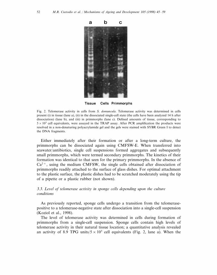
Fig. 2. Telomerase activity in cells from S. domuncula. Telomerase activity was determined in cellspresent (i) in tissue (lane a), (ii) in the dissociated single-cell state (the cells have been analyzed 14 h afterdissociation) (lane b), and (iii) in primmorphs (lane c). Defined amounts of tissue, corresponding to5×103 cell equivalents, were assayed in the TRAP assay. After PCR amplification the products wereresolved in a non-denaturing polyacrylamide gel and the gels were stained with SYBR Green I to detectthe DNA fragments.
Either immediately after their formation or after a long-term culture, theprimmorphs can be dissociated again using CMFSW-E. When transferred intoseawater/antibiotics, single cell suspensions formed aggregates and subsequentlysmall primmorphs, which were termed secondary primmorphs. The kinetics of theirformation was identical to that seen for the primary primmorphs. In the absence ofCa2+, using the medium CMFSW, the single cells obtained after dissociation ofprimmorphs readily attached to the surface of glass dishes. For optimal attachmentto the plastic surface, the plastic dishes had to be scratched moderately using the tipof a pipette or a plastic rubber (not shown).
3.3. Le6el of telomerase acti6ity in sponge cells depending upon the cultureconditions
As previously reported, sponge cells undergo a transition from the telomerase-positive to a telomerase-negative state after dissociation into a single-cell suspension(Koziol et al., 1998).
The level of telomerase activity was determined in cells during formation ofprimmorphs from a single-cell suspension. Sponge cells contain high levels oftelomerase activity in their natural tissue location; a quantitative analysis revealedan activity of 8.9 TPG units/5×103 cell equivalents (Fig. 2, lane a). When the

M.R. Custodio et al. / Mechanisms of Ageing and De6elopment 105 (1998) 45–59 53
telomerase activity was determined in cells that had been left for 14 h in thedissociated single-cell state, the enzyme level dropped to 0.9 TPG units/5×103 cells(Fig. 2, lane b). However, when cells from primmorphs (10 days after formationfrom single cells) were used for the analysis, a telomerase activity of 4.7 TPGunits/5×103 cells was observed (Fig. 2, lane c). These data confirm that cells losetheir telomerase activity when removed from tissues. As postulated earlier, singlecells will recover after formation of tissue-like organisation into primmorphs, andturn from the telomerase-negative to the telomerase-positive state (Koziol et al.,1998; Wagner et al., 1998).
3.4. Immunocytochemical detection of BrdU incorporation in cells of primmorphs
The BrdU-labelling and detection assay (Gratzner, 1982) was used to demon-strate that the cells organised into the primmorphs regain the capacity to prolifer-ate. As a measure of proliferation, the cells were incubated for 12 h in the presenceof BrdU. The incorporation of BrdU into DNA was detected by using ananti-BrdU monoclonal antibody as described in Section 2.
The BrdU-positive cells undergoing DNA synthesis stained brown in their nuclei(Fig. 3B–D). In a control assay, the antibody against BrdU was omitted, and underthis condition no staining is observed (Fig. 3A).
Fig. 3. Immunocytochemical detection of proliferating (BrdU-labelled) cells from primmorphs of S.domuncula. After incubation of the primmorphs with BrdU the cells were dissociated and subjected tostaining with anti-BrdU monoclonal antibody as described in Section 2. The dark brownish stainednuclei are those which incorporated BrdU (B–D); in D the arrow marks a BrdU-positive cell and thearrow head a BrdU-negative cell. A shows a control in which the antibody against BrdU was omitted.A and B, magnification ×20; C, ×40; D, ×125.

M.R. Custodio et al. / Mechanisms of Ageing and De6elopment 105 (1998) 45–5954
Table 1Analysis of cells for DNA synthesis applying the BrdU-labelling and detection assay
Percentage of BrdU-positive cells (%)Cells analyzed
Dissociated cells (after 24 h) 06.5Aggregates (after 24 h)
Primary primmorphs (3 days) 33.822.3Primary primmorphs (1 month)
A single cell suspension was incubated with BrdU and the incorporated nucleotides were visualizedimmunologically using anti-BrdU monoclonal antibody. The percentage of BrdU-positive cells is given.The analysis was performed with (i) dissociated cells which had been kept for 1 day in CMFSW-E, (ii)cell aggregates formed from single cells after 1 day in culture with seawater, (iii) primary primmorphsafter formation for 3 days and (iv) primary primmorphs after 1 month in culture. Per assay 300 cellswere counted.
Suspensions of dissociated cells, which had been kept for 1 day in CMFSWshowed no cells with active DNA synthesis (Table 1). The percentage of BrdU-pos-itive cells in cell aggregates formed from single cells after 1 day in culture was low,reaching only 6.5%. In contrast, the number of DNA-synthesising and proliferatingcells present in primmorphs was high. As summarised in Table 1, the number ofBrdU-positive cells in primary primmorphs was 33.8% and in ‘older’ primmorphs,after 1 month in culture, 22.3%. These data document that cells incorporated andreorganised into tissue-like primmorphs undergo DNA synthesis and subsequentcell division.
4. Discussion
The major objective of the present study was the development of experimentalconditions that support a long-term sponge cell culture in vitro, in order to studycontrols of cell proliferation and differentiation. As outlined in Section 1, so far thesponge cells were only maintained in vitro for a limited period of time. Inexperiments in our laboratory until now we also failed to compose a culturemedium for single cells in suspension culture.
In a rational approach we could demonstrate that after dissociation the spongecells lose the telomerase activity and in consequence their intriguing property toproliferate and to constitute (to a large extent) immortal cell populations (Koziol etal., 1998; Wagner et al., 1998Fig. 4). In addition, in a series of trials we failed toidentify supplements for the culture medium supplying the growth factors known tobe required for invertebrate and lower vertebrate cells to grow (data not shown).Based on these facts, we postulated that for an efficient culture of dividing spongecells, the following prerequisites are necessary: (i) viable cells, (ii) provision ofcell-cell and/or cell-matrix contacts, and (iii) suitable nutrients presented in a formthat can be used by sponge cells. These criteria have been met with the establish-ment of primmorph cultures.

M.R. Custodio et al. / Mechanisms of Ageing and De6elopment 105 (1998) 45–59 55
In order to obtain viable cells, we have used dissociation procedures based on theelimination of bivalent cations, shown in previous studies to mediate sponge cellsadhesion (reviewed in Muller et al., 1988). This method could be applied withsuccess both to primary sponge tissues and to primmorphs.
Sponge cells live inside or on an organic structured matrix (Simpson, 1984) andare expected to be anchorage-dependent. Simultaneously, the attachment onto asolid substrate of sponge cell diamorphs, external buds or gemmules has beenshown to be strictly required for the sponge tissue reorganisation and morphogen-esis (reviewed in Borojevic, 1971). Accordingly, previous attempts to establishisolated sponge cell cultures have used artificial solid substrates, associated or notwith biogenous molecules such as collagen, in order to provide the requiredconditions for anchorage of sponge cells in vitro. In the present study, besides themechanical support for cell attachment, the culture model using primmorphsestablishes the cell-cell contacts as well as the cell contacts with the endogenousextracellular matrix. The presence of specific collagen types, a major structuralelement required for cell adhesion and migration, has been thoroughly documentedin sponges (Garrone, 1985). The existence of the adhesive glycoprotein fibronectin,known from higher invertebrates and vertebrates to mediate cellular interactionswith the extracellular matrix (Hynes, 1990), has been proven—until now—only byimmune cross-reactivity with heterologous antibodies, and by isolation of a putativecDNA encoding this polypeptide from G. cydonium (Pahler et al., 1998a). Thepresence of morphogens in primmorphs has to be postulated, in order to under-stand the precise arrangement of proliferating cells in these bodies. Recently a
Fig. 4. Proposed scheme for the explanation of the transition of telomerase-positive- (T+) to telomerase-negative cells (T−) with the consequence of apoptotic cell death. It is outlined that exogenous and/orendogenous factor(s), cause the transition from T+ − to T−-cells; during this process the apoptoticprogram is initiated. The apoptotic cells expose both phosphatidyserine (PS) and phosphatidylinositol(PI); these phospholipids bind to T+-cells which express on their surface receptors composed ofscavenger receptor cysteine-rich repeats (SRCR). Further details are given in Section 4.

M.R. Custodio et al. / Mechanisms of Ageing and De6elopment 105 (1998) 45–5956
cDNA, encoding the potential morphogen endothelial-monocyte-activatingpolypeptide, has been also isolated from the sponge G. cydonium (Pahler et al.,1998b). The proposed model of in vitro culture should open the possibility to studythe biological activity of such molecules.
The sponge cells assembled in primmorphs become telomerase-positive and showDNA synthesis and hence regain the prerequisites for cell growth. One cause forthis transition must be seen in the fact that the cells have recovered the physiolog-ical contact to the neighbouring cells and/or to the homologous extracellular matrixand in consequence have arranged a functional organisation. This is similar tocultures of embryoid bodies from mammalian embryonic stem cells, which providethe conditions for both proliferation and differentiation of totipotent and telom-erase positive early embryo cells (Keller, 1995). Three-dimensional cell cultures arefrequently required for experimental cell differentiation models of higher animals,but not for cell proliferation (Muller-Klieser, 1997), and the fact that sponge cellsrequire this mode of culture for proliferation remains to be explained.
Sponge nutritional physiology is poorly known. Most studies have shown thatphagocytosis is probably the major input pathway for nutritional materials intosponges (Willenz et al., 1986; Van de Vyver et al., 1990), although the presence ofspecific cell categories that store glycogen and lipids would suggest internal circula-tion of energy-transport molecules such as glucose (Boury-Esnault, 1977). It is wellestablished that archeocytes, choanocytes or spherulous cells are active in phagocy-tosis of cells and debris (Simpson, 1984), and this finding is supported also byenzymatic data (Krasko et al., 1997). Phagocytosis of cell debris and cells undergo-ing apoptosis is mediated by receptors, including scavenger receptors with cysteine-rich repeats (Krieger and Herz, 1994; Fig. 4). Receptors belonging to this familyhave been identified in sponges (Pancer et al., 1997; Pahler et al., 1998a). Atpresent, no experimental data are available on factors which might be involved inthe expression of these receptors.
Knowing from earlier studies that sponges live in a symbiotic and commensalicrelationship with bacteria (Muller et al., 1981; Althoff et al., 1998) and/or algae(Vacelet, 1971; Gilbert and Allen, 1973) it was reasonable to develop a procedureby which sponge cells retain the ability to maintain the symbiotic relationship tobacteria and—if present—also to algae. Therefore, the cells were dissociated andallowed to reaggreate under conditions which facilitate reaggregation of spongecells with their potential symbionts.
Finally, similar to the ability of sponges to extrude debris or unused spicules, theprimmorphs extruded cell debris observed to be deposited at the bottom, aroundthe free unattached primmorphs, and this phenomenon may be relevant for themaintenance of the cell viability in primmorphs.
In conclusion, it can be suggested that both apoptotic cells and bacteria or otherorganisms are eliminated via binding to scavenger receptors and serve as suitablenutrients for the support of the cell metabolism. In addition, sponge cells appar-ently require cell-cell contacts for DNA synthesis and growth. The primmorphsystem described here can be considered to be a powerful novel model to studybasic mechanisms of cell proliferation and cell death; it can be applied e.g. to the
M.R. Custodio et al. / Mechanisms of Ageing and De6elopment 105 (1998) 45–59 57
analysis of molecular events causing the transition from telomerase-positive totelomerase-negative cells. In addition, this system may be used in the future asbioreactor to produce bioactive compounds from sponges.
Acknowledgements
This work was supported by grants from the Bundesministerium fur Bildung undForschung (Project ‘Cell Culture of Sponges’; project no. 03F0197A) and fromCNPq and PRONEX of the Brazilian Government.
References
Althoff, K., Schutt, C., Krasko, A., Steffen, R., Batel, R., Muller, W.E.G., 1998. Evidence for asymbiosis between bacteria of the genus Rhodobacter and the marine sponge Halichondria panicea :harbor also for putatively-toxic bacteria? Mar. Biol. 130, 529–536.
Beckstead, J.H., 1985. Optimal antigen localization in human tissues using aldehydefixed plastic-embed-ded sections. J. Histochem. Cytochem. 9, 954–958.
Belair, C.D., Yeager, T.R., Lopez, P.M., Reznikoff, C.A., 1997. Telomerase activity: a biomarker of cellproliferation, not malignant transformation. Proc. Natl. Acad. Sci. USA 94, 13677–13682.
Bergquist, P.L., 1978. Sponges. Hutchinson, London, 268 pp.Biesalski, H.K., Doepner, G., Tzimas, G., Gamulin, V., Schroder, H.C., Batel, R., Nau, H., Muller,
W.E.G., 1992. Modulation of myb gene expression in sponges by retinoic acid. Oncogene 7,1765–1774.
Borojevic, R., 1970. Differenciation cellulaire dans l’embryogenese et morphogenese ches les spongiaires.In: Fry, W.G. (Ed.), The Biology of Porifera. Academic Press, London, pp. 467–490.
Borojevic, R., 1971. Le comportement des cellules d’eponges lors des processus morphogenetiques. Ann.Biol. 10, 533–539.
Borojevic, R., Levi, C., 1964. Etude au microscope electronique des cellules de l’eponge Ophlitaspongiaserriata (Grant) au cours de la reorganisation apres dissociation. Z. Zellforsch. Mikroskop. Anat. 64,708–725.
Borojevic, R., Fry, W.G., Jones, W.C., Levi, C., Rasmont, R., Sara, M., Vacelet, J., 1967. Areassessment of the terminology for sponges. Bull. Mus. Natl. Hist. Nat. Paris 39, 1225–1235.
Boury-Esnault, N., 1977. A cell type in sponge involved in the metabolism of glycogen: the gray cells.Cell Tissue Res. 175, 523–539.
Curtis, A.S.G., 1962. Pattern and mechanism in the reaggregation of sponges. Nature 196, 245–248.Custodio, M.R., Imsiecke, G., Borojevic, R., Rinkevich, B., Rogerson, A., Muller, W.E.G., 1995.
Evolution of cell adhesion systems: evidence for Arg-Gly-Asp mediated adhesion in the protozoanNeoparamoeba aestuarina. J. Protozool. Eukary. Res. 42, 721–724.
De-Flora, S., Bagnasco, M., Bennicelli, C., Camoirano, A., Bojnemirski, A., Kurelec, B., 1995.Biotransformation of genotoxic agents in marine sponges. Mechanisms and modulation. Mutagenesis10, 357–364.
Freshney, R.I., 1994. Culture of Animal Cells, 3rd ed. Wiley-Liss, New York, 486 pp.Garrone, R., 1985. The collagen of the Porifera. In: Bairati, A., Garrone, R. (Eds.), Biology of
Invertebrate and Lower Vertebrate Collagens. Plenum, London, pp. 157–175.Gilbert, J.J., Allen, H.L., 1973. Chlorophyll and primary productivity of some green, freshwater
sponges. Int. Rev. Ges. Hydrobiol. 58, 633–658.Gratzner, H.G., 1982. Monoclonal antibody to 5-bromo- and 5-iododeoxyuridine: a new reagent for
detection of DNA replication. Science 218, 474–475.Hynes, R.P., 1990. Fibronectins. Springer, New York.

M.R. Custodio et al. / Mechanisms of Ageing and De6elopment 105 (1998) 45–5958
Ilan, M., Contini, H., Carmeli, S., Rinkevich, B., 1996. Progress towards cell cultures from marinesponges that produce bioactive compounds. J. Mar. Biotechnol. 4, 145–149.
Imsiecke, G., Borojevic, R., Muller, W.E.G., 1994. Retinoic acid acts as a morphogen in freshwatersponges. Invertebr. Reproduc. Dev. 26, 89–98.
Imsiecke, G., Custodio, M., Borojevic, R., Steffen, R., Moustafa, M.A., Muller, W.E.G., 1995a.Genome size and chromosomes in marine sponges (Suberites domuncula, Geodia cydonium). Cell Biol.Int. 19, 995–1000.
Imsiecke, G., Steffen, R., Custodio, M.R., Borojevic, R., Muller, W.E.G., 1995b. Formation of spiculesby sclerocytes from the freshwater sponge Ephydatia muelleri in short-term cultures in vitro. In VitroCell. Dev. Biol. 31, 528–535.
Keller, G.M., 1995. In vitro differentiation of embryonic stem cells. Curr. Opin. Cell Biol. 7, 862–869.Kim, N.W., Piatyszek, M.A., Prowse, K.R., Harley, C.B., West, M.D., Ho, P.L.C., Coviello, G.M.,
Wright, W.E., Weinrich, S.L., Shay, J.W., 1994. Specific association of human telomerase activitywith immortal cells and cancer. Science 266, 2011–2014.
Klautau, M., Custodio, M.R., Borojevic, R., 1994. In vitro culture of primary cell lines from marinesponges. In: van Soest, R., Balkema, A.A. (Eds.), Sponges in Time and Space. Brookfield,Rotterdam, pp. 401–406.
Koziol, C., Borojevic, R., Steffen, R., Muller, W.E.G., 1998. Sponges (Porifera) model systems to studythe shift from immortal to senescent somatic cells: the telomerase activity in somatic cells. Mech.Ageing Dev. 100, 107–120.
Krasko, A., Gamulin, V., Seack, J., Steffen, R., Schroder, H.C., Muller, W.E.G., 1997. Cathepsin, amajor protease of the marine sponge Geodia cydonium : purification of the enzyme and molecularcloning of cDNA. Mol. Mar. Biol. Biotechnol. 6, 296–307.
Krieger, M., Herz, J., 1994. Structures and functions of multiligand lipoprotein receptors: macrophagescavenger receptors and LDL receptor-related protein (LRP). Annu. Rev. Biochem. 63, 601–637.
Lange, T.V., 1998. Telomeres and senescence: ending the debate. Science 279, 334–335.Lehnert, H., Reitner, J., 1997. Lebensdauer und Regeneration bei Ceratoporella nicholsoni (Hickson,
1911) and Spirastrella (Acanthochaetetes) wellsi (Hartman and Goreau, 1975). Geol. Bl. NO-Bayern47, 265–272.
Martoja, R., Martoja, M., 1967. Initiation aux Techniques de l’Histologie Animale, 1st ed. Masson,Paris.
Molecular Probes, 1996. Product information sheet: SYBR Green I nucleic acid gel stain, MP 756707/16/96. Molecular Probes, Leiden, Netherlands.
Muller, W.E.G., 1982. Cell membranes in sponges. Int. Rev. Cytol. 77, 129–181.Muller, W.E.G., 1998a. Molecular phylogeny of eumetazoa: experimental evidence for monophyly of
animals based on genes in sponges (Porifera). Prog. Mol. Subcell. Biol. 19, 98–132.Muller, W.E.G., 1998b. Origin of metazoa: sponges as living fossils. Naturwissenschaften 85, 11–25.Muller, W.E.G., 1995. Molecular phylogeny of metazoa (animals): monophyletic origin. Naturwis-
senschaften 82, 321–329.Muller, W.E.G., Schacke, H., 1996. Characterization of the receptor protein-tyrosine kinase gene from
the marine sponge Geodia cydonium. Prog. Mol. Subcell. Biol. 17, 183–208.Muller, W.E.G., Zahn, R.K., Kurelec, B., Lucu, C., Muller, I., Uhlenbruck, G., 1981. Lectin, a possible
basis for symbiosis between bacteria and sponges. J. Bacteriol. 145, 548–558.Muller, W.E.G., Diehl-Seifert, B., Gramzow, M., Friese, U., Renneisen, K., Schroder, H.C., 1988.
Interrelation between extracellular adhesion proteins and extracellular matrix in reaggregation ofdissociated sponge cells. Int. Rev. Cytol. 111, 211–229.
Muller-Klieser, W., 1997. Three-dimensional cell cultures: from molecular mechanisms to clinicalapplications. Am. J. Physiol. 273, C1109–1123.
Oncor, 1996. TRAPeze telomerase detection kit, catalog no. S7700-Kit, 2nd ed. Oncor, Gaithersburg,MD.
Osinga, R., Tramper, J., Wijffels, R.H., 1998. Cultivation of marine sponges for metabolite production:application for biotechnology? Trends Biotechnol. 16, 130–134.
Pahler, S., Blumbach, B., Muller, I.M., Muller, W.E.G., 1998a. A putative multiadhesive basal laminaprotein from the marine sponge Geodia cydonium : cloning of the cDNA encoding a fibronectin-, anSRCR- as well as a complement control protein module. J. Exp. Zool. (in press).

M.R. Custodio et al. / Mechanisms of Ageing and De6elopment 105 (1998) 45–59 59
Pahler, S., Krasko, A., Schutze, J., Muller, I.M., Muller, W.E.G., 1998b. Isolation and characterizationof a cDNA encoding a potential morphogen from the marine sponge Geodia cydonium, that isconserved in higher metazoans. Proc. R. Soc. London B. 265, 421–425.
Pancer, Z., Munkner, J., Muller, I., Muller, W.E.G., 1997. A novel member of an ancient superfamily:sponge (Geodia cydonium, Porifera) putative protein that features scavenger receptor cysteine-richrepeats. Gene 193, 211–218.
Pomponi, S.A., Willoughby, R., 1994. Sponge cell culture for production of bioactive metabolites. In:van Soest, R., Balkema, A.A. (Eds.), Sponges in Time and Space. Brookfield, Rotterdam, pp.395–400.
Romeis, B., 1989. Mikroskopische Technik. Urban und Schwarzenberg, Munchen.Rottmann, M., Schroder, H.C., Gramzow, M., Renneisen, K., Kurelec, B., Dorn, A., Friese, U., Muller,
W.E.G., 1987. Specific phosphorylation of proteins in pore complex-laminae from the sponge Geodiacydonium by the homologous aggregation factor and phorbol ester. Role of protein kinase C in thephosphorylation of DNA topoisomerase II. EMBO J. 6, 3939–3944.
Simpson, T.L., 1984. The Cell Biology of Sponges. Springer, New York.Vacelet, J., 1971. Etude en microcopie electronique de l’association entre une Cyanophycee
Chroococcale et une eponge du genre Verongia. J. Microsc. 12, 363–380.Van de Vyver, G., Vray, B., Belauane, S., Toussaint, D., 1990. Efficiency and selectivity of microorgan-
ism retention by Ephydatia flu6iatilis. In: Rutzler, K., Macintyre, V.V., Smith, K.P. (Eds.), NewPerspectives in Sponge Biology. Smithsonian Institution Press, Washington, DC, pp. 511–515.
Wagner, C., Steffen, R., Koziol, C., Batel, R., Lacorn, M., Steinhart, H., Simat, T., Muller, W.E.G.,1998. Apoptosis in marine sponges: a biomarker for environmental stress (cadmium and bacteria).Mar. Biol. 131, 411–421.
Willenz, P., Vray, B., Maillard, M.P., Van de Vyver, G., 1986. A quantitative study of the retention ofradioactively labelled E. coli by the freshwater sponge Ephydatia flu6iatilis. Physiol. Zool. 59,495–504.
Wilson, H.V., 1907. On some phenomena of coalescence and regeneration in sponges. J. Exp. Zool. 5,245–257.
.