principles of protein protein interactions: what are the...
TRANSCRIPT

Principles of Protein −Protein Interactions: What are the Preferred Ways ForProteins To Interact?
Ozlem Keskin,*,† Attila Gursoy,† Buyong Ma,‡ and Ruth Nussinov*,‡,§
Koc University, Center for Computational Biology and Bioinformatics and College of Engineering, Rumelifeneri Yolu, 34450 Sariyer Istanbul, Turkey;Basic Research Program, SAIC−Frederick, Inc., Center for Cancer Research Nanobiology Program, NCIsFrederick, Frederick, Maryland 21702; and
Sackler Institute of Molecular Medicine, Department of Human Genetics and Molecular Medicine, Sackler School of Medicine, Tel Aviv University,Tel Aviv 69978, Israel
Received July 13, 2007
Contents1. Introduction 1
1.1. Protein−Protein Interactions: TowardFunctional Prediction and Drug Design
1
1.2. Proteins are Flexible Molecules Even ThoughWe Frequently Treat Them as Rigid
3
1.3. Proteins Interact through Their Surfaces 52. Cooperativity in Protein Folding and in
Protein−Protein Associations5
3. Protein−Protein Interfaces Have PreferredOrganization
6
3.1. Description of Protein−Protein Interfaces 63.2. Some Amino Acids at the Interface Are Hot
Spots Since They Contribute Significantly tothe Stability of the Protein−ProteinAssociation
7
3.3. Protein Binding Sites Can Be Described asConsisting of a Combination ofSelf-Contained Modules, or Hot Regions
8
3.4. Hot Spots Tend to Occur in Preorganized(Complemented) Pockets That DisappearUpon Binding
9
3.5. There Are Favorable Organizations inProtein−Protein Interactions
9
4. Different Protein Partners May Share SimilarBinding Sites
10
5. Obligatory and Transient Complexes 116. Disordered Proteins: A Major Component of
Protein−Protein Interactions12
7. Systems Biology and the Chemistry ofProtein−Protein Interactions
12
7.1. Are There Any Structural Features ThatDistinguish Highly Interactive Proteins fromLoners?
13
7.1.1. Interface Size and Binding Modes 137.1.2. Protein Fold 137.1.3. Structural and/or Sequence Repeats 137.1.4. Function 137.1.5. Residue Propensities and Conservation 13
7.2. Interfaces of Shared Proteins 14
7.3. Chemistry of the Interactions: How AreSubtle Differences Distinguished?
15
8. Allostery 159. Large Assemblies 16
10. Crystal Interfaces 1611. Concluding Remarks: Preferred Organization in
Protein Interactions17
12. Acknowledgment 1813. References 18
1. Introduction
1.1. Protein −Protein Interactions: TowardFunctional Prediction and Drug Design
Proteins are the working horse of the cellular machinery.They are responsible for diverse functions ranging frommolecular motors to signaling. They catalyze reactions,transport, form the building blocks of viral capsids, traversethe membranes to yield regulated channels, and transmit theinformation from the DNA to the RNA. They synthesize newmolecules, and they are responsible for their degradation.Proteins are the vehicles of the immune response and of viralentry into cells. The broad recognition of their involvementin all cellular processes has led to focused efforts to predicttheir functions from sequences, and if available, from theirstructures (e.g., refs 1-6). A practical way to predict proteinfunction is through identification of the binding partners.Since the vast majority of protein chores in living cells aremediated by protein-protein interactions, if the function ofat least one of the components with which the proteininteracts is identified, it is expected to facilitate its functionaland pathway assignment. Through the network of protein-protein interactions, we can map cellular pathways and theirintricate cross-connectivity (e.g., refs 7-11). Since twoprotein partners cannot simultaneously bind at the same (oroverlapping) site, discovery of the ways in which proteinsassociate should assist in inferring their dynamic regulation.Identification of protein-protein interactions is at the heartof functional genomics. Prediction of protein-protein in-teractions is also crucial for drug discovery. Knowledge ofthe pathway and its topology, length, and dynamics shouldprovide useful information for forecasting side effects.
While it is important to predict protein associations, it isa daunting task. Some associations are obligatory, whereasothers are transient, continuously forming and dissociating.12-18
From the physical chemical standpoint, any two proteins can
* Correspondence should be addressed to R. Nussinov and O. Keskin atNCIsFrederick, Bldg. 469, Rm. 151, Frederick, MD 21702. Tel.: (301)846-5579.Fax: (301)846-5598.E-mail: [email protected]@ncifcrf.gov.† Center for Computational Biology and Bioinformatics and College ofEngineering.‡ NCIsFrederick.§ Tel Aviv University.
10.1021/cr040409x CCC: $71.00 © xxxx American Chemical SocietyPAGE EST: 20Published on Web 03/21/2008

interact. The question is under what conditions and at whichstrength. Protein-protein interactions are largely driven bythe hydrophobic effect.19-21 Hydrogen bonds and electrostaticinteractions play crucial roles,22-25 and covalent bonds arealso important. The physical chemical principles of protein-protein interactions are general, and many of the interactionsobserved in vitro are the outcome of experimental overex-pression or of crystal effects, complicating functional predic-tion. The Gibbs free energy upon complex formation (alsocalled binding free energy) can be evaluated directly fromthe equilibrium constant of the reaction (usually denoted as
Ka andKd, for association or dissociation constants) to assesshow stable the interactions are. These constants are functionsof the concentrations of the free protein and the complexedform at thermodynamic equilibrium. TheKd is wide (betweenMicromolar and Picomolar) in protein-protein interaction,resulting in free energy changes (∆Ga) of -6 to -19 kcal/mol. Both enthalpic (∆H) and entropic (∆S) contributionsare temperature dependent in the Gibbs free energy. Theformation of the complex is said to be enthalpy driven if∆H is negative (favoring association) and∆S is negative(disfavoring association) and entropy driven otherwise.26
To be able to predict protein-protein interactions, thereis a need to figure out the chemical aspects of theirassociations.27-36 These range from shape complementarity
Currently, Ozlem Keskin is an associate professor in the Chemical andBiological Engineering Department and Center for Computational Biologyand Bioinformatics at Koc University, Istanbul. Her work focuses oncomputational biology and bioinformatics on understanding the physicalprinciples and dynamics of macromolecular systems, basically theprinciples of protein−protein interactions and prediction of interactions.Before her career in Koc University, she was a postdoctoral fellow at theNational Cancer Institute-National Institutes of Health, U.S.A., during 1999−2001. She received her Ph.D. degree in Chemical Engineering in 1999,at Bogazici University, Istanbul. She received UNESCO-L’OREAL Co-Sponsored Fellowship Award for Young Women in Life Sciences, 2005;Turkish Academy of Sciences (TUBA) Distinguished Young InvestigatorAward, 2006; best Ph.D. Dissertation Award, 1999, Bogazici University;and International Integrated Graduate Research Fellowship from Scientificand Technical Research Council of Turkey (TUBITAK) 1997−1999.
Attila Gursoy is an associate professor in the Computer EngineeringDepartment, Koc University, Istanbul, Turkey. He received his Ph.D. degreefrom University of Illinois at Urbana−Champaign in Computer Science in1994. He joined Theoretical Biophysics Group in Beckman Institute,University of Illinois, as a postdoctoral research associate, where hecontributed significantly to the development of NAMD, a parallel moleculardynamics simulation program. Dr. Gursoy worked at Bilkent University,Ankara, Turkey, as an assistant professor, and since 2002, he is withComputer Engineering Department and Center for Computational Biology,Koc University. Dr. Gursoy’s research interests are in the area ofcomputational biology, particularly protein interactions and high-performance algorithms for computational biology.
Buyong Ma received his B.Eng. degree in Polymer Chemistry from theHefei University of Technology in 1984. He received his Ph.D. degree inPhysical Chemistry from the University of Georgia in 1995. From 1995 to1998, he was a postdoctoral researcher with Professor N. L. Allinger,working in the field of molecular mechanics. In 1998, he joined the NationalCancer Institute (NCI) as a Research Fellow in Professor Ruth Nussinov’sgroup. He was a research scientist in Locus Pharmaceuticals from 2002to 2003. In 2003, he returned to NCI as a senior scientist at SAIC−Frederick, Inc. His research interests cover computational approaches toprotein−protein interaction, protein-nucleic acid interaction, and proteinaggregation.
Ruth Nussinov received her Ph.D. in 1977, from the BiochemistryDepartment at Rutgers University, and did postdoctoral work in theStructural Chemistry Department of the Weizmann Institute. Subsequentlyshe was at the Chemistry Department at Berkeley, the BiochemistryDepartment at Harvard, and the NIH. In 1984 she joined Tel AvivUniversity. In 1990 she became a Professor in the Department of HumanGenetics, at the Medical School. In 1985, she accepted a concurrentposition at the National Cancer Institute of the NIH, where she is a SeniorPrincipal Investigator heading the Computational Structural Biology Group.She has authored over 300 scientific papers. Her interests largely focuson protein folding, protein−protein interactions, amyloid conformations,and large multimolecular associations with the goal of understanding theprotein structure−function relationship.
B Chemical Reviews Keskin et al.

to the organization37 and the relative contributions of thephysical/chemical components to their stability. Proteinsinteract through their interfaces. Interfaces consist of interact-ing residues that belong to two different chains, alongwith residues in their spatial vicinity. Thus, interfaces consistof fragments of each of the chains and some isolatedresidues. Figure 1 illustrates some examples of protein-protein interfaces. To analyze protein-protein interactions,residues (or atoms) that are in contact across the two-chain interface are studied. In addition, residues in theirvicinity are also inspected to explore the chemical effectsof their supporting matrix.32,38-42 At the same time, itbehooves us to remember that proteins are flexible. Proteinsthat are free in solution exist in ensembles of intercon-verting conformations. Backbones and side-chains move. Inaddition, native proteins frequently populate distinctminima that are separated by low, yet not so easy tosurmount, barriers. These conformers lie on the rugged
bottom of the funnel, reflecting multiple conformationalstates and allosteric effects.43 Conformational and dynamicallosteric effects are the outcome of binding to othermolecules, proteins, small molecules, or nucleic acids,leading to population shifts. Such allosteric effects are thehallmarks of functional regulation. Depending on the extentof the conformational change in the binding site, they maymislead predictions of protein-protein interactions. In view-ing proteins as static structures, the properties of a particularpopulation are explored. Yet, if we consider hub proteins,proteins with shared binding sites, or proteins involved inregulation, different populations may preferentially associatewith different partners.
A large fraction of cellular proteins are estimated to be“natively disordered”, i.e., unstable in solution.44-46 Thestructures of disordered proteins are not “random”. Rather,the disordered state has a significant residual structure.47-50
In the “disordered” state, a protein exists in an ensemble ofconformers. In many cases, these regions constitute onlycertain parts or domains of the whole protein. Disorderedproteins are believed to account for a large fraction of allcellular proteins and to play roles in cell-cycle control, signaltransduction, transcriptional and translational regulation, andlarge macromolecular complexes.51 While disordered on theirown, their native conformation is stabilized upon binding.The global fold of disordered proteins does not change uponbinding to different partners; however, local conformationalvariability can be observed, inevitably complicating thepredictions of protein interactions.
The overriding reasons for the heightened interest inprotein-protein interactions are that better understandingand better quantization of the key features controlling theinteractions should lead to higher success in the pre-diction of protein associations.28,52,53This would assist in theelucidation of cellular pathways and in drug design. Itwill also assist in figuring out the effects of crucial mu-tations, which are often clustered in binding sites, as inp53.54,55
Below, we aim to provide an overview of the principlesof protein-protein interactions. Within this framework, wehighlight what we consider are key components in thequestion of “what are the preferred ways for proteins tointeract”. The goal is to be able to predicthow the proteinswill interact. Our assumption is that the structures areavailable and that there are experimental data that the proteinsdo interact. In the absence of such data, docking the structuresof any pair of proteins will always find a matching patch ofsurface that may appear favorable.56-58
1.2. Proteins are Flexible Molecules Even ThoughWe Frequently Treat Them as Rigid
When carrying out an analysis of protein-protein bindinginterfaces, the routine procedure is to examine the complexesas they are available in their crystal structures. Hence, theprotein is treated as a rigid molecule in that crystalconformation. Yet, the conformation observed in the complexis not necessarily the one that prevails in solution.59-62
Moreover, depending on its binding state, i.e., whether it isalready bound to another protein (or ligand) although atanother binding site, different prevailing conformationalstates may be populated.63-66 Figure 2A illustrates the freeenergy landscape and the shift in the populations and,consequently, in the prevailing binding-site shape uponbinding to another protein at another site.
Figure 1. Illustration of protein-protein interfaces. (A) The figurerepresents two interacting proteins (human glutathione S-transferase,PDB ID: 10gs, Chains A and B). The two chains are colored yellowand cyan. Interacting residues from the two chains are shown withsurface representation in order to emphasize the complementarity,while the rest of the proteins are illustrated with ribbon representa-tions. (B) The details of the interface of mouse monoclonal antibodyD1.3 (PDB ID: 1kir, Chains A (yellow) and B (cyan)). The H-bondbetween Gln38 in Chain A and Gln 39 in Chain B and the saltbridge between Arg96 in Chain A and Glu98 in Chain B arehighlighted.
Principles of Protein−Protein Interactions Chemical Reviews C

Further, the crystal structure used in the prediction ofthe protein-protein interaction is likely to also be affectedby the crystallization conditions.61 The crystal struc-ture presents a homogeneous population of one conformer,whereas other conformers are not accounted for. Forexample, importin has different conformations in dif-ferent complexes (Figure 2B). The existence of popu-lations of such conformers is reflected in the crystallization
time scales. Molecular dynamics simulations assist inthe sampling; however, the sampling is a function of thebarrier heights between the different populations and ofthe simulation time scales. Hence, the small backbone andthe side-chain movements are likely to be sampled; however,distinct conformers even with a limited conformationalchange may not be visited in the simulations, presenting aproblem in the analysis and prediction of the preferred
Figure 2. (A) The free energy landscape of a protein may change upon binding to another protein. Binding may induce a shift in thedistribution of the populations of the conformational states of the protein; consequently, the relative population of the conformer with analtered binding site shape at another location on the protein surface may increase. The solid black line refers to the free energy landscape,and the dashed red line refers to the relative populations. (I) Distribution of the substates of the protein conformations, presenting severalbinding possibilities. (II) When a ligand binds at the first binding site, it shifts the conformational energy landscape and the distribution ofthe populations to favor selective binding at a second, allosteric site. (III) The final dominant conformer recognizes both ligands. (B)Conformational variability is very important for importin to mediate nucleo-cytoplasmic transportation. Shown here are the superimposition(left panel) of three crystal structures of importin in the free state (red ribbon, left panel, PDB ID: 1gcj), bound to RanGTP (green ribbon,left panel, PDB ID: 1ibr; RanGTP is represented by ribbon and surface dots), and bound to nucleoporin (blue ribbon, left panel, PDB ID:1f59). The bound/unbound conformational states are coupled with the importin functions of cargo binding and release by RanGTP binding.The importin conformations in the three crystal structures differ significantly in their binding sites with an overall rmsd around 3.5 Å. Insolution, SAXS revealed much larger conformational variations.224
D Chemical Reviews Keskin et al.
interactions. Figure 3 presents a few examples of complexedversus free protein molecules.
1.3. Proteins Interact through Their SurfacesProteins interact through their surfaces. Consequently,
analyses usually focus on protein surfaces. To identify theresidues and atom groups that line the surfaces, it is essentialto have the structures of the proteins. The determination ofwhich residues and atoms are on the surface is usually carriedout through calculations of the surface area that is accessibleto the solvent.32,39,67,68Figure 4 illustrates some binary proteincomplexes. In each complex, one protein is colored purpleand the accompanying protein is in yellow. Both side-chainand backbone atoms can be on the surface, interacting withsolvent molecules. If the molecule interacts with anotherprotein molecule, atoms on the surface of one molecule willinteract with atoms on the surface of the partner protein. Tounderstand the nature of the intermolecular interaction,various properties of the protein-protein interface areexamined, for example, the surface area that is buried bythe interacting molecules and what fraction is nonpolar; thehydrogen bonds across the interface and the salt bridges;buried water molecules; the composition of the interface;residue conservation; the strength of the interaction; residuesthat contribute significantly to the free energy of binding;the shape of the binding interface; and the types of secondarystructures.17,37,40,69-72 Figure 1 presents a few examples ofprotein-protein interaction interfaces, highlighting some ofthese features. Yet, while all of these properties are essential,they provide insufficient description of the binding. This canbe best judged by the difficulties in the correct prediction ofprotein-protein associations and in accounting for mutationaleffects.
The major features of the interaction vary substantiallyamong proteins. These depend on the protein surface at thebinding site, on protein stability, and on the distribution ofthe protein conformational substates, as well as the typesand locations of the conformational changes that are in-volved. This is the major reason why the availability of theprotein structures and the description of their surfaces areinsufficient for an accurate prediction of protein-proteininteractions. Despite the detailed chemical description of theprotein molecular surface, our ability to correctly assess apossible association is limited. Hence, while the availabilityof the protein structures is essential for prediction of theprotein-protein complex and an estimation of its stability,in the absence of additional biochemical data, the problemis still extremely difficult and predictions cannot be consid-ered reliable. A potential exception that increases theconfidence level is when the proteins present complementarysurface patches similar to those shown to interact (e.g., ref1). However, here too computational predictions are merecandidates for the experiment to test.
2. Cooperativity in Protein Folding and inProtein −Protein Associations
CooperatiVity is nonindependence. It is generally acceptedthat proteins fold cooperatively. If proteins were to foldnoncooperatively, in order to reach the global minimum theywould need to perform an exhaustive search of the confor-mational space. However, the time scales that are involvedin an exhaustive search are not physiologically relevant. Thischallenging question of the physical basis of cooperativitythrough which proteins would avoid an exhaustive searchhas been the focus of considerable research (e.g., refs 73and 74). Cooperativity derives from the hydrophobic effect,
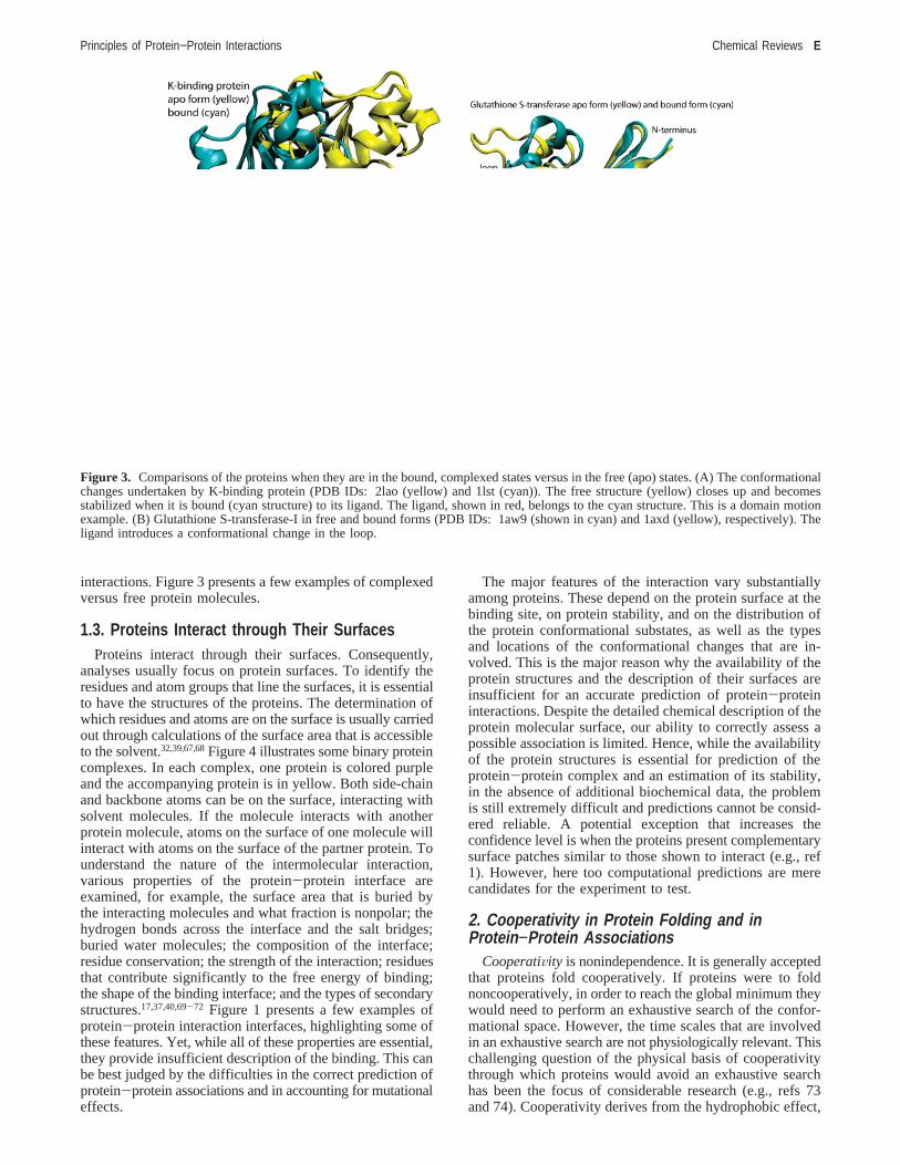
Figure 3. Comparisons of the proteins when they are in the bound, complexed states versus in the free (apo) states. (A) The conformationalchanges undertaken by K-binding protein (PDB IDs: 2lao (yellow) and 1lst (cyan)). The free structure (yellow) closes up and becomesstabilized when it is bound (cyan structure) to its ligand. The ligand, shown in red, belongs to the cyan structure. This is a domain motionexample. (B) Glutathione S-transferase-I in free and bound forms (PDB IDs: 1aw9 (shown in cyan) and 1axd (yellow), respectively). Theligand introduces a conformational change in the loop.
Principles of Protein−Protein Interactions Chemical Reviews E

the driving force in a single-chain protein folding.73 Proteinsthat are approximated by a two-state transition correspondto an all-or-none description of protein folding displayedby cooperatively folding hydrophobic folding units. Such abehavior is typically observed in small globular proteinsconsisting of one hydrophobic unit; on the other hand, largerchains do not fold cooperatively into a single hydrophobicunit. The hydrophobic folding units that are observed at theinterfaces of two-state complexessimilarly suggest thecooperative nature of the two-chain protein folding, also theoutcome of the hydrophobic effect.75 Thus, cooperativityimplies preferred protein folding pathways.
To understand cooperativity, we need to think of thesystem as a cohesive unit, where the parts do not behaveindependently of each other. The behavior of the system isthe outcome of the properties of the system as a whole, ratherthan the sum of the properties of the individual components.In our case, the thermodynamic stability of the protein-protein complex is not a simple summation of the individualcontributions of each of the residues or of the pairs ofresidues; rather, residues that are in direct spatial contact,or in close contact through a few tightly packed intermediateresidues, impact the stability of the association in a nonad-
ditive manner. Substitution of a tightly packed residue wouldinevitably affect the interactions of its neighboring residues.Thus, a mutation affects the stability of the complex sincethe interactions will change; however, at the same time, sincethe residue is tightly packed in the native complex, itssubstitution will also impact the stability of the complexindirectly, through the changes of the interactions of itsneighbors. This may occur if a large residue is substitutedby a smaller residue leading to side-chain (and backbone)movements to fill the “hole” that is created; by contrast, ifa smaller residue is substituted by a larger one, the neighbor-ing residues’ contacts will change to allow accommodationof the inserted residue in the tight environment.76,77 Theextent and direction of the impact depends on the type andenvironment of the substitutions. Either way, this wouldaffect the stability of the complex beyond the direct alteredinteractions of the mutated residue. This implies that, if wesimultaneously mutate two contacting or spatially nearbyresidues in a tightly packed environment, the change in thestability would not be the sum of the measured changes ofeach one separately. The measured change in the thermo-dynamic stability upon a mutation of asingleresidue alreadyimplicitly takes into account changes in the interactions ofits closely packed neighboring residues. Hence, a summationof the substitutions of two residues that are in spatialproximity may overestimate (or underestimate) the totalcontribution. On the other hand, if the protein-proteininterface can be separated into units, the impact of mutationsin each of these is independent and these can be summed.That is, these contributions are noncooperative. Such effectshave been shown in a range of systems.42,78-82 The affinitymaturation process through which proteins evolve to bindwith increased affinity has been shown to be a particularlyuseful system for studies of cooperative effects at the residuelevel.81 Cooperative effects complicate the estimation of thestability of the interactions, since the free energy change upona mutation already implicitly accounts for some of the effectsof the neighboring residues as well, making the accuracy ofthe per residue (or per chemical group) parametrization lessaccurate.
3. Protein −Protein Interfaces Have PreferredOrganization
3.1. Description of Protein −Protein InterfacesAbove, we have discussed attributes that hamper predic-
tions of protein associations. Among these, we highlightedprotein flexibility, the existence of ensembles with distinctconformations separated by barriers, the difficulties encoun-tered by the presence of even partial disorder, and thecooperativity in protein-protein association. Are there anyattributes of protein-protein interactions that may assist inthe prediction? For example, is there a property thatdistinguishes interfaces from the rest of the protein surface?If there were such a property, it could a priori be used towarda prediction, allowing us to focus on the binding sites, thusreducing the conformational search. Toward this aim, variousdata sets of protein-protein interfaces have been derived,divided into groups, and analyzed.83-88 Homodimers, whichare frequentlypermanent complexes, were mostly analyzedseparately from heterodimers. Homodimeric interfaces re-semble protein cores.19,20 They are typically large, arehydrophobic as measured by high values of nonpolar buriedsurface areas, and show good complementarity between the
Figure 4. Several examples of crystal structures of binary proteincomplexes. The interfaces are highlighted with boxes. In part A,the two glutathione S-transferase complexes (PDB IDs: 10gs and1b48) are homologous; they use similar interfaces to bind eachother. In part B, the two complexes, cytochrome C and neuropep-tide/membrane protein (PDB IDs: 1bbh and 1rso) are not relatedevolutionarily, yet the interface architecture is similar. Part Crepresents two complexes (dynein light chain 8 (PDB ID: 1f95AB)and 4-oxalocrotonate tautomerase (PDB ID: 1otfAE)) where onlyone side of the interface has similar architectures, the accompanyingsides are unrelated. The similar side belongs to the magenta chains.
F Chemical Reviews Keskin et al.

two chains. These interfaces can often be distinguished fromthe remainder of the protein surface. In contrast, this is notthe case for heterocomplexes, where the chains differ fromeach other. Yet these largely nonpermanent complexes arethe interfaces we would, in particular, like to be able topredict, since the structures of homodimeric proteins areusually obtained in the complex state. Heterocomplexes can-not be distinguished by the extent of their hydrophobicity.89-93
Jones and Thornton94 have compared the residue typesweighted by their accessible surface areas. They haveobserved that large hydrophobic and uncharged polar residueswere more frequent in the interfaces of heterocomplexes ascompared to the rest of the surface. Charged residues weremore frequent on the exposed, noninterface surface. Theyhave further divided the surface into patches. Analysis ofthese has illustrated that interface patches are more planar,and their residues have larger accessible surface areas. Forsome interfaces, the geometric and electrostatic complemen-tarity is important, and a small fraction of the interfaceresidues may make a large contribution to the bindingenergy.89 Thus, no single physicochemical property distin-guishes sufficiently well interfaces from the remainder ofthe surface; on the other hand, all hydrophobicities, solvationenergies, and relative solvent accessible areas and residuecompositions show trends that differ in the interfaces versusthe rest of the protein surface.52
Residue conservation was also observed to be higher ininterfaces as compared to the rest of the surface.71 Quanti-fication of the conservation through calculation of sequenceentropies complements existing methods.90 It was furtherfound that central interface residues were more conservedthan peripheral ones.89 Li et al.95 examined the hydrophobic-ity in the center of the interface versus its periphery. Tomeasure the hydrophobicity at the center, they replacedburied phenylalanine by smaller hydrophobic residues instructures of antibody-antigen complexes, obtaining anestimated energy change of 46 cal/mol per Å.2 Ofran andRost96 observed that six types of protein-protein interfacesdiffered significantly from each other in their residuecomposition and interaction preferences. Janin and co-workers have suggested dividing the interface into cores andtheir surrounded rims and have used it to differentiatebetween biological interfaces and nonspecific crystal packingones.27,97 Nevertheless, while these trends may assist in theprediction, they too are insufficient.
3.2. Some Amino Acids at the Interface Are HotSpots Since They Contribute Significantly to theStability of the Protein −Protein Association
Are there residues in the interface that contribute domi-nantly to the binding free energy of the protein-proteincomplex or do all residues contribute roughly equally? Infolding, some residues in the protein core have been shownto be important for the stability of the protein. Does the samehold for protein-protein association? To address this ques-tion, Wells and his colleagues have carried out alaninescanning.98 Residues in the interface were systematicallyreplaced by alanine, and the difference in the binding freeenergy (∆∆G) between the wild type and each mutant wasmeasured. They have defined a hot spot as a residue whosesubstitution by alanine leads to a significant (∆∆G g 2 kcal/mol) drop in the binding free energy.76 Clackson et al.99
provided structural data coupled with binding and kineticanalysis of these mutants and proposed that hot spots are
“assembled cooperatively” and that many residues contributeindirectly to binding. They suggested that several hydro-phobic residues serve to orient key tryptophan residues andthat the electrostatic contacts (receptor Arg43 to humangrowth hormone) were less important than the intramolecularpacking of its alkyl chain with Trp169. Sundberg et al.100
have correlated the detailed structural effects of hot spotsubstitution with the energetics of binding.
While identification of hot spots is crucial, exhaustivescreening is still very expensive. Thus, to date, only a limitednumber of interfaces have been screened for residue hotspots. Thorn and Bogan101 compiled experimentally assessedhot spots from the literature. This compilation facilitated thedevelopment of computational strategies to screen protein-protein interfaces with the goal of identifying the hotspots.102,103 Since structure conservation is expected topositively correlate with the stability constraints acting on aposition in a protein, hot spots are expected to correlate withstructurally conserved residues. Consistently,104,105it has beenshown that the alanine scanning mutagenesis data assembledby Bogan and Thorn101,106 correlate well with residueconservation. Thus, “computational hot spots” correlate withexperimental ones, suggesting that hot spots may be identi-fied based on their structural conservation and sequenceidentity. Residue conservation, particularly if it is a me-thionine or a tryptophan, suggests that it is likely to be a hotspot.
Bogan and Thorn postulated that it is the burial of a hotspot in a hydrophobic environment that leads to its majorstabilizing contribution.106Further investigation has illustratedthat packing along the interface is not homogeneous and thatthe hot spots are located within the densely packed areas.32
This explains why these residues contribute dominantly tothe stability of the complex and why they are conserved. Areplacement of a residue under such circumstances isdifficult: substitution by a smaller residue would create holes,while substitution by a larger residue would lead to stericclashes. It is striking that, in the complexes where bothprotein partners were alanine-scanned, the∆∆G of a hot spotcorrelates remarkably well with the local packing density.29
Analysis of structurally conserved and experimental hotspot residues illustrates that they tend to be coupled acrossthe interface more than expected by random distribution.Charge-charge couples are disfavored, and the total numberof hydrogen bonds and salt bridges contributed by hot spotsis as expected. At first glance this appears surprising, sinceelectrostatic interactions and hydrogen bonds are well-knownto be crucial to the stability of protein-protein complexes.Further, the high success rate of the simple physical modelsin the prediction of the hot spots binding energy contributionclearly illustrates the important role of electrostatic interac-tions and hydrogen bonds in the hot spots contributions. Thissuggests that the charged/polar residues may act through awater-exclusion mechanism. Since the hot spots are locatedwithin highly packed regions, water molecules are easilyremoved upon binding, leading to strengthened electrostaticcontributions of charge-charge interactions. This explanationis consistent with the insightful Bogan and Thorn106 proposi-tion of a hydrophobic “O-ring” around the hot spots.
Thus, to conclude, as we noted above, estimation of thestability of a candidate complex by computationally scanningits interface with the goal of quantifying the association maybe inaccurate, given potential hot spot cooperativity.32,33,42
Summation of∆∆G for hot spots may overestimate the
Principles of Protein−Protein Interactions Chemical Reviews G

binding free energy. Nevertheless, since hot spots cor-relate with residue conservation and they tend to be coupledacross the two sides of the interface, these measures canassist in the prediction. Furthermore, their properties, asdescribed in the next two sections below, makethem potentially useful attributes in the prediction, althoughto date these properties have not been used in predictionstrategies.
3.3. Protein Binding Sites Can Be Described asConsisting of a Combination of Self-ContainedModules, or Hot Regions
Hot spots tend to occur in clusters. Within the cluster,the tightly packed hot spots are in contact with eachother and form a network of interactions (Figure 5) constitut-ing hot regions.32,33 This organization implies that,within a cluster, the contributions of the hot spots to thestability of the complex are cooperative; however, thecontributions of independent clusters are additive. Such aconclusion is further supported by the double mutantcycle analysis.42,107 For the barnase-barstar interface,it was observed that the coupling energy between tworesidues decreases with the distance between them.Residues within a distance of 10 Å are defined asmodules. Residues located within a module may be coopera-tive, while residues located in different modules are addi-tive.41,42
At greater distances, the effects of mutations are additive,and the energetics of the interactions are independent of eachother. This organization reenforces our conclusion above:the binding free energy is not a simple summation of thesingle hot spot residue contributions; however, that is thecase for hot spots within the samehot region.
Protein binding sites have been described either in termsof the residues that take part in the interaction with the
binding partner or in terms of the binding area patch. Herewe describe protein binding sites as a combination of “hotregions”. This description is not merely semantic; rather, itrepresents a new view of macromolecular binding. A“classical” description that employs single amino acids thatinteract across the interface implies that the contributions ofsingle residues to the stability of the protein-proteinassociation are additive. At the other extreme, a “patch”definition usually refers to the area over which the intermo-lecular interactions extend. In contrast to both views, we viewthe binding interface as consisting of independent regions.Each region is tightly packed. The amino acids that contributedominantly to the stability are clustered within these regions.Their tightly packed environment rationalizes their highcontributions and the observation that they are stronglyconserved by evolution. The clustered hot spot residues forma network of conserved interactions. The implications of sucha description are that, within a hot region, the contributionsof the hot spot residues to the stability of the complex arecooperatiVe. On the other hand, since the regions areindependent of each other, the contributions of the hot regionsareadditiVe.
Such a description suggests that, in between the tightlypacked hot regions, packing is not optimal, allowing binding-site flexibility. One clear advantage of such a model is thatit highlights the similarity between protein folding andprotein binding. The cooperative contributions of conservedresidues in the tightly packed protein cores have long beenknown to be a hallmark of protein folding. Thus, here weargue that protein-protein interactions might be understoodin terms of hot-region organization. We stress that a hotregion includes residues from both chains, which form anetwork of interactions (Figure 5).
Figure 5. Crystal structure of a complex displaying the hot regions between two M chains of the human muscle L-lactate dehydrogenase(PDB ID: 1i10). Two interacting chains are shown in yellow and cyan. The hot spot residues (red) are shown in ball and stick representation.There are two hot regions in this interface of the homodimer. The figure illustrates that hot spots are in contact with each other and forma network of interactions forminghot regions. The bottom hot region is composed of residues Ser183, Val205, Val179, Gly178 from ChainC and Val269 and Ile293 from Chain A of the complex.
H Chemical Reviews Keskin et al.

3.4. Hot Spots Tend to Occur in Preorganized(Complemented) Pockets That Disappear UponBinding
The protein surface is not flat. It is studded with pockets,crevices, and indentations.68 In the unbound state, dependingon their sizes and shapes, these imperfections of the proteinsurface may be occupied by water.108 In the bound state, thewater may or may not be replaced by the partner proteinmolecule. Unfilled pockets are those that remain unfilled bythe protein partner. Complemented pockets are pockets thatdisappear upon binding, representing tightly fit regions.39 Thequestion arises as to whether there is a preference for thehot spot residues to occur in a specific geometry. Since thehot spots are tightly packed, they are strongly favored to belocated in complemented pockets and are disfavored inunfilled pockets. Interestingly, however, complementedpockets often pre-exist binding. In 16 of 18 protein-proteincomplexes with complemented pockets whose unboundstructures were available, the pockets were identified to pre-exist in the unbound structures.39 Figure 6 presents such anexample. The root-mean-squared deviations of the atomslining the pockets between the bound and unbound stateswere observed to be as small as 0.9 Å, suggesting that suchpockets constitute features of the populated native state. Thus,these pockets are usually alreadypreorganized in theunbound state, prior to the protein complexation. The findingthat key residues have preferred states is in agreement withthe observations of Rajamani et al.109 that some key residuesact as “ready-made” recognition motifs by acquiring native-like conformation prior to binding. The conferred rigidityin the unbound state minimizes the entropic cost on binding,whereas the surrounding residues form a flexible cushion.The studies of Smith et al.110 further reinforce these conclu-sions: the fluctuations that they observed in a set of 41proteins that form binary complexes took parts of themolecules into regions of conformational space close to thebound state; however, at no point in their simulations doeseach protein as whole sample the complete bound state. Asin Rajamani et al., in simulations in the absence of thebinding partner, the core interface residues presented atendency to be less mobile (either measured by the size ofthe fluctuation or by its entropy) than the rest of the surface,while the peripheral interface residues were more mobile.This result, obtained across 40 of the 41 proteins, suggestsdifferent roles for these regions in protein recognition andbinding. In a recent study, we compared the mobility ofconserved and nonconserved residues in 17 protein-proteininterfaces by performing molecular dynamics simulations.111
Figure 7 presents the results from our simulations illustratingthis interesting hallmark of protein-protein interactions. Theresults further suggest that docking algorithms may treat theseregions differently in the docking process and substantiatethe feasibility of targeting hot spots in drug design.112
3.5. There Are Favorable Organizations inProtein −Protein Interactions
The molecular architecture of protein-protein bindingsites, which can be defined as the secondary structuralorganization, have been reviewed recently.37 While theinterfaces are heterogeneous in terms of size, shape, andchemical composition, amino acid sequence order-indepen-dent structural alignment procedures are able to cluster thelarge set of interfaces (>20 000) from different protein
Figure 6. Illustration of pockets in protein interfaces. (A) Theupper panel shows the Cyclin A protein in bound (left) andfree (right) forms. For clarity, the residues except the pro-truding ones from the accompanying protein (cyclin depen-dent kinase) are not shown in the complexed form. (PDBIDs of the complex and monomer are 1fin and 1vin, respectively).The bottom figure shows the details of the pocket. The red resi-dues (belonging to CDK) protrude into the pocket. The samepocket exists in the free form as shown in the boxed rectan-gular region of the apo form. (B) The top figure shows tri-osephosphate isomerase in complex form (PDB ID: 1b9b).Red, blue, and green residues are the protruding residues belong-ing to the left (pink) protein. The bottom figure displaysthe pocket and the protruding residues in detail.
Principles of Protein−Protein Interactions Chemical Reviews I

families into a small set of groups (∼3 500 clusters),113 withsimilar architectures. Studies of these clusters have shownthat interfaces sharing similar scaffolds may derive fromglobally different structures and belong to functionallydifferent protein families.114 This, however, is not surprising,as it is well-known that proteins with similar structures canhave different functions.115 Different structures whose as-sociations lead to similar interface architectural motifs areparticularly interesting: these similar-interfaces, dissimilar-protein folds fall into different families (according to theSCOP classification).116 In Figure 8a, the interfaces and theglobal protein architectures are similar; in Figure 8b, the3-dimensional structures of the monomers are different, yettheir interfaces have similar architectures. A real case is givenin Figure 4B. Two complexes, cytochrome C and neuropep-tide/membrane protein, are not related evolutionarily, yettheir interface architectures are similar. Thus, as in monomerstructures, evolution has reutilized “good” favorable motifs,leading to preferred architectures. These interface motifsresemble those of protein chains. Despite the absence ofchain connections, global features of the architectural motifsthat are present in monomers recur in the interfaces, reflectingthe limited set of the folding patterns. However, the detailsof the architectural motifs may vary. In particular, the extentof the similarity correlates with the consideration of howthe interface has been formed: whether the proteins cofold(two-state folders) or fold separately (three-state folders).20
Architectures of interfaces derived from two-state complexes,i.e., where the chains fold cooperatively, are similar to thosein protein cores, as judged by the quality of their geometricsuperposition. On the other hand, three-state interfaces,representing binding of already folded molecules, manifesta larger variability and resemble the monomer architectureonly in general outline.20,75 The origin of the differencebetween the monomers and the three-state interfaces can beunderstood in terms of the different nature of the foldingand the binding that are involved. Whereas in the former alldegrees of freedom are available to the backbone to
maximize favorable interactions, in rigid body three-statebinding, only six degrees of freedom are allowed.20 Examplesinclude four-helix bundles, extensions ofâ-sheets across theinterface, two-helices packed against each other,â-sand-wiches, etc.13,113,114
Thus, like protein folds, protein-protein interfaces havepreferred architectures. Since the number of secondarystructure organizations is limited because of the restrictedfreedom upon secondary structure formation,117 these pre-organized secondary structure motifs may be important inlimiting the conformational space, key to protein association.On the practical side, similar to schemes for predictions ofprotein structures by threading through available folds, alibrary of protein-protein interaction architectures mayprovide patterns for modeling protein-protein associations,assisting in docking predictions. However, a large portionof protein-protein interfaces are formed by disordered loopspresenting a difficulty in such modeling strategies.
4. Different Protein Partners May Share SimilarBinding Sites
Preferred organization is further observed in the reutiliza-tion of given binding sites by different partners.13 The recentincrease in the number of protein structures, the additionalexperimental results of protein-protein interactions, and theconstruction of maps of protein interactions for someorganisms all consistently indicate that some proteins arecentrally connected, whereas others are at the edges of themap. The centrally connected hub proteins may interact witha large number of proteins.118,119Genomic maps indicate thatsome proteins have as many as tens of connections. Whilethis may be an overestimate, nonetheless it does suggestmultiple interactions, beyond the possibility of the surfaceproviding as many separate, isolated sites. Thus, whereassome binding sites are distinct, it may be expected that others
Figure 7. Flexibility of conserved and nonconserved residues inthe interfaces. Each point represents a different complex. Seventeencomplexes are shown (the first eight and last five points are forhomodimers and enzyme-inhibitor complexes, respectively; themiddle points correspond to antibodies). The flexibility of residuesover 5 ns molecular dynamics simulations of the complexes111 arecompared to determine the difference in the dynamic behavior ofconserved and nonconserved interface residues. First, the averagermsd of each residue in the interface is calculated over the entiresimulation time. Before calculating the residue side-chain rmsdvalues, all heavy backbone atoms (N, CR, C, O) of the interfaceresidues are aligned with the initial structure at the beginning ofthe simulations to avoid systematic errors caused by translationalmotions. Side-chain rmsd values are obtained by comparing eachframe during the simulations with the structure at the beginning ofthe simulations after the equilibration step. The red and blue linesrepresent the flexibility of conserved and nonconserved residues,respectively. RMSD units in Angstroms.
Figure 8. Schematic representation of the interfaces and the globalarchitectures of protein complexes. Part (A) shows cases whereboth the interfaces and the global architectures are similar; ProteinA is homologous to Protein A′ and B is homologous to B′. In Part(B), the three-dimensional structures of the monomers are different,yet their interfaces have similar architectures. Proteins A and Care non-homologous, as are proteins B and D.
J Chemical Reviews Keskin et al.

may bind different molecules at the same location. Thissuggests that there are binding sites that are multiplyreutilized, albeit with different affinities. Furthermore, for afew cases, there are documented examples with crystalstructures, like the Elongin B/Elongin C/VHL and ElonginB/Elongin C/SOCS2.120,121Beckett has recently highlighted“functional switches” in transcriptional regulation,122 focusingon the ability of proteins to bind alternative proteins at thesame binding site. Figure 9 presents one such example. Atable illustrating similar binding sites among proteins withglobally different structures and with different functions wasprovided in our previous study.13 To create this table, wehave used the data set of structurally and sequentiallynonredundant protein-protein interfaces.113 The clusteredbinding sites in this table provide a set of structurally similarsites that bind different partners. For theâ-catenin, Beck-ett122,123observed that similar interactions are responsible forbinding to the different partners. This is expected, since thehot spots are those residues conserved in the protein families.Our analysis of the data set validates this observation. Figure10 highlights conserved interactions of a given site wheninteracting with multiple partners. This observation suggeststhat these optimized local interactions involve the preorga-nized conserved hot spots. On the other hand, their actualcontributions are likely to be functionally modulated. Thus,
while the patterns of the local interactions are similar inmultipartners and in single partners, the multipartners havebeen optimized by evolution to accommodate different ligandshapes, sizes, and composition.
5. Obligatory and Transient Complexes
Protein complexes have been classified into obligatory,or permanent, and transient.17,30,124,125Obligatory protein-protein complexes are formed by proteins that only functionwhen associated in the complex. Homodimers provide a niceexample for obligatory complexes; however, many otherproteins consisting of heteromultimers may also fall into thiscategory. By contrast, formation of transient complexesdepends on the functional state of the partners. Examplesinclude enzyme-inhibitor, hormone-receptor, and signal-ing-effector types of interactions. In recent years, consider-able attention has focused on the distinction between the twotypes of complexes.42 The relative contributions of thephysical interactions differ between the two. Obligatoryassociations are in general tighter, with a stronger hydro-phobic effect, better packing, and fewer structural watermolecules trapped between the monomers, and they manifestbetter shape complementarity. In contrast, the interfaces inthe transient complexes are generally less extensive and more
Figure 9. Example of multiple proteins binding at the same site on the protein surface, dimerization cofactor of hepatocyte nuclear factor(DCoH). DCoH serves as an enzyme and a transcription coactivator. The left figure is the crystal structure of hepatocyte nuclear factordimerization domain, HNF-1R, bound to a DCoH dimer (PDB ID: 1F93, Chains A, B of DCoH, and Chains E, F of HNF-1R). In orderto act as a coactivator, DCoH binds to HNF 1R. The figure on the right displays the enzymatic form of the protein DCoH forming dimersof dimers (shown Chains A, B, C, and D, PDB ID: 1DCH).
Figure 10. Shared binding sites. The figure highlights the conserved interactions of a given site when interacting with multiple partners.The yellow protein is the antibody interacting with a peptide and protein G (PDB IDs: 1dn2 and 1fcc). The residues shown in red belongto the antibody and they are utilized to form H-bonds with both partners.
Principles of Protein−Protein Interactions Chemical Reviews K

polar/charged, and the surfaces of the interacting proteins attheir interface are not as optimized, leading to weakerassociations with the exception of some enzyme-inhibitorcomplexes.30,124,125Quantifying these differences is importantsince many predictive protein-protein schemes use knowl-edge-based scoring parameters derived from the combineddata set of complexes.
Interestingly, analysis of the interfaces of both types ofcomplexes illustrates that residues in the interfaces of obligatecomplexes tend to evolve at a relatively slower rate, whichallows the protein-partners to coevolve. By contrast, the lesstight transient partners illustrate increased rate of mutationsat the interface and no evidence of correlated mutations.15
6. Disordered Proteins: A Major Component ofProtein −Protein Interactions
While the presence of “disordered” proteins has beenrecognized for a long time, in recent years they have drawnincreasing attention. Disordered proteins (or “intrinsicallyunstructured” proteins) lack a stable, well-defined structureunder physiological conditions, existing in a continuum ofconformations from the less to the more structured states.47-50
Natively unstructured proteins undergoing a disorder-to-ordertransition upon binding their partner, and stable monomericproteins, which exist as multimers in their crystal form butnot in solution, provide examples of two vastly differentscenarios. There are two major reasons for the recentheightened interest in the disordered protein state: First, alarge number of proteins have now been identified to belongto this category, with a diverse functional spectrum. Second,the disordered state is analogous to the denatured state.Comprehension of the protein-folding reaction necessitatesknowledge of the ensembles of the folded and the denaturedstates under different conditions. The lack of understandingof the denatured state impedes understanding of the foldingprocess.
Natively unstructured proteins have a broad range offunctions,44-46,51 including regulation of transcription andtranslation, cellular signaling, phosphorylation, regulation oflarge multimolecular self-assemblies, and small moleculestorage.45 Analysis of the structural characteristics of com-plexes of natively unstructured proteins, ribosomal proteins,two-state and three-state complexes, and crystal-packingdimers has suggested that ordered monomers can be distin-guished from disordered monomers on the basis of the per-residue surface and interface areas, which are significantlysmaller for ordered proteins.126 With this scale, two-statedimers (where the monomers unfold upon dimer separation)and ribosomal proteins resemble disordered proteins. On theother hand, crystal-packing dimers, whose monomers arestable in solution, fall into the ordered protein category.While there is a continuum in the distributions, nevertheless,the per-residue scale measures the confidence in the deter-mination of whether a protein can exist as a stable monomer.Disordered proteins lack a strong hydrophobic core and arecomposed of highly polar surface area.
Molecules or regions displaying disorder have beenconsidered inherently unstructured. Yet prevailing conforma-tions still exist, with population times higher than those ofother conformations.47-49 Disordered molecules are theoutcome of rugged energy landscapes away from the nativestate. Ruggedness has a biological function, creating adistribution of conformers that bind via conformationalselection, driving association, and multimolecular complex
formation. A rugged energy landscape modulates the life-times of different conformers, depending on the biologicalfunction.
Disordered functional proteins provide evidence that thefunction of a protein and its properties are not only decidedby its static folded three-dimensional structure; they aredetermined by the distribution and redistribution of theconformational substates. Enumeration127 of all stericallyallowed conformations for short polyalanine chains consis-tently shows that, in the denatured state, not all conformationsare accessible. Even for alanines, local steric effects beyondnearest neighbors already restrict significantly the confor-mational space. For variable-sequence chains with bulkierside-chains, this effect is likely to be enhanced, biasing thelocal conformations.127-131 Preferred conformation impliesthat there is no need to search for the favored binding partnerover broad space in time-scales not biologically relevant.Hence, the fact that binding is fast implies selection: theconformation is already there. With its binding, the equilib-rium shifts in its favor, further driving the reaction. Asbinding and folding are similar processes with similarunderlying principles, this principle applies to disorderedmolecules in binding and to unstable, conformationallyfluctuating building blocks in folding. Folding and bindingimply selection, rationalizing rugged energy landscapes awayfrom the native conformations. However, local conforma-tional diversity can be expected, allowing latitude in theassociations, depending on the binding partner.
7. Systems Biology and the Chemistry ofProtein −Protein Interactions
Proteins function in cellular processes. Unfortunately, forthe vast majority of the proteins participating in these, thereare no structural data; only databases citing experiments thatinfer which proteins interact and sequence information allowfor prediction of protein-protein interactions based onvarious schemes, such as coevolution,132 orthologous rela-tionship,133 or, for example, based on domain combina-tions,134 to name a few. In the absence of structures, it is notpossible to address the chemistry of the interactions. Nev-ertheless, by crossing the structural data available in thePDB135 with the connectivity data for the yeast map,136-139
we have obtained a data set of proteins that have completestructures and interactivity data. The problem is, however,that, even for these, the interfaces are largely unknown. Evenif interfaces are available, they are not necessarily thosewhich play transient regulatory roles in the network; rather,they may belong to the protein multimeric permanentinteractions. Bearing these caveats in mind, it is neverthelessinteresting to look into the structural/chemical properties ofthe central versus the edge proteins. By definition, a centralprotein has a large number of interacting partners, whereasa loner has one or very few. Developed organisms typicallyhave a more centralized network topology. Topologiesconsisting of highly connected proteins are functionallyadvantageous, leading to higher efficiency and inherentlysuperior regulation. In this respect, it is interesting to notethat the human genome has a fewer number of genes ascompared to some lower organisms, implying that ourgenome is more flexible and functionally more complex. Itis now clear that one gene can specify more than one protein,with gene expression regulated by different factors at thedifferent levels of control. A more highly connected mapimplies more proteins binding at a shared site. This leads to
L Chemical Reviews Keskin et al.
the question of whether there are any structural features thatcharacterize such proteins and binding sites, making themincreasingly central in the network as compared to highlyspecific ones.
7.1. Are There Any Structural Features ThatDistinguish Highly Interactive Proteins fromLoners?
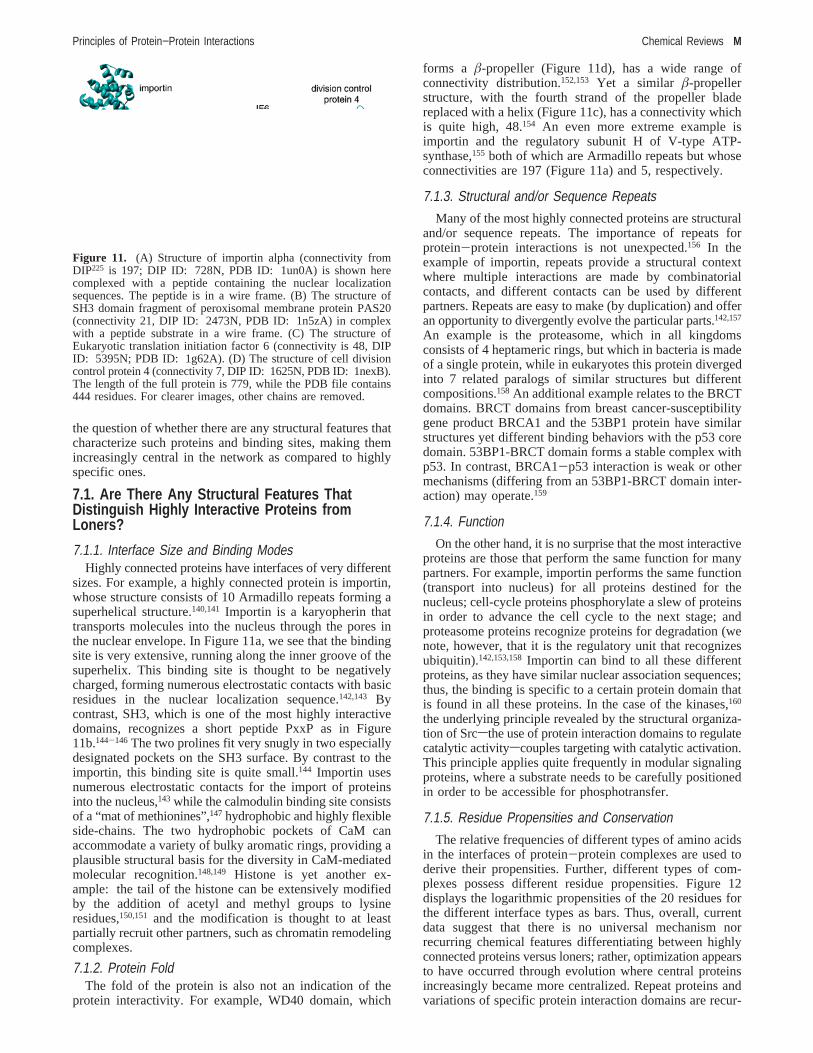
7.1.1. Interface Size and Binding ModesHighly connected proteins have interfaces of very different
sizes. For example, a highly connected protein is importin,whose structure consists of 10 Armadillo repeats forming asuperhelical structure.140,141 Importin is a karyopherin thattransports molecules into the nucleus through the pores inthe nuclear envelope. In Figure 11a, we see that the bindingsite is very extensive, running along the inner groove of thesuperhelix. This binding site is thought to be negativelycharged, forming numerous electrostatic contacts with basicresidues in the nuclear localization sequence.142,143 Bycontrast, SH3, which is one of the most highly interactivedomains, recognizes a short peptide PxxP as in Figure11b.144-146 The two prolines fit very snugly in two especiallydesignated pockets on the SH3 surface. By contrast to theimportin, this binding site is quite small.144 Importin usesnumerous electrostatic contacts for the import of proteinsinto the nucleus,143 while the calmodulin binding site consistsof a “mat of methionines”,147 hydrophobic and highly flexibleside-chains. The two hydrophobic pockets of CaM canaccommodate a variety of bulky aromatic rings, providing aplausible structural basis for the diversity in CaM-mediatedmolecular recognition.148,149 Histone is yet another ex-ample: the tail of the histone can be extensively modifiedby the addition of acetyl and methyl groups to lysineresidues,150,151 and the modification is thought to at leastpartially recruit other partners, such as chromatin remodelingcomplexes.
7.1.2. Protein FoldThe fold of the protein is also not an indication of the
protein interactivity. For example, WD40 domain, which
forms a â-propeller (Figure 11d), has a wide range ofconnectivity distribution.152,153 Yet a similar â-propellerstructure, with the fourth strand of the propeller bladereplaced with a helix (Figure 11c), has a connectivity whichis quite high, 48.154 An even more extreme example isimportin and the regulatory subunit H of V-type ATP-synthase,155 both of which are Armadillo repeats but whoseconnectivities are 197 (Figure 11a) and 5, respectively.
7.1.3. Structural and/or Sequence Repeats
Many of the most highly connected proteins are structuraland/or sequence repeats. The importance of repeats forprotein-protein interactions is not unexpected.156 In theexample of importin, repeats provide a structural contextwhere multiple interactions are made by combinatorialcontacts, and different contacts can be used by differentpartners. Repeats are easy to make (by duplication) and offeran opportunity to divergently evolve the particular parts.142,157
An example is the proteasome, which in all kingdomsconsists of 4 heptameric rings, but which in bacteria is madeof a single protein, while in eukaryotes this protein divergedinto 7 related paralogs of similar structures but differentcompositions.158 An additional example relates to the BRCTdomains. BRCT domains from breast cancer-susceptibilitygene product BRCA1 and the 53BP1 protein have similarstructures yet different binding behaviors with the p53 coredomain. 53BP1-BRCT domain forms a stable complex withp53. In contrast, BRCA1-p53 interaction is weak or othermechanisms (differing from an 53BP1-BRCT domain inter-action) may operate.159
7.1.4. Function
On the other hand, it is no surprise that the most interactiveproteins are those that perform the same function for manypartners. For example, importin performs the same function(transport into nucleus) for all proteins destined for thenucleus; cell-cycle proteins phosphorylate a slew of proteinsin order to advance the cell cycle to the next stage; andproteasome proteins recognize proteins for degradation (wenote, however, that it is the regulatory unit that recognizesubiquitin).142,153,158Importin can bind to all these differentproteins, as they have similar nuclear association sequences;thus, the binding is specific to a certain protein domain thatis found in all these proteins. In the case of the kinases,160
the underlying principle revealed by the structural organiza-tion of Srcsthe use of protein interaction domains to regulatecatalytic activityscouples targeting with catalytic activation.This principle applies quite frequently in modular signalingproteins, where a substrate needs to be carefully positionedin order to be accessible for phosphotransfer.
7.1.5. Residue Propensities and Conservation
The relative frequencies of different types of amino acidsin the interfaces of protein-protein complexes are used toderive their propensities. Further, different types of com-plexes possess different residue propensities. Figure 12displays the logarithmic propensities of the 20 residues forthe different interface types as bars. Thus, overall, currentdata suggest that there is no universal mechanism norrecurring chemical features differentiating between highlyconnected proteins versus loners; rather, optimization appearsto have occurred through evolution where central proteinsincreasingly became more centralized. Repeat proteins andvariations of specific protein interaction domains are recur-
Figure 11. (A) Structure of importin alpha (connectivity fromDIP225 is 197; DIP ID: 728N, PDB ID: 1un0A) is shown herecomplexed with a peptide containing the nuclear localizationsequences. The peptide is in a wire frame. (B) The structure ofSH3 domain fragment of peroxisomal membrane protein PAS20(connectivity 21, DIP ID: 2473N, PDB ID: 1n5zA) in complexwith a peptide substrate in a wire frame. (C) The structure ofEukaryotic translation initiation factor 6 (connectivity is 48, DIPID: 5395N; PDB ID: 1g62A). (D) The structure of cell divisioncontrol protein 4 (connectivity 7, DIP ID: 1625N, PDB ID: 1nexB).The length of the full protein is 779, while the PDB file contains444 residues. For clearer images, other chains are removed.
Principles of Protein−Protein Interactions Chemical Reviews M

ring themes. Nevertheless, as described in the next section,by studying a data set of shared binding sites and itscomparison with specific interacting pairs, some trends areobserved.
7.2. Interfaces of Shared ProteinsFor a protein to be a hub, it must be involved in more
than a single complex;136,161,162therefore, hub proteins areshared proteins that can act as linkers of cellular processes,joining complexes into higher order networks. Dandekar andco-workers162 investigated the properties of shared proteincomponents in six sets of protein complexes. They concludedthat many of the shared proteins appear to be primarilyregulatory links in cellular processes acting as peripheralcomponents of protein interaction networks.162
Different properties of intermolecular interfaces can havea strong effect in modulating binding affinity and specificityof molecular recognition. Comparison of the flexibilities ofhomologous proteins across species suggested that, as thespecies gets more complex, its proteins become moreflexible.163 Ekman et al.164 observed that multiple and repeatdomains are enriched in hub proteins. At the same time, thereis evidence that proteins whose function requires a numberof specific interactions evolve slowly.15,165-168 Thus, bindingregions with high specificity evolve more slowly than thosewith lower specificity; this in turn may suggest that additionalcentral links evolve faster than the unique links of loners.
Understanding how a given site binds to different bindingsites may shed light on identifying the mechanism of protein
interactions. To look into this question, we have assembleda data set of protein-protein interfaces from the PDB.87 Weclustered interfaces where one side of the interface is similarbut the second, complementary, side is different.13 Suchsimilar interfaces interacting with different binding sites canbe defined as shared binding sites. Inspection of theconnectivity of these clusters confirms that the proteins withshared binding sites have higher numbers of interactions withother proteins (∼13)13 compared with the average connectiv-ity number in yeast interactome (∼5).169We find that proteinswith common binding-site motifs preferentially use conservedinteractions at similar interface locations, despite the differentpartners. Our analysis of multipartner interfaces furtherindicates that proteins that use common interface motifs tobind to other proteins have smaller interfaces than complexeswith specific partners. The average accessible surface area(ASA) of multiprotein interfaces is 1235 Å,2 compared tothe 1967 Å2 ASA of the other types. It appears that, with alarge interface, it would be more difficult to bind to differentcomplementary sites. Multipartner interfaces are not as wellpacked and organized as other proteins. The geometricalmatching is not as optimized, and there are water molecules,allowing variability in the interactions. In particular, weobserve that multipartner interfaces preferentially consist ofR helices. Helices appear as the major vehicle through whichsimilar binding sites are able to bind different partners.Helices at multipartner binding sites allow alternate variableways to achieve favorable binding, depending on the side-chain identities. They allow more dynamics in the optimiza-tion of the helical associations as compared to extension ofâ-sheets. It will be of interest to examine whether centrallylocated proteins with multiple proteins binding at the samesites are enriched inR-helical folds as compared to the edgeproteins. Figure 13 displays two proteins: one edge and onehub protein. The left figure is a receptor activator of nuclearfactor kappa B-ligand (PDB ID: 1iqa). This protein has 3connections according to MINT database. The right figureis an aequorin (PDB ID: 1ej3) with 57 interactions with otherproteins.
Figure 12. Logarithmic propensities of the contacting residues inthe different interface types. A positive value indicates a favorablepropensity in the interfaces as compared to the rest of the protein,whereas a negative propensity indicates that it is less likely to findthe particular residue in the interfaces compared to the rest of theprotein. Here, Types 1, 2, and 3 refer to different types of complexesaccording to our definition.13,32,113,114In order to separate interfacesinto different types, we used the data set of structurally andsequentially nonredundant protein-protein interfaces.113 The dataset was created by extracting all existing interfaces between twoprotein chains obtained from higher complexes of proteins. Theseinterfaces are compared structurally with a sequence- and order-independent algorithm. Interfaces sharing similar architectures areclustered. We divided the 103 clusters into 3 types: In Type 1clusters, the global folds of the parent chains are similar and thefunctions of the members of the cluster are also similar (Figure4A). In Type 2 clusters, members often do not share similarfunctions and do not have globally similar structures (Figure 4B).Members of Type 3 clusters only have one side of their chainsaligned. Thus, members of a Type 3 cluster have similar bindingsites on one side of the interface, but the partner proteins aredifferent (Figure 4C). Here, all member interfaces have dissimilarfunctions. The listings of the three types were given previ-ously.13,32,113,114The data was obtained from 358 Type 1, 94 Type2, and 367 Type 3 complexes.
Figure 13. Crystal structures of two proteins: one edge and onehub. The left figure is a receptor activator of nuclear factor kappaB-ligand (PDB ID: 1iqa, Chains A and B). This (edge) proteinhas 3 connections according to MINT database. The right figure isan aequorin (PDB ID: 1ej3, Chains A and B) with 57 interactionswith other proteins.
N Chemical Reviews Keskin et al.
7.3. Chemistry of the Interactions: How AreSubtle Differences Distinguished?
Given the similarities between features of protein-proteininterfaces, the question arises: how does nature neverthelessdistinguish subtle differences, and what happens if nature’schoice fails.159 An insight into these questions should assistin figuring out the principles of protein-protein interactionsand in predicting the preferred ways in which proteinsinteract.
The BRCT domain from the breast cancer-susceptibilitygene product BRCA1 noted above is a good example.BRCA1 relates to 45% of the families with inherited breastcancers and 90% of the families with inherited breast andovarian cancers.170,171 BRCA1 encodes a large protein of1863 amino acids, with a zinc-finger RING domain N-terminal and tandem BRCT (BRCA1 C-terminal) domains.BRCT was first identified in BRCA1 as∼95 amino acidtandem repeats172 and has been found in many proteins, suchas p53-binding protein, 53BP1,173,174 the base excisionresponse scaffold protein, XRCC1, and DNA ligase IV,175
many of which appear to participate in cell-cycle checkpointsor DNA repair in many species.176 BRCA1 stimulates p53transcriptional activity.177-180 It was reported to associate withp53 with two interaction domains: the central disorderedregion of BRCA1 interacting with C-terminal domain ofp53,181 and there are some in vitro studies suggesting thatBRCT domain of BRCA1 binds to the core domain of p53.177
53BP1-p53 interactions were observed directly by X-raycrystallography of the 53BP1-p53 complex. The 53BP1-p53 binding site partially overlaps the p53 DNA-binding site,thus inhibiting the DNA-binding activities of p53.182
Both 53BP1 and human BRCA1 have two BRCT repeats,with high structural similarities, even though the sequenceidentity is only 19%. Each repeat consists of fourâ-strandsand four R-helices, with the exception that one of theR-helices is disordered in the C-terminal repeat of BRCA1.The BRCT region of 53BP1 (taken from the 53BP1-p53complex, PDB ID: 1kzy) superimposed (by Swiss-Pdb-Viewer http://www.expasy.org/spdbv/ on the crystal structureof BRCA1 BRCT, PDB ID: 1jnx), gives a root-mean-squared deviation of 1.44 Å for 133 out of 211 BRCA1 CRatoms, including all eightâ-strands and seven of eightR-helices. The N-terminal repeat (repeat 1) of 53BP1 andBRCA1 gives an rmsd of 1.38 Å (for 69 out of 88 CR atoms),and the C-terminal repeat (repeat 2) has an rmsd of 1.25 Å(for 60 out of 94 CR atoms). The sequence identities ofrepeats 1 and 2 are 24% and 17%, respectively. The leastconserved region is the linker between repeats 1 and 2, witha low 10% identity. Except for the linker, the region involvedin 53BP1 bound to p53, includingR3A throughR4A, has astriking structural conservation with the corresponding regionof BRCA1, with an rmsd of 0.58 Å for all 23 CR atoms.The sequence identity of this region (26%) is also higherthan that in the other regions. Yet despite the structuralconservation, the p53 core domain interacts with the BRCTdomains of 53BP1 and BRCA1 proteins to different extents.Isothermal titration calorimetry, analytical ultracentrifugation,and analytical size-exclusion chromatography confirmed thep53 core domain interactions with the BRCT domain of53BP1 protein but not with the BRCA1 BRCT domain.183
While it is possible that these biophysical methods are notsensitive enough, it does imply that, if there is an interactionbetween BRCA1 BRCT domain and p53 core domain, it isvery weak, or that there is no interaction with this BRCT
domain repeat and the interaction is with the second repeat.Hence, within the global similarity, the complex details ofthe structure and the chemistry lead to such selectivedifferentiation. On the other hand, in silico mutations in thefirst repeat may stabilize the interactions, possibly leadingto a non-native, diseased state.159
8. AllosteryAllostery is a key in regulation; it has a crucial role in
practically all proteins: in hubs and loners. Allostery involvescoupling of conformational and dynamic changes betweentwosnearby or widely separatedsbinding sites. Proteins arenot rigid as it appears when looking at crystal or averagedNMR structures.62,63 Hydrogen/deuterium (H/D) exchangeclearly indicates that native proteins exist as statisticalensembles184-186 distinguished by locally unfolded region-(s) in the binding sites or elsewhere. Elber and Karplus havedemonstrated that the potential energy surface of myoglobinis characterized by a large number of thermally accessibleminima around the native structure.187,188These observationssuggest that the Gibbs energy of stabilization is not equallydistributed in the structure. Since local unfolding occurs inthe functional state, its significance is beyond protein foldingper se. There is experimental and theoretical support thatbinding at one site effectively can shift the population,showing conformational and dynamic changes at some othersites. Structural perturbation at any site leads to a redistribu-tion of the populations. One source of structural perturbationis the binding of inhibitors (or effectors). Other sourcesinclude mutations, binding to sister molecules, binding tonucleic acids or to small molecules, changes in pH, ionicstrength, temperature, and covalent modification such asphosphorylation and acetylation, discussed above. Redis-tributed conformations are not a manifestation unique toallostery. Rather, they are physical attributes of proteins.Allostery derives from populations. Thus, there is no well-defined path, nor a distinct series of steps that moleculesfollow. Rather than every single molecule undergoing a seriesof steps to reach the conformational change observed in thesnapshot of a shape of a site that is far away,what we obserVeis the outcome of the ensemble. The perturbations at one sitedo not yield a homogeneous distribution. Since some portionsof the molecule are less stable than others, these parts willmanifest larger variability. When thought of in these terms,allosteric activation should not produce an alternate rigidbinding site shape fits the ligand (substrate or protein).Rather, the perturbation upon effector binding leads to aredistribution of the ensemble, which would be largelyreflected in binding sites which are a priori less stable.Nevertheless, the “active” conformer is also present in thepresumably “inactive” ensemble, albeit at a lower concentra-tion. Upon binding, there is an equilibrium shift in itsdirection, further driving the binding reaction.
p53 presents a relevant example of protein allostery. Thelast 30 residues of the C-terminal domain were proposed tonegatively regulate DNA binding by an allosteric mechanism(reviewed in ref 189). This hypothesis was based on theobservation that the interaction of p53 with a short oligo-nucleotide containing a consensus p53-binding site is greatlyenhanced either by the deletion of the C-terminal basic region(30 residues) or by binding of the antibody PAb421 to thesame region.190 This was confirmed by a study showing thatp53 transcriptional activity is activated by PAb421 in cells.191
Recent studies have demonstrated that, within the context
Principles of Protein−Protein Interactions Chemical Reviews O

of chromatin or supercoiled DNA, the C-terminal domainmay actually facilitate binding of the core to its target DNAsequence by providing an additional anchorage to specificDNA sites via nonspecific DNA binding.192-194 Cross-talkbetween the different p53 domains has also been indicatedin earlier studies, showing that destabilization of the coreby mutation (R273H) inhibits the transactivation activity ofthe N-terminal domain.195 NMR studies confirm that the N-and C-terminal domains have an impact on the thermody-namic instability of the p53 tetramer.196 However, how thecross-talk between p53 domains occurs is still unclear. Asimilar situation is observed in other tumor suppressors thatserve as hub proteins, such as pVHL or suppressors of thecytokine signaling (SOCS) family,120 which function as keyregulators at all levels of cellular pathways. Binding of thepVHL to the elongin B-elongin C complex leads to aconformational change that allows it to bind to the HIF;197
in contrast, without the pVHL binding to the elonginC/elongin B, the pVHL has not been observed to bind toHIF.
Although allostery plays a role in protein-protein interac-tions in general, it is likely to play a particularly importantrole in central shared proteins. The conformational changemay or may not be large. However, even if small, it maylead to distinct minima at the bottom of the protein foldingfunnel; the low barrier heights may nevertheless be physi-ologically sufficiently high to necessitate a change in theconditions to allow surpassing them. Such a change may bethe binding of another protein, leading to the population shift.Distinct minima with small conformational changes mayexplain the more centralized nature of the cellular networkand how central regulatory proteins are able to bind anastonishingly large number of different partners. In manycases, such as in p53 and pVHL, the allosteric communica-tion is transmitted via cross-talk between domains.
9. Large AssembliesAccurate determination of the structures of protein mol-
ecules and their complexes constitute major challenges inthe biological sciences. Availability of the structures wouldfacilitate drug design, identification of the origin of mis-function, and disease. It would provide crucial assistance tothe prediction of protein function. Yet despite the broadrecognition of the importance of the knowledge of thestructures, experimental structure determination and com-putational prediction still face immense hurdles. Proteinsnever function when they exist isolated in solution. Functiondictates molecular associations. The sizes of the assembliescan be very large. This arrangement effectively increases thelocal concentrations of the reactants/products in enzymepathways, provides effective regulation and control of cellularprocesses, and leads to structural scaffolds and regulatedmolecular machines. Large assemblies are an advantage ofcomplex and robust systems. To understand how they workat the molecular level, it is essential to have the interactionsbetween the components and their spatial organization. Thestructure is also crucial if we are to figure out how themachine performs its work and how the regulation isachieved. The structure is crucial in order to understand theconformational switches and alternate potential bindingmodes.
The main reaction pathways in the living cell are carriedout by functional modules, namely, macromolecular ma-chines with compact structure or the multimolecular en-
sembles that change their composition and spatial organi-zation during function.198 Function relies on spatial sequestra-tion, chemical specificity, and time series of the dynamicensembles. The addition of the individual components resultsin systemic properties that could not be predicted byconsidering the components individually.199
The molecular ensembles constitute compact, specific, andtransient functional modules. Type III secretion systems(TTSSs) constitute one example. TTSSs are multiproteinmacromolecular “machines” that have a central function inthe virulence of many Gram-negative pathogens.200 TheTTSSs directly mediate the secretion and translocation ofbacterial effector proteins into the cytoplasm of eukaryoticcells. The 20 unique structural components constituting thissecretion apparatus are largely conserved among animal andplant pathogens and are evolutionarily related to proteins inthe flagellar-specific export system. Electron microscopyrevealed a “needle-shaped” morphology of the TTSS. The1.8 Å crystal structure of EscJ from enteropathogenicEscherichia coli(EPEC), a member of the YscJ/PrgK familywhose oligomerization represents one of the earliest eventsin TTSS assembly, provided the detailed structural charac-teristics and the organization of these protein components.200
Molecular modeling has indicated that EscJ could form alarge 24-subunit “ring” superstructure with extensive grooves,ridges, and electrostatic features. Electron microscopy,labeling, and mass spectrometry studies on the orthologousSalmonella typhimuriumPrgK within the context of theassembled TTSS support the stoichiometry, membraneassociation, and surface accessibility of the modeled ring.
Another example is the nuclear pore complex.200-202 Thenuclear pore complex (NPC) consists of multiple copies of∼30 different proteins (nucleoporins, nups). They form achannel in the nuclear envelope that mediates macromolecu-lar transport between the cytosol and the nucleus. Only<5%of the nup residues are currently available in experimentallydetermined structures, and consequently, very little is knownabout the detailed structure of the NPC. Nevertheless, acombined computational and biochemical approach was usedto assign folds for∼95% of the residues in the yeast andvertebrate nups. The assigned folds suggest simplicity in thecomposition and modularity in the architecture of all eu-karyotic NPCs, reflected in the presence of only 8-fold types;three of the most frequent folds account for∼85% of theresidues. The modularity in architecture is reflected in thehierarchical and symmetrical organization. These partitionthe predicted nup folds into three groups: the transmembranegroup with transmembrane helices and a cadherin fold; thecentral scaffold group consisting ofâ-propeller andR-sole-noid folds; and the peripheral FG group containing predomi-nantly the FG repeats and the coiled-coil fold. These led tothe suggestion that the small number of fold types in theNPC and their internal symmetries evolved through extensivemotif and gene duplication from a simple precursor set ofonly a few proteins.
10. Crystal InterfacesA considerable effort has been directed at differentiating
between the “real” biological interfaces and interfaces thatare the outcome of crystal effects.30 This is an important issuesince potential functions and other chemical and functionalattributes are derived from the protein-protein complexesin the PDB. Analysis of a manually curated crystal interfacedata set has illustrated that the average area of crystal-packing
P Chemical Reviews Keskin et al.

interfaces is only 570 Å2 per interface. Nonetheless, somecrystals contain pairwise interfaces comparable in size tothose of protein-protein complexes. Large packing interfacesare often associated with twofold symmetry elements forming“crystal dimers” that may be mistaken for real dimers. Toidentify structural features other than size that distinguishbetween the two, Janin et al. selected from a set of crystalsof monomeric proteins 188 packing interfaces with>800Å2 including 105 with twofold symmetry.27 Their resultsshowed that, on average, these large crystal-packing inter-faces are standard size and with similar nonpolar fraction asin complexes. Because homodimers have a larger averagefraction of nonpolar buried surface area, the chemicalcomposition of the interface may distinguish between realand crystal dimers; however, the distributions of individualvalues overlap. The amino acid compositions reflect similartrends; however, these are not sufficiently distinct to removeambiguities between the crystal and the biological inter-faces.27,203,204Our results also show that crystal interfacescan either be unique or share similar patterns with biologicalinterfaces. However, for the majority of the interfaces, thereare no details in the literature to elucidate their real tertiarystructure. Therefore, it is not clear whether some of theinterfaces that share the same chemical and structural featureswith crystal interfaces are indeed “real” interfaces, or perhapsthey, too, are crystal interfaces. A conclusive solution forthis problem has not yet been found.
A relevant example of the complexity and relevance ofthe problem can be gauged from examination of the p53crystal structures.205-207 The first crystal structure obtainedby Pavletich et al.205 has presented a trimer structure for thep53 proteins with interfaces differing from the expectedsymmetric associations of the p53 dimer binding to the DNA.Crystal structures with symmetric associations were onlypublished in 2006,206,207 12 years after the nonsymmetrictrimeric crystal organization. Detailed high-resolution struc-ture of the full native p53 tetramer-DNA has yet to bedetermined experimentally, probably in a supramolecularassociation given its disordered regions.
11. Concluding Remarks: Preferred Organizationin Protein Interactions
Considerable statistics have accumulated over the yearson protein binding sites and protein-protein interactions.Studies have been carried out on data sets of structures,focusing on particular complexes and on their dynamics.Protein-protein interactions have been studied in binaryassociations and within the framework of the cellularnetworks. Much progress has been made in our understandingof the types of associations, permanent and transient; theconformational transitions; and the ordered and disorderedstates.
Currently we know that protein-protein interactions arelargely driven by the hydrophobic effect. Nevertheless,although the hydrophobic effect plays a dominant role inprotein-protein binding, it is not as strong as that observedin the interior of protein monomers, and its extent is variable.The binding site is not necessarily at the largest patch ofhydrophobic surface. At the interface, there are higherproportions of buried charged and polar residues as comparedto protein cores, suggesting that hydrogen bonds and ion pairscontribute more to the stability of protein binding than tothat of protein folding. Residue conservation has also beenobserved to tend to be higher at binding sites as compared
to other protein surface areas. However, such observationsare insufficient to assist in predicting protein-proteininteractions. Protein binding sites have neither the largesttotal buried surface area nor the most extensive nonpolarburied surface area. They cannot be uniquely distinguishedby their electrostatic characteristics, as observed by param-eters such as unsatisfied buried charges, or the number ofhydrogen bonds. Although the geometry of molecularsurfaces has provided clues to binding sites on enzymesurfaces, which are often shaped as the largest or deepestclefts on the surface, none were found for protein-proteinbinding sites. On the other hand, a description of bindingsites in terms of preferred residue, and particularlyregionandarchitectural organizations, may lead to classificationstrategies assisting in predictions of the preferred ways forproteins to interact. Within the recurring favorable architec-tures, there are preferred cooperative hot spot organizations.A combination of all the mentioned features can be used todistinguish the location of interfaces with an average successrate of 75%.40,87,208,209
Preferred organization is a key in chemistry and in proteinscience, whether in amyloid microfilaments or in globularprotein-protein associations. Evolution reutilizes favorablepatterns and modulates these toward different functions. Thewell-recognized fact that protein architectures do not spanthe entire conformational space and certain topologies aredisallowed has led to the imaginative proposition of usingthe limited repertoire in folding strategies.117,210Despite theabsence of the chain connectivity between the interactingpartners, nature appears to similarly follow these preferredorganizations in protein-protein associations. Within thesescaffolds, functional hot spot residues are conserved. Theenergetic contributions of the hot spots derive from their localnetworked organization in tightly packed “hot” regions.Between these, packing is less optimal, allowing flexibilityand binding to multiplesdifferentspartners. A self-containedhot region organization offers many advantages and may alsoplay a role in binding sites to other molecules such as DNA,RNA, and small molecules.
The proposition that an interface can be divided into partsor patches is not new. Jones and Thornton analyzed protein-protein interaction sites using surface patches, defined interms of solvation potential, residue propensity, hydropho-bicity, planarity, protrusion, and accessible surface area.31,94,211
Shanahan and Thornton analyzed the conservation of surfacepatch polarity.72 Surface complementarity in complexes hasbeen estimated using patches. The so-called Ile-44 surfacepatch of ubiquitin binds to the alpha3 helix of the GATdomain, which is responsible for ubiquitin binding andubiquitination.212,213 Surface patches containing basic andaromatic residues were detected in domains of the La proteinthat interacts with RNA. These account for the cooperativebinding of short oligonucleotides. A surface patch with twoexposed tryptophan residues that interface with lipid bilayerswas noted for the GM2-activator protein. Using a patchanalysis, side-chain conformational entropy at protein-protein interfaces has also been performed.214 These consti-tute only a few examples. Thus, while patch definitions vary,it has been recognized that a binding site or an interface canbe divided into parts. Here, however, our definition is interms of continuous paths of interacting residues withindensely packed neighborhoods, leading to cooperative effects.
A recent review215 summarized the challenges in modelingstructures and protein interactions by sequence and structure.
Principles of Protein−Protein Interactions Chemical Reviews Q

Sali and co-workers216 discussed the localization of proteinbinding sites within families of proteins. They observed that72% of the 1847 SCOP domains have binding sites at similarpositions, that is, members of that domain family have theirbinding regions at or around the same positions. Their findingcan assist in describing the functional diversity of protein-protein interactions, as well as introducing spatial constraintsin modeling protein assemblies. Similarly, Aloy et al.217
analyzed the relationship between sequence similarity andbinding orientation and showed that the geometry of theinteractions tends to be conserved between highly similarpairs. On the other hand, Henschel et al.218 investigatedbinding at equivalent sites between nonhomologous proteinswhen interacting with a common partner. They found that,of all nonhomologous domains that bind with a commoninteraction partner, 4.2% use the same interface of the samecommon interaction partner (excluding immunoglobulins andproteases). Aytuna et al.1 employed a bottom-up approach,combining structure and sequence conservation in proteininterfaces to predict protein-protein interactions. Runningthe algorithm on a template data set of 67 known interfacesand a sequentially nonredundant data set of 6170 proteinstructures, they found a number of potential interactions,which they further verified with experimental data.87 Theseindicate that there is progress toward this profound problemof predicting protein-protein interaction.
So,what is the preferred way for proteins to interact? Ina thought-provoking comment already some years ago, vanRegenmortel219 argued that analyzing the interactions be-tween biological molecules cannot be reduced to the descrip-tion of (static) molecular structures. Integrated functionalapproaches need to consider the binding partner and the timecomponent of the interaction. The function of a protein andits properties are decided not only by the static folded three-dimensional structure but also by the distribution andredistributions of the populations of its conformational anddynamic substates under different (physical or binding)environments.65 Such mechanisms provide multiple pathwaysand allow a single molecular surface to interact withnumerous structurally distinct binding partners, accommodat-ing mutations through shifts in the dynamic energy landscape,and, as such, are evolutionarily advantageous. Yet thedistribution of the conformations is not homogeneous, andthe protein topology dictates the more dynamic regions.220-223
Future work along integrative lines will provide insight intothis profound protein-protein interaction problem.
12. Acknowledgments
We thank members of the group for discussions andsuggestions. This project has been funded in whole or inpart with federal funds from the National Cancer Institute,National Institutes of Health, under Contract No. N01-CO-12400 and TUBITAK (Research Grant No. 104T504). O.K.has been granted with Turkish Academy of Sciences YoungInvestigator Programme (TUBA-GEBIP). The content of thispublication does not necessarily reflect the views or policiesof the Department of Health and Human Services, nor doesmention of trade names, commercial products, or organiza-tions imply endorsement by the U.S. Government. Thisresearch was supported (in part) by the Intramural ResearchProgram of the NIH, National Cancer Institute, Center forCancer Research.
13. References(1) (1) Aytuna, A. S.; Gursoy, A.; Keskin, O.Bioinformatics2005, 21,
2850.(2) Malmstrom, L.; Riffle, M.; Strauss, C. E.; Chivian, D.; Davis, T.
N.; Bonneau, R.; Baker, D.PLoS Biol.2007, 5, e76.(3) Nariai, N.; Kolaczyk, E. D.; Kasif, S.PLoS ONE2007, 2, e337.(4) Punta, M.; Forrest, L. R.; Bigelow, H.; Kernytsky, A.; Liu, J.; Rost,
B. Methods2007, 41, 460.(5) Sharan, R.; Ulitsky, I.; Shamir, R.Mol. Syst. Biol.2007, 3, 88.(6) Watson, J. D.; Sanderson, S.; Ezersky, A.; Savchenko, A.; Edwards,
A.; Orengo, C.; Joachimiak, A.; Laskowski, R. A.; Thornton, J. M.J. Mol. Biol. 2007, 367, 1511.
(7) Ewing, R. M.; Chu, P.; Elisma, F.; Li, H.; Taylor, P.; Climie, S.;McBroom-Cerajewski, L.; Robinson, M. D.; O’Connor, L.; Li, M.;Taylor, R.; Dharsee, M.; Ho, Y.; Heilbut, A.; Moore, L.; Zhang, S.;Ornatsky, O.; Bukhman, Y. V.; Ethier, M.; Sheng, Y.; Vasilescu, J.;Abu-Farha, M.; Lambert, J. P.; Duewel, H. S.; Stewart, II; Kuehl,B.; Hogue, K.; Colwill, K.; Gladwish, K.; Muskat, B.; Kinach, R.;Adams, S. L.; Moran, M. F.; Morin, G. B.; Topaloglou, T.; Figeys,D. Mol. Syst. Biol.2007, 3, 89.
(8) Hart, G. T.; Ramani, A. K.; Marcotte, E. M.Genome Biol.2006, 7,120.
(9) Kim, P. M.; Lu, L. J.; Xia, Y.; Gerstein, M. B.Science2006, 314,1938.
(10) Rachlin, J.; Cohen, D. D.; Cantor, C.; Kasif, S.Mol. Syst. Biol.2006,2, 66.
(11) Yip, A. M.; Horvath, S.BMC Bioinformatics2007, 8, 22.(12) Cho, K. I.; Lee, K.; Lee, K. H.; Kim, D.; Lee, D.Proteins2006, 65,
593.(13) Keskin, O.; Nussinov, R.Structure2007, 15, 341.(14) Maerkl, S. J.; Quake, S. R.Science2007, 315, 233.(15) Mintseris, J.; Weng, Z.Proc. Natl. Acad. Sci. U.S.A.2005, 102,
10930.(16) Morell, M.; Espargaro, A.; Aviles, F. X.; Ventura, S.Proteomics
2007, 7, 1023.(17) Nooren, I. M.; Thornton, J. M.J. Mol. Biol. 2003, 325, 991.(18) Sprinzak, E.; Altuvia, Y.; Margalit, H.Proc. Natl. Acad. Sci. U.S.A.
2006, 103, 14718.(19) Tsai, C. J.; Lin, S. L.; Wolfson, H. J.; Nussinov, R.Protein Sci.
1997, 6, 53.(20) Tsai, C. J.; Xu, D.; Nussinov, R.Protein Sci.1997, 6, 1793.(21) Young, L.; Jernigan, R. L.; Covell, D. G.Protein Sci.1994, 3, 717.(22) Norel, R.; Sheinerman, F.; Petrey, D.; Honig, B.Protein Sci.2001,
10, 2147.(23) Sheinerman, F. B.; Honig, B.J. Mol. Biol. 2002, 318, 161.(24) Sheinerman, F. B.; Norel, R.; Honig, B.Curr. Opin. Struct. Biol.
2000, 10, 153.(25) Xu, D.; Lin, S. L.; Nussinov, R.J. Mol. Biol. 1997, 265, 68.(26) Kleanthous, C.Protein-protein recognition; Oxford University
Press: Oxford, New York, 2000.(27) Bahadur, R. P.; Chakrabarti, P.; Rodier, F.; Janin, J.J. Mol. Biol.
2004, 336, 943.(28) Halperin, I.; Ma, B.; Wolfson, H.; Nussinov, R.Proteins2002, 47,
409.(29) Halperin, I.; Wolfson, H.; Nussinov, R.Structure2004, 12, 1027.(30) Janin, J.; Rodier, F.; Chakrabarti, P.; Bahadur, R. P.Acta Crystallogr.,
Sect. D2007, 63, 1.(31) Jones, S.; Thornton, J. M.J. Mol. Biol. 1997, 272, 133.(32) Keskin, O.; Ma, B.; Nussinov, R.J. Mol. Biol. 2005, 345, 1281.(33) Keskin, O.; Ma, B.; Rogale, K.; Gunasekaran, K.; Nussinov, R.Phys.
Biol. 2005, 2, S24.(34) Laskowski, R. A.; Luscombe, N. M.; Swindells, M. B.; Thornton, J.
M. Protein Sci.1996, 5, 2438.(35) Mintz, S.; Shulman-Peleg, A.; Wolfson, H. J.; Nussinov, R.Proteins
2005, 61, 6.(36) Nooren, I. M.; Thornton, J. M.EMBO J.2003, 22, 3486.(37) Reichmann, D.; Rahat, O.; Cohen, M.; Neuvirth, H.; Schreiber, G.
Curr. Opin. Struct. Biol.2007, 17, 67.(38) Haliloglu, T.; Keskin, O.; Ma, B.; Nussinov, R.Biophys. J.2005,
88, 1552.(39) Li, X.; Keskin, O.; Ma, B.; Nussinov, R.; Liang, J.J. Mol. Biol.
2004, 344, 781.(40) Neuvirth, H.; Raz, R.; Schreiber, G.J. Mol. Biol. 2004, 338, 181.(41) Reichmann, D.; Cohen, M.; Abramovich, R.; Dym, O.; Lim, D.;
Strynadka, N. C.; Schreiber, G.J. Mol. Biol. 2007, 365, 663.(42) Reichmann, D.; Rahat, O.; Albeck, S.; Meged, R.; Dym, O.;
Schreiber, G.Proc. Natl. Acad. Sci. U.S.A.2005, 102, 57.(43) Gunasekaran, K.; Ma, B.; Nussinov, R.Proteins2004, 57, 433.(44) Chong, P. A.; Ozdamar, B.; Wrana, J. L.; Forman-Kay, J. D.J. Biol.
Chem.2004, 279, 40707.(45) Dyson, H. J.; Wright, P. E.Nat. ReV. Mol. Cell Biol. 2005, 6, 197.(46) Dyson, H. J.; Wright, P. E.IUBMB Life 2006, 58, 107.
R Chemical Reviews Keskin et al.

(47) Gunasekaran, K.; Tsai, C. J.; Kumar, S.; Zanuy, D.; Nussinov, R.Trends Biochem. Sci.2003, 28, 81.
(48) Kortemme, T.; Kelly, M. J.; Kay, L. E.; Forman-Kay, J.; Serrano,L. J. Mol. Biol. 2000, 297, 1217.
(49) Mittag, T.; Forman-Kay, J. D.Curr. Opin. Struct. Biol.2007, 17, 3.(50) Shortle, D.; Ackerman, M. S.Science2001, 293, 487.(51) Dosztanyi, Z.; Chen, J.; Dunker, A. K.; Simon, I.; Tompa, P.J.
Proteome Res.2006, 5, 2985.(52) Camacho, C. J.; Ma, H.; Champ, P. C.Proteins2006, 63, 868.(53) Camacho, C. J.; Vajda, S.Curr. Opin. Struct. Biol.2002, 12, 36.(54) Joerger, A. C.; Fersht, A. R.Oncogene2007, 26, 2226.(55) Kim, E.; Deppert, W.Oncogene2007, 26, 2185.(56) Inbar, Y.; Benyamini, H.; Nussinov, R.; Wolfson, H. J.Phys. Biol.
2005, 2, S156.(57) Inbar, Y.; Benyamini, H.; Nussinov, R.; Wolfson, H. J.J. Mol. Biol.
2005, 349, 435.(58) Schneidman-Duhovny, D.; Nussinov, R.; Wolfson, H. J.Curr. Med.
Chem.2004, 11, 91.(59) Fuentes, E. J.; Gilmore, S. A.; Mauldin, R. V.; Lee, A. L.J. Mol.
Biol. 2006, 364, 337.(60) James, L. C.; Roversi, P.; Tawfik, D. S.Science2003, 299, 1362.(61) Lindner, A. B.; Eshhar, Z.; Tawfik, D. S.J. Mol. Biol. 1999, 285,
421.(62) Ma, B.; Kumar, S.; Tsai, C. J.; Nussinov, R.Protein Eng.1999, 12,
713.(63) Demchenko, A. P.J. Mol. Recognit.2001, 14, 42.(64) Fetler, L.; Kantrowitz, E. R.; Vachette, P.Proc. Natl. Acad. Sci. U.S.A.
2007, 104, 495.(65) Kumar, S.; Ma, B.; Tsai, C. J.; Sinha, N.; Nussinov, R.Protein Sci.
2000, 9, 10.(66) Popovych, N.; Sun, S.; Ebright, R. H.; Kalodimos, C. G.Nat. Struct.
Mol. Biol. 2006, 13, 831.(67) Binkowski, T. A.; Joachimiak, A.; Liang, J.Protein Sci.2005, 14,
2972.(68) Dundas, J.; Ouyang, Z.; Tseng, J.; Binkowski, A.; Turpaz, Y.; Liang,
J. Nucleic Acids Res.2006, 34, W116.(69) Glaser, F.; Rosenberg, Y.; Kessel, A.; Pupko, T.; Ben-Tal, N.
Proteins.2005, 58, 610.(70) Jones, S.; Marin, A.; Thornton, J. M.Protein Eng.2000, 13, 77.(71) Nimrod, G.; Glaser, F.; Steinberg, D.; Ben-Tal, N.; Pupko, T.
Bioinformatics2005, 21 (Suppl 1), i328.(72) Shanahan, H. P.; Thornton, J. M.Biopolymers2005, 78, 318.(73) Dill, K. A.; Fiebig, K. M.; Chan, H. S.Proc. Natl. Acad. Sci. U.S.A.
1993, 90, 1942.(74) Kolinski, A.; Galazka, W.; Skolnick, J.Proteins1996, 26, 271.(75) Tsai, C. J.; Nussinov, R.Protein Sci.1997, 6, 1426.(76) Clackson, T.; Wells, J. A.Science1995, 267, 383.(77) Karpusas, M.; Baase, W. A.; Matsumura, M.; Matthews, B. W.Proc.
Natl. Acad. Sci. U.S.A.1989, 86, 8237.(78) Albeck, S.; Unger, R.; Schreiber, G.J. Mol. Biol. 2000, 298, 503.(79) Pal, G.; Ultsch, M. H.; Clark, K. P.; Currell, B.; Kossiakoff, A. A.;
Sidhu, S. S.J. Mol. Biol. 2005, 347, 489.(80) Teufel, D. P.; Kao, R. Y.; Acharya, K. R.; Shapiro, R.Biochemistry
2003, 42, 1451.(81) Yang, J.; Swaminathan, C. P.; Huang, Y.; Guan, R.; Cho, S.; Kieke,
M. C.; Kranz, D. M.; Mariuzza, R. A.; Sundberg, E. J.J. Biol. Chem.2003, 278, 50412.
(82) Yang, P. L.; Schultz, P. G.J. Mol. Biol. 1999, 294, 1191.(83) De, S.; Krishnadev, O.; Srinivasan, N.; Rekha, N.BMC Struct. Biol.
2005, 5, 15.(84) Gao, Y.; Wang, R.; Lai, L.J. Mol. Model.2004, 10, 44.(85) Kim, W. K.; Henschel, A.; Winter, C.; Schroeder, M.PLoS Comput.
Biol. 2006, 2, e124.(86) Lu, H.; Lu, L.; Skolnick, J.Biophys. J.2003, 84, 1895.(87) Ogmen, U.; Keskin, O.; Aytuna, A. S.; Nussinov, R.; Gursoy, A.
Nucleic Acids Res.2005, 33, W331.(88) Saha, R. P.; Bahadur, R. P.; Chakrabarti, P.J. Proteome Res.2005,
4, 1600.(89) Bordner, A. J.; Abagyan, R.Proteins2005, 60, 353.(90) Elcock, A. H.; McCammon, J. A.Proc. Natl. Acad. Sci. U.S.A.2001,
98, 2990.(91) Korn, A. P.; Burnett, R. M.Proteins1991, 9, 37.(92) Kufareva, I.; Budagyan, L.; Raush, E.; Totrov, M.; Abagyan, R.
Proteins2007, 67, 400.(93) Lo Conte, L.; Chothia, C.; Janin, J.J. Mol. Biol. 1999, 285, 2177.(94) Jones, S.; Thornton, J. M.J. Mol. Biol. 1997, 272, 121.(95) Li, Y.; Huang, Y.; Swaminathan, C. P.; Smith-Gill, S. J.; Mariuzza,
R. A. Structure2005, 13, 297.(96) Ofran, Y.; Rost, B.J. Mol. Biol. 2003, 325, 377.(97) Bahadur, R. P.; Chakrabarti, P.; Rodier, F.; Janin, J.Proteins2003,
53, 708.(98) Cunningham, B. C.; Wells, J. A.Science1989, 244, 1081.
(99) Clackson, T.; Ultsch, M. H.; Wells, J. A.; de Vos, A. M.J. Mol.Biol. 1998, 277, 1111.
(100) Cho, S.; Swaminathan, C. P.; Yang, J.; Kerzic, M. C.; Guan, R.;Kieke, M. C.; Kranz, D. M.; Mariuzza, R. A.; Sundberg, E. J.Structure2005, 13, 1775.
(101) Thorn, K. S.; Bogan, A. A.Bioinformatics2001, 17, 284.(102) Guerois, R.; Nielsen, J. E.; Serrano, L.J. Mol. Biol.2002, 320, 369.(103) Kortemme, T.; Baker, D.Proc. Natl. Acad. Sci. U.S.A.2002, 99,
14116.(104) Hu, Z.; Ma, B.; Wolfson, H.; Nussinov, R.Proteins2000, 39, 331.(105) Ma, B.; Elkayam, T.; Wolfson, H.; Nussinov, R.Proc. Natl. Acad.
Sci. U.S.A.2003, 100, 5772.(106) Bogan, A. A.; Thorn, K. S.J. Mol. Biol. 1998, 280, 1.(107) Schreiber, G.; Fersht, A. R.J. Mol. Biol. 1995, 248, 478.(108) Clodfelter, K. H.; Waxman, D. J.; Vajda, S.Biochemistry2006, 45,
9393.(109) Rajamani, D.; Thiel, S.; Vajda, S.; Camacho, C. J.Proc. Natl. Acad.
Sci. U.S.A.2004, 101, 11287.(110) Smith, G. R.; Sternberg, M. J.; Bates, P. A.J. Mol. Biol.2005, 347,
1077.(111) Erdemli, S. B.; Yogurtcu, O. N.; Turkay, M.; Nussinov, R.; Keskin,
O. Biophys. J.2008.(112) Landon, M. R.; Lancia, D. R., Jr.; Yu, J.; Thiel, S. C.; Vajda, S.J.
Med. Chem.2007, 50, 1231.(113) Keskin, O.; Tsai, C. J.; Wolfson, H.; Nussinov, R.Protein Sci.2004,
13, 1043.(114) Keskin, O.; Nussinov, R.Protein Eng. Des. Sel.2005, 18, 11.(115) Moult, J.; Melamud, E.Curr. Opin. Struct. Biol.2000, 10, 384.(116) Andreeva, A.; Howorth, D.; Brenner, S. E.; Hubbard, T. J.; Chothia,
C.; Murzin, A. G.Nucleic Acids Res.2004, 32, D226.(117) Finkelstein, A. V.; Ptitsyn, O. B.Prog. Biophys. Mol. Biol.1987,
50, 171.(118) Kohn, K. W.; Aladjem, M. I.Mol. Syst. Biol.2006, 2, 2006 0002.(119) Kohn, K. W.; Aladjem, M. I.; Weinstein, J. N.; Pommier, Y.Mol.
Biol. Cell 2006, 17, 1.(120) Bullock, A. N.; Debreczeni, J. E.; Edwards, A. M.; Sundstrom, M.;
Knapp, S.Proc. Natl. Acad. Sci. U.S.A.2006, 103, 7637.(121) Kamura, T.; Maenaka, K.; Kotoshiba, S.; Matsumoto, M.; Kohda,
D.; Conaway, R. C.; Conaway, J. W.; Nakayama, K. I.Genes DeV.2004, 18, 3055.
(122) Beckett, D.Biochemistry2004, 43, 7983.(123) Beckett, D.Phys. Biol.2005, 2, S67.(124) Block, P.; Paern, J.; Hullermeier, E.; Sanschagrin, P.; Sotriffer, C.
A.; Klebe, G.Proteins2006, 65, 607.(125) Winter, C.; Henschel, A.; Kim, W. K.; Schroeder, M.Nucleic Acids
Res.2006, 34, D310.(126) Gunasekaran, K.; Tsai, C. J.; Nussinov, R.J. Mol. Biol. 2004, 341,
1327.(127) Pappu, R. V.; Srinivasan, R.; Rose, G. D.Proc. Natl. Acad. Sci. U.S.A.
2000, 97, 12565.(128) Baldwin, R. L.; Zimm, B. H.Proc. Natl. Acad. Sci. U.S.A.2000, 97,
12391.(129) Tran, H. T.; Wang, X.; Pappu, R. V.Biochemistry2005, 44, 11369.(130) Wang, X.; Vitalis, A.; Wyczalkowski, M. A.; Pappu, R. V.Proteins
2006, 63, 297.(131) Keskin, O.; Yuret, D.; Gursoy, A.; Turkay, M.; Erman, B.Proteins
2004, 55, 992.(132) Bowers, P. M.; Pellegrini, M.; Thompson, M. J.; Fierro, J.; Yeates,
T. O.; Eisenberg, D.Genome Biol.2004, 5, R35.(133) Huang, T. W.; Tien, A. C.; Huang, W. S.; Lee, Y. C.; Peng, C. L.;
Tseng, H. H.; Kao, C. Y.; Huang, C. Y.Bioinformatics2004, 20,3273.
(134) Han, D. S.; Kim, H. S.; Jang, W. H.; Lee, S. D.; Suh, J. K.NucleicAcids Res.2004, 32, 6312.
(135) Berman, H.; Henrick, K.; Nakamura, H.; Markley, J. L.Nucleic AcidsRes.2007, 35, D301.
(136) Gavin, A. C.; Bosche, M.; Krause, R.; Grandi, P.; Marzioch, M.;Bauer, A.; Schultz, J.; Rick, J. M.; Michon, A. M.; Cruciat, C. M.;Remor, M.; Hofert, C.; Schelder, M.; Brajenovic, M.; Ruffner, H.;Merino, A.; Klein, K.; Hudak, M.; Dickson, D.; Rudi, T.; Gnau, V.;Bauch, A.; Bastuck, S.; Huhse, B.; Leutwein, C.; Heurtier, M. A.;Copley, R. R.; Edelmann, A.; Querfurth, E.; Rybin, V.; Drewes, G.;Raida, M.; Bouwmeester, T.; Bork, P.; Seraphin, B.; Kuster, B.;Neubauer, G.; Superti-Furga, G.Nature2002, 415, 141.
(137) Ho, Y.; Gruhler, A.; Heilbut, A.; Bader, G. D.; Moore, L.; Adams,S. L.; Millar, A.; Taylor, P.; Bennett, K.; Boutilier, K.; Yang, L.;Wolting, C.; Donaldson, I.; Schandorff, S.; Shewnarane, J.; Vo, M.;Taggart, J.; Goudreault, M.; Muskat, B.; Alfarano, C.; Dewar, D.;Lin, Z.; Michalickova, K.; Willems, A. R.; Sassi, H.; Nielsen, P. A.;Rasmussen, K. J.; Andersen, J. R.; Johansen, L. E.; Hansen, L. H.;Jespersen, H.; Podtelejnikov, A.; Nielsen, E.; Crawford, J.; Poulsen,V.; Sorensen, B. D.; Matthiesen, J.; Hendrickson, R. C.; Gleeson,
Principles of Protein−Protein Interactions Chemical Reviews S

F.; Pawson, T.; Moran, M. F.; Durocher, D.; Mann, M.; Hogue, C.W.; Figeys, D.; Tyers, M.Nature2002, 415, 180.
(138) Salwinski, L.; Miller, C. S.; Smith, A. J.; Pettit, F. K.; Bowie, J. U.;Eisenberg, D.Nucleic Acids Res.2004, 32, D449.
(139) Uetz, P.; Giot, L.; Cagney, G.; Mansfield, T. A.; Judson, R. S.; Knight,J. R.; Lockshon, D.; Narayan, V.; Srinivasan, M.; Pochart, P.;Qureshi-Emili, A.; Li, Y.; Godwin, B.; Conover, D.; Kalbfleisch,T.; Vijayadamodar, G.; Yang, M.; Johnston, M.; Fields, S.; Rothberg,J. M. Nature2000, 403, 623.
(140) Fontes, M. R.; Teh, T.; Kobe, B.J. Mol. Biol. 2000, 297, 1183.(141) Kobe, B.Nat. Struct. Biol.1999, 6, 388.(142) Bayliss, R.; Littlewood, T.; Strawn, L. A.; Wente, S. R.; Stewart,
M. J. Biol. Chem.2002, 277, 50597.(143) Cingolani, G.; Bednenko, J.; Gillespie, M. T.; Gerace, L.Mol. Cell
2002, 10, 1345.(144) Fazi, B.; Cope, M. J.; Douangamath, A.; Ferracuti, S.; Schirwitz,
K.; Zucconi, A.; Drubin, D. G.; Wilmanns, M.; Cesareni, G.;Castagnoli, L.J. Biol. Chem.2002, 277, 5290.
(145) Gao, Y. G.; Yan, X. Z.; Song, A. X.; Chang, Y. G.; Gao, X. C.;Jiang, N.; Zhang, Q.; Hu, H. Y.Structure2006, 14, 1755.
(146) Lewitzky, M.; Harkiolaki, M.; Domart, M. C.; Jones, E. Y.; Feller,S. M. J. Biol. Chem.2004, 279, 28724.
(147) Yuan, T.; Vogel, H. J.J. Biol. Chem.1998, 273, 30328.(148) Ishida, H.; Nakashima, K.; Kumaki, Y.; Nakata, M.; Hikichi, K.;
Yazawa, M.Biochemistry2002, 41, 15536.(149) Zhang, M.; Yuan, T.Biochem. Cell. Biol.1998, 76, 313.(150) Garcia, B. A.; Hake, S. B.; Diaz, R. L.; Kauer, M.; Morris, S. A.;
Recht, J.; Shabanowitz, J.; Mishra, N.; Strahl, B. D.; Allis, C. D.;Hunt, D. F.J. Biol. Chem.2007, 282, 7641.
(151) Taverna, S. D.; Ueberheide, B. M.; Liu, Y.; Tackett, A. J.; Diaz, R.L.; Shabanowitz, J.; Chait, B. T.; Hunt, D. F.; Allis, C. D.Proc.Natl. Acad. Sci. U.S.A.2007, 104, 2086.
(152) Orlicky, S.; Tang, X.; Willems, A.; Tyers, M.; Sicheri, F.Cell 2003,112, 243.
(153) Smith, T. F.; Gaitatzes, C.; Saxena, K.; Neer, E. J.Trends Biochem.Sci.1999, 24, 181.
(154) Groft, C. M.; Beckmann, R.; Sali, A.; Burley, S. K.Nat. Struct. Biol.2000, 7, 1156.
(155) Sagermann, M.; Stevens, T. H.; Matthews, B. W.Proc. Natl. Acad.Sci. U.S.A.2001, 98, 7134.
(156) Main, E. R.; Lowe, A. R.; Mochrie, S. G.; Jackson, S. E.; Regan, L.Curr. Opin. Struct. Biol.2005, 15, 464.
(157) Magliery, T. J.; Regan, L.BMC Bioinformatics2005, 6, 240.(158) Voges, D.; Zwickl, P.; Baumeister, W.Annu. ReV. Biochem.1999,
68, 1015.(159) Liu, J.; Pan, Y.; Ma, B.; Nussinov, R.Structure2006, 14, 1811.(160) Boggon, T. J.; Eck, M. J.Oncogene2004, 23, 7918.(161) Krogan, N. J.; Peng, W. T.; Cagney, G.; Robinson, M. D.; Haw, R.;
Zhong, G.; Guo, X.; Zhang, X.; Canadien, V.; Richards, D. P.;Beattie, B. K.; Lalev, A.; Zhang, W.; Davierwala, A. P.; Mnaimneh,S.; Starostine, A.; Tikuisis, A. P.; Grigull, J.; Datta, N.; Bray, J. E.;Hughes, T. R.; Emili, A.; Greenblatt, J. F.Mol. Cell 2004, 13, 225.
(162) Krause, R.; von Mering, C.; Bork, P.; Dandekar, T.Bioessays2004,26, 1333.
(163) Demirel, M. C.; Keskin, O.J. Biomol. Struct. Dyn.2005, 22, 381.(164) Ekman, D.; Light, S.; Bjorklund, A. K.; Elofsson, A.Genome Biol.
2006, 7, R45.(165) Bloom, J. D.; Adami, C.BMC EVol. Biol. 2003, 3, 21.(166) Caffrey, D. R.; Somaroo, S.; Hughes, J. D.; Mintseris, J.; Huang, E.
S. Protein Sci.2004, 13, 190.(167) Fraser, H. B.; Hirsh, A. E.; Steinmetz, L. M.; Scharfe, C.; Feldman,
M. W. Science2002, 296, 750.(168) Jordan, I. K.; Wolf, Y. I.; Koonin, E. V.BMC EVol. Biol. 2003, 3,
1.(169) Grigoriev, A.Nucleic Acids Res.2003, 31, 4157.(170) Futreal, P. A.; Liu, Q.; Shattuck-Eidens, D.; Cochran, C.; Harshman,
K.; Tavtigian, S.; Bennett, L. M.; Haugen-Strano, A.; Swensen, J.;Miki, Y., et al. Science1994, 266, 120.
(171) Miki, Y.; Swensen, J.; Shattuck-Eidens, D.; Futreal, P. A.; Harshman,K.; Tavtigian, S.; Liu, Q.; Cochran, C.; Bennett, L. M.; Ding, W., etal. Science1994, 266, 66.
(172) Koonin, E. V.; Altschul, S. F.; Bork, P.Nat. Genet.1996, 13, 266.(173) Derbyshire, D. J.; Basu, B. P.; Serpell, L. C.; Joo, W. S.; Date, T.;
Iwabuchi, K.; Doherty, A. J.EMBO J.2002, 21, 3863.(174) Joo, W. S.; Jeffrey, P. D.; Cantor, S. B.; Finnin, M. S.; Livingston,
D. M.; Pavletich, N. P.Genes DeV. 2002, 16, 583.(175) Sibanda, B. L.; Critchlow, S. E.; Begun, J.; Pei, X. Y.; Jackson, S.
P.; Blundell, T. L.; Pellegrini, L.Nat. Struct. Biol.2001, 8, 1015.(176) Glover, J. N.; Williams, R. S.; Lee, M. S.Trends Biochem. Sci.2004,
29, 579.(177) Chai, Y. L.; Cui, J.; Shao, N.; Shyam, E.; Reddy, P.; Rao, V. N.
Oncogene1999, 18, 263.(178) Hartman, A. R.; Ford, J. M.J. Mol. Med.2003, 81, 700.
(179) MacLachlan, T. K.; Takimoto, R.; El-Deiry, W. S.Mol. Cell. Biol.2002, 22, 4280.
(180) Navaraj, A.; Mori, T.; El-Deiry, W. S.Cancer Biol. Ther.2005, 4,1409.
(181) Mark, W. Y.; Liao, J. C.; Lu, Y.; Ayed, A.; Laister, R.; Szymczyna,B.; Chakrabartty, A.; Arrowsmith, C. H.J. Mol. Biol. 2005, 345,275.
(182) Iwabuchi, K.; Bartel, P. L.; Li, B.; Marraccino, R.; Fields, S.Proc.Natl. Acad. Sci. U.S.A.1994, 91, 6098.
(183) Ekblad, C. M.; Friedler, A.; Veprintsev, D.; Weinberg, R. L.; Itzhaki,L. S. Protein Sci.2004, 13, 617.
(184) Bai, Y.; Sosnick, T. R.; Mayne, L.; Englander, S. W.Science1995,269, 192.
(185) Kim, K. S.; Woodward, C.Biochemistry1993, 32, 9609.(186) Woodward, C.Trends Biochem. Sci.1993, 18, 359.(187) Elber, R.Biophys. J.2007, 92, L85.(188) Elber, R.; Karplus, M.Science1987, 235, 318.(189) Selivanova, G.; Wiman, K. G.Oncogene2007, 26, 2243.(190) Hupp, T. R.; Lane, D. P.Curr. Biol. 1994, 4, 865.(191) Lu, X.; Lane, D. P.Cell 1993, 75, 765.(192) Espinosa, J. M.; Emerson, B. M.Mol. Cell 2001, 8, 57.(193) McKinney, K.; Mattia, M.; Gottifredi, V.; Prives, C.Mol. Cell 2004,
16, 413.(194) McKinney, K.; Prives, C.Mol. Cell. Biol. 2002, 22, 6797.(195) Fields, S.; Jang, S. K.Science1990, 249, 1046.(196) Bell, S.; Klein, C.; Muller, L.; Hansen, S.; Buchner, J.J. Mol. Biol.
2002, 322, 917.(197) Paltoglou, S.; Roberts, B. J.Oncogene2007, 26, 604.(198) Hofmann, K. P.; Spahn, C. M.; Heinrich, R.; Heinemann, U.Trends
Biochem. Sci.2006, 31, 497.(199) Bork, P.; Serrano, L.Cell 2005, 121, 507.(200) Yip, C. K.; Kimbrough, T. G.; Felise, H. B.; Vuckovic, M.; Thomas,
N. A.; Pfuetzner, R. A.; Frey, E. A.; Finlay, B. B.; Miller, S. I.;Strynadka, N. C.Nature2005, 435, 702.
(201) Alber, F.; Dokudovskaya, S.; Veenhoff, L. M.; Zhang, W.;Kipper, J.; Devos, D.; Suprapto, A.; Karni-Schmidt, O.;Williams, R.; Chait, B. T.; Rout, M. P.; Sali, A.Nature2007, 450,683.
(202) Alber, F.; Dokudovskaya, S.; Veenhoff, L. M.; Zhang, W.; Kipper,J.; Devos, D.; Suprapto, A.; Karni-Schmidt, O.; Williams, R.; Chait,B. T.; Sali, A.; Rout, M. P.Nature2007, 450, 695.
(203) Mintseris, J.; Weng, Z.Proteins2003, 53, 629.(204) Ponstingl, H.; Henrick, K.; Thornton, J. M.Proteins 2000, 41,
47.(205) Cho, Y.; Gorina, S.; Jeffrey, P. D.; Pavletich, N. P.Science1994,
265, 346.(206) Ho, W. C.; Fitzgerald, M. X.; Marmorstein, R.J. Biol. Chem.2006,
281, 20494.(207) Kitayner, M.; Rozenberg, H.; Kessler, N.; Rabinovich, D.; Shaulov,
L.; Haran, T. E.; Shakked, Z.Mol. Cell 2006, 22, 741.(208) Bradford, J. R.; Needham, C. J.; Bulpitt, A. J.; Westhead, D. R.J.
Mol. Biol. 2006, 362, 365.(209) Liang, S.; Zhang, C.; Liu, S.; Zhou, Y.Nucleic Acids Res.2006, 34,
3698.(210) Bowie, J. U.; Luthy, R.; Eisenberg, D.Science1991, 253, 164.(211) Jones, S.; Thornton, J. M.Curr. Opin. Chem. Biol.2004, 8, 3.(212) Bilodeau, P. S.; Winistorfer, S. C.; Allaman, M. M.; Surendhran,
K.; Kearney, W. R.; Robertson, A. D.; Piper, R. C.J. Biol. Chem.2004, 279, 54808.
(213) Zhu, G.; Zhai, P.; He, X.; Wakeham, N.; Rodgers, K.; Li, G.; Tang,J.; Zhang, X. C.EMBO J.2004, 23, 3909.
(214) Wright, C. S.; Zhao, Q.; Rastinejad, F.J. Mol. Biol.2003, 331, 951.(215) Schueler-Furman, O.; Wang, C.; Bradley, P.; Misura, K.; Baker, D.
Science2005, 310, 638.(216) Korkin, D.; Davis, F. P.; Alber, F.; Luong, T.; Shen, M. Y.;
Lucic, V.; Kennedy, M. B.; Sali, A.PLoS Comput. Biol.2006, 2,e153.
(217) Aloy, P.; Ceulemans, H.; Stark, A.; Russell, R. B.J. Mol. Biol.2003,332, 989.
(218) Henschel, A.; Kim, W. K.; Schroeder, M.Bioinformatics2006, 22,550.
(219) Van Regenmortel, M. H.J. Mol. Recognit.1999, 12, 1.(220) Keskin, O.; Jernigan, R. L.; Bahar, I.Biophys. J.2000, 78, 2093.(221) Ma, B.; Shatsky, M.; Wolfson, H. J.; Nussinov, R.Protein Sci.2002,
11, 184.(222) Ma, B.; Wolfson, H. J.; Nussinov, R.Curr. Opin. Struct. Biol.2001,
11, 364.(223) Sinha, N.; Nussinov, R.Proc. Natl. Acad. Sci. U.S.A.2001, 98, 3139.(224) Fukuhara, N.; Fernandez, E.; Ebert, J.; Conti, E.; Svergun, D.J. Biol.
Chem.2004, 279, 2176.(225) Xenarios, I.; Rice, D. W.; Salwinski, L.; Baron, M. K.; Marcotte, E.
M.; Eisenberg, D.Nucleic Acids Res.2000, 28, 289.
CR040409X
T Chemical Reviews PAGE EST: 20 Keskin et al.