procedure for emergency cerclage gestational and neonatal
TRANSCRIPT
Page 1/16
Gestational and Neonatal Outcomes of a New Three-StepProcedure for Emergency CerclageManuel Gómez Castellano
Servicio de Obstetricia y Ginecología. Hospital Regional Universitario de MálagaLorena Sabonet Morente ( [email protected] )
Servicio de Obstetricia y Ginecología. Hospital Regional Universitario de MálagaErnesto González Mesa
Servicio de Obstetricia y Ginecología. Hospital Regional Universitario de MálagaJesús S Jiménez Lopez
Servicio de Obstetricia y Ginecología. Hospital Regional Universitario de Málaga
Research Article
Keywords: cervical incompetence, emergency cerclage, preterm birth
Posted Date: August 23rd, 2021
DOI: https://doi.org/10.21203/rs.3.rs-710497/v1
License: This work is licensed under a Creative Commons Attribution 4.0 International License. Read Full License
Page 2/16
AbstractBackground: The objective of our study was to evaluate a new technique for emergency cerclage performed in a cohortof patients with cervical incompetence in the second trimester of pregnancy. Design: Prospective observational study inRegional university hospital. Population: Twenty-four pregnant women at 15 to 24 weeks gestation with cervicaldilatation and bursa prolapse
Methods: Depending on the clinical condition of the patient, a new emergency cerclage was performed with a technicalconsisting of a �rst cerclage in a purse-string and a second occlusive cerclage located inferiorly to the �rst one. Thetechnique ended with the performance of a cervical cleisis, depending on the presence or absence of prolapse of thevaginal bag. This procedure is called the Three-Step Procedure for Emergency Cerclage (TSEC). Outcome measure: Thelatency period to delivery
Results: Latency from procedure to delivery, pregnancy duration, infant birth weight, rate of premature amniorrhexis. Themean latency from procedure to delivery was 14 weeks + 6 days, the mean weight of the newborns was 2550 g, and themean age at delivery was 35 weeks. The neonatal survival rate was 95.8%. The rate of premature amniorrhexis <34weeks was 8.3% (two cases) with successful perinatal outcomes. There were signi�cant differences (p < 0.05) betweengroups when we sub-divided the cohort in terms of history of conization, preterm delivery, and bursal prolapse. Themultivariate regression model showed that best predictor variables for latency to delivery were cervical dilatation atdiagnosis, the use of the three step cerclage, cervical length after the procedure, and gestational age at diagnosis.
Conclusion: The excellent results obtained with the TSEC procedure in terms of the latency from procedure to delivery,gestational age at delivery, birth weight, and few reported complications, highlight the importance of collecting new dataon this promising novel procedure.
BackgroundPreterm delivery accounts for 70–80% of neonatal mortality, and intensive care, support after hospital discharge, andcare during childhood impose a great cost on health systems in developed countries (1,2,3,4). Women who present withcervical shortening are considered at high risk of preterm delivery (5). Although the incidence of cervical insu�ciency isdi�cult to establish due to the lack of clear diagnostic criteria, this pathology could be responsible for approximately10–25% of gestational losses in the second trimester (6).
Cervical insu�ciency is de�ned by an open cervical ostium and bulging amniotic membranes, without pain or othersymptoms of threatened abortion (external bleeding or uterine contractions) (1). Multiple observational studies havehighlighted the e�cacy of emergency cerclage in these patients, evidenced by prolonged pregnancy duration. Both abibliographic review published by Namouz (7) and a recent meta-analysis by Chatzakis (8) concluded that emergencycerclage in pregnant women with painless cervical dilatation decreases preterm births, prolongs pregnancy, anddecreases neonatal deaths and fetal losses without increasing the risk of chorioamnionitis and preterm prematurerupture of membranes.
Various techniques have been described for performing cerclage, including that described by Shirodkar (9) andMcDonald (10) in the 1950s, which are still performed today. Shirodkar initially introduced a transvaginal cervicalcerclage to be performed after detection of a gradually yielding cervix, even on women in the seventh month ofpregnancy. McDonald performed transvaginal cerclage on women with cervical dilatation and bulging amnioticmembranes during the second trimester of pregnancy (10). Throughout the years, modi�cations to the originaltechniques have been introduced (11), including the level of the cerclage (12) and the suture materials used (13).
Page 3/16
There is considerable controversy regarding the cerclage success rate and maternal-fetal outcomes. Bayrak et al. (14)reported that in 27 patients who received emergency McDonald cerclage, 63% reached week 28 with the averageprolongation of pregnancy being 64 days. Curti et al. (15) reported on 37 women between 17–27 weeks’ gestation withcervical dilatation. A Shirodkar cerclage was performed and the average prolongation of pregnancy was 43 days and theaverage gestational age at delivery was 29 weeks. Studies have been carried out to compare the bene�ts of varioustechniques ; however, no technique has been found to be superior to the others. Basbug in a recent study ,compared thee�cacy of modi�ed Shirodkar and McDonald rescue cerclage techniques in women with singleton pregnancies. Thestudy sample included 47 women who presented cervical incompetence and cervical dilatation with fetal membranesprolapsed into the vagina. The outcomes were compared by cerclage technique used, Shirodkar or McDonald. Bothgroups had similar delivery rates after 28, 32, and 37 weeks, and concluded that the effects of the McDonald andmodi�ed Shirodkar cerclage procedures on prolonging pregnancy and improving the live birth rate were similar. (16)
Following a review of the literature on perinatal outcomes in patients who required emergency cerclage, we found thatdespite the treatments applied, the rates of prematurity and related complications remain high, and none of the cerclagetechniques described so far have shown to be superior to the others. This is why we see the need to continue with theresearch into new technical modi�cations that can improve these results.
This study aimed to evaluate pregnancy outcomes in pregnant patients with cervical dilatation and prolapsed fetalmembranes in the second trimester of pregnancy who were treated with a novel three step procedure for emergencycerclage (TSEC) developed at our hospital.
MethodsStudy design
Prospective observational study. During the 2015–2020 period, all pregnant women between 15 and 24 weeks ofgestation who presented with clinical cervical modi�cation with or without membrane prolapse, or who were diagnosedwith this condition after ultrasound diagnosis, were offered the option of undergoing emergency cerclage to prevent apreterm delivery.
Study population
Twenty-four pregnant women at 15 to 24 weeks gestation with cervical dilatation and bursa prolapse.
This procedure was offered only when one of the following clinical criterion was met: 1) cervical length <15 mm,measured with a 2.5–6.0 MHz transvaginal ultrasound probe (measurement had to be taken in the lithotomy positionwith the bladder completely empty, the shortest measurement was recorded) or 2) examination �nding of a cervicaldilatation between 1–4 cm, with visualization of the membranes at or exceeding the level of the external cervical os.Cerclage was not performed in the presence of fetal anomalies, bacterial vaginosis, uterine contractions, pretermpremature rupture of membranes, active labor, and clinical symptoms or laboratory �ndings that suggestchorioamnionitis.
All patients underwent a 24-h observation period to rule out chorioamnionitis, bacterial vaginosis, and active labor beforethe surgery. In addition, vaginal ovules with clorhexidina were used 24 h before the operation.
Premature rupture of membranes (PROM) was determined using Actim® PROM (Medix Biochemica, Espoo, Finland) orby the direct identi�cation of amniotic �uid thru the cervix and sonographic characteristics of oligohydramnios oranhydramnios. Chorioamnionitis was de�ned by fever >38ºC, signi�cantly elevated maternal serum leukocyte count
Page 4/16
(>15000/mm3) (11), and the combination of positive amniotic Gram stain and glucose <5 mg/dl. Bacterial vaginosiswas diagnosed by purulent vaginal discharge detected during the speculum examination on admission. Active labor wasde�ned as the presence of regular uterine contractions, three or more in 10 minutes with cervical modi�cations.
Patients who underwent the procedure were informed that the surgeon may choose to make adaptations to theconventional cerclage techniques depending on the clinical conditions.
All patients provided the necessary consent to undergo cerclage and for participation in the study. This study wasapproved by the Ethical Committee of the Hospital Regional Universitario de Málaga.
Operative procedure
After making the diagnosis of cervical incompetence, our group decided to apply a modi�cation to the cerclagetechnique consisting of:
1. A �rst cerclage in a purse-string suture to ensure the reduction of the bag
2. A subsequent occlusive cerclage to ensure good cervical competence
3. To this technique a posterior cervical cleisis would be added depending on the cervical dilation and the degree ofprolapse (TSEC)
The TSEC was reserved for those patients with dilation greater than 3 cm with a prolapsed bag, while the McDonald-typecerclage was performed in those patients with greater cervical length and less dilation, reserving double cerclage withoutcleisis for the remaining patients.
The technique for performing the TSEC was performed as follows:
1. The cervix was exposed using Sim’s specula, after washing and asepsis of the vagina with chlorhexidine.
2. The bag was reduced using a swab impregnated with sterile lubricant, until both cervical lips were exposed(normally the lower lip proves more di�cult) and then pulled up using a Foester clamp.
3. A Foley catheter was prepared and cut at the distal end, at the level of the upper edge of the in�ated balloon. It wasimportant not to leave any edges that could damage the amniotic sac.
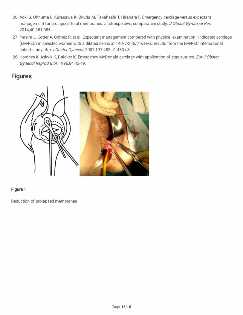
4. The Foley catheter was inserted and then �lled depending on the degree of cervical dilation and the stage of theprocedure. To do this, a third assistant helped to increase or reduce the drainage �ow, depending on the stage of theprocedure. This initially corrected the prolapse. It was then reduced in order to cross the cervical canal and later,once past the internal cervical os, the volume was increased again to �x the reduction of the bag and to facilitatesafe cerclage (Fig. 1).
5. The �rst cerclage was performed using Prolene 1. A purse-string suture was placed as cranially as possible and asclose as possible to the level of the internal cervical os, with care taken not to damage the bladder. The suture wasapplied super�cially, without going too deep into the cervical stroma, since the aim of this step is to keep the bagreduced once the Foley catheter had been removed, and to leave a segment of the cervix free on which to perform asecond cerclage, so conglutination is completely guaranteed (Fig. 2).
�. The tobacco pouch seam was then closed while the Foley catheter was simultaneously de�ated and removed,ensuring that the cervix was completely closed.
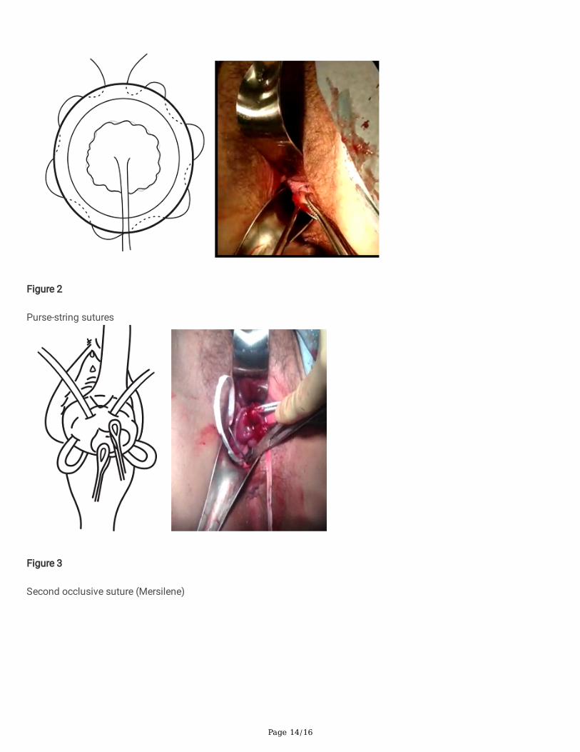
7. A second cerclage was performed with Mersilene tape, which was attached approximately 1 cm below the previouscerclage. The stitches in this suture were designed to conglutinate the cervix, including the anterior and posteriorlips at both commissures (8 to 11 o’clock on the left edge and 4 to 1 o’clock on the right edge). The point applied on
Page 5/16
the lower edge extended from 7 to 5 o’clock, and it was positioned as cranially as possible. The knot was located at12 o'clock (Fig. 3)
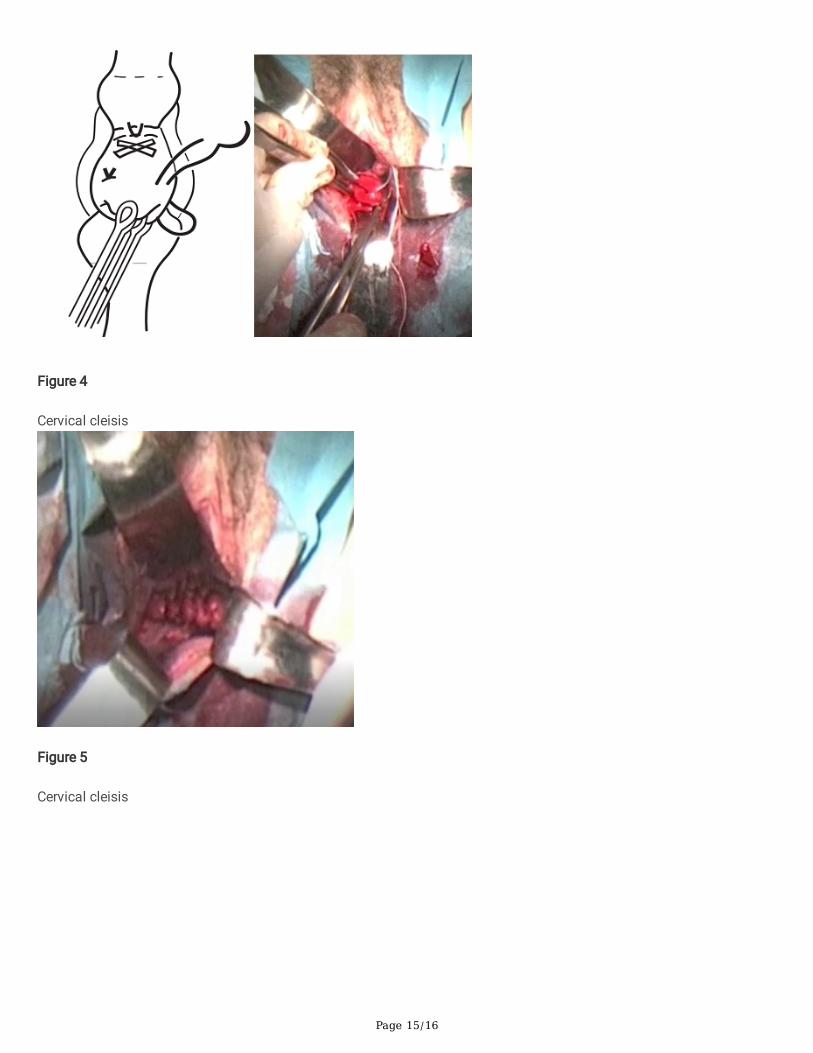
�. The cervix was closed at the cervical os using Vycril 0. Two double stitches were applied in both commissures(Fig4), plus a third in the central area (Fig. 5).
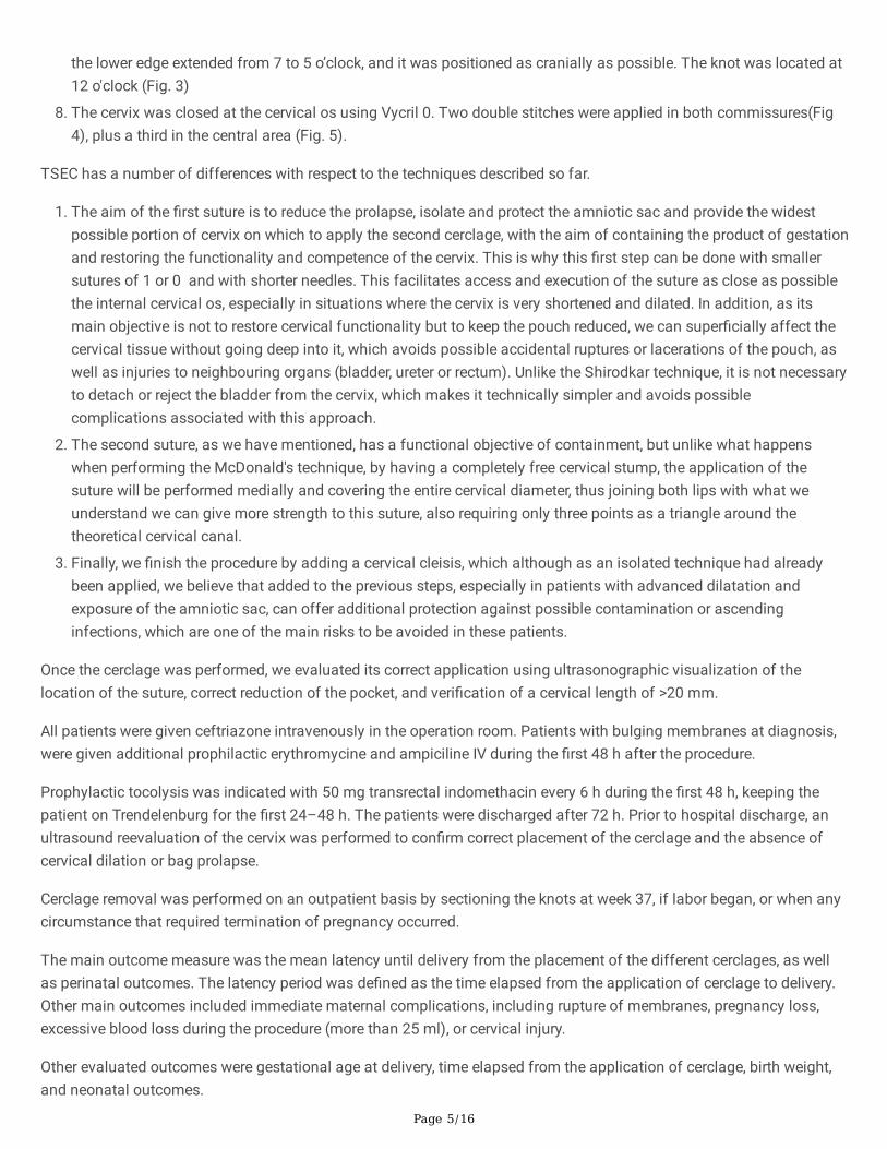
TSEC has a number of differences with respect to the techniques described so far.
1. The aim of the �rst suture is to reduce the prolapse, isolate and protect the amniotic sac and provide the widestpossible portion of cervix on which to apply the second cerclage, with the aim of containing the product of gestationand restoring the functionality and competence of the cervix. This is why this �rst step can be done with smallersutures of 1 or 0 and with shorter needles. This facilitates access and execution of the suture as close as possiblethe internal cervical os, especially in situations where the cervix is very shortened and dilated. In addition, as itsmain objective is not to restore cervical functionality but to keep the pouch reduced, we can super�cially affect thecervical tissue without going deep into it, which avoids possible accidental ruptures or lacerations of the pouch, aswell as injuries to neighbouring organs (bladder, ureter or rectum). Unlike the Shirodkar technique, it is not necessaryto detach or reject the bladder from the cervix, which makes it technically simpler and avoids possiblecomplications associated with this approach.
2. The second suture, as we have mentioned, has a functional objective of containment, but unlike what happenswhen performing the McDonald's technique, by having a completely free cervical stump, the application of thesuture will be performed medially and covering the entire cervical diameter, thus joining both lips with what weunderstand we can give more strength to this suture, also requiring only three points as a triangle around thetheoretical cervical canal.
3. Finally, we �nish the procedure by adding a cervical cleisis, which although as an isolated technique had alreadybeen applied, we believe that added to the previous steps, especially in patients with advanced dilatation andexposure of the amniotic sac, can offer additional protection against possible contamination or ascendinginfections, which are one of the main risks to be avoided in these patients.
Once the cerclage was performed, we evaluated its correct application using ultrasonographic visualization of thelocation of the suture, correct reduction of the pocket, and veri�cation of a cervical length of >20 mm.
All patients were given ceftriazone intravenously in the operation room. Patients with bulging membranes at diagnosis,were given additional prophilactic erythromycine and ampiciline IV during the �rst 48 h after the procedure.
Prophylactic tocolysis was indicated with 50 mg transrectal indomethacin every 6 h during the �rst 48 h, keeping thepatient on Trendelenburg for the �rst 24–48 h. The patients were discharged after 72 h. Prior to hospital discharge, anultrasound reevaluation of the cervix was performed to con�rm correct placement of the cerclage and the absence ofcervical dilation or bag prolapse.
Cerclage removal was performed on an outpatient basis by sectioning the knots at week 37, if labor began, or when anycircumstance that required termination of pregnancy occurred.
The main outcome measure was the mean latency until delivery from the placement of the different cerclages, as wellas perinatal outcomes. The latency period was de�ned as the time elapsed from the application of cerclage to delivery.Other main outcomes included immediate maternal complications, including rupture of membranes, pregnancy loss,excessive blood loss during the procedure (more than 25 ml), or cervical injury.
Other evaluated outcomes were gestational age at delivery, time elapsed from the application of cerclage, birth weight,and neonatal outcomes.
Page 6/16
Statistical Analyses
We performed an initial analysis of the frequency distribution of the independent variables. Subsequently, a bivariateanalysis was performed to identify associations between variables. For bivariate analyses, we used the independentsample t-test to compare the mean values in two groups/categories of women when conditions of normality werepresent, and a Mann Whitney U test for the rest of cases. For comparisons between a greater number of groups, we usedeither a single-factor ANOVA or the non-parametric Kruskal–Wallis test according to the conditions of homoscedasticitythat were evaluated using Levene's test. The chi-square test was used to compare qualitative variables. To analyze therelationship between quantitative variables, the Pearson´s correlation coe�cient was used. The signi�cance level wasset at p < 0.05. We used logistic regression models to predict the results of the main dependent variable, latency todelivery. The models were constructed using the Intro procedure, including the sociodemographic and obstetric variablesthat were shown �rst to be signi�cantly associated, using the typical stopping p-value thresholds for explanatorymodeling.(17)
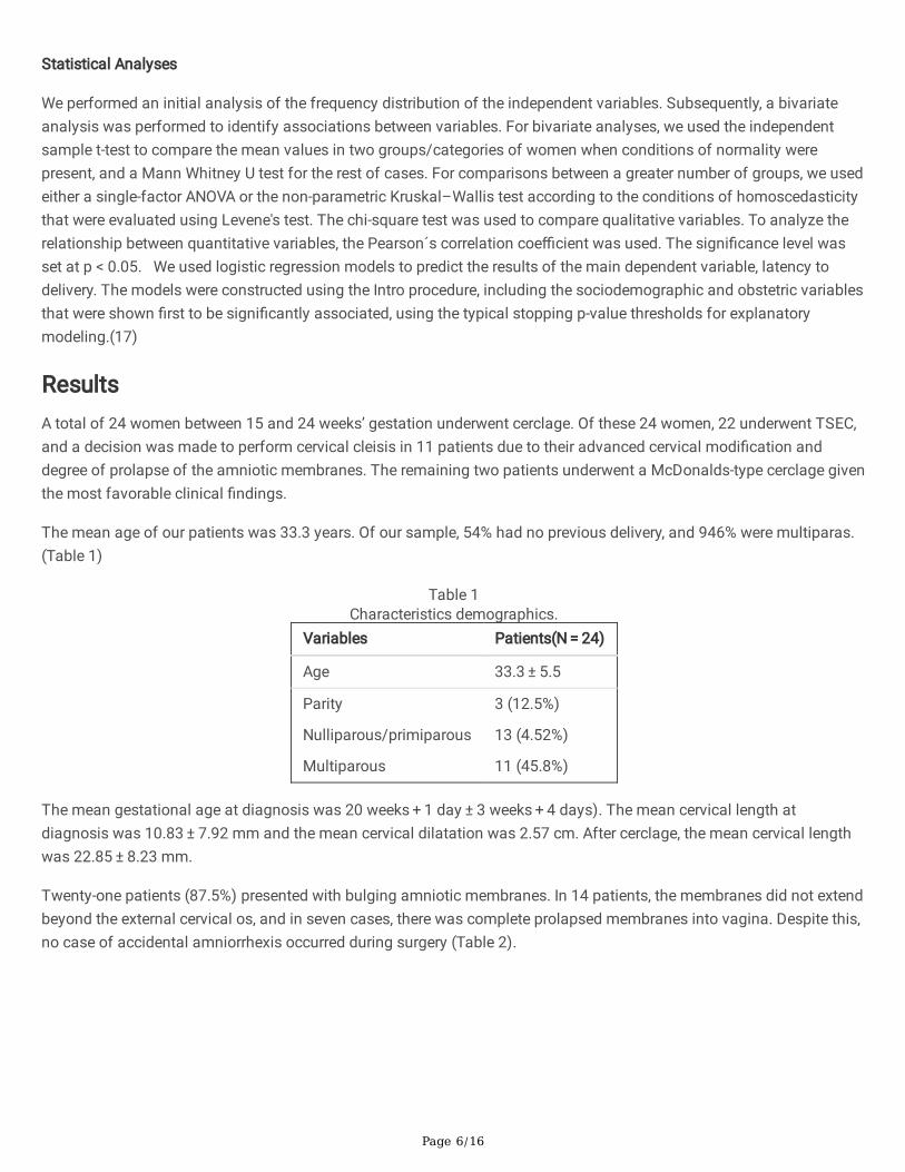
ResultsA total of 24 women between 15 and 24 weeks’ gestation underwent cerclage. Of these 24 women, 22 underwent TSEC,and a decision was made to perform cervical cleisis in 11 patients due to their advanced cervical modi�cation anddegree of prolapse of the amniotic membranes. The remaining two patients underwent a McDonalds-type cerclage giventhe most favorable clinical �ndings.
The mean age of our patients was 33.3 years. Of our sample, 54% had no previous delivery, and 946% were multiparas.(Table 1)
Table 1Characteristics demographics.
Variables Patients(N = 24)
Age 33.3 ± 5.5
Parity
Nulliparous/primiparous
Multiparous
3 (12.5%)
13 (4.52%)
11 (45.8%)
The mean gestational age at diagnosis was 20 weeks + 1 day ± 3 weeks + 4 days). The mean cervical length atdiagnosis was 10.83 ± 7.92 mm and the mean cervical dilatation was 2.57 cm. After cerclage, the mean cervical lengthwas 22.85 ± 8.23 mm.
Twenty-one patients (87.5%) presented with bulging amniotic membranes. In 14 patients, the membranes did not extendbeyond the external cervical os, and in seven cases, there was complete prolapsed membranes into vagina. Despite this,no case of accidental amniorrhexis occurred during surgery (Table 2).
Page 7/16
Table 2Characteristics of our patients (n = 24)
N (%)
Previous abortion 1 (45.8%)
Previous conization 3 (12.5%)
Previous premature delivery 5 (20.8%)
Previous chorioamnionitis 2 (8.3%)
Previous cervical incompetence 3 (12.5%)
Previous prolapsed membranes 3 (12.5%)
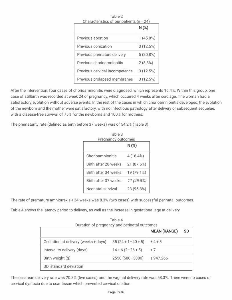
After the intervention, four cases of chorioamnionitis were diagnosed, which represents 16.4%. Within this group, onecase of stillbirth was recorded at week 24 of pregnancy, which occurred 4 weeks after cerclage. The woman had asatisfactory evolution without adverse events. In the rest of the cases in which chorioamnionitis developed, the evolutionof the newborn and the mother were satisfactory, with no infectious pathology after delivery or subsequent sequelae,with a disease-free survival of 75% for the newborns and 100% for mothers.
The prematurity rate (de�ned as birth before 37 weeks) was of 54.2% (Table 3).
Table 3Pregnancy outcomes
N (%)
Chorioamnionitis 4 (16.4%)
Birth after 28 weeks 21 (87.5%)
Birth after 34 weeks 19 (79.1%)
Birth after 37 weeks 11 (45.8%)
Neonatal survival 23 (95.8%)
The rate of premature amniorrexis < 34 weeks was 8.3% (two cases) with successful perinatal outcomes.
Table 4 shows the latency period to delivery, as well as the increase in gestational age at delivery.
Table 4Duration of pregnancy and perinatal outcomes
MEAN (RANGE) SD
Gestation at delivery (weeks + days) 35 (24 + 1–40 + 5) ± 4 + 5
Interval to delivery (days) 14 + 6 (2–26 + 5) ± 7
Birth weight (g) 2550 (580–3880) ± 947.266
SD, standard deviation
The cesarean delivery rate was 20.8% (�ve cases) and the vaginal delivery rate was 58.3%. There were no cases ofcervical dystocia due to scar tissue which prevented cervical dilation.
Page 8/16
We observed signi�cant positive correlations between cervical length after the intervention and gestational age (g.a.) atdelivery and between cervical length at diagnosis and latency with r = 0.48 (p < 0.05). On the other hand, negativesigni�cant correlations were found between dilatation at diagnosis and g.a. at delivery, and latency duration with r =-0.68 (p < 0.001).
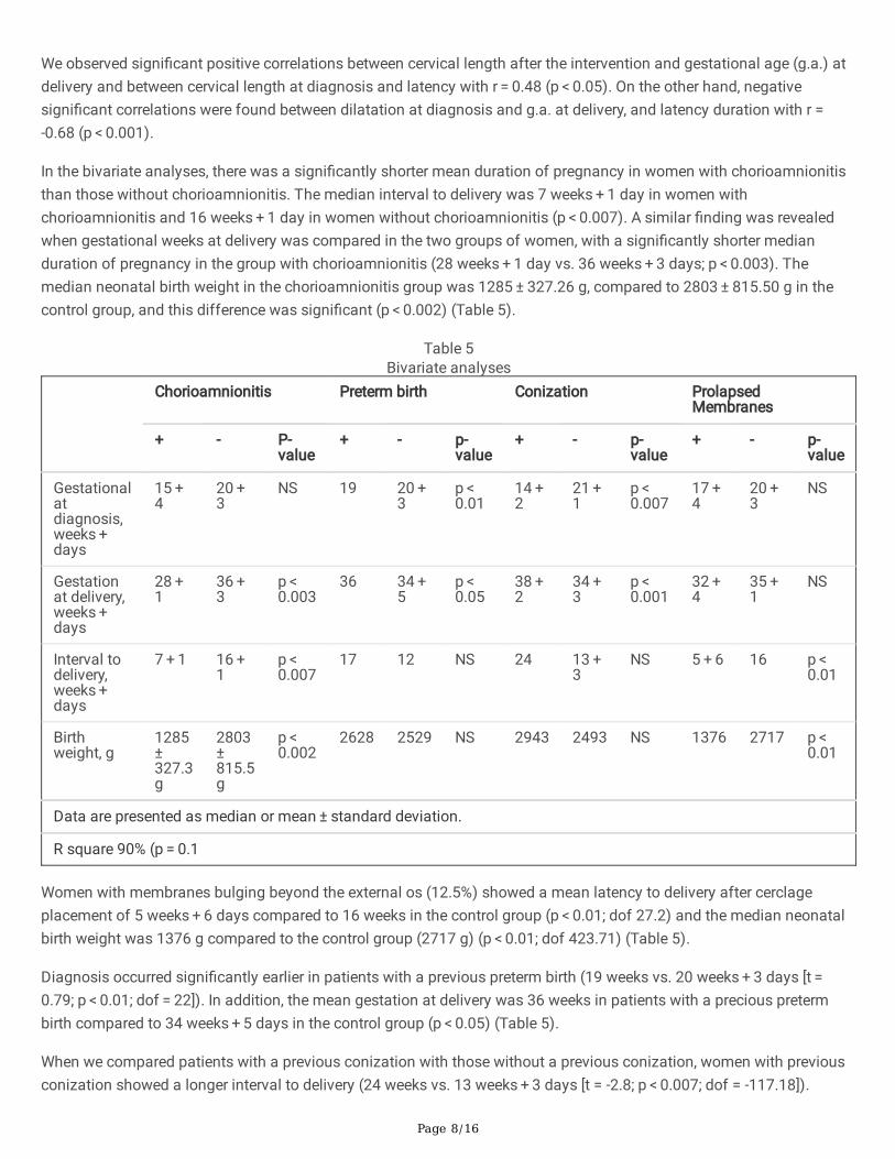
In the bivariate analyses, there was a signi�cantly shorter mean duration of pregnancy in women with chorioamnionitisthan those without chorioamnionitis. The median interval to delivery was 7 weeks + 1 day in women withchorioamnionitis and 16 weeks + 1 day in women without chorioamnionitis (p < 0.007). A similar �nding was revealedwhen gestational weeks at delivery was compared in the two groups of women, with a signi�cantly shorter medianduration of pregnancy in the group with chorioamnionitis (28 weeks + 1 day vs. 36 weeks + 3 days; p < 0.003). Themedian neonatal birth weight in the chorioamnionitis group was 1285 ± 327.26 g, compared to 2803 ± 815.50 g in thecontrol group, and this difference was signi�cant (p < 0.002) (Table 5).
Table 5Bivariate analyses
Chorioamnionitis Preterm birth Conization ProlapsedMembranes
+ - P-value
+ - p-value
+ - p-value
+ - p-value
Gestationalatdiagnosis,weeks + days
15 + 4
20 + 3
NS 19 20 + 3
p < 0.01
14 + 2
21 + 1
p < 0.007
17 + 4
20 + 3
NS
Gestationat delivery,weeks + days
28 + 1
36 + 3
p < 0.003
36 34 + 5
p < 0.05
38 + 2
34 + 3
p < 0.001
32 + 4
35 + 1
NS
Interval todelivery,weeks + days
7 + 1 16 + 1
p < 0.007
17 12 NS 24 13 + 3
NS 5 + 6 16 p < 0.01
Birthweight, g
1285 ± 327.3g
2803 ± 815.5g
p < 0.002
2628 2529 NS 2943 2493 NS 1376 2717 p < 0.01
Data are presented as median or mean ± standard deviation.
R square 90% (p = 0.1
Women with membranes bulging beyond the external os (12.5%) showed a mean latency to delivery after cerclageplacement of 5 weeks + 6 days compared to 16 weeks in the control group (p < 0.01; dof 27.2) and the median neonatalbirth weight was 1376 g compared to the control group (2717 g) (p < 0.01; dof 423.71) (Table 5).
Diagnosis occurred signi�cantly earlier in patients with a previous preterm birth (19 weeks vs. 20 weeks + 3 days [t = 0.79; p < 0.01; dof = 22]). In addition, the mean gestation at delivery was 36 weeks in patients with a precious pretermbirth compared to 34 weeks + 5 days in the control group (p < 0.05) (Table 5).
When we compared patients with a previous conization with those without a previous conization, women with previousconization showed a longer interval to delivery (24 weeks vs. 13 weeks + 3 days [t = -2.8; p < 0.007; dof = -117.18]).
Page 9/16
Similarly, women with a previous conization showed a longer total gestation compared to women with no conization (38weeks + 2 days vs. 34 weeks + 3 days [t = -1.3; p < 0.001; dof 21.49) (Table 5)
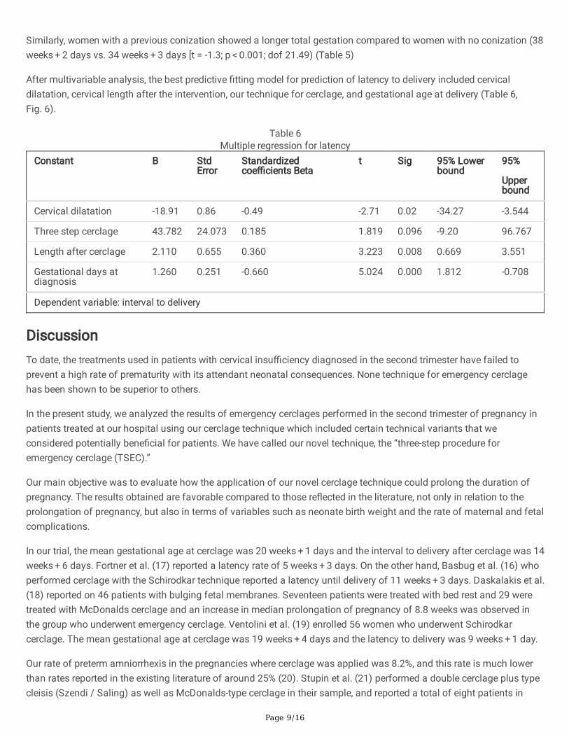
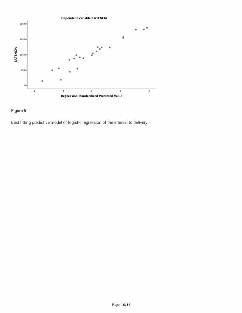
After multivariable analysis, the best predictive �tting model for prediction of latency to delivery included cervicaldilatation, cervical length after the intervention, our technique for cerclage, and gestational age at delivery (Table 6,Fig. 6).
Table 6Multiple regression for latency
Constant B StdError
Standardizedcoe�cients Beta
t Sig 95% Lowerbound
95%
Upperbound
Cervical dilatation -18.91 0.86 -0.49 -2.71 0.02 -34.27 -3.544
Three step cerclage 43.782 24.073 0.185 1.819 0.096 -9.20 96.767
Length after cerclage 2.110 0.655 0.360 3.223 0.008 0.669 3.551
Gestational days atdiagnosis
1.260 0.251 -0.660 5.024 0.000 1.812 -0.708
Dependent variable: interval to delivery
DiscussionTo date, the treatments used in patients with cervical insu�ciency diagnosed in the second trimester have failed toprevent a high rate of prematurity with its attendant neonatal consequences. None technique for emergency cerclagehas been shown to be superior to others.
In the present study, we analyzed the results of emergency cerclages performed in the second trimester of pregnancy inpatients treated at our hospital using our cerclage technique which included certain technical variants that weconsidered potentially bene�cial for patients. We have called our novel technique, the “three-step procedure foremergency cerclage (TSEC).”
Our main objective was to evaluate how the application of our novel cerclage technique could prolong the duration ofpregnancy. The results obtained are favorable compared to those re�ected in the literature, not only in relation to theprolongation of pregnancy, but also in terms of variables such as neonate birth weight and the rate of maternal and fetalcomplications.
In our trial, the mean gestational age at cerclage was 20 weeks + 1 days and the interval to delivery after cerclage was 14weeks + 6 days. Fortner et al. (17) reported a latency rate of 5 weeks + 3 days. On the other hand, Basbug et al. (16) whoperformed cerclage with the Schirodkar technique reported a latency until delivery of 11 weeks + 3 days. Daskalakis et al.(18) reported on 46 patients with bulging fetal membranes. Seventeen patients were treated with bed rest and 29 weretreated with McDonalds cerclage and an increase in median prolongation of pregnancy of 8.8 weeks was observed inthe group who underwent emergency cerclage. Ventolini et al. (19) enrolled 56 women who underwent Schirodkarcerclage. The mean gestational age at cerclage was 19 weeks + 4 days and the latency to delivery was 9 weeks + 1 day.
Our rate of preterm amniorrhexis in the pregnancies where cerclage was applied was 8.2%, and this rate is much lowerthan rates reported in the existing literature of around 25% (20). Stupin et al. (21) performed a double cerclage plus typecleisis (Szendi / Saling) as well as McDonalds-type cerclage in their sample, and reported a total of eight patients in
Page 10/16
whom amniorrhexis occurred during the technique, as well as three cases of cervical lacerations during the removal ofthe cerclage. In our study, there were no cases of accidental amniorrhexis during the procedure or any notablecomplications in its removal, nor did we observe any in�uence on the evolution of the subsequent delivery.
Gupta et al. (22) reported a chorioamnionitis rate of 46% and Abo-Yaoub et al. (23) reported a rate of 16.2%. Likewise,Freire et al. (24) reported a rate of 23.5%. Our chorioamnionitis rate was 16%, which is lower than that reported in otherstudies. Analysis of the cases that developed clinical chorioamnionitis revealed signi�cant differences in terms of theduration of pregnancy (7 weeks + 1 days vs. 16 weeks + 2 days), indicating that this is the main determining factor forpredicting the �nal results in our cerclages.
One of the most outstanding �ndings was that in conized patients, the results were very satisfactory with latency time todelivery even higher than that in non-conized patients. Even though our sample was very limited, the differences foundbetween the two groups are signi�cant, and we suggest that this type of cerclage could be particularly bene�cial in thisgroup of patients.
It is especially important to evaluate how cerclage is able to prevent extreme prematurity in these patients. In his study,Aoki (25) succeeded in prolonging pregnancies beyond 28 weeks in 66% of his patients. Bayrak (14) reported similarnumbers (63%), as did Pereira (62%) (26). In our study, 87.5% of the pregnant women who underwent cerclage managedto exceed 28 weeks of gestation.
There are very few reports on pregnancies that reach full-term. Ventollini (19) reported that 23% of pregnancies exceeded38 weeks; however, Basbug (16) reported a lower �gure of 13.6%. In our sample, 11 patients exceeded 37 weeks ofgestation, which represents 45.8% of the total.
Birth weight is an important indicator of perinatal outcomes, and there are many reports on this in the literature. Onestudy that stands out is that by Hordnes (27) which was published in 1996 and reported an average birth weight of 2252g; however, it should be noted that the number of patients in that study was small. However, larger and more recentstudies, such as the study by Fortner (19) reported an average birth weight of 1190 g. In 2010, Gupta (22) reported anaverage birth weight of 1937 g. The results of our study are very favorable since we found an average birth weight of2550 g.
After analysing all the above data, we can conclude that using our TSEC technique we could obtain a longer latencyperiod that would result in lower prematurity rates. These results would be especially bene�cial in patients with poorprognosis (prolapsed membranes, cervical dilatation, conization patients), in which most of the studies have beenunsatisfactory. It is an easily applicable technique and we �nd of particular interest the low or null incidence ofcomplications during its performance. The e�cacy of the technique and the low or null incidence of complications hasled to a drastic reduction in the hospital stay of these patients, making TSEC a bene�cial technique from a cost-effectivepoint of view.
ConclusionThe cerclage is a technique of proven usefulness in patients with cervical dilation in the second trimester of pregnancyfor the prevention of preterm delivery. It prolongs pregnancy and prevents poor perinatal outcomes derived fromprematurity.
So far, no technique has been proven as superior to the rest. In this study, we have presented our results of our noveltechnique which introduces certain modi�cations—the TSEC.
Page 11/16
Most of the studies to date, and the present study, report on a small number of recruited patients. This fact, together withthe fact that most studies, including the present study, are observational, prevents us from obtaining de�nitiveconclusions. These are the main limitations of this research, and larger studies are required to con�rm our �ndings.
Despite this, the good results in terms of latency until delivery after cerclage, gestational age at delivery, birth weight, andminimization of complications reported in our work compels us to continue to evaluate the results of our techniquewhich may provide signi�cantly better outcomes for neonates and mothers.
AbbreviationsTSEC: Three-Step Procedure for Emergency Cerclage, PROM: Premature rupture of membranes.
DeclarationsEthics approval and consent to participate: The research has been carried out in accordance with the Declaration ofHelsinki.This original contribution was approved by the ethics committee of Regional University Hospital ( Málaga.Sapin) and the consent for participation was obtained in writing. The committee’s reference number is 15032021/50.
Consent for publication The authors have obtained the patients’ signed consent to publication. This work has not beenpublished before and it is not under consideration for publication anywhere else.
Availability of data and materials. The datasets used to support the fndings of this study are available from HospitalRegional Universitario Málaga Spain upon reasonable request and signing a data access agreement subject to approvalfrom the study principal investigators.
Competing interests: In this study, there is no con�icts of interest
Funding: This article has not been funded by any public/private external entity or any commercial organization. Thisarticle has not undergone peer-review by any funding body.
Authors' contributions: The information related to the cases can be obtained from the corresponding authors (MGC andLSM) upon reasonable request. MGC and LSM also data collection and manuscript writing. EGM and JSJL., editing andwriting The data were collected during the patients’ hospital admission by the investigators who provided directassistance. These data are stored in the computerized support system of electronic medical records of the Andalusianhealth system.
Acknowledgments: None.
Author details: Work center of all authors: 1º Obstetric and Gynecology Service, Hospital Regional Universitary Málaga,Avd Arroyo de los Angeles S/N, Málaga, Spain 29011. 2º Ernesto González Mesa. Malaga University. Spain.
References1. Lawn J, McCarthy BJ, Ross SR, The healthy newborn: a reference manual for program managers. Atlanta: Center for
Disease Control and Prevention, 2001.
2. Martin JA, Hamilton BE, Osterman MJ, et al. Births: �nal data for 2013. Natl Vital Stat Rep. 2015,64:1-65.
3. Draper ES, Manktelow B, Field DJ, James D. Prediction of survival for preterm births by weight and gestational age:retrospective population based study. BMJ. 1999,319:1093-1097.
4. Rennie JM. A review of the risk of being born too soon. Fetal Matern Med Rev. 2002,13:157-168.
Page 12/16
5. Iams JD, Johnson FF, Sonek J, Sachs L, Gebauer C, Samuels P. Cervical competence as a continuum: a study ofultrasonographic cervical length and obstetric performance. Am J Obstet Gynecol. 1995,172:1097-1103.
�. Stromme WB, Haywa EW. Intrauterine fetal death in the second trimester. Am J Obstet Gynecol. 1963,85:223-233.
7. Namouz S, Porat S Okun N, Windrim R, Farine, D. Emergency cerclage: literature review. Obstet Gynecol Surv. 2013,68:379-388.
�. Chatzakis C, Efthymiou A, Sotiriadis A, Makrydimas G. Emergency cerclage in singleton pregnancies with painlesscervical dilatation: A meta-analysis. Acta Obstet Gynecol Scand. 2020,99:1444-1457.
9. Shirodkar VN. A new method of operative treatment for habitual abortions in the second trimester of pregnancy.Antiseptic. 1955,52:299-300.
10. McDonald IA. Suture of the cervix for inevitable miscarriage. J Obstet Gynaecol Br Emp. 1957,64:346-350.
11. Althuisius SM, Dekker GA, Hummel P, Bekedam DJ, van Geijn HP. Final results of the Cervical IncompetencePrevention Randomized Cerclage Trial (CIPRACT): therapeutic cerclage with bed rest versus bed rest alone. Am JObstet Gynecol. 2001,185:1106-1112.
12. Berghella V, Ludmir J, Simonazzi G, Owen J. Transvaginal cervical cerclage: Evidence for perioperative managementstrategies. Am J Obstet Gynecol. 2013,209:181-192.
13. Sato Y, Hidaka N, Nakano T, et al. E�cacy of an Emergency Cervical Cerclage Using Absorbable Mono�lamentSutures. J Pregnancy. 2018,2018:4049792.
14. Bayrak M, Gul A, Goynumer G. Rescue cerclege when foetal membranes prolapse into the vagina. J Obstet Gynaecol(Lahore). 2017,37:471-475.
15. Curti A, Simonazzi G, Farina A, Mehmeti H, Facchineti F, Rizzo N. Exam-indicated cerclage in patients with fetalmembranes at or beyond external os: a retrospective evaluation. J Obstet Gynaecol Res. 2012,38:1352-1357.
1�. Basbug A, Bayrak M, Dogan O, Kaya A, Goynumer G. McDonald versus modi�ed Shirodkar rescue cerclage inwomen with prolapsed fetal membranes. J Matern Fetal Neonatal Med. 2020,33:1075-1079.
17. Variable Selection in Multiple Regression | Introduction to Statistics | JMP [Internet]. [cited 2021 May 12]. Availablefrom: https://www.jmp.com/en_us/statistics-knowledge-portal/what-is-multiple-regression/variable-selection.html
1�. Fortner KB, Fitzpatrick CB, Grotegut CA, et al. Cervical dilation as a predictor of pregnancy outcome followingemergency cerclage. J Matern Fetal Neonatal Med. 2012,25:1884-1888.
19. Daskalakis G, Papantoniou N, Mesogitis S, et al. Management of cervical insu�ciency and bulging fetalmembranes. Obstet Gynecol. 2006,107:221-226.
20. Ventolini G, Genrich TJ, Roth J, et al. Pregnancy outcome after placement of ‘rescue’ Shirodkar cerclage. J Perinatol.2009,29:276-279.
21. Uzun Clingir I, Sayin C, Sutcu H, et al. Does emergency cerclage really work in patients with advanced cervicaldilatation. J Gynecol Obstet Hum Reprod. 2019, 48:387-390.
22. Stupin JH, David M, Siedentopf JP, et al. Emergency cerclage versus bed rest for amniotic sac prolapse before 27gestational weeks. A retrospective, comparative study of 161 women. Eur J Obstet Gynecol Reprod Biol.2008,139:32-37.
23. Gupta M, Emary K, Impey L. Emergency cervical cerclage: predictors of success. J Matern Fetal Neonatal Med.2010,23:670-674.
24. Abo-Yaqoub S, Mohammed AB, Saleh H. The effect of second trimester emergency cervical cerclage on perinataloutcome. J Matern Fetal Neonatal Med. 2012,25:1746-1749.
25. Freire Costa MM, Amorin Filho AG, De Barros MF, et al. Emergency cerclage: gestational and neonataloutcomes. Rev Assoc Med Bras. 2019,65:598-602.
Page 13/16
2�. Aoki S, Ohnuma E, Kurasawa K, Okuda M, Takahashi T, Hirahara F. Emergency cerclage versus expectantmanagement for prolapsed fetal membranes: a retrospective, comparative study. J Obstet Gynaecol Res.2014,40:381-386.
27. Pereira L, Cotter A, Gómez R, et al. Expectant management compared with physical examination–indicated cerclage(EM-PEC) in selected women with a dilated cervix at 140/7-256/7 weeks: results from the EM-PEC internationalcohort study. Am J Obstet Gynecol. 2007,197:483.e1-483.e8.
2�. Hordnes K, Askvik K, Dalaker K. Emergency McDonald cerclage with application of stay sutures. Eur J ObstetGynecol Reprod Biol. 1996,64:43-49.
Figures
Figure 1
Reduction of prolapsed membranes
Page 14/16
Figure 2
Purse-string sutures
Figure 3
Second occlusive suture (Mersilene)
Page 15/16
Figure 4
Cervical cleisis
Figure 5
Cervical cleisis
Page 16/16
Figure 6
Best �tting predictive model of logistic regression of the interval to delivery