proceedings of the 8th international students conference modern
TRANSCRIPT

Proceedings of the
th International Students Conference
“Modern Analytical Chemistry”
8


Prague, 24 25 September 2012—
Edited by Karel Nesměrák
Charles University in Prague, Faculty of Science
Prague 2012
Proceedings of the
8th International Students Conference
Modern Analytical Chemistry“ ”

CATALOGUING-IN-PUBLICATION – NATIONAL LIBRARY OF THE CZECH REPUBLICKATALOGIZACE V KNIZE – NÁRODNÍ KNIHOVNA ČR
Modern Analytical Chemistry (2012 : Praha, Česko)Proceedings of the 8th International Students Conference
Modern Analytical Chemistry : Prague, 24–25 September 2012
543 – Analytic
“ ”
ká chemie [10]
/edited by Karel Nesměrák. – 1st ed. – Prague : CharlesUniversity in Prague, Faculty of Science, 2012. – 104 s.ISBN 978-80-7444-017-5
543analytical chemistryproceedings of conferencesanalytická chemiesborníky konferencí
543 – Analytical chemistry [10]
12
▪
▪
▪
▪
The Proceedings publication was supported by the research projectMSM0021620857 f the Ministry of Education of the Czech Republic.
The electronic version of the Proceedings is available at the conference webpage:
o
http://www.natur.cuni.cz/isc-mac/
© Charles University in Prague, Faculty of Science, 20 .
ISBN 978-80-7444-017-5

Preface
Dear friends and colleagues,
The AP Czech, HPST, Quinta Analytica, and Shimadzu companies are cordiallythanked, not only for their ponsorship, but for their continuoussupport and cooperation in many of our other activities.
We wish you success in the presentation of your contributions, vivid discu-ssions with the audience and your colleagues, pleasant social encounters and nicestay in the city of Prague.
Prof. RNDr. Věra Pacáková, CSc. RNDr. Karel Nesměrák, Ph.D.
Welcome t the 8th International Students Conference “Modern AnalyticalChemistry”. We are pleased that more than thirty young scientists from four countries attend the conference. We hope that the conference, like in previous years,will be interesting and successful event. It will become a platform for
presentation of new scientific results and will show the further directions ofresearch in the field of analytical chemistry. We are convinced that the conferenceoffers many possibilities for improv the presentation skills, provides thefloor for discussion and exchange of experiences and opinions, and helps tomaster the English language to all the participants.
kind financial s
o-
, challenging,the
ement of
Proceedings of th ISC Modern Analytical Chemistry Prague 201the 8 2▪ 3

Sponzors
The organiz of th International Students Conference “Modern AnalyticalChemistry” gratefully acknowledge the generous sponsorship of followingcompanies:
ers 8s
http://www. .cz/quinta
http://www.shimadzu.cz/
4 the 8 2Proceedings of th ISC Modern Analytical Chemistry Prague 201▪
http://www. .cz/apczech
http://www. .cz/hpst

Programme
The conference is held at the Institute of Chemistry, Faculty of Science, CharlesUniversity in Prague (Hlavova 8, 128 43 Prague 2) in the main lecture hall(Brauner’s Lecture Theatre). Oral presentations are minutes includingdiscussion and speakers are asked to download their Power Point presentation onthe local computer in the lecture hall before the start of the session. The coffeebreaks are held in the lecture hall.
: 0–9: 0
9:00–9: 0
9: 0–9: 0(p. )
9: 0– . 0(p. )
: 0–10: 0
(p. )10: 0–10: 0
(p. )
1 : 0–11: 0
(p. )11: 0–11: 0
(p. )11: 0–1 : 0
(p. )1 : 0–12: 0
(p. )
twenty
8 3 0
2
chairperson: Michal Tatarkovič2 4 Dziubakiewicz E.:
114 10 0 Pažitná A.:
1310 0 2 Krejčová Z.:
162 4 Čížková A.: –
19
chairperson: Magda Staňková1 0 2 Bursová M.:
212 4 Liebherr R.B.:
244 2 0 Prchal V.:
282 0 2 Hengerics Szabó A.:
33
Monday, September 24, 2012
Registration of participants
ceremony, welcoming address
Session 1
Session 2
Opening
▪
▪
Influence of charge distribution on electro-
phoretic separation of bacterial cells
The enantiomer distribution of major chiral volatile
organic compounds in Slovak monofloral honeys
Voltammetric DNA biosensor for the detection of DNA
damage caused by nitrated polycyclic aromatic hydrocar
bons
Single drop microextraction method application to
essential oils analysis in real samples of herbal tea
Preconcentration of aromatic and polyaromatic
amines with bell-shaped extraction device assisted liquid-liquid
microextraction
Analysis of individual enzyme molecules in femtoliter
arrays
Use of alkanethiol self-assembled monolayer modified
electrodes in voltammetric analysis
Analysis of breath volatiles by inside needle
capillary adsorption trap-gas chromatography
-
10: 0–1 : 0
12: 0–13: 0
2 1 0
2 2
Coffee Break
Lunch
Proceedings of th ISC Modern Analytical Chemistry Prague 201the 8 2▪ 5

Session 3
Session 4
Session 5
▪
▪
▪
chairperson: Miroslava BursováPomastowski P.:
36Markechová D.:
38Zavázalová J.: -
41Staňková M.:
45
4 4 0
chairperson: Natalia Denderz0 2 Kozlík P.:
472 5 4 Sedlmeier A.:
495 4 0 Walczak J.:
53
chairperson: Petr KozlíkDenderz N.:
55Jaćkowska M.:
58Klusáčková M.:
60Tatarkovič M.:
63
13:20–13:40(p. )
13:40–14:00
(p. )14:00–14:20
(p. )14:20–14:40
(p. )
1 : 0–15: 0
15: 0–15: 0(p. )
15: 0–1 : 0(p. )
1 : 0–16: 0(p. )
9:20–9:40(p. )
9:40–10:00(p. )
10:00–10:20(p. )
10:20–10:40(p. )
Influence of heterogeneity biocolloids surface on
their electrophoretic separation
Fluorescence spectroscopy combined with parallel
factor analysis for determination of geographical origin of juniper-
flavoured spirit drinks
Utilization of boron-doped diamond thin film elec
trode in electroanalysis of selected derivatives of amino derivatives
of polycyclic aromatic hydrocarbons
Effect of crosslinking monomer on the efficiency of
polymethacrylate monolithic columns
Study of separation of selected pteridines in hydrophilic
interaction liquid chromatography
Surface modification of upconverting luminescent
nanoparticles for bioconjugation
SPME-LC/MS for the analysis of selected biologically
active compounds
Temperature effect on the sorption selectivity of some
phenolic acids on molecularly imprinted stationary phases
Functionalized dendrimer stationary phases on silica
gel for ion chromatography
E -
s h b p
c d
-
lectrocatalytic oxidation of unsaturated hydro
carbons mediated by phthalocyanine derivate
Chiroptical pectroscopy of uman lood lasma in
linical iagnosis
Coffee Break
n
16: 0–1 : 0
1 : 0
10:40–11:00
0 6 3
7 0
Sponsors’ presentations
Coffee Break
Get-Together Party
Tuesday, September 25, 2012
6 the 8 2Proceedings of th ISC Modern Analytical Chemistry Prague 201▪

Session 6
Session 7
Session 8
Closing Ceremony
▪
▪
▪
chairperson: Pomastowski.:
65á M.: -
67Novotný V.:
72ński D.:
77
chairperson: Jonas MarkKrasulová J.:
79Novosad L.: -
82Rumlová T.:
-84
Němcová V.:
87
4 4 0
chairperson: Alexandra Hengerics Szabó0 2 Rybínová M.: -
-89
2 5 4 Podolec P.: -93
5 4 0 Vojta J.:96
0 6 1
Pawel11:00–11:20 Mark J
(p. )11:20–11:40 Dendisov
(p. )11:40–12:00
(p. )12:00–12:20 Grzywi
on-line (p. )
13:20–13:40(p. )
13:40–14:00(p. )
14:00–14:20
(p. )14:20–14:40
(p. )
1 : 0–15: 0
15: 0–15: 0
(p. )15: 0–1 : 0
(p. )1 : 0–16: 0
(p. )
16: 0–1 : 0
Fast non-aqueous capillary electrophoresis in short
capillaries with amperometric end-column detection
Voltammetric etermination of Fomesafen and Fluoro
difen in drinking water and river water
Determination of ycotoxins by icro-SPE oupled
with icro-HPLC/LIF
Analytical methods used in the studies on chemical
ecology of termites
Analytical capabilities of plasma pencil in atomic emis
sion spectrometry
New types of carbon composite film electrodes based on
various metal substrates for determination of genotoxic environ
mental pollutants
HPLC-UV method for the determination of genotoxic
4-nitroindane in the mixture of selected nitrated polycyclic aro
matic hydrocarbons
Determination of selenium using photochemical vola
tile compounds generation coupled with atomic absorption spectro
metry detection
Determination of sevoflurane and hexafluoroisopro
panol from plasma by GC-MS/MS
New capillary monolithic column for isocratic separation of
small molecules
Spectroelectrochemical study of 4-aminobenzene
thiol adsorbed on gold, silver and copper nanostructured surfaces
d -
m m c
m
-
12:20–1 : 03 2 Lunch
Coffee Break
Proceedings of th ISC Modern Analytical Chemistry Prague 201the 8 2▪ 7


Contributions


Microorganisms are biocolloids that can change their surface properties based on
interactions with each other and the surrounding environment. This change
in surface characteristics dictate how bacterial cells may interact when forming
aggregates and/or adhesion of bacterial cells. These phenomena are relatively
complicated problem because no comprehensive theory describing every aspect
of bacterial cells interaction has been created yet. Explanation of the mechanism
forming aggregates and biofilms is very important also during electrophoretic
separation.
The cell wall of bacteria is composed mainly of proteins, phospholipids,
teichoic acid, teichuronic acid and lipopolysaccharides. These macromolecules
contribute to bacterial surface charge due to the ionization of proton-active
functional groups, such as carboxyl, phosphate, amino or hydroxyl groups and the
adsorption of the ions from the solution.
In this work physicochemical surface characteristics of bacteria were mea-
sured to establish their role in bacterial adhesion and aggregation on the basis on
electrophoretic behavior of different clinical strains of Gram-positive and Gram-
-negative bacteria. Properties of bacterial cell wall surfaces were investigated by
the combination of electrochemical and spectroscopic techniques allow to obtain
Influence of Charge Distribution
on Electrophoretic Separation
of Bacterial Cells
E D B BWELINA ZIUBAKIEWICZ, OGUSŁAW USZEWSKI
Department of Environmental Chemistry and Bioanalytics, Faculty of Chemistry,
Nicolaus Copernicus University
Gagarin Str. 7, 87-100 Torun, Poland, [email protected]�
Keywords
bacterial cell
electrophoresis
physicochemical surface characteristics
Proceedings of th ISC Modern Analytical Chemistry Prague 201the 8 2▪ 11

concentration and protonation/deprotonation of the specific functional groups
on the microorganism cell surface.
The results obtained help understand the electrophoretic separation behavior
and allow to obtain more information about the charge on the surface of bacterial
cell. Furthermore they are useful for describing the fundamental mechanisms
involved in bacterial aggregation and adhesion to solid surfaces.
Acknowledgments
This work was supported by the National Science Centre (Narodowe Centrum Nauki, abbr. NCN,Warsaw, Poland) Grant No. N N204 369040.
12 the 8 2Proceedings of th ISC Modern Analytical Chemistry Prague 201▪

Honey is one of the most important natural products that are used in food
industry, in cosmetics and in medicine. One can distinguish between monofloral
and polyfloral honeys. Monofloral honey is usually more appreciated, since it
primarily originates from the nectar of one type of flower. Monofloral honeys
differ in taste, flavor as well as in color depending on properties of primary nectar
sources.
A volatile organic compound in honey is a mixture of different components
containing various chemical functional groups that are usually present at low
concentration levels. There are several ways how to extract -
from honey. The most popular is solid phase microextraction (SPME). This
technique can be easily automated, but optimization of working conditions (type
of SPME fibres, sorption temperature, sorption time and desorption temperature)
requires more time. Currently, it is mostly used for characterization of honey
profiles [1–4].
Many organic compounds present in honey are chiral, so they can exist as two
enantiomers. The determination of enantiomer composition of chiral compounds
can be used for evaluation of adulteration and manipulation with non-food
commodities or for distinguishing between natural and synthetic compounds.
Chiral compounds occur in the nature as a pure enantiomers or mixture of
volatile organic com
pound
volatile organic compound
Proceedings of th ISC Modern Analytical Chemistry Prague 201the 8 2▪ 13
The Enantiomer Distribution of Major
Chiral Volatile Organic Compounds in
Slovak Monofloral Honeys
A P I Š J DLEXANDRA AŽITNÁ, VAN PÁNIK, ANA ŽÚROVÁ
Institute of Analytical Chemistry, Faculty of Chemical and Food Technology, Slovak University of
Technology, Radlinského 9, SK-812 37 Bratislava, Slovak Republic, [email protected]�
Keywords
honey
multidimensional gas chromatography
volatile organic compounds

enantiomers with specific ratio. Any changes in these ratios may indicate
manipulation with products or addition of synthetically produced chemicals.
In this work, the distribution of enantiomers of selected chiral volatile organic
compounds in 45 monofloral honey samples was studied by GC. The volatile
organic compounds were extracted from Slovakian rapeseed, acacia, sunflower
basswood and raspberry honeys by solid phase microextraction followed by
GC-MS analysis. Afterwards, chiral compounds present at higher concentration
level were selected from more than 230 organic compounds found in studied
honey samples for determination of their enantiomer ratios. It was found that one
dimesnsional GC with chiral stationary phases shows excellent efficiency towards
enantiomer separations; however resolved enantiomers often coelute with
another non-chiral or already separated enantiomer of another organic com-
pound. Thus, two dimensional GC with two independent thermostats was used to
determine correct enantiomer ratios. Finally, the enantiomer ratios of linalool,
and linalool oxides, hotrienol and four isomers of lilac aldehydes were
determined. It was found, that distribution of enantiomers in honey samples
partially depends on their botanical origin. The significant differences in enan-
tiomer ratios of lilac aldehyde isomer B and hotrienol were observed for acacia
honey that allows us to distinguish this type of honey from other ones.
cis- trans-
14 the 8 2Proceedings of th ISC Modern Analytical Chemistry Prague 201▪
Fig. .1 GC-MS chromatogram of honey sample with marked compounds for which the enantiomerratio was determined.

Significantly different enantiomer ratio of -linalool oxide was found for sun-
flower honeys.
cis
Acknowledgments
References
111
26
228
126
This work was supported by the project VEGA n. 1/0972/12
–
[1] Kaskoniene V., Venskutonis P.R., Ceksteryte V.: (2008), 988–997.
[2] Soria A. C., Martinez-Castro I., Sanz J.: (2003), 793 801.
[3] Soria A.C., Sanz J., Martinez-Castro I.: (2009), 579 590.
[4] Plutowska B., Chmiel T., Dymerski T., Wardencki W.: (2011), 1288 1298.
and by project Separation of
enantiomers in food matrices by multidimensional gas chromatography Program to support young
researchers.
Food Chem.
J. Sep. Sci.
Eur. Food Res. Technol.
Food Chem.
–
–
–
Proceedings of th ISC Modern Analytical Chemistry Prague 201the 8 2▪ 15

Nitrated polycyclic aromatic hydrocarbons (NPAHs) are derivatives of polycyclic
aromatic hydrocarbons, which contain two or more fused aromatic rings made of
carbon and hydrogen atoms. originate
primarily as direct or indirect products of incomplete combustion, only a few
are produced industrially. have
been detected in the emissions of kerosene heaters, fuel gas and LPG burners used
for heating and cooking at home, as well as in the fumes of cooking oils [1].
administered by various routes are
rapidly absorbed and metabolized. Many bacteria reduce NPAHs to mutagenic
amino derivatives of polycyclic aromatic hydrocarbons. Nitroreduction by intes-
tinal microflora plays a major role in the metabolism of NPAHs in mammals [2].
Although the genotoxic effects of the NPAHs are already well known more than
30 years, an electrochemical research on their interactions with DNA was
realized quite recently [3]. Electrochemical analysis of DNA offers a number of
approaches in DNA damage detection as well as in sensing of DNA damaging
agents in the environment. Adenine and cytosine residues in DNA produce
Nitrated polycyclic aromatic hydrocarbons
NPAHs Nitrated polycyclic aromatic hydrocarbons
Nitrated polycyclic aromatic hydrocarbons
in vitro
Voltammetric DNA Biosensor for the
Detection of DNA Damage Caused by
Nitrated Polycyclic Aromatic
Hydrocarbons
Z K V V J BUZANA REJČOVÁ, LASTIMIL YSKOČIL, IŘÍ AREK
Department of Analytical Chemistry, Faculty of Science, Charles University in Prague,
Hlavova 2030/8, 128 43 Prague 2, Czech Republic, [email protected]�
Keywords
DNA biosensor
hanging mercury drop electrode
nitrated polycyclic aromatic hydrocarbons
2-nitrofluorene
voltammetry
16 the 8 2Proceedings of th ISC Modern Analytical Chemistry Prague 201▪

reduction signals, while guanine residues yield anodic signals due to oxidation of
the guanine reduction product. For the detection of these signals, voltammetric
techniques with a hanging mercury drop electrode are used most frequently. By
the observation of the changes in the intensity of such signals, the measure of DNA
damage can be determined. Moreover, on the basis of the shift in the potential of
voltammetric peaks of the analyte with the concentration of DNA present in the
incubation solution, the type of damaging binding to DNA can be estimated [4].
In this contribution, 2-nitrofluorene has been chosen as a model repre-
sentative of NPAHs. The interaction of 2-nitrofluorene with a double-stranded calf
thymus DNA has been studied using the as
an electrochemical sensor. Two types of DNA damage were investigated and
electrochemically detected: The DNA damage caused by the direct interaction
with 2-nitrofluorene and by short-lived radicals generated by the electro-
chemical reduction of the nitro group in 2-nitrofluorene. After reductive acti-
vation of 2-nitrofluorene, the oxidative DNA damage induced by the reactive
species was evaluated from the height of cathodic peak of cytosine and adenine
using cyclic voltammetry. The electrochemical reduction of 2-nitrofluorene
(during the first 4-electron reduction step of nitro group reduction to hydroxyl-
amino group) generates short-lived radicals that interact with DNA causing
damage [5].
For the study of direct interaction, two approaches of DNA damage detection
were used: Utilization of the signals of 2-nitrofluorene interacting with DNA non-
-covalently using hanging mercury drop electrode (DNA titration technique)
and at the DNA modified hanging mercury drop electrode after DNA incu-
bation, right at the electrode surface using adsorptive transfer stripping
technique. At the titration technique, DNA was preincubated with 2-nitrofluorene
and, subsequently, the interaction was studied by means of differential pulse
voltammetry. Using both detection techniques, the differences in the electro-
chemical behaviors of the 2-nitrofluorene molecules in the absence and presence
of DNA were quite obvious. Direct interaction of DNA with 2-nitrofluorene results
in the formation of a –
nitrofluorene
hanging mercury drop electrode
DNA 2-nitrofluorene complex. The mutual interaction was
interpreted as an intercalation between the DNA base pairs. By addition of DNA,
the peak of 2- decreased and shifted toward the less negative
potentials [6].
(i)
(ii)
(i)
(ii)
Proceedings of th ISC Modern Analytical Chemistry Prague 201the 8 2▪ 17

Acknowledgments
References
Financial support of this work, provided by The Ministry of Education, Youth and Sports of the CzechRepublic (Project MSM 0021620857), by the Charles University in Prague (Project UNCE 2012/44and SVV 2012-263204 ), is gratefully acknowledged.
World Health Organization:http://www.inchem.org/documents/ehc/ehc/ehc229.htm, accessed 25.6.2012.
[2] Möller L., Lax I., Eriksson L. C.: (1993), 309–315.[3] Vyskočil V., Labuda J., Barek J.: (2010), 233 241.[4] Paleček E., Wang J., Scheller F.: Amsterdam,
Elsevier 2005.[5] Tocher J.: (1997), 485 487.[6] Fojta M.: (2002), 1449 1463.
[1] Selected Nitro- and Nitro-Oxy-Polycyclic Aromatic Hydrocarbons.
Environ. Health Persp.
Anal. Bioanal. Chem.
Electrochemistry of Nucleic Acids and Proteins.
Gen. Pharmacol.
Electroanalysis
101
397
28
14
–
––
18 the 8 2Proceedings of th ISC Modern Analytical Chemistry Prague 201▪

Practical application of single-drop microextraction method is documented. This
was realised in direct immersion mode (DI-SDME) for analysis of the essential oils
components in soft drinks. There were ten samples of herbal tea and five ice tea
samples analysed. Results were compared with the values obtained by a hydro-
distillation method. All extracts were analysed by gas chromatography with flame
ionization detector. Also an optimization of the beverage (tea) preparation and
sensoric evaluation of selected herbal teas were performed.
For the DI-SDME method the simplified validation procedure was performed.
Limits of detection and limits of quantification were evaluated together with
repeatability and linearity tests. Repeatability was performed via intra-day (one
day) and inter-day (one week) assays. The values RSD (relative standard
deviations) ranged from 0.40 to 12.71% for intra-day and from 1.55 to 14.96% for
inter-day repeatability.
Optimization of the beverages preparation was also realised. Portion of the
sample (1, 2, 3, 4, 5, 7 and 9 g), water temperature (50, 70, 90 and 100 °C),
extraction time (3, 5, 10, 30 and 60 minutes) and sample volume used for analysis
(6, 8, 10, 12 and 15 mL) were tested. Suitable conditions were found as follows:
4.0 g of herbal sample was transfused with 250 mL of boiled tap water pre-cooled
to 90 °C. After 10 minutes the infusion was filtered and tempered to laboratory
temperature (approx. 25 °C).
of
Single–Drop Microextraction Method
Application to Essential Oils Analysis in
Real Samples of Herbal Tea
A Č M A K VNDREA ÍŽKOVÁ, ARTIN DAM, AREL ENTURA
Department of Analytical Chemistry, Faculty of Chemical Technology, University of Pardubice,
Studentská 573, 532 10Pardubice, Czech Republic, [email protected]�
Keywords
essential oils
herbal beverages
single drop microextraction
Proceedings of th ISC Modern Analytical Chemistry Prague 201the 8 2▪ 19

Sensory analysis of herbal tea was done as well. Five herbal tea samples were
selected. It was Slim line, Immunostim, Digestion, Sleep and nerves and Lipton
Alps. Clarity, colour, smell, taste and overall impression of all tea were assessed.
Tasters did not know in advance what kind of herbal tea is evaluated. The best
rated herbal tea (i.e. Digestion tea) was voted according to obtained results. That
tea was then prepared at various temperatures and at different embedding
leaching time. As a five types of preparation procedure the extraction temperature
70 °C for time 10 and 20 min, temperature 90 °C for 5 and 20 min and 5 min at
100 °C were selected. Top rated tea was prepared with 90 °C water at leaching time
5 min.
Fifteen samples of soft drinks were analysed (ten samples of herbal tea and five
samples of ice tea) by DI-SDME method. Essential oils were extracted into the
organic solvent drop immersed under the surface of liquid sample. Microdrop of
the extraction solvent was pushed to the point of the sloping microsyringe needle.
The obtained extracts were analysed by gas chromatography with flame ioni-
zation detector. Extraction phase was moved into the GC injection port at split
ration 1:10 at temperature 250 °C. Detector temperature was maintained at
250 °C and inlet column pressure was set to 50 kPa. Nitrogen was used as carrier
gas and capillary column SLB-5ms (30 m × 0.32 mm, 1 μm film thickness) was
used for separation. Column temperature program was as follows: 0–6 min at
80 ° C and then increased by 6 °C/min to the final temperature 250 °C. To deter-
mine the qualitative and quantitative composition of herbal ice tea samples the
standard addition method was used.
20 the 8 2Proceedings of th ISC Modern Analytical Chemistry Prague 201▪

The objective of the presented work is a preconcentration of selected aromatic
and polyaromatic amines by a new extraction technique called bell-shaped
extraction device assisted liquid-liquid microextraction [1]. Afterwards, the ex-
tracted analytes have been determined by GC-MS.
Aromatic and polyaromatic amines are widely occurring in the nature and are
substances of concern due to their toxicity and persistence in the environment.
They are also useful industrial chemicals (pesticides, explosives, epoxy polymers,
etc.) and highly toxic to human, some of them are classified as carcinogens. Owing
to their high solubility in water, they can easily permeate through soil and conta-
minate groundwater [2].
Extraction method called bell-shaped extraction device assisted liquid-liquid
microextraction was used for a preconcentraction of aromatic and polyaromatic
amines. The principle of the technique is an application of a special bell-shaped
extraction device (Fig. 1) which allows application and withdrawal of very small
volume of the extracting solvent (50–300 L). An intense mixing of the aqueous
sample creates distinct vortex on which surface the extraction solvent floats
‘
’
μ
Preconcentration of Aromatic and
Polyaromatic Amines with Bell-shaped
Extraction Device Assisted Liquid-
-Liquid Microextraction
M B R ČIROSLAVA URSOVÁ, ADOMÍR ABALA
Department of Analytical Chemistry, Faculty of Science, Charles University in Prague,
Albertov 6, 128 43 Prague 2, Czech Republic, b� [email protected]
Keywords
aromatic and polyaromatic amines
bell-shaped extraction device assisted liquid-liquid microextraction
GC-MS
response surface method
Proceedings of th ISC Modern Analytical Chemistry Prague 201the 8 2▪ 21

without escaping from the interior of bell-shaped extraction device. After the
extraction, bell-shaped extraction device provides an easy withdrawal of the
extraction solvent by microsyringe. The important condition is application of
organic solvent lighter than water.
The microextraction procedure was optimized with the response surface
method [3]. The aim of response surface method is finding of a polynomial model
equation, which describes the dependence of the defined response on the selected
experimental parameters, in this case-the extraction time, the volume and type of
solvent, the ionic strength (sodium chloride), the stirring rate and the diameter of
extraction vial. The sum of the relative peak areas and the sum of the absolute peak
areas of all analytes were used as the analytical response. The statistical software
Minitab 16 was used for optimization of the microextraction method.
The optimal conditions were found as follows: the extraction time 30 min, the
volume of toluene 170 μL, stirring rate 986 rpm, no addition of NaCl and the
diameter of vial 1.9 cm. The enrichment factors of aromatic and polyaromatic
amines were in range of 50–100, comparable with other microextraction techni-
ques like single drop micro extraction or hollow fiber liquid-liquid micro-
extraction.
The bell-shaped extraction device and the bell-shaped extraction device liquid-
-liquid microextraction methods were patented in Industrial Property Office in
Czech Republic for commercial applications.
22 the 8 2Proceedings of th ISC Modern Analytical Chemistry Prague 201▪
1
2
3
Fig. .1 The bell-shaped extraction device inglass vial: (1) bell-shaped extractiondevice, (2) organic phase, (3) aqueousphase.

Acknowledgments
References
Financial support from the Grant Agency of Charles University in Prague (GA UK-21210), theMinistry of Education, Youth and Sports of the Czech Republic, project MSM 0021620857 andSVV 2012-265201 is gratefully acknowledged.
Čabala R., Bursová M.: (2012), 24–29.[2] Reddy-Noone, Jain A., Verma K.K.: (2007), 684–691.[3] Bezerra M.A., Santelli R.E., Oliveira E.P.,Villar L.S., Escaleira L.A.: (2008), 965–977.
[1] J. Chromatogr. A
Talanta
Talanta
1230
73
76
Proceedings of th ISC Modern Analytical Chemistry Prague 201the 8 2▪ 23

Traditionally, enzymatic processes are studied on a macroscopic scale. Whereas
conventional bulk phase experiments reflect the mean performance of a total
population they omit the contribution of individual molecules. The development
of new technologies for enzyme studies at the single-molecule level has pro-
foundly extended the knowledge of enzyme mechanisms. Apparently identical
enzyme molecules have been observed to possess distinct and different turnover
rates due to varying molecular conformations and posttranslational modifi-
cations [1].
A useful method to isolate and exploit single enzyme molecules is to enclose
them individually in the reaction chambers of a high-density array of 50 000
femtoliter (fL) wells, embedded in a fused silica slide (Fig. 1). The dilute enzyme
solution is combined with an excess of the substrate and enclosed in the array. On
the basis of Poisson statistics an appropriate enzyme concentration is calculated
to maximize the number of fL chambers that contain a single enzyme molecule
only [2]. Poisson distribution
(1)
where is the number of enzyme molecules enclosed in one reaction chamber,
and is the .
ν
μ mean number of enzyme molecules per chamber
Analysis of Individual Enzyme
Molecules in Femtoliter Arrays
R B. L F C. V H H. GAPHAELA IEBHERR, RANZISKA OGL, ANS ORRIS
Institute of Analytical Chemistry, Chemo- and Biosensors, University of Regensburg,
, 930 53 Regensburg, Germany, [email protected]ätsstraße 31 �
Keywords
enzymes
femtoliter arrays
single-molecule studies
microscopy
luminescence
24 the 8 2Proceedings of th ISC Modern Analytical Chemistry Prague 201▪

The enzyme activity is usually monitored through a fluorogenic reaction.
Within a small reaction chamber a single enzyme molecule can generate a high
local concentration of fluorescent product, sufficient to yield a detectable
fluorescence signal that can be read out by fluorescence microscopy.
In the directed enzyme evolution, the amino-acid sequence of an established
enzyme, the wildtype, is altered by point mutations. The evolved enzymes are then
screened for new substrate specifities. Whereas the wildtype enzyme shows the
highest enzyme activity for the substrate A, the evolved enzyme is intended to
finally show the highest turnover rate for substrate B. Within the directed
evolution process enzyme species are formed that display some activity for both
substrates A and B. Those, so called generalists, are similar to the primordial
enzymes which possessed a high metabolic flexibility hence a broad substrate
specifity to compensate for the low gene content of ancient cells (cf. patchwork
hypothesis) [3, 4]. According to the patchwork hypothesis, generalists should
show a broad substrate specifity, hence a broad distribution of different and
distinct activities in their enzyme population.
Single enzyme studies in femtoliter arrays provide the opportunity to verify
this assumption by investigating both the enzyme activities of the generalists and
the wildtype enzymes. The respective enzyme as well as a fluorogenic substrate is
enclosed in the femtoliter array, where the substrate is cleaved, yielding a fluore-
scent product. The increasing fluorescence intensities are plotted against the
detection time and from the linear part of the curve the turnover rate of
the individual enzyme molecules can be determined.
Fig. .1 TEM image ofa high-density femtoliterarray of 50 000 wellsetched into the surface ofa fused silica slide.
Proceedings of th ISC Modern Analytical Chemistry Prague 201the 8 2▪ 25

From the standard deviation and the median of the recorded enzyme activities
the coefficient of variation for the wildtype and the evolved variant can be
calculated
the patchwork hypothesis generalist should show a higher
coefficient of variation hence a broader distribution of different and distinct
activities in its enzyme population than the wildtype enzyme.
cv
(2)
To support the
Fig. 2. Detection of theactivity of single enzymemolecules in femtoliterarrays.
Femtoliter scale technologies give the opportunity to develop new forms of
bioanalytical assays to replace antiquated, elaborate or expensive methods. One
example is the invention of a novel, ultra-sensitive protease assay for the endo-
peptidase renin with the potential to replace current state of the art analytic
methods for renin detection like radioimmunoassays or FRET-assays [5, 6 ].
Renin is a monomeric endopeptidase that initiates the renin-angiotensin
cascade. It specifically cleaves the N-terminal part of the glycoprotein angio-
tensinogen, resulting in the decapeptide angiotensin I which is further processed
to the octapeptide angiotensin II. Angiotensin II induces vasoconstriction, renal
sodium and water retention, which leads to elevated blood pressure [7].
26 the 8 2Proceedings of th ISC Modern Analytical Chemistry Prague 201▪

Protease analysis on the macroscopic scale relies on the conventional analogue
read-out mode at which the substrate conversion in the entire reaction volume is
monitored. In contrast, single molecule measurements operate with a digital
read-out mode, wherein all chambers with active protease molecules are counted.
Renin analysis in the reaction chambers of a femtoliter array hence enables the
reduction of the limit of detection. Additionally, femtoliter scale technologies
allow for renin detection without surface attachment, reducing steric hind-
rance [8].
The ambition for the novel renin assay within a femtoliter array is to reduce the
limit of detection of renin in raw blood samples and thus to facilitate the diagno-
stics of renin. For this purpose angiotensin I responsive antibodies are immo-
bilized on the surface of the femtoliter wells. The blood raw extract containing
renin together with the substrate angiotensinogen is then enclosed in the reaction
chambers. After cleavage the angiotensin I decapeptide will be bound by the
immobilized antibodies. Subsequently, the wells are treated with fluorophor-
-labeled angiotensin I. The labeled and the non-labeled decapeptides compete for
the antibody binding sites resulting in a sensitive competitive immunoassay.
References
[1] , D.R.: ––
––
–
–
[ ] D.R.: –
Rissin D.M., Gorris H.H. Walt (2008), 5349 5353.[2] Gorris H.H., Rissin D.M., Walt D.R.: (2007), 17680 17685.[3] Ycas M.: (1974), 145 160.
R.A.
[6] Gorris H.H., Bade S., Röckendorf N., Albers E., Schmidt M.A., Franek M., Frey A.:(2009), 1580 1586.
8 Gorris H.H., Walt (2008), 3880 3895.
J Am Chem Soc
Proc. Natl. Acad. Sci. USA
J. Theor. Biol.
Anal. Chem.
Angew Chem Int Ed
. . . .
. . . .
130
104
44
81
49
[4] Jensen : (1976), 409 425.[5] Castrop H., Hocherl K., Kurtz A., Schweda F., Todorov V., Wagner C.: (2010),
607 673.
[7] Gorris H.H.: , accepted June, 2012.
Annu. Rev. Microbiol.
Physiol. Rev.
J. Med. Chem.
30
90
Proceedings of th ISC Modern Analytical Chemistry Prague 201the 8 2▪ 27

With stricter legislation in many countries around the world the need for new ana-
lytical methods for determinations of various pollutants arises. Those methods
should be preferably sensitive, cheap, fast, easy to perform and without the need
for complex instrumentation. Using modern electroanalytical methods are per-
fect approach in such case. The main goal of this project is to develop novel method
for simultaneous determination of various nitrated compounds (pesticides and
explosives) using voltammetric techniques. Electrochemical peaks of these com-
pounds frequently overlap – thus the determination is not always possible or is
not accurate enough. The workaround for such cases is utilizing electrodes with
chemically modified surface. This self-assembled monolayer (SAM) significantly
changes sensitivity of the electrode towards different analytes, differentiating the
signals (by means of peak potential shift and/or change of peak current). In theory
such SAM works as a sieve that changes the accessibility of the electrode surface
for different analytes.
To investigate this behavior, two similar substances (with overlapping electro-
chemical signals) were selected: 1-nitrobenzene, and 1,3-dinitrobenzene (99%
and 97% respectively, supplied by Sigma-Aldrich). The SAMs investigated were
formed by ethanethiol, octanethiol and octadecanethiol (97%, 98.5%, and 98%
Use of Alkanethiol Self-Assembled
Monolayer Modified Electrodes in
Voltammetric Analysis
V P V V J BÍT RCHAL, LASTIMIL YSKOČIL, IŘÍ AREK
UNESCO Laboratory of Environmental Electrochemistry,
Hlavova 2030/8 [email protected]
Department of Analytical Chemistry,
Faculty of Science, Charles University in Prague,
, 128 43 Prague 2, The Czech Republic,�
Keywords
hanging mercury drop electrode
self-assembled monolayer
square-wave voltammetry
28 the 8 2Proceedings of th ISC Modern Analytical Chemistry Prague 201▪

Fig. .1 Square-wave voltammograms of 1-nitrobenzene and 1,3-dinitrobenzene on unmodifiedHMDE at pH* = 7,0. Peaks of both substances are overlapping: (1) 1,3-dinitrobenzene, (2) 1-nitro-benzene, (3) the mixture of both. The black line represents blank signal of the supporting electrolyte.
respectively, Sigma-Aldrich). Stock solutions of all these substances (concen-
tration of 1×10 mol L ) were prepared by dissolving these in pure methanol.
Voltammetric measurements were performed using a Metrohm 663 VA stand with
hanging mercury drop electrode (HMDE) controlled by Autolab PGstat 10 poten-
tiostat connected to three-electrode system: HMDE working electrode, Ag/AgCl
(3 mol L ) reference electrode with platinum wire auxiliary electrode. All
measurements were recorded using Ecochemie NOVA 1.8 software. For voltam-
metric measurements square-wave voltammetry was used with following
parameters [1]: frequency 15 Hz, amplitude –25 mV and step potential –4.05 mV.
Supporting electrolyte always consisted of 5 mL Britton-Robinson buffer solution
of given pH, 4 mL of pure methanol, and 1 mL of stock solution of analyte. The
SAMs were prepared by immersing the mercury drop into alkanethiol solution for
given time and then rinsed in pure methanol. After these two steps electrode was
ready for voltammetric analysis [2].
–3 –1
–1
It was found that the voltametric signals of the model substances move linearly
in the range of pH = 2 to pH = 8 on bare electrode. First the substances were
measured on bare HMDE (see Fig. 1). Measurements were then performed in
strongly acidic pH = 2.0 and neutral pH = 7.0. In this range the peaks of both
Proceedings of th ISC Modern Analytical Chemistry Prague 201the 8 2▪ 29

substances overlap. First, the SAM modified HMDE was examined using all three
SAM forming agents – to find out if the time of the immersion of the electrode has
some significant effect on the voltammetric signal. The SAM formed by ethane-
thiol and octanethiol shown sharp desorption peaks (values of –0.3 V for ethane-
thiol to –0.65 V for octanethiol), overlapping the signals of the studied compounds
(for ethanethiol desorption occurs even before the signals of the studied sub-
stances). Thus these two SAMs formed by those agents were not thoroughly
investigated further. Octadecanethiol shown some better behaviour, since the
desorption occurs in way more negative potential values not affecting the signals
of the studied compounds. It was found out that the desorption potential is depen-
dent on the pH (shifts towards more negative potentials with increasing pH), and
also (and more importantly) on the chain length of the modification agent used –
with increasing length of the alkyl chain the desorption potential rapidly
increases. Though during octadecanethiol measurements a new peak was obser-
ved at potential of –0.6 V.
The dependency of the peak current on the time of immersion of the HMDE was
investigated. Three voltammetric scans were performed with immersion time of
15 s, 60 s and 300 s. The peak current increased non-linearly with time elapsed,
with l tter two to such extent that this new peak could interfere with the studied
compounds. Hence for further measurements the immersion time of 15 s was
selected. Then the dependency of concentration of the modification agent on the
peak current was investigated results show that the concentration in the range of
1×10 mol L to 1×10 mol L has a little effect so the SAM is always formed, not
depending on the concentration SAM forming agent.
Then the voltammteric measurements on HMDE modified with octade-
canethiol in the presence of studied compounds were carried out (Fig. 2). Most
important fact is that all voltammetric peaks were shifted towards more negative
potentials by –0.2 V the reduction of the analytes on the electrode surface is more
difficult (higher potential of the HDME is needed for reduction to occur). 1-nitro-
benzene shown interesting results – this substance normally shows one reduction
peak though in presence on the modified HMDE it presented with two peaks.
Possible explanation – there is another electrode process underway, which is not
yet fully understood.
This fundamental study evaluated usage of the alkanethiol SAMs created on
surface of hanging mercury drop electrode – with potential application for
a
–3 6–1 – –1
30 the 8 2Proceedings of th ISC Modern Analytical Chemistry Prague 201▪

determination of industrially important nitrated organic compounds. Aliphatic
alkanethiols alter the signal of the model substances significantly, though with
limited future usage (due to desorptions of the SAM itself). Different concen-
trations of the SAM forming substances do not affect the creation of the SAM – it is
always formed in the presence of such compound. The future development of the
novel method is either by using different thiols (e.g., hydroxy terminated) or by
using different working electrodes – like solid gold electrodes (which show
intense extension of the potential window when modified with SAMs) [3], or
mercury meniscus modified silver solid amalgam electrode.
Acknowledgments
Financial support by The Ministry of Education, Youth and Sports of the Czech Republic (ProjectMSM 0021620857), by the Charles University in Prague (Projects UNCE 2012/44 and SVV 2012-265201), and The Grant Agency of the Czech Republic (Project P206/12/G151) is gratefullyacknowledged.
Fig. 2. -Square-wave voltammograms of model substances on HMDE modified with 1-octadecanethiol , . Electrochemical signal of self-assembled monolayer itself overlapswith the signal of model substances: (1) ( ) -
( ) ( ) (b
at pH* = 7,0 mod. time 15 ssupporting electrolyte on modified HMDE, 2 1-nitro
benzene, 3 1,3-dinitrobenzene, 4 the mixture of both lack line represents the signal of blank onunmodified electrode). Most important fact shown here is the peak potential shift of all substancestowards more negative values (approx. –0,2 V) compared to unmodified HMDE.
Proceedings of th ISC Modern Analytical Chemistry Prague 201the 8 2▪ 31

References
[1] Bozic R. G., West A. C., Levicky R.: (2008), 509–515.
[3] Carrillo I., Quintana M. C., Esteva A. M., Hernandéz L., Hernandéz P.: (2008),2614 2620.
Sensor Actuat. B-Chem.
Electroanalysis
133
20
[2] Buoninsegni T., Herrero R., Moncelli M. R.: (1998), 33–42.
–
J. Electroanal. Chem. 452
32 the 8 2Proceedings of th ISC Modern Analytical Chemistry Prague 201▪

The aim of this work was to develop a new solventless microextraction technique,
which in combination with GC-MS can be used for analysis of volatile organic
metabolites in exhaled breath samples.
Volatile organic compounds have been proposed to be contained in exhaled
breath, their concentration pattern serving for identification of lung carcinoma,
breast carcinoma and rejection of foreign tissue after heart transplant rejection or
other human diseases. Exhaled breath analysis as a clinical tool requires reliable
identification and quantification of the ppb–ppt concentrations present and
proper understanding of the basic biochemical mechanisms that generate these
trace components [1].
The changing concentration of most trace gases in the breath represents the
main problem of determination. The reproducibility of measurements is difficult
at such low concentrations. Due to the low concentration of substances in exhaled
breath sometimes preconcentration is needed before the analysis to increase the
amount of volatile organic compounds in the sample [2, 3]. There are many
techniques available for preconcentration, such as adsorption of the analytes and
Analysis of Breath Volatiles by Inside
Needle Capillary Adsorption Trap-Gas
Chromatography (INCAT-GC)
A H S , P P , R K ,V G. B
LEXANDRA ENGERICS ZABÓ ETER ODOLEC ÓBERT UBINEC
ICTOR EREZKIN
a a a
b
a
b
Institute of Chemistry, Faculty of Natural Sciences, Comenius University,
Mlynská dolina CH-2, 842 15 Bratislava, Slovakia, [email protected]
A. V. Topchiev Institute of Petrochemical Synthesis, Russian Academy of Science,
Leninsky Prosp. 29, 119991 Moscow, Russia
�
Keywords
exhaled breath
GC-MS
INCAT
solventless extraction
Proceedings of th ISC Modern Analytical Chemistry Prague 201the 8 2▪ 33

their subsequent thermal desorption and determination by gas chromatography-
-mass spectrometry. Adsorbents in adsorption devices must be chosen carefully
to avoid the memory effect and the breakthrough of the analytes. Organic poly-
mers, activated carbon, various types of graphitized carbon blacks and carbon
molecular sieves are used for preconcentration of organic compounds present in
human exhaled breath. High humidity of the exhaled breath samples can com-
plicate the sampling to a great extent. These methodological problems are
partially solved by improving sampling and analytical techniques [4].
The most frequent method used for exhaled breath analysis is GC-MS. Different
ways of preconcentration of exhaled breath samples and subsequent GC-MS
analysis represent a reliable and sensitive method for volatile organic compounds
analysis. First results suggest that GC-MS can be a useful tool in the future for the
diagnosis of various diseases [5]. Gas chromatography-mass spectrometry and
GC with tandem MS (GC-MS/MS) in hyphenation with off-line sample collection of
relatively large amounts of samples and preconcentration are sufficiently sen-
sitive for the analysis of compounds present in human breath at ppt concentration
levels [6].
We developed a device suitable for the sampling of exhaled breath, what
contains a wide range of volatile organic compounds. A newly designed three-
-layered needle capillary adsorption trap device packed with Chromosorb W
coated with 20% methyl silicone OV-1, Carbopack X and Carboxen 1000, as
sorbent materials inside the full volume of stainless steel needle was used for
sampling, preconcentration and injection of volatile analytes from breath samples
into the gas chromatograph.
Exhaled breath samples were collected into inert Tedlar bags. Active sampling
method was used by drawing a constant sample volume through the pump into
the INCAT (Inside Needle Capillary Adsorption Trap) device, where analytes were
trapped by the sorbents. An injection port with a modified metal liner was used to
desorb analytes trapped in the needle trap device.
The possibility of using the developed technique for the analysis of volatile
organic compounds in exhaled breath was tested. Concentration of substances in
ambient air, working environment, consumed food and beverages may affect the
determination of some endogenous molecules.
The main advantages of INCAT device compared to other solventless extraction
methods are robustness of the device, simplicity and the possibility of sampling
substances with a wide range of volatility and polarity.
34 the 8 2Proceedings of th ISC Modern Analytical Chemistry Prague 201▪

Acknowledgments
References
This publication is the result of the project implementations: Amplification of the Centre ofExcellence on Green Chemistry Methods and Processes (CEGreenII) ITMS: 26240120025 sup-ported by the Research & Development Operational Programme funded by the ERDF. Work was alsosupported by the Slovak Research and Development Agency under the contract Nos. APVV-0416-10,and APVV-0665-10.
Amann A., Španěl P., Smith D.: (2007), 115–129.[2] Di Francesco F., Fuoco R., Trivella M.G., Ceccarini A.: (2005), 405 410.[3] Libardoni M., Stevens P.T., Waite J.H., Sacks R.: (2006), 13 21.[4] Ochiai N., Takino M., Daishima S., Cardin D.B.: (2001), 67 75.[5] Buszewski B., Kesy M., Ligor T., Amann A.: (2007), 553 566.[6] Chambers S.T., Syhre M., Murdoch D.R., McCartin F., Epton M.J.: (2009), 468 476.
‘’
[1] Mini Rev. Med. Chem.
Microchem. J.
J. Chromatogr. B
J. Chromatog. B
Biomed. Chromatogr.
Med. Mycol.
7
79
842
762
21
47
––
––
–
Proceedings of th ISC Modern Analytical Chemistry Prague 201the 8 2▪ 35

Most microorganisms are species pathogenic to humans, animals. Modern micro-
biological analysis in hospitals, medical centers based on a time-consuming inocu-
lation methods by which it is possible to make the necessary antibiograms. There
are diagnostic microbiological laboratories, which for the identification of patho-
genic fungi by using the PCR technique. An alternative to time consuming and
expensive methods for the identification of microorganisms can become a cheap
and fast electrophoretic analysis of microorganisms. Characterization of the sur-
face microorganisms – which are biocolloids – is necessary to understand, yet
unclear, their behavior during electrophoretic analysis. Knowledge of the func-
tional groups responsible for the aggregation and adhesion to the surface allowed
understand the mechanism of electrophoretic separation and surface modifi-
cations to improve the selectivity of the separation process. The aim of this study
was the identification and participation the dominant functional groups on the
surface of the modification of surface functional groups
in order to eliminate the adhesion to the surface of the capillary wall and
optimization of conditions for the electrophoretic analysis of the yeast.
Saccharomyces cerevisia,
Influence of Heterogeneity
Biocolloids Surface on Their
Electrophoretic Separation
P P E D B BAWEŁ OMASTOWSKI, WELINA ZIUBAKIEWICZ, OGUSŁAW USZEWSKI
Department of Environmental Chemistry and Bioanalytics, Faculty of Chemistry,
Nicolaus Copernicus University
Gagarin Str. 7, 87-100 Torun, Poland, [email protected]�
Keywords
biocolloid
potentiometric titration
yeast
zeta potential
zone capillary electrophoresis
36 the 8 2Proceedings of th ISC Modern Analytical Chemistry Prague 201▪

Acknowledgments
This work was supported by the National Science Centre (Narodowe Centrum Nauki, abbr. NCNPoland) Grants No. N N204 369040, N N204 369440 and CHEMO-KOP no. 02/2011.
,,
Proceedings of th ISC Modern Analytical Chemistry Prague 201the 8 2▪ 37

The composition of juniper-flavoured spirit drinks is influenced by many factors
related to the specific production area: origin of ethyl alcohol, species
and spirit making practices. For this reason, there is a need for a rapid method for
determining the geographical origin of products to protect regional designations
and reassure consumers. Although juniper-flavoured spirit drinks are well known
and widely consumed (gin is the most popular), there are only few studies on their
volatile/semivolatile composition and sensory profiles available. Recently, infor
mation about the sensory profile of four London Dry Gins and two gins with
geographical indications was presented. In addition, the sensory results were in
agreement with the composition of gin volatile fraction obtained by headspace
solid phase microextraction coupled with gas chromatography/mass spectro
metry. Only limited data on the nonvolatile (aromatic) components of leaves,
twigs, and berries of juniper are reported to date. Eugenol, methoxyeugenol,
totarol, flavonoids, biflavonoids, coumarins, and chlorophyll are the best known
aromatic molecules in juniper berries. These components are also possible fluo
Juniperus
-
-
- -
-
Fluorescence Spectroscopy Combined
with Parallel Factor Analysis (PARAFAC)
for Determination of Geographical
Origin of Juniper- lavoured Spirit
Drinks
F
D M P M J SIANA ARKECHOVÁ, AVEL ÁJEK, ANA ÁDECKÁ
Institute of Analytical Chemistry, Faculty of Chemical and Food Technology, Slovak University of
Technology, Radlinského 9, SK-812 37 Bratislava, Slovakia, d� [email protected]
Keywords
beverage
fluorescence
juniper drinks
parallel factor analysis
38 the 8 2Proceedings of th ISC Modern Analytical Chemistry Prague 201▪

rescent molecules in juniper-flavoured spirit drinks. Indeed totarol was deter
mined in commercial distilled gins recently.
This study suggests the use of fluorescence spectroscopy combined with para-
llel factor analysis (PARAFAC) for distinguishing between commercial samples of
juniper-flavoured spirit drinks. Distillates of different geographical indications
included drinks produced in Belgium, Czech Republic, Germany, Slovakia and UK.
The other samples were Slovak commercial brands of juniper-flavoured spirit
drinks . Fluorescence spectra were recorded using a Perkin-Elmer LS 50 Lumine-
scence spectrometer equipped with a Xenon lamp. Samples were placed in
10 mm × 10 mm × 45 mm quartz cell. Excitation and emission slits were both set at
5 nm. The spectrometer was interfaced to a computer supplied with FL Data
Manager Software (Perkin-Elmer) for spectral acquisition and data processing.
Fluorescence emission spectra were recorded from 250 to 700 nm (increment
0.5 nm), repeatedly, at excitation wavelengths from 200 to 500 nm, spaced by 5 nm
interval in the excitation domain. Thus it was possible to record one fluorescence
excitation–emission matrix, i.e. set of emission spectra recorded at several exci-
tation wavelengths, for each sample. E s were plotted
using Windows-based software OriginPro 7.5 (OriginLab, USA, 2002).
is a commonly used method for modeling fluorescence
excitation-emission data The fluorescence signals are decomposed into
tri-linear components according to the number of fluorophores present in the
samples
(1)
where is the intensity of the measured light for sample at emission wavelength
and excitation wavelength and is the error term. The th score for the th
component is denoted by and is related to the concentration of fluorophore in
sample ; and represent the th and th matrix element of the th emission
and excitation loading, respectively. Microsoft Office Excel 2003, Statistica version
7.0 (StatSoft, USA, 2004), MATLAB version 7.0 (The MathWorks Inc., USA, 2005)
and The N-way toolbox for MATLAB were used for statistical analysis. No pre-
processing of the data was used in PARAFAC. Parallel factor analysis models of
fluorescence landscapes were estimated with one to four components. Based
on split-half experiments and investigation of the residuals, the PARAFAC model
-
X
F
x i
j k e i- f-
a f
i b c j- k- f-
-
‘
’
xcitation–emission matrixe
Parallel factor analysis
ijk
ijk
if
if kf
Proceedings of th ISC Modern Analytical Chemistry Prague 201the 8 2▪ 39

with three components was considered optimal, i.e. three different fluorescence
phenomena were found present in the spirits in this investigation. The model
explained 99.6% of the variation within the dataset, with variation explained by
each component decreasing sequentially from component 1 to component 3.
Relatively high residual errors remained at low excitation wavelengths
( 240 nm), however, this characteristic do not necessarily invalidate a model.
This study shows that juniper-flavoured spirit drinks can be distinguished
using differences in their fluorescence spectra. Differentiation between samples is
in part possible by visual inspection of the spectra however it is accomplished by
multivariate data analysis methods more easy. Although the molecular species
responsible for the observed spectral features are unknown, pattern recognition
techniques applied to a spectral dataset allow information applicable to obtaining
a satisfactory differentiation of the juniper spirits according to their geographical
origin. Thus fluorescence spectroscopy offers a promising approach for the
authentication of juniper-flavoured spirit drinks as no sample preparation is
required, and data acquisition and analysis are relatively simple.
<λex
Acknowledgements
This research was supported by the Scientific Grant Agency of the Ministry of Education of Slovak
Republic and the Slovak Academy of Sciences VEGA No 1/0182/11.
40 the 8 2Proceedings of th ISC Modern Analytical Chemistry Prague 201▪

Aminobiphenyls and aminonaphthalenes belong to aminoderivatives of poly
cyclic aromatic hydrocarbons, significant pollutants of working and living envir
onment. Aminoderivatives of polycyclic aromatic hydrocarbons may have
carcinogenic, mutagenic, and teratogenic effects. 4-aminobiphenyl and 2-amino
naphthalene are proven human carcinogens [1], for 1-aminonaphthalene muta
genic effects have been verified [2].
-
-
-
-
In general, amino group at aromatic skeleton can easily undergo electro-
chemical oxidation; therefore modern electroanalytical detection methods repre-
sent a suitable tool for monitoring of aminoderivatives of polycyclic aromatic
hydrocarbons in various environmental and biological matrices. Boron-doped
diamond thin film is a popular electrode material with favorable mechanical and
electrochemical properties, such as wide potential window in cathodic and anodic
region, mechanical and chemical stability, low residual current and biocompa-
tibility [3, 4]. Since its introduction in electroanalysis in 1992 [3] it has been
Utilization f Boron-Doped Diamond
Thin Film Electrode n Electroanalysis
f Selected Derivatives f Amino
Derivatives f Polycyclic Aromatic
Hydrocarbons
o
i
o o
o
J Z J B K PAROSLAVA AVÁZALOVÁ, IŘÍ AREK, AROLINA ECKOVÁ
UNESCO Laboratory of Environmental Electrochemistry,
Department of Analytical Chemistry,
Faculty of Science, Charles University in Prague,
Hlavova 8, 128 43 Prague 2, Czech Republic,�
Keywords
aminobiphenyls
aminonaphthalenes
amperometry
boron-doped diamond film electrode
voltammetry
Proceedings of th ISC Modern Analytical Chemistry Prague 201the 8 2▪ 41

successfully employed in electroanalysis of oxidizable and reducible pharma-
ceuticals, agrochemicals, environmental pollutants including aminoderivatives of
polycyclic aromatic hydrocarbons [5, 6] and nitrated polycyclic aromatic hydro-
carbons [7], and other biologically active organic compounds [4].
In this study, a differential pulse voltammetric method was optimized for the
determination of 2-aminobiphenyl, 4-aminobiphenyl, 1-aminonaphthalene, and
2-aminonaphthalene using anodically oxidized boron-doped diamond electrode.
Further, submicromolar limits of determination were obtained for amperometric
detection of these analytes at boron-doped diamond film electrode in wall-jet
arrangement after their separation using HPLC on a reversed phase.
The 1×10 mol L stock solutions of 2-aminobiphenyl, 4-aminobiphenyl (both
Sigma-Aldrich, 97%), 1-aminonaphthalene (Sigma-Aldrich, 98%), and 1-amino
naphthalene (Sigma-Aldrich, 95%) were prepared by dissolving of exact mass of
each compound in deionized water (Millipore Q-plus System, Millipore, USA).
Britton-Robinson buffers were prepared by mixing a solution of phosphoric,
acetic and boric acid (concentration of each 0.04 mol L ) with an appropriate
amount of 0.2 mol L sodium hydroxide solution (all p.a., Lach-Ner, Czech
Republic). Acetonitrile (HPLC grade, Merck, Germany) was used as the organic
part of the mobile phase. The aqueous part of the mobile phase was phosphate
buffer consisting of 0.01 mol L disodium hydrogen phosphate (p.a., Lachema,
Czech Republic), its pH was adjusted by the addition of concentrated phosphoric
acid (p.a., Lach-Ner, Czech Republic).
Voltammetric measurements were carried out using a computer controlled
EcoTribo Polarograph with Polar Pro software (version 5.1, Polaro-Sensors, Czech
Republic). Differential pulse voltammetry (DPV) with a pulse height of +50 mV,
pulse width of 100 ms and scan rate of 20 mV s were used. For HPLC (gradient
pump BETA 10, degasser DG 3014 on-line, SAPPHIRE UV-VIS detector, all ECOM,
Czech Republic) with amperometric detection (potentiostat ADLC 2, Laboratorní
přístroje, Czech Republic), the column LichroCART 125 4 Purospher
(Merck, Germany), mobile phase consisting of acetonitrile and
0.01 mol L phosphate buffer pH = 3.0 (40:60, v/v), and detection potential +1.0 V
were used. All measurements were performed in a three-electrode arrangement,
using a silver chloride reference electrode (Ag|AgCl, 3 mol L KCl) and a platinum
wire auxiliary electrode (both Monokrystaly Turnov, Czech Republic). A boron
doped diamond microcrystalline film electrode (prepared at Michigan State
–4 1
®
–
–
–
–
–
–
–
-
–
1
1
1
1
®
1
1
STAR
RP-18e (5 μm)
42 the 8 2Proceedings of th ISC Modern Analytical Chemistry Prague 201▪

University, East Lansing, USA) with active geometric area of 12.6 mm in disc
arrangement was used as the working electrode in both, voltammetric and
amperometric detection modes [7].
Britton-Robinson buffer pH = 2.0 12.0.
The optimal pH of Britton-Robinson buffer is 7.0 for 1-aminonaphthalene, 2-ami
nonaphthalene, and 2-aminobiphenyl, and pH = 9.0 for 4-aminobiphenyl. Limits
of determination were obtained in the range from 0.25 for 4-amino
biphenyl to 2.96 for 1-aminonaphthalene using these optimum
conditions.
in the case that the difference of peaks
potentials of particular analytes was higher than ca 150 mV, e.g. for the pair
of 2-aminobiphenyl and 4-aminobiphenyl using Britton-Robinson buffer
pH = 12.0 as supporting electrolyte. Parameters of calibration dependencies of
2-aminobiphenyl for changing concentration of 2-aminobiphenyl and constant
concentration of 4-aminobiphenyl and vice versa with micromolar detection
limits were obtained.
The boron-doped diamond film electrode in wall-jet arrangement was also
employed for amperometric determination of studied analytes in HPLC with
electrochemical detection. Their separation was completed in 11 minutes. The
optimum detection potential +1.0 V was chosen on the basis of the highest signal-
to-background ratio evaluated from hydrodynamic voltammograms. Concen
tration dependencies were linear in the concetration range from 0.02 to
10 and limits of determination were obtained in the range from
0.06 for 2-aminonaphthalene to 0.20 for 2-aminobiphenyl. Prac
tical applicability of the proposed methods will be demonstrated on the
determination of mixtures of aminobiphenyls and aminonaphthalenes in the dye
2
Firstly, differential pulse voltammetry was used for the investigation of
passivation of electrode surface in the presence of 1-aminonaphthalene, 2-amino-
naphthalene, 2-aminobiphenyl, for 4-aminobiphenyl and to study the influence of
pH on signals of these analytes. The problem with passivation of electrode surface
in the presence of aminonaphthalenes was visualized by consecutive decrease of
their peak heights at repetitive DP voltammograms and solved by applying an
activation program consisting of stirring and applying the potential of +2.4 V for
15 s on working electrode in measured solution. The influence of pH on signals of
analytes was measured in the media of –
-
Further, an attempt was made to use DPV for simultaneous determination of
studied analytes. It succeeded only
,
‘ ’
- -
μmol L
μmol L
μmol L
μmol L μmol L
–
–
–
– –
1
1
1
1 1
-
-
Proceedings of th ISC Modern Analytical Chemistry Prague 201the 8 2▪ 43

44 the 8 2Proceedings of th ISC Modern Analytical Chemistry Prague 201▪
sunset yellow (E 110) after their preliminary separation and preconcentration
using solid-phase extraction.
Acknowledgments
References
The research was supported by the Ministry of Education, Youth and Sports of the Czech Republic(Project MSM 0021620857), Charles University in Prague (Project SVV 2012-265201), and GrantAgency of the Czech Republic (Project P206/12/G151). JZ thanks to the Faculty of Science, CharlesUniversity in Prague (project STARS) for financial support.
IARC Working Group on the Evaluation of Carcinogenic Risks to Humans:(IARC onographs on the valuation of
arcinogenic isks to umans 99) Lyon, IARC 2010.[2] Cheung Y., Lewis D. F. V., Ridd T. I., Gray T. J. B., Ioannides C.: (1997), 115 127.[3] Fujishima A., Einaga Y., Rao T. N., Tryk D. A.: Amsterdam, Elsevier
2005.[4] Peckova K., Musilova J., Barek J.: (2009), 148 172.[5] Barek J., Jandova K., Peckova K., Zima J.: (2007), 421 426.[6] Maixnerová L., Barek J., Pecková K.: (2012), 649 658.[7] Cizek K., Barek J., Fischer J., Peckova K., Zima J.: (2007), 1295 1299.
[1]M E
C R H , vol. .–
Some romatic
mines, rganic yes, and elated xposures
Toxicology
Diamond Electrochemistry.
Crit. Rev. Anal. Chem.
Talanta
Electroanalysis
Electroanalysis
A
A O D R E .
118
39
74
24
19
––
––

Miniaturization in liquid chromatography with micro-bore or capillary chromato
graphic columns allows lower volumes of mobile phases, and of samples to be
used. Monolithic stationary phases formed by a single piece of highly porous
material are very suitable for miniaturization.
Organic polymer monolithic columns in capillary or micro-bore format can be
prepared by polymerization in a fused silica capillary. The polymerization
mixture generally contains a crosslinking monomer, a functional monomer, poro
gen solvents and an initiator of radical polymerization. Optimization of compo
sition of the individual components in the polymerization mixture allows straight
forward control of hydrodynamic and separation properties of columns such as
porosity, efficiency, and selectivity.
Separation of low-molecular compounds on organic polymer monolithic
columns has been not as easy to achieve as with silicagel monolithic columns
because of relatively low efficiency of polymethacrylate or polystyrene monolithic
columns prepared in traditional way. The objection of the present work was
increasing the efficiency of separation of low molecular compounds by varying
the chemistry of crosslinking monomers with two methacrylate units.
-
-
-
-
in-situ
Effect of rosslinking onomer
on the fficiency of olymethacrylate
onolithic olumns
C M
E P
M C
M S P J J U V ŠAGDA TAŇKOVÁ, AVEL ANDERA, IŘÍ RBAN, ERONIKA KEŘÍKOVÁ
Department of Analytical Chemistry, Faculty of Chemical Technology, University of Pardubice,
Studentska 573, 532 10 Pardubice, Czech Republic, [email protected]�
Keywords
c
l
l
o
t
apillary columns
iquid chromatography
ow-molecular compounds
rganic polymer monolith
wo-dimensional chromatography
Proceedings of th ISC Modern Analytical Chemistry Prague 201the 8 2▪ 45
-
-
-
-
-
The generic polymerization mixture contained lauryl methacrylate as func
tional monomer with ethylene dimethacrylate as a crosslinking monomer. The
crosslinker was replaced by dimethacrylates with longer alkyl or oxyethylene
chains between the two end methacrylate units. These columns have been opti
mized for application in reversed-phase chromatography.
Because the separation of highly polar compounds in reversed-phase chro
matography is very difficult, hydrophobic lauryl methacrylate monomer was
replaced by zwitterionic -dimethyl- -metacryloxyethyl- -(3-sulfopropyl)
ammonium betaine functional monomer for hydrophilic interaction chromato
graphy, where polar compounds provide sufficient retention. The optimized
column afford very fast isocratic separation of low-molecular non-polar and polar
compounds in less than two minutes and show efficiencies of 70 000 theoretical
plates/meter.
The result of this work confirmed significant effect of the length and chemistry
of the chain in the crosslinking monomer on the efficiency of polymethacrylate
capillary columns and importance of its optimization to obtain suitable pore
morphology enabling fast and efficient isocratic separations of low molecular
compounds.
The optimized columns have been tested for reversed-phase and hydrophilic
interaction separations of complex mixture of flavones and phenolic acids in one-
dimensional and comprehensive two-dimensional liquid chromatography.
N,N N N
Acknowledgments
The financial support of GACR project P206/12/0398 is gratefully acknowledged.
46 the 8 2Proceedings of th ISC Modern Analytical Chemistry Prague 201▪

Hydrophilic interaction liquid chromatography (HILIC) is a technique that has
attracted increasing attention since it offers an alternative approach for sepa-
ration of highly polar compounds. The primary retention mechanism in HILIC is
believed to be analyte partitioning between the bulk eluent and the water-rich
layer that is partially immobilized on the surface of the stationary phase [1]. The
other suggested mechanism involves ionic, hydrogen-bonding, hydrophobic and
hydrophilic interactions [2–5]. The predominant retention mechanism in HILIC
separation is not unequivocal and it can differ when different analytes and/or
different stationary and mobile phases are applied [6].
Pteridines or pterins represent one of the families of pigmentary colours of
insect cuticle but some of them are also important eye pigments. They produce a
variety of colours, ranging from white (leucopterin), or red (erythropterin) over
yellow (xanthopterin) to fluorescent blue under ultraviolet light (biopterin). In
addition, pteridines are important metabolically as co-factors of enzymes asso-
ciated with growth and differentiation; they may act as controlling agents in these
processes [7]. According to their structure it seems that they could be separated in
HILIC mode.
The main aim of this study was testing retention properties and separation of
selected pteridines, namely leucopterin, isoxanthopterin, xanthopterin,
Study of Separation of Selected
Pteridines in Hydrophilic Interaction
Liquid Chromatography
P K Z BETR OZLÍK, UZANA OSÁKOVÁ
Department of Analytical Chemistry, Faculty of Science, Charles University in Prague,
Albertov 6, 128 43 Prague 2, Czech Republic, [email protected]�
Keywords
hydrophilic interaction liquid chromatography
mass spectrometry
pteridines
Proceedings of th ISC Modern Analytical Chemistry Prague 201the 8 2▪ 47

biopterin, neopterin and erythropterin on two different statioanary phases:
Atlantis HILIC Silica (Waters, 4.6 150 mm, 3 μm) and× ZIC -HILIC (Merck,
4.6×150 mm, 3.5 μm). The effects of the ratio of organic and aqueous parts of
mobile phase, buffer pH and concentration on retention and separation of pteri-
dines were investigated. Model designed by SnyderSoczewinski [8] was used for
an indication whether partitioning or adsorption is the dominating retention
mechanism. Tandem mass spectrometry was used as a selectivity and sensitivity
detector. Mass spectrometric detection was performed in reaction monitoring
mode where the precursor and product ions were selected for each analyte indivi-
dually. Based on the obtained results, it can be concluded that Atlantis HILIC Silica
column is not suitable for separation of studied analytes. On the other hand,
ZIC -HILIC column provided sufficient retention and selectivity for separation of
selected pteridines (results will be included in presentation).
®
Acknowledgments
References
The financial support of the Grant Agency of Charles University in Prague, SVV 2012-265201, theMinistry of Education, Youth and Sports of the Czech Republic, project MSM0021620857, the GrantAgency of the Czech Republic project P505/11/1459 as well as the Norwegian Financial Mechanism,project CZ0116, is gratefully acknowledged.
[1] Alpert A.J.: (1990) 177–196.[2] Berthod A., Chang S.S.C., Kullman J.P.S., Armstrong D.W.: (1998), 1001 1012.[3] McCalley D.V., Neue U.D.: (2008), 225 229.
[5] Jandera J., Urban J., Škeříková V., Langmaier P., Kubíčková R., Planeta J.:(2010), 22 33.
[7] Porcar M., Bel Y., Socha R., Němec V., Ferré J.: (1996), 193 197.
J. Chromatogr. A
Talanta
J. Chromatogr. A
J. Chromatogr. A
J. Chromatogr.
499 ,
47
1192
1217
724
––
[4] Buszewski B., Bocian S., Dziubakiewicz E.: (2010), 1529–1537.
–[6] Hao Z., Xiao B., Weng N.: (2008), 1449–1464.
–[8] Snyder L.R, Poppe H.: (1980), 363–414.
J. Sep. Sci.
J. Sep. Sci.
J. Chromatogr.
33
31
184
®
48 the 8 2Proceedings of th ISC Modern Analytical Chemistry Prague 201▪

Fluorescence and phosphorescence are based on the absorption of photons with
short wavelengths and the emission of photons with a longer wavelength (Stokes
processes). Anti-Stokes processes, by contrast, are characterized by short-wave
length emission after long-wavelength excitation. The process of photon upcon
version (UC), as an example, includes the sequential absorption of two or more
photons (mostly near infrared) and the emission at shorter wavelength (VIS). The
population of the emitting state occurs solely over metastable discrete energy
levels with a relative high efficiency compared to simultaneous two-photon
absorption and second-harmonic generation, two other Anti-Stokes processes
[1, 2].
Several characteristics of upconversion emphasize their suitability for bio-
logical applications. There are at least two emission peaks suitable for referenced
measurements, i.e. measurements are independent of excitation power
an additional reference material,
-
Also, excitation using near infrared instead of ultraviolet or visible results
-
-
,
and there
is no need for UCLNPs are non-toxic unlike heavy
metal-containing quantum dots, and do not photobleach unlike organic fluoro
phores.
Surface Modification of Upconverting
Luminescent Nanoparticles (UCLNPs)
for Bioconjugation
A S S W H H GNDREAS EDLMEIER, TEFAN ILHELM, ANS ORRIS.
Institute of Analytical Chemistry, Chemo- and Biosensors, University of Regensburg,
, 930 53 Regensburg, Germany,
Universitätsstraße 31
�
Keywords
capsid
encapsulation
nanoparticles
pconversion
virus-like particles
u
Proceedings of th ISC Modern Analytical Chemistry Prague 201the 8 2▪ 49

in several advantages like deep penetration into biological tissue, low
of biomolecules and low photodamage of tissue [3].
Most materials displaying photon upconversion consist of a host material with
low phonon energy and one or more dopants enabling the upconversion process.
Oxides and halides were used as host materials with fluorides showing the lowest
phonon energy. The dopants of choice for high upconversion efficiency are tri-
valent lanthanide ions which determine the absorption and emission wavelengths
of the host/dopants system. Ytterbium (Yb ) absorbs light of 980 nm and
transfers the energy to the emitting ions. These can be erbium (Er ) or holmium
(Ho ) which entails emission in the green and red spectrum or thulium (Tm )
with emission bands in the blue and red spectrum. A host/dopants system sho-
wing the highest upconversion efficiency is NaYF :Yb, Er (Fig. 1). All these
materials can either be in the bulk phase or in the nanoscale (upconverting
luminescent nanoparticle UCLNP, Fig. 1) [4].
There are many fields of application of the UCLNPs in biological and medicinal
science. There were several studies in this regard like photodynamic therapy [5],
bioimaging [6], detection and FRET based sensing [3]. Another
application of high interest is their use as thermometers in the nanoscale [7]. The
feasibility and the temperature resolution attainable were studied recently by our
working group with a ratiometric evaluation method.
Biocompatibility includes a low cyto-
toxicity and the prevention of any detrimental effects on the biological
background
autofluorescence
High biocompatibility of the nanoparticles and low tendendcy to aggregate are
requirements for biological applications.
3+
4
3+
3+ 3+
in vivo in vitro
50 the 8 2Proceedings of th ISC Modern Analytical Chemistry Prague 201▪
Fig. .1 Left: Right: .Emission spectrum of NaYF :Yb, Er nanoparticles. TEM image of UCLNPs4
400 450 500 550 600 650 700 750
0
100
200
300
400
500
600
700
800
900
1000
Inte
nsity
[a.u
.]
Wavelength [nm]

environment like denaturation of proteins or the addition of proteins to the
particle surface. Aggregation of upconverting luminescent nanoparticles may
occur due to surface interactions and a low stability of the dispersion. This process
can result in precipitation and, thus, in sedimentation, a decreased spatial reso-
lution or even in the uselessness of the particles. Therefore, a stable colloid is one
of the main challenges during the synthesis and subsequent surface modification
of the particles. The prevention of aggregation as well as high compatibility can
probably be achieved by surface modifications like a silica shell around the
particles or ligand exchanges [8]. Another approach to obtain a biocompatible
particle is building a biomimetic shell. A promising material for this method is
viral capsids, an outer part of a virus.
Capsids are the protein cage of virions, the extracellular form of a virus,
containing the viral DNA/RNA. They are built by self-assembly of the protein
subunits, the so called capsomers, around the nucleation grain , i.e. the DNA/RNA.
Their morphology, i.e. their outer appearance, depends on the symmetry of the
capsid. For example, helical symmetry results in a rod-shaped form and icosa-
hedrons are quasi-spherical. These almost spherical constructs have an inner
cavity where the genomic material is stored. This cavity is the target for different
synthetic modifications or functionalities like encapsulation or reaction
chambers [9].
The replacement of the material by non-genomic material, e.g. functionalized
polymer, enzyme, nanoparticles, forms a so called virus-like particle (VLPs). There
are several approaches to achieve this replacement: the synthesis of the non-
-genomic material inside the cavity after enclosing the starting materials in the
assembled capsid, the self-assembly of the capsomers around the non-genomic
material (encapsulation) or the covalent bonding of the material to the capsomers
and the following self-assembly [9].
We have employed an icosahedral virus (Fig. 2) for the encapsulation of photon
upconverting nanoparticles. In general, non-genomic material suitable for
encapsulation has to meet two requirements. First, the material must not be
larger than the cavity of the virus although small deviations to larger diameters
are acceptable. Second, it has to have a negatively charged surface to imitate the
DNA/RNA, i.e. the nucleation grain . The synthesis of VLPs with gold nano-
particles [10] and quantum dots [11] was successfully carried out employing the
second approach.
‘
‘ ’
’
Proceedings of th ISC Modern Analytical Chemistry Prague 201the 8 2▪ 51
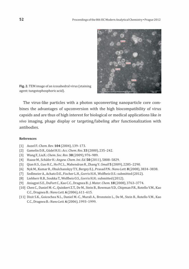
52 the 8 2Proceedings of th ISC Modern Analytical Chemistry Prague 201▪
The virus-like particles with a photon upconverting nanoparticle core com-
bines the advantages of upconversion with the high biocompatibility of virus
capsids and are thus of high interest for biological or medical applications like
imaging, phage display or targeting/labeling after functionalization with
antibodies.
in
vivo
References
[1] Auzel F.: (2004), 139–173.[2] Gamelin D.R., Güdel H.U.: (2000), 235 242.[3] Wang F., LiuX.: (2009), 976 989.[4] Haase M., Schäfer H.: (2011), 5808 5829.[5] Qian H.S., Guo H.C., Ho P.C.L., Mahendran R., Zhang Y.: (2009), 2285 2290.[6] Nyk M., Kumar R., Ohulchanskyy T.Y., Bergey E.J., Prasad P.N.: (2008), 3834 3838.[7] Sedlmeier A., Achatz D.E., Fischer L.H., Gorris H.H., Wolfbeis O.S.: submitted (2012).[8] Liebherr R.B., Soukka T., Wolfbeis O.S., Gorris H.H.: submitted (2012).[9] Aniagyei S.E., DuFort C., Kao C.C., Dragnea B.: (2008), 3763 3774.[10] Chen C., Daniel M.-C., Quinkert Z.T., De M., Stein B., Bowman V.D., Chipman P.R., Rotello V.M., Kao
C.C., Dragnea B.: (2006), 611 615.[11] Dixit S.K., Goicochea N.L., Daniel M.-C., Murali A., Bronstein L., De M., Stein B., Rotello V.M., Kao
C.C., Dragnea B.: (2006), 1993 1999.
Chem. Rev.
Acc. Chem. Res.
Chem. Soc. Rev.
Angew. Chem. Int. Ed.
Small
Nano Lett.
J. Mater. Chem.
Nano Lett.
Nano Lett.
104
33
38
50
5
8
18
6
6
––
––
–
–
–
–
Fig. 2. TEM image of an icosahedral virus (stainingagent: tungstophosphoric acid).

Metabolomics applications require sample preparation methods that are fast,
reproducible and able to extract a wide range of analytes of differing polarities.
The measurement of drug levels in biological fluids is the corner stone for drug
discovery and development as well as for pharmacodynamic, pharmacokinetic
studies and drug monitoring. The most relevant matrices to be analyzed for this
purpose are plasma or blood, due to providing a good correlation between their
concentration and pharmacological effects. Sample preparation is frequently
done and this is often a limiting step to perform fast bioanalysis. The intro
duction of sample pretreatment would greatly speed up the analyses. The
choice of an appropriate sampleextraction technique is very important step for
metabolomic studies. For metabolomic analyses, the analytical technique must
therefore be really suitable for a diverse range of small endogenous metabolites in
various concentrations. solid phase microextraction (SPME) provides an
interesting new complement to the range of technologies currently being em
ployed for analysis of living systems. S can
match the selectivity and sensitivity for improved time resolution of sampling.
Polypyrrole, polythiophene, and poly(3-alikilothiopenes) SPME coatings with
a molecular imprint were used and evaluated their ability to extract selected
off-line -
on-line
In vivo
-
in vivo olid phase microextraction
SPME-LC/MS for the Analysis of
Selected Biologically Active Compounds
n
J W M S B BUSTYNA ALCZAK, AŁGORZATA ZULTKA, OGUSŁAW USZEWSKI
Department of Environmental Chemistry and Bioanalytics, Faculty of Chemistry,
Nicolaus Copernicus University
Gagarin Str. 7, 87-100 Torun, Poland, [email protected]�
Keywords
antibiotics
drug monitoring
high performance liquid chromatography
mass spectrometry
solid phase microextraction
Proceedings of th ISC Modern Analytical Chemistry Prague 201the 8 2▪ 53

drugs from different classes with different physicochemical properties and of
widely varying polarities. Mass spectrometric parameters were optimized for
target compound in positive ion mode over the / 100–1000 range. Quantitation
was done using multiple reaction monitoring mode to monitor precursor ion at
[M+H] to product ion transition of / 366 → 349 for amoxicillin, 332 → 288 for
ciprofloxacin, 478 → 322 for gentamycin, 338 → 296 for linezolid, 172 → 128 for
metronidazole. The results demonstrate the potential of SPME as a useful
sample preparation tool for chromatographic based metabolomics drug monito-
ring in the biomedical application from patients receiving therapeutic dosages.
m z
m z
in vivo
+
Acknowledgments
This work was supported by National Science Center (Cracow, Poland), no. 2011/01/N/ST4/03178and CHEMO-KOP no. 49/2011.
54 the 8 2Proceedings of th ISC Modern Analytical Chemistry Prague 201▪

Recently, a great deal of emphasis has been placed on the development of molecu-
larly imprinted polymers. The term molecularly imprinted polymers stands for
very attractive man-made materials with defined molecular recognition and
predetermined selectivity for the specific analyte, named template [1, 2].
In the presence of the template there are creating synthetic recognition sites,
tailor-made by the copolymerization of crosslinking monomer and at least one
functional monomer. After a polymerization process the template molecule is
removed leaving three-dimensional cavities. Prepared binding sites possess
a steric (in size and shape) and chemical (spatial arrangement of complementary
functional groups) memory for one structure or group of structures on which was
designed [3].
At present time, the importance of molecularly imprinted polymers is growing
up due to many reasons. Their advantage is that they are very selective, cheap,
easy to prepare and they have special chemical and physical properties, which
guarantee them a good stability over a wide range of experimental conditions and
solvents [1].
Temperature Effect on the Sorption
Selectivity of Some Phenolic Acids
on Molecularly Imprinted Stationary
Phases
N D J LATALIA ENDERZ, OZEF EHOTAY
Institute of Analytical Chemistry, Faculty of Chemical and Food Technology,
Slovak University of Technology in Bratislava,
Radlinskeho 9, 812 37 Bratislava, Slovak Republic [email protected]�
Keywords
liquid chromatography
molecularly imprinted polymers
phenolic acids
an t Hoff equationv ’
Proceedings of th ISC Modern Analytical Chemistry Prague 201the 8 2▪ 55

The large interest of molecularly imprinted polymers has progressed mainly in
chemistry and biology, but its preparation is of importance also for philosophy
of science because it enables to get closer to the nature. Currently, there are a lot of
examples of molecularly imprinted polymers application as stationary phases,
sorbents, or synthetic receptors in liquid chromatography, capillary electro-
phoresis, capillary electrochromatography, solid-phase extraction, solid-phase
microextraction or chemical sensors [4–8].
Our work was devoted to determination of temperature-dependence of reten-
tion factors of tested analytes in acetonitrile (porogen) and methanolic eluents.
By examining the temperature dependence of retention we can determine the
retention mechanisms as enthalpically or entropically driven [9]. The extent in
the retention of analytes on stationary phases under given conditions is deter-
mined by the retention factors, ki and the distribution of the solute between the
mobile and stationary phases is define
is specified as a partitioning
process between two phases, the temperature dependence on retention should be
modelled by the van t Hoff plot [10, 12]
(1)
where is the retention factor, is the partial molar enthalpy of transfer
, is the partial molar entropy of transfer , is the gas
constant , is the absolute temperature and is the phase
ratio (the ratio of stationary and mobile phase volumes, and respectively,
within the column) [4].
The aim of present study was an examination of thermodynamic parameters
of the molecular recognition in order to understand the sorption mechanisms on
prepared molecularly imprinted polymers. As templates the following phenolic
compounds were used: gallic, gentisic, syringic, protocatechuic, 4-hydroxy-
benzoic, and vanillic acids (Fig. 1). The functional monomer was acrylamide.
d by the standard free energy change, ΔG
k
T
V V
i
i
°.
Combination of these two quantities over a sufficiently broad temperature range
is commonly used to investigate recognition mechanisms in chromatography as
a van’t Hoff analysis. If the chromatographic retention
’
Δ
Δ
φ
H
S
i
i[kJ mol ] [J mol K ]
[8,314 J mol K ] [K],
–1 –1 –1
–1 –1
R
S M
56 the 8 2Proceedings of th ISC Modern Analytical Chemistry Prague 201▪

Acknowledgments
References
This work was supported by the VEGA grants No. 1/0164/11.
1 Gholivand M.B., Karimian N., Malekzadeh G.: (2012), 513–520.[2] Valtchev M., Palm B.S., Schiller M., Steinfeld U.: (2009), 722 728.[3] Hall A., Lanza-Sellergren F., Manesiotis P., Sellergren B.: (2005), 9 14.[4] Guo Z., Hao T., Shi L., Gai P., Duan J., Wang S., Gan N.: (2012), 1092 1097.[5] Xu Z.X., Gao H.J., Zhang L.M., Chen X.Q., Qiao X.G.: (2011), R69 R75.[6] Denderz N., Lehotay J., Čižmárik J.: (2012), 77 83.[7] Quaglia M., De Lorenzi E., Sulitzky C., Caccialanza G., Sellergren B.: (2003),
952 957.[8] Turiel E., Martin-Esteban A.: (2009), 3278 3284.[9] Ogden P.B., Coym J.W.: (2011), 2936 2943.[10] Haidacher D., Vailaya A., Horváth C.: (1996), 2290 2295.[11] Coym J.W.: (2008), 1712 1718.[12] Chester T.L., Coym J.W.: (2003), 101 111.
[ ] Talanta
J. Hazard. Mater.
Anal. Chim. Acta
Food Chem.
J. Food Sci.
J. Chrom. A
Electrophoresis
J. Sep. Sci.
J. Chrom. A
PNAS
J. Sep. Sci.
J. Chrom. A
89
170
538
132
76
1235
24
32
1218
93
31
1003
––
––
–
––
––
––
Proceedings of th ISC Modern Analytical Chemistry Prague 201the 8 2▪ 57
Fig. .1 Structures of analytes used in the study: (a) gallic acid, (b) gentisic acid, (c) protocatechuicacid, (d) 4-hydroxybenzoic acid, (e) vanillic acid, (f) syringic acid, (g) quercetin, and (h) diperodon.

Ion chromatography is one of the most often applied techniques in the labora-
tories all over the world. Since ion chromatography offers many possibilities of
application it is used in different branches of current analytical chemistry. This
method is fast, cheap, giving good reproducibility of the results, and what is the
most important, providing the qualitative and quantitative results as well.
To develop this method it is necessary to look for new solutions reduce of the
detection limits, improving the efficiency of analytical systems, the quality of
the separation of the components of a mixture and selectivity. At present search
modern stationary phases of high ion exchange capacity, which permit for the
analysis of samples large concentration and contain considerably disproportions
in concentrations of particular ions. The aim of current science is to build systems
simulating the behaviour of real ones. Recently very popular are dendrimer-
-bonded silica surfaces. Unlike classical polymers, dendrimers have a high degree
of molecular uniformity, narrow molecular weight distribution, specific size and
shape characteristic and a highly-functionalised terminal surface which features
render them suitable candidates for the production of homogenous, highly fun-
ctionalised chromatographic surfaces.
The main purpose of this paper was to present the method to obtain new
stationary phase for the ion chromatography. A novel ion-exchanger was prepared
Functionalized Dendrimer
Stationary Phases on Silica Gel
for Ion Chromatography
M J B BAGDALENA AĆKOWSKA, OGUSŁAW USZEWSKI
Department of Environmental Chemistry and Bioanalytics, Faculty of Chemistry,
Nicolaus Copernicus University
Gagarin Str. 7, 87-100 Torun, Poland, [email protected]�
Keywords
anion exchanger
dendrimer-bonded silica surfaces
ion chromatography
58 the 8 2Proceedings of th ISC Modern Analytical Chemistry Prague 201▪

by chemically modification of surface of silica gel carrier. A multilayer structure
(Joshua Tree) was formed during the process. As the monomers: 1, 4-butanediol
diglycidyl ether and methylamine were used. The new anion exchanger for the
determination of inorganic anions was subjected physico-chemical characteri-
zation: Si CP/MAS NMR and FT-IR and elemental analysis.29
Acknowledgments
This work was supported by European Social Found, Polish National Budget, Kuyavian-PomeranianVoivodeship Budget (within Sectoral Operational Programme Human Resources) Krok wprzyszłość IV and Nicolaus Copernicus University grant no. 1115-Ch.
‘’
Proceedings of th ISC Modern Analytical Chemistry Prague 201the 8 2▪ 59

The oxidation of ethylene and propylene respectively, mediated by thin layer
-tetramethyltetra-3,4-pyridinoporphyrazinocobalt, simply called
CoTmt-3,4-ppa, deposited on basal plane of highly ordered pyrolytic graphite
(HOPG) was studied in aqueous phosphate buffer phosphate solution by cyclic
voltammetry, backscattering spectroscopy and atomic force microscopy. Our
interest has been focused on optimizing conditions of electrocatalytic oxidation
unsaturated hydrocarbon on electrode modified by pyridinoporphyrazine.
The complex CoTmt-3,4-ppa as a heterocyclic derivate of metallophthalo-
cyanines [1, 2] is water soluble due to the positive charge localized on the porphy-
razine cycle and shows lower tendency to form aggregates in solution [1, 3].
CoTmt-3,4-ppa can form thin electrically conducting films on the electrode sur-
face after reduction of the central metal [4, 5]. It can act as an electron transfer
mediator between the electrode surface and analyte in solution in our case of
ethylene and propylene. In this work we modified electrode surface by two
different deposition techniques: adsorption and electrochemical deposition, that
just lead to the deposition of an insoluble film on electrode surface.
N,N´,N´´,N´´´
Electrocatalytic Oxidation of
Unsaturated Hydrocarbons Mediated
by Phthalocyanine Derivate
MONIKA LUSÁČKOVÁ AVEL ANDAK , P Ja, b a
a
b
J. Heyrovský Institute of Physical Chemistry, Academy of Sciences of the Czech Republic, v.v.i.,
Dolejškova 3, 182 23 Prague 8, Czech Republic [email protected]�
Department of Analytical Chemistry, Faculty of Science, Charles University in Prague,
Albertov 6, 128 43 Prague 2, Czech Republic
Keywords
atomic force microscopy
backscattering spectroscopy
cyclic voltammetry
phthalocyanine
unsaturated hydrocarbons
60 the 8 2Proceedings of th ISC Modern Analytical Chemistry Prague 201▪

The highly ordered pyrolytic graphite used in this work as a working electrode
is a special type of graphite material [6] with the advantage of well-defined
structure, atomically flat surface, easily renewable surface by removing several
layers of the surface with using adhesive (Scotch) tape. It represents an excellent
electrode material suitable for characterization by atomic force microscopy
respectively.
We demonstrate that HOPG with deposited thin layer CoTmt-3,4-ppa displays
electrocatalytic activity towards the oxidation of both ethylene and propylene,
respectively. We report on the interaction of ethylene and propylene with reduced
form of the CoTmt-3,4-ppa complex involving metal centre and ligand catalytic
activity. During cyclic voltammetry on modified electrode surface irreversible
anodic peaks of ethylene and propylene were observed in the potential range
corresponding to redox couple of the complex CoTmt-3,4-ppa. Influence of pH and
thickness of the deposit on electrode surface on oxidation unsaturated hydro-
carbon were observed. The electrochemical behavior has been completed by
backscattering spectroscopy that confirms the interaction of CoTmt-3,4-ppa
with ethylene and propylene. The change in nanomorphology of the surface
reacting with unsaturated hydrocarbon (Fig. 1b) compared to argon atmosphere
(Fig. 1a) and the thickness of the film on the electrode surface have been examined
by atomic force microscopy.
in
situ
ex situ
(a) (b)
Fig. .1 Ex situ AFM image of the basal plane HOPG modified by CoTmt-3,4-ppa with succesive subjectto (a) argon atmosphere, comparated to (b) propylene atmosphere.
Proceedings of th ISC Modern Analytical Chemistry Prague 201the 8 2▪ 61

Acknowledgments
References
We are grateful to Yu-Hong Tse, York University, Toronto for supply of CoTmtppa used in this work.This work was supported by the research project MSM0021620857, grant project SVV-2012--265201 of Charles University in Prague.
[2] Zhang J., Tse Y.H., Pietro W.J., Lever A.B.P.: (1996), 203 211.[3] Thamae M.A., Nyokong T.: (1999), 126 135.[4] Tse Y.H., Janda P., Lever A.B.P.: (1994), 384 390.[5] Janda P., Weber J., Dunsch L., Lever A.B.P.: (1996), 960 965.[6] Chang H., Bard A.J.: (1991), 1143 1153.
[1] Chen J., Zhang J., Tse Y.H., Janda P., Christendat D., Lever A.B.P.:(2006), 1238–1248.
––
––
–
J. Porphyrins Phthalocyanines 10
J. Electroanal. Chem.
J. Electroanal. Chem.
Anal. Chem.
Anal. Chem.
Langmuir
406
470
66
68
7
62 the 8 2Proceedings of th ISC Modern Analytical Chemistry Prague 201▪

Biological fluids, such as blood plasma, serum and serum ultrafiltrate [1] have
been widely investigated by molecular spectroscopy methods in order to esta
blish new, minimally-invasive, rapid, reliable and relatively inexpensive screening
methods in the clinical diagnosis of various diseases neurodegenerative, can
cerous and others [2 4]. Blood plasma probably has the highest potential because
it is relatively easy to obtain and contains many dissolved peptides, proteins and
other biomolecules, among others those causing or indicating several diseases
[2 4]. The methods newly introduced in the last decade for such investigations are
Raman, infrared and fluorescence spectroscopy [1 6]. However, to the best of our
knowledge, the advanced chiroptical variants (ECD, -
, , of above mentioned methods have never
been used for analyzing the real clinical blood-based samples. Chiroptical me-
thods are among the few methods inherently sensitive to the 3D structure of chiral
biomolecules [7, 8] that is a big advantage compared to their common (unpolari-
zed) variants. Furthermore, chiroptical methods have probably a potential for the
detection of slight conformational and stereochemical changes in the secondary
and tertiary structure of biomolecules, which occur during the above mentioned
diseases.
We tested possibilities of chiroptical methods for the prospective diagnosis of
cancer and neurodegenerative diseases, based on the analysis of blood plasma
-
– -
Electronic Circular Dichro
ism and ROA Raman Optical Activity)
–
–
–
Chiroptical pectroscopy of uman
lood lasma in linical iagnosis
S H
B P C D
MICHAL ATARKOVIČ, LADIMÍR ETNIČKAT V S
Department of Analytical Chemistry, Institute of hemical echnology Prague,
Technická 5, 166 28 Prague 6, Czech Republic
C T
Keywords
blood plasma
cancer
chiroptical spectroscopy
spectral marker
Proceedings of th ISC Modern Analytical Chemistry Prague 201the 8 2▪ 63

samples. The blood of healthy controls and patients with colon cancer was
collected by venipuncture and plasma was immediately frozen ( 75 °C). The ECD
spectra were measured directly after thawing frozen samples at ambient tempe
rature and also after dilution them by a phosphate buffer (pH = 7.4) in 0.01 mm
quartz cuvette. Unfortunately, the blood plasma had a very strong fluorescence
background in ROA/Raman spectroscopy; therefore, we had developped the
methodology how to suppress it [9]. We were able to reduce fluorescence back
ground ten times. However, the spectral baselines had to be further improved by
correction using Fourier filtering procedure [10]. The ECD and ROA spectra were
evaluated by multidimensional statistical methods (PCA, LDA).
Our preliminary results suggest that the human blood plasma can be success
fully measured by both the ECD and ROA methods. We observed the spectral
differences between the blood samples of healthy controls and the colon cancer
patients. Therefore, these methods can potentially be useful for following
research in the development of new, minimally-invasive screening methods of
clinical diagnostics of colon cancer and maybe other diseases in future.
–
-
-
-
Acknowledgments
References
The work was supported by the Prague Operational Programme for Competitiveness (Grant NoCZ.2.16/3.1.00/22197); the Ministry of Health of the Czech Republic (Grant No NT13259); theMinistry of Education, Youth, and Sports of the Czech Republic (Grant No MSM6046137307);Specific University Research MSMT No 21/2012 - A1_FCHI_2012_003 and A2_FCHI_2012_015.
Lasch P., Kneipp J.: Wiley 2008.2 Burns D.H., Rosendahl S., Bandilla D., Maes O.C., Chertkow H.M., Schipper H.M..:
(2009), 391 397.
4 Ahmed S.S.S.J., Santosh W., Kumar S., Thanka Christlet T.H.: (2010), 181 188.5 Mizuno A., Kitajima H., Kawauchi K., Muraishi S., Ozaki Y.: (1994),
25 29.6 Huang Z., McWilliams A., Lui H., McLean D.I., Lam S., Zeng H.: (2003),
1047 1052.7 Berova N., Nakanishi K., Woody R.: Wiley 2000.8 Barron L.D., Hecht L., Blanch E.W., Bell A.F.: (2000), 1 49.9 Tatarkovič M., Fišar Z., Raboch J., Jirák R. Setnička V.: (2012), in press10 Člupek M., Matějka P., Volka K.: (2007), 1174 1179
.
[1][ ]
–
[ ][ ]
[ ]
[ ][ ][ ] , .[ ] .
Biomedical ibrational pectroscopy.
J. Alzheimer s
Dis.
Vib. Spectrosc.
J. Raman Spectrosc.
Int. J. Cancer
Circular ichroism rinciples and pplications.
Prog. Biophys. Mol. Biol.
Chirality
J. Raman Spectrosc.
V S
’
D . P A
17
53
25
107
73
38
[ ]–
–
–
–
–
–
3 Griebe M., Daffertshofer M., Stroick M., Syren M., Ahmad-Nejad P., Neumaier M., Backhaus J.,Hennerici M.G., Fatar M.: (2007), 29 33.Neurosci. Lett. 420
64 the 8 2Proceedings of th ISC Modern Analytical Chemistry Prague 201▪

Research on separations using short conventional capillary pathways at high field
strengths is receiving increased interest due to the possibility of achieving rapid
and inexpensive analyses with potential for high-throughput applications. The
concept can be especially useful for monitoring reaction kinetics or for imple-
mentation into multidimensional systems. Coupling the short capillaries to
electrochemical detection offers the possibility to construct highly miniaturized
and independent analysis devices as power consumption and component size are
comparatively low [1, 2].
A system for combining the advantages of short capillary electrophoresis and
amperometric detection within an automated device is presented. An electro-
chemical cell made from PTFE is used to align short capillaries (4 to 10 cm) with
homemade Pt-microelectrodes in the end-column detection mode. The use of
non-aqueous acetonitrile based buffer systems offers some advantages like better
electrochemical response and faster separations. In order to achieve high-
-throughput separations, the cell is placed in a homemade autosampling unit and
injection occurs in a vertical positioning of the capillary. As the injection plug
Fast Non-Aqueous Capillary
Electrophoresis in Short Capillaries
with Amperometric End-Column
Detection
J M F M MONAS ARK, RANK- ICHAEL ATYSIK
Institute of Analytical chemistry, Chemo- and Biosensors, University of Regensburg,
Universitätsstrasse 31, 93040 Regensburg, Germany [email protected],�
Keywords
amperometric detection
end-column
fast separation
high-throughput
non-aqueous capillary electrophoresis
Proceedings of th ISC Modern Analytical Chemistry Prague 201the 8 2▪ 65

length has to be kept at a narrow level, especially using such short capillaries,
different injection protocols (for example tapered capillary tips) are investigated.
In addition to that, a range of different capillary inner diameters (5 to 50 µm) were
tested and evaluated for applicability. A variety of effects like potential shift under
influence of high voltage fields are examined and the method is optimized using
a model system consisting of ferrocene, its derivatives and other electro-
chemically active substances. In this manner, a continuous sequence of fast and
reproducible electrophoretic measurements is possible. This method will allow in
the near future the construction of a miniaturized, fully portable device with
point-of-care applicability.
References
[1] Opekar F., Coufal P., Stulik K.: (2009), 4487–4499.[2] Matysik F. M.: (2010), 961 965.
Chem Rev.
Anal. Bioanal. Chem.
109
397 –
66 the 8 2Proceedings of th ISC Modern Analytical Chemistry Prague 201▪

4-aminobenzenethiol can form self-assembled mono-
layers via its thiol group on appropriate metal surfaces
(Fig. 1). The Surface-Enhanced Raman Scattering
spectroscopy (SERS) has become an appropriate tech-
nique to study layers on metal/solution surface/inter-
face. Raman spectra of analytes adsorbed on nano-
structured surfaces of coinage metals can be enhanced
by factor 10 or higher [1, 2], but only under certain
conditions: The analyte molecules have to be adsorbed to
the surface or located in its close vicinity. Roughness of
metal surface has to be in nano- and/or micro-scale level.
5
Spectroelectrochemical Study
of 4-Aminobenzenethiol Adsorbed
on Gold, Silver and Copper
Nanostructured Surfaces
MARCELA ENDISOVÁ, UKÁŠ AVRÁNEK, AVEL ATĚJKAD L H P M
Department of Analytical Chemistry, Institute of Chemical Technology Prague,
Technická 5, 166 28 Prague 6, Czech Republic [email protected]�
Keywords
4-aminobenzenethiol
spectroelectrochemistry
SERS
in-situ
Proceedings of th ISC Modern Analytical Chemistry Prague 201the 8 2▪ 67
Fig. .1 Bound of 4-amino-benzenethiol on a metalsurface.
The suitable method for approaching nanostructured surface is electrochemical
roughening using cathodic metal coating of suitable substrate or oxidation-
-reduction cycles when both surface morphology and the corresponding enhan-
cement factor are changing. The enhancement factor depends on several effects,
e.g. applied potential, excitation wavelength, surface coverage, and surface
morphology [3]. The resulting enhancements of surface Raman signals of
adsorbed molecules arise from two main aspects: physical and chemical
enhancement mechanisms. The so-called electromagnetic mechanism is

considered as the main contribution to the SERS effect. Chemical enhancement
can be caused for example by photoinduced charge transfer mechanism,
tautomerization of benzoid or the charge tunnelling enhancement mechanism
[4]. Raman bands corresponding to the modes perpendicular to the surface are
significantly enhanced. Thiol group of 4-aminobenzenethiol interacts strongly
with the metal surface and covalent bond is formed usually between surface
atoms and sulphur atom [4]. Furthermore, 4-aminobenzenethiol can react to a
new azobenzene compound by catalytic coupling reaction on silver nanoparticles
[5, 6]. A similar behaviour was observed probably on gold nanofeatures [5]. The
formation of the azobenzene should be indicated by observation of intense bands
about 1142, 1392 and 1439 cm .
The nature and re-orientation of molecule adsorbed on SERS-active nanostruc-
tured surfaces (Au, Ag, or Cu) can be readily determined by electrochemical
surface-enhanced Raman scattering spectroscopy [7]. The combination of SERS
spectroscopy and electrochemistry allows much more comprehensive studying of
adsorption processes, enabling to tune the conditions of analyte sorption onto
metal surface by changing the experimental electrochemical parameters. In this
study, the electrochemical cell was used for electrochemical SERS study of
4-aminobenzenethiol adsorbed on gold, silver and copper substrates to demon-
strate that adsorption of 4-aminobenzenethiol is influenced by applied electrode
potential and formation of azobenzene complex depends evidently on the kind of
metal used. In our previous study aimed at enhancement factor calculation [1] we
revealed that any azo-complex do not form from 4-aminobenzenethiol adsorbed
on copper surface at 1064-nm excitation which can be explained either by the
influence of the metal or the low energy excitation.
SERS-active large-scaled substrates were prepared by electrochemical (catho-
dic) metal coating of massive platinum target in two-electrode arrangement from
electrochemical bath contained corresponding ions. Current sequences (based on
previously published procedures [8–10]) include from two to five individual steps
(Table 1). Surface morphologies were modified using oxidation-reduction cycle
application.
All SERS spectra were collected using Raman spectrometer Dimension
P2 (Lambda Systems, USA). The laser beam (785 nm) irradiating the surface of
target through glass window was focused using a micrometer positioning device
developed at our department. Measurement conditions as laser power and
integration time were optimized for each tested metal. Number of repeated
accumulations was always ten. Spectra were recorded with resolution better than
8 cm .
–1
–1
in-situ
68 the 8 2Proceedings of th ISC Modern Analytical Chemistry Prague 201▪

All measurements were performed in an electrode cell (Fig. 2) which was
equipped with a salt bridge suitable for various reference electrodes, platinum
plate and contact for SERS-active working electrode (massive metal target with
diameter up to 10 mm coated with various SERS-active metal layers). Cyclic series
of potentials were applied to working electrode in direction from ca. +100 mV to
ca. –1100 mV and backward with a step of 100 mV; SERS spectra were recorded at
each (stabilized during data accumulation) applied potential. Raman probe head
was connected to the spectrometer via fibre optics. Electrolyte (aqueous
0.1 K SO ) contained 4-aminobenzenethiol (5×10 mol L ).
In SERS spectra recorded on from 100 to –1100 mV we
can observed maximal spectral intensity at potential values about –600 mV and
lower. The bands of azobenzene compound at ca. 1142 and 1390 cm are clearly
mol L–12 4
–5 –1
–1
in-situ gold substrate
Proceedings of th ISC Modern Analytical Chemistry Prague 201the 8 2▪ 69
Table 1
Current sequences used for large-scaled substrates preparation.
Substrate Au Ag Cu
Bath [Au(CN) ] [Ag(NH ) ] [Cu(NH ) ]
Step , mA , min mA min mA min
1 15 5 5 5 10 10
2 10 10 10 15 20 10
3 5 15 – 30 10
4 40 10
5 50 10
2– + 2+
3 2 3 4
I t I, t, I, t,
–
– – – –
– – – –
Fig. 2. Spectroelectrochemical cell

evident (Fig. 3). Intensity of other characteristic vibration modes assigned to
4-aminobenzenethiol (e.g., at about 1080 cm assigned to C S vibration mode)
increased to potential values about 600 mV and then increased very gradually.
From the observations we can conclude that adsorption of 4-aminobenzenethiol
is irreversible process, while azobenzene complex formation depends reversibly
on potential. In spectra measured on we can observe similar
behaviour as in the case of gold substrate. Band intensities characteristic for
azobenzene formation increased up to 700 mV and then decreased close to noise
level. Bands intensity assigned to 4-aminobenzenethiol vibration modes increa
sed too but their intensity did not decrease to the noise level. In the case of
no azobenzene bands (Fig. ) were observed at any electrode potential.
That means that photon-driven charge transfer mechanism does not contribute to
the chemical mechanism and 4-aminobenzenethiol molecules are adsorbed to the
copper substrate unchanged.
In the work we confirm formation of azo-complex produced by catalytic
coupling reaction in the cases of gold and silver surfaces. The formation of the
complex is not observed in the case of copper surface even at shorter wavelengths
(785 nm) than 1064 nm. Thus, there is a crucial role of the metal on the photo-
catalytic formation of azobenzene surface complex.
–1 –
–
–
-
3
silver substrate
copper
substrate
Fig. 3. In-situ surface-enhanced Raman scattering spectra of adsorbed ongold, silver, or copper surfaces measured at –700 mV.
4-aminobenzenethiol
70 the 8 2Proceedings of th ISC Modern Analytical Chemistry Prague 201▪

Proceedings of th ISC Modern Analytical Chemistry Prague 201the 8 2▪ 71
Acknowledgments
References
Financial support from specific university research (MSMT no. 21/2012 – A2_FCHI_2012_013) wasgratefully acknowledged.
Dendisová-Vyškovská M., Prokopec V., Člupek M., Matějka P.: (2012),181–186.
[2] Otto A., Mrozek I., Grabhorn H., Akemann W.: (1992), 1143 1212.[3] Brolo A. G., Irish D. E., Smith B. D.: (1997), 29 44.[4] Wu D. Y., Liu X. M., Huang Y. F., Ren B., Xu X., Tian Z. Q.: (2009), 18212 18222.[5] Canpean V., Iosin M., Astilean S.: (2010), 277 282.[6] Wu D. Y., Zhao L. B., Liu X. M., Huang R., Huang Y. F., Ren B., Tian Z. Q.: (2011),
2520 2522.[7] Roguska A., Kudelski A., Pisarek M., Lewandowska M., Kurzydłowski K. J., Janik-Czachor M.:
(2009), 2820 2824.[8] Prokopec V., Cejkova J., Matejka P., Hasal P.: (2008), 601 607.[9] Cejkova J., Prokopec V., Brazdova S., Kokaislova A., Matejka P., Stepanek F.:
(2009), 7864 7870.[10] Prokopec V., Dendisová-Vyškovská M., Kokaislová A., Čejková J., Člupek M., Matějka P.:
(2011), 410 419.
[1] J. Raman Spectrosc.
J. Phys.-Condes. Matter
J. Mol. Struct.
J. Phys. Chem. C
Chem. Phys. Lett.
Chem. Commun.
Surf. Sci.
Surf. Interface Anal.
Appl. Surf. Sci.
J. Mol.
Struct.
43
4
405
113
500
47
603
40
255
993
––
––
–
––
–
–

Introduction
The diphenyl ether herbicides Fomesafen and Fluorodifen, that are a focus of this
work, are potent herbicides [1, 2]. As other diphenyl ethers they have unfortu-
nately some unpleasant side-effects, including mast cell activation, teratogenesis
and endocrine disrupting activity [3–5]. In addition, some of their metabolites are
known to be possible mutagens [6, 7]. Compounds with similar structure exhibit
toxicity toward aquatic organisms and interfere with blood formation [4]. The
herbicide Fomesafen has been developed for the preemergence protection
of beans, soybeans and cotton against weeds [8]. Although appropriate methods
for the determination of diphenyl ether herbicides in the environment already
exist, it still makes sense to develop new voltammetric methods. This is because
substances of our interest do exhibit electrochemical activity [9] and electro-
chemical methods are cheap, easy and have low detection limits [10]. Silver solid
amalgam electrodes poses these characteristics and have already proved suitable
for the determination of trace amounts of pollutants [11, 12]. Our goal in this work
is to develop sensitive and reliable methods for the determination of Fomesafen
and Fluorodifen at (m-AgSAE)meniscus modified silver solid amalgam electrode
Voltammetric Determination
of Fomesafen and Fluorodifen
in Drinking Water and River Water
V N J M E K J BÍT OVOTNÝ, AN AŠKA, TELA OUKLÍKOVÁ, IŘÍ AREK
UNESCO Laboratory of Environmental Electrochemistry, Department of Analytical Chemistry,
Faculty of Science, Charles University in Prague,
Albertov 6/2030, 128 43 Prague 2, Czech Republic, [email protected]�
Keywords
Fomesafen
Fluorodifen
voltammetry
water
72 the 8 2Proceedings of th ISC Modern Analytical Chemistry Prague 201▪

by finding the optimal conditions for the determination and testing the reliability
of the method.
Fomesafen (98.7 %) and Fluorodifen (99.6 %) were used. The stock solution of
Fomesafen ( = 1×10 mol L ) has been prepared by dissolving 0 0439 g of
Fomesafen in 100 mL of methanol. The stock solution of Fluorodifen (
) has been prepared by dissolving 0 03290 g of Fluorodifen in
100 mL of methanol. Both were purchased from SigmaAldrich Laborchemikalien,
Steinheim, Germany. Other used chemicals: boric acid, acetic acid (99%), phos
phoric acid (85%), sodium hydroxide, potassium chloride all substances p.a.,
Lachema Brno, Czech Republic. Methanol p.a. (Merck, Germany) and deionized
water (Millipore, USA) were used as solvents.
Measurements of pH were performed on a Jenway 3510 (Jenway, Essex, Great
Britain) pH-meter with a combined glass membrane electrode (type 924 005) The
electrode was calibrated by standard buffer solutions in water. The UV-VIS
measurements were done using a HP 8453 (Hewlett Packard, Great Britain)
diode-array spectrophotometer with a fused quartz cuvette with a thickness of
1 mm. Palmsens Electrochemical Sensor Interface (Palm Instruments BV, Ruiter-
camp, The Netherlands) and the PalmsensPC software were used for all voltam-
metric measurements. The software was running under the Windows XP (Micro-
soft Corp.) operating system.
Pulses of width of 100 ms and height of 50 mV were used while performing
DPV. A polarization rate of 20 mV s , and potential resolution of 5 mV were used.
All measurements were performed using a three electrode system. A silver
chloride electrode (1 mol L KCl) type RAE 113, (Monokrystaly, Turnov, Czech
Republic), a platinum wire auxiliary electrode and a meniscus modified silver
solid amalgam electrode (Polaro Sensors, Prague, Czech Republic). The vacuum
manifold and Lchrolut RP-18 E cartridges were purchased from Merck, Darm-
stadt, Germany.
Chemicals
Apparatus
c
c
–3 1
–1
–1
–
–3 –1
.
=
= 1×10 mol L .
-
Proceedings of th ISC Modern Analytical Chemistry Prague 201the 8 2▪ 73
Procedures
Results and Discussion
The stability of stock solutions was checked by UV-VIS spectrophotometric mea-
surements. The stock solutions were stable for the duration of the experiments.
The phosphate buffer of the desired pH was prepared by mixing the appropriate
amounts of 1 mol L sodium hydroxide and 0.5 phosphoric acid. Passi
vation of the electrode during DPV measurements of solutions containing Fome
safen was resolved by activating the electrode by 300 0,1 s pulses between the
potentials 0 V and 500 mV. Passivation of the electrode while measuring Fluoro
difen was resolved by activating the electrode by 300 0,1 s pulses between the
potentials 200 mV and 1100 mV. Measured solutions of Fomesafen were
prepared by mixing 1 mL phosphate buffer pH 2 9 mL drinking water and
then adding the appropitate amount of Fomesafen stock solution in methanol.
Measured solutions of Fluorodifen were prepared by mixing 3 mL methanol
eluate with 2 mL methanol and 5 mL BR buffer pH 6 . Values of points in
calibration curves are arithmetic averages of measurements done in newly
prepared solutions. Error bars are the standard deviations of the same data.
Detection limits are calculated according to the formula
=3.3 / (1)
where is the standard deviation of 10 measurements of the lowest concentration
when the signal can still be evaluated, and is the slope of calibration curve in the
vicinity of that concentration [13]. The SPE cartidges were conditioned by passing
10 mL methanol and deionised water. 100 mL of sample of drinking water spiked
with Fluorodifen was then passed through them. In order to elute the cartridges
3 mL of methanol was used.
The voltammograms of Fomesafen in a mixture of phosphate buffer pH = 2.0 and
drinking water (1:9) can be found in Fig. 1. The calibration dependence of Fome-
safen in the concentration range from 1×10 mol L to 2 10 mol L is in the
inset. The calibration dependence is linear. Lower concentrations could not be
rached because the signal is not meaningfully reproducible below 2 10 mol L
concentration.
–1
–6 1 7 1
mol L -
-
×
– -
×
– –
= .0 with
= .0
three
×
×
–1
– – –
–7 –1
LOD S
S
σ
σ
74 the 8 2Proceedings of th ISC Modern Analytical Chemistry Prague 201▪

The calibration dependence of Fluorodifen is linear and the intercept is not
significantly different from zero, and detection limit is below 2×10 mol L .
A method for the determination of Fomesafen in drinking water in the concen-
tration range 2–100×10 mol L has been developed. The resulting calibration
dependence is linear. The limit of detection achieved is 1.5×10 mol L . A method
for the determination of Fluorodifen in drinking water using SPE in the
concentration range has been developed. The limit of detection
achieved is below 1 . The calibration dependence is linear. The
results for the determination of Fomesafen in drinking water in the concentration
range and the methods for direct determination of Fomesafen
and SPE assisted determination of Fluorodifen in river water will be presented.
–7 1
–6 –1
–6 –1
–
–7 –1
–7 –1
–7 –1
Conclusions
2–10×10 mol L
.0×10 mol L
2–10×10 mol L
Acknowledgments
Financial support of this work, provided by The Ministry of Education, Youth and Sports of the CzechRepublic (project MSM 0021620857), Technological Agency of the Czech Republic (projectTA01020565), Charles University in Prague (project SVV 2012-265201), and Grant Agency of theCzech Republic (project P206/12/G151), is gratefully acknowledged.
-100 -200 -300 -400 -500 -600
-5
-10
-15
-20
-25
-30
0 2 4 6 8 100
-3
-6
-9
-12
c [ mol.L
-1
]2
4
5
6
3
I [nA]
E [mV]
1
I [nA]
Fig. .1 Difference pulse voltammograms of Fomesafen at meniscus modified silver solid amalgamelectrode in a mixture of phosphate buffer pH = 2.0 and drinking water 1:9. Fomesafen concentrationin solution 0 (1), 2 (2), 4 (3), 6 (4), 8 (5) and . The corresponding calibration straightline is in the inset.
10 (6) μmol·L–1
Proceedings of th ISC Modern Analytical Chemistry Prague 201the 8 2▪ 75

References
[1] Jacobs J. M., Jacobs N. J., Sherman T. D., Duke S. O.: (1991), 197–203.[2] Hallahan B. J., Camilleri P., Smith A., Bowyer J. R.: (1992), 1211 1216.[3] Mastorakos G., Karoutsou E. I., Mizamtsidi M., Creatsas G.: (2007), 219 237.[4] Teshima R., Nakamura R., Nakajima O., Hachisuka A., Sawada J.-I.: (2004),
277 283.[5] Takayasu H., Nakazawa N., Montedonico S., Puri P.: (2007), 426 430.[6] Draper W. M., Casida J. E.: (1983), 227 231.[7] Tanaka Y., Iwasaki H., Kitamori S.: (1996), 15 20.[8] Santos J. B., Jakelaitis A., Silva A. A., Costa M. D., Manabe A., Silva M. C. S.:
(2006), 284 289.[9] Rupp E. B., Zuman P., Šestáková I., Horák V.: (1992), 2016 2021.[10] Barek J., Fischer J., Navrátil T., Pecková K., Yosypchuk B., Zima J.: (2007),
2003 2014.[11] Yosypchuk B., Barek J.: (2009), 189 203.[12] Novotný V., Barek J.: (2009), 217 223.[13] Hayashi Y., Matsuda R., Ito K., Nishimura W., Imai K., Maeda M.: (2005), 167 169.
Plant Physiol.
Plant Physiol.
Endocrine
Toxicol. Lett.
J. Pediatr. Surg.
J. Agric. Food Chem.
Water Sci. Technol.
Weed Research
J. Agric. Food Chem.
Electroanalysis
Crit. Rev. Anal. Chem.
Chem. Listy
Anal. Sci.
97
100
31
150
42
31
34
46
40
19
39
103
21
––
––
––
––
––
––
76 the 8 2Proceedings of th ISC Modern Analytical Chemistry Prague 201▪

Aflatoxins are a type of pathogens which can be found in agricultural food pro-
ducts. These mycotoxins are secondary metabolites produced by several species
of fungi, such as (which produces aflatoxin B and aflatoxin B )
and (which produces aflatoxin B , B , G and G ). Aflatoxins
are known to be mutagenic, carcinogenic and teratogenic compounds. The intake
of these toxins over a long period of time in very low concentrations may be highly
dangerous. It has been estimated that mycotoxin contamination may effect as
much as 25% of the world s food crops each year.
There have been elaborated several methods for determination of aflatoxins in
food. The aim of this work was develop an innovative system of micro-SPE coupled
with micro-HPLC/LIF. The designed system consists of two pumps delive
ring the mobile phases, a set of capillaries (extraction, separation, connecting
capillaries) and a LIF detector (comprising a 355 nm laser, a photomultiplier and
an optical system of our own design). The capillary extraction columns were
either packed with C phase or molecularly imprinted polymeric monoliths. The
main separation column was a 200 μm i.d. capillary packed with a cholesterol
phase. Also, an attempt to synthesize a cholesteryl methacrylate monolithic
column has been made.
Aspergillus flavus
Aspergillus parasiticus
on-line -
, ,
1 2
18
1 2 1 2
’
Determination of Mycotoxins
by Micro-SPE Coupled
with Micro-HPLC/LIF
on-line
D G M S R G KB B
AMIAN RZYWIŃSKI, ICHAŁ ZUMSKI, ENATA ADZAŁA- OPCIUCH,
OGUSŁAW USZEWSKI
Department of Environmental Chemistry and Bioanalytics, Faculty of Chemistry,
Nicolaus Copernicus University
Gagarin Str. 7, 87-100 Torun, Poland, [email protected]�
Keywords
aflatoxins
micro-SPE
micro-HPLC/LIF
Proceedings of th ISC Modern Analytical Chemistry Prague 201the 8 2▪ 77

Monolithic capillary extraction columns have been prepared in fused-silica
capillaries in two-step process. First, a poly-TRIM monolith was photopoly-
merized, then it was modified by photografting by the mixture of methacrylic acid
(MAA) as a functional monomer, ethylene dimethacrylate (EDMA) as a cross-
-linking agent, 5,7-dimethoxycoumarin (5,7-DMC) as a template, isooctane/to-
luene as a porogenic solvent and 2,2-azobis-(2-methylpropionitrile) (AIBN) as an
initiator. Different thermal conditions and concentrations of the grafting mixture
were tested. The extraction capillary columns were evaluated in the terms of their
hydrodynamic and chromatographic properties. Retention coefficients for afla-
toxin B and 5,7-DMC were calculated in order to determine the selectivity and
imprinting factor.
The on-line micro-SPE/micro-HPLC-LIF system allowed to extract and
separate four aflatoxins from an aqueous sample and detect them on the picogram
level. It was clearly demonstrated that MIP polymers are able to bind aflatoxins
more effectively than NIP. The obtained experimental results successfully high-
light the effectiveness of this method in routine analysis of environmental
samples.
1
Acknowledgments
The work was financially supported by the Ministry of Science (Warsaw, Poland) grant numberN N204 012038 and National Science Center (Krakow, Poland) grant no 2011/01/B/ST4/00543.
78 the 8 2Proceedings of th ISC Modern Analytical Chemistry Prague 201▪

Chemical signals represent the main communication tool in termites. In view of
the fact that termites are living in the permanent darkness of their nests and
tunnels, all aspects of their social life are regulated by pheromones. Chemistry is
also involved in termite defence; up to now, nearly 400 defensive compounds from
different chemical classes were identified in termites, produced namely by the
unique defensive gland of soldiers, the frontal gland [1]. In our current project, we
aim at chemical communication and defensive chemistry of several termite
genera from the families Rhinotermitidae and Serritermitidae. In my presen-
tation, I review our recent results on the trail-following communication in the
Neotropical termite (Serritermitidae) and the defensive
chemistry of African sand termite (Rhinotermitidae).
The results are presented with emphasis on modern methods of analytical
chemistry used to detect and identify semiochemicals and defensive compounds.
Firstly, we investigated the trail-following pheromone in
The trail-following pheromones, studied in more than 60 termite
Glossotermes oculatus
Psammotermes hybostoma
Glossotermes
oculatus.
Analytical Methods Used in the Studies
on Chemical Ecology of Termites
J K , R H , P Ž ,P J , J Š , B K , Z B ,I V
ANA RASULOVÁ OBERT ANUS ETR ÁČEK
AVEL IROŠ AN OBOTNÍK LANKA ALINOVÁ UZANA OSÁKOVÁ
RENA ALTEROVÁ
a, b , D S -DAVID ILLAM USSÈSb b a, b
b b b a
b
a
b
Department of Analytical Chemistry, Faculty of Science, Charles University in Prague,
Albertov 6, 128 43 Prague 2, Czech Republic
Infochemicals, Institute of Organic Chemistry and Biochemistry, Academy of Sciences
of the Czech Republic,
Flemingovo nám. 2, 166 10 Prague 6, Czech Republic [email protected]�
Keywords
defensive compounds
GC-MS
GC×GC-MS
termites
trail-following pheromone
Proceedings of th ISC Modern Analytical Chemistry Prague 201the 8 2▪ 79

species, belong to the most frequent chemical signals used by termites and their
diversity is surprisingly low, with only 8 compounds being identified so far [2].
Using a combination of two-dimensional gas chromatography, preparative GC,
electrophysiology and behavioural bioassays, we identified (10 ,13 )-nonadeca-
-10,13-dien-2-one as the trail-following pheromone of [3].
Fragmentation pattern of EI mass spectrum, DMDS derivatization and knowledge
of termite biosynthesis enabled us to propose the final structure which was sub-
sequently confirmed by synthesis of the compound. The biological activity was
verified in bioassays and the quantity of pheromone (hundreds of picograms to
units of nanograms) in the sternal gland of one worker was determined using
three different approaches.
Secondly, we studied the composition of the frontal gland secretion of soldiers
in Using GC-MS analyses we indicated altogether 30
sesquiterpene hydrocarbons or their oxygenated derivatives in nine colonies
from five localities [4]. Identification of sesquiterpenes is difficult due to similarity
of their mass spectra so we compared retention indices and fragmentation
patterns with published data [5] or with commercial or natural standards using
SPME. The comparison of chemical diversity between particular colonies has
revealed a considerable geographical variability.
I review our findings on the trail-following pheromone of
and on the chemical composition of the defensive secretion in
These findings belong to our current project focused on
chemical ecology of critical termite genera from families Rhinotermitidae and
Serritermitidae.
Z Z
Glossotermes oculatus
Psammotermes hybostoma.
Glossotermes
oculatus Psammo-
termes hybostoma.
Acknowledgments
References
We are grateful for financial support to the Czech Science Foundation (No. P506/10/1570), to theAcademy of Sciences, Czech Republic (RVO:61388963) and to the Grant Agency of the CharlesUniversity in Prague, projects SVV 2012265201 and MSM 0021620857.
Šobotník J., Jirošová A., Hanus R.: (2010), 1012 1021.[2] Bordereau C., Pasteels J.M.: Pheromones and chemical ecology of dispersal and foraging in
termites. In: Bignell D.E., Roisin Y., Lo N. (edits).Dordrecht, Springer 2011. p. 279–320.
[3] Hanus R., Šobotník J., Krasulová J., Jiroš P., Žáček P., Kalinová B., Dolejšová K., Cvačka J.,Bourguignon T., Roisin Y., Lacez M., Sillam-Duss (2012), 55 63.
[1]
ès D.:
J. Insect Physiol. –
Biology of Termites, a Modern Synthesis.
Chem. Senses.
56
37 –
80 the 8 2Proceedings of th ISC Modern Analytical Chemistry Prague 201▪

[4 ichý M., Valterová I.:(2012), 557–565.
] Krasulová J., Hanus R., Kutalová K., Šobotník J., Sillam-Dussès D., T J. Chem.
Ecol. 38
[5] Adams R. P.:Allured Publishing Corporation 2007.
Identification of Essential Oil Components by Gas Chromatography/Mass
Spectrometry.
Proceedings of th ISC Modern Analytical Chemistry Prague 201the 8 2▪ 81

Plasma pencil is a modern and unique tool developed for surface treatment of
some archaeological artefacts. In fact it is a narrow quartz tube with an atmos-
pheric pressure argon or helium radiofrequency (13.56 MHz) plasma jet. This
plasma pencil was modified for chemical analysis of liquids by a fused inlet for
sample aerosol introduction from the nebulizer into the plasma gas stream. This is
a big challenge for development of a new low-cost analytical method of elements
determination. Possibilities can be upgraded towards chemical analysis or to
control the admixture composition. The admixture may then be helpful for sample
treatment. The top of this work is the fact that the excitation capabilities of the
discharge were proven. For this purpose sets of calibration standard water solu-
tions containing Li, Ca, Mg, Zn, Cu ions, construction of calibration dependences in
the range of concentrations from 1 to 100 mg/L and calculation of instrumental
detection limits (3 A sample was introduced by
a system consisted of a peristaltic pump delivering 0.6 mL/min of the aqueous
solution into a Scott chamber with a concentric nebulizer with efficiency about
6%. Carrier gas argon (0.3 L/min) flowing from the nebulizer transported this
created aerosol perpendicularly into the main argon stream (4 L/min) flowing
σ) for these elements were done.
Analytical Capabilities of Plasma Pencil
in Atomic Emission Spectrometry
L N , A H , P S ,V O , V K
UKÁŠ OVOSÁD LEŠ RDLIČKA AVEL LAVÍČEK
ÍTĚZSLAV TRUBA IKTOR ANICKÝ
a a, * b
a a
a
b
Central European Institute of Technology, CEITEC MU, Department of Chemistry, Faculty of
Science, Masaryk University, Kamenice 753/5, 62500 or Kotlářská 2, 61137 Brno, Czech Republic,
Department of Physical Electronics, Faculty of Science, Masaryk University,
Kotlářská 2, 61137 Brno, Czech Republic
�
Keywords
elements determination
low-cost
plasma pencil
sample introduction
82 the 8 2Proceedings of th ISC Modern Analytical Chemistry Prague 201▪

directly to the plasma pencil. The electrical input power coming into the plasma
should be between 120 and 140 W, which is necessary to maintain discharge
stability in the case of water and sample load. Electron number densities and
rotational temperatures (relatively low about 1000 K) for the discharge under and
without water load were calculated using H line and OH spectra. Found excitation
temperatures from argon lines ranged from 3000 to 4200 K. Intensities of the lines
were acquired in 10 lateral positions along the tube from the aerosol inlet towards
the discharge tail. Intensities of analytical lines were acquired in the same posi-
tions and an optimal position providing the best signal-to-noise ratio was for each
line intensity was determined. These results are promising and mean that the
analytical application of plasma pencil as a Liquid Chromatography detector or
instead of e. g. Inductively Coupled Plasma Optical Emission Spectrometry
(ICP-OES) for non-ultra trace analysis may be possible. Plasma pencil has lower
investment and operational costs and thus possible higher economical profit-
ability than ICP-OES.
β
Acknowledgments
This work was supported by the projects CEITEC – Central European Institute of Technology(CZ.1.05/1.1.00/02.0068) from European Regional Development Fund, and MSM0021622411 ofthe Ministry of Education, Youth and Sports of the Czech Republic.
Proceedings of th ISC Modern Analytical Chemistry Prague 201the 8 2▪ 83

A non-traditional carbon composite film electrode (CCFE) was developed as an
alternative to commercially available screen-printed carbon electrodes compa-
tible with the concept of green analytical chemistry [1]. The main advantage of
the CCFE is primarily its simple, fast and inexpensive preparation (the surface of
classical solid working electrode is covered with carbon ink suspension a mix
ture of carbon powder and polystyrene dis in an organic solvent – which is
evaporated to dryness). The film can be easily removed by wiping it off by a filter
paper and reformed by immersing the electrode surface into the carbon ink cre-
ated from a carbon powder homogenously mixed with some polymer dissolved in
a solvent in an exact ratio. Renewing the film is a way how to eliminate the influ-
ence of the electrode history completely. The another advantages are its wide
potential window in both cathodic and anodic regions (ca. 3 V span), high
sensitivity and low noise and also the non-toxicity as compared with mercury
electrodes [1].
In this contribution, electrochemical properties of the CCFEs based on non-
-traditional substrates for the carbon composite film deposition (aluminum,
copper and tin) were investigated by cyclic voltammetry, and the electroanalytical
‘ ’
– -
olved
New Types of Carbon Composite Film
Electrodes Based on Various Metal
Substrates for Determination of
Genotoxic Environmental Pollutants
T R V V J BEREZA UMLOVÁ, LASTIMIL YSKOČIL, IŘÍ AREK
UNESCO Laboratory of Environmental Electrochemistry,
Hlavova 2030/8 [email protected]
Department of Analytical Chemistry,
Faculty of Science, Charles University in Prague,
, 128 43 Prague 2, The Czech Republic,�
Keywords
carbon composite film electrodes
electrochemistry
voltammetry
84 the 8 2Proceedings of th ISC Modern Analytical Chemistry Prague 201▪

applicability of such sensors was tested by differential pulse voltammetry (DPV)
using 4-nitrophenol as a model analyte. 4-nitrophenol is known as a hazardous
substance, and it has a high environmental impact due to its toxicity and persis-
tence in the environment; the contamination generally comes from chemical
industry and agricultural practices [2]. 4-nitrophenol is one of the nitrophenols,
which are registered in the U.S. Environmental Protection Agency s list of priority
pollutants [3, 4]. Nitrophenols are major degradation products from organo-
phosphorus pesticides [5].
For voltammetric measurements, an appropriate amount of 4-nitrophenol
stock solution in deinozed water was measured into a voltammetric vessel and
filled up to 10.0 mL with buffer of appropriate pH. Oxygen was
removed from the measured solution by bubbling with nitrogen for five minutes.
All curves were measured five times.
At the beginning, the electrodes from different metal substrates were made in
our laboratory. Namely, the aluminium, copper, and tin substrates (disc diameters
5 00 mm) were used for the CCFEs preparation. Then, the electrochemical beha-
vior of 4-nitrophenol was tested using these electrodes.
’
.
4-Nitrophenol (ReagentPlus , ≥99%, CAS Registry number: 100-02-07) was
supplied by Sigma-Aldrich, CZ. A stock solution of 1×10 mol L was prepared by
dissolving an exactly weighed amount of substance in deionized water. Diluted
solutions were prepared by exact dilution of the stock solution with Britton-
-Robinson buffer solutions. The solutions were stored in refrigerator. Britton-
-Robinson buffer solutions were prepared by mixing a 0.04 mol L solution of
phosphoric acid (Lach-Ner, CZ), acetic acid (Lachema, CZ) and boric acid (Lache-
ma, CZ) with an appropriate amount of 0.2 mol L sodium hydroxide (Lach-Ner,
CZ). Deionized water was produced by Milli-Q Plus system (Millipore, USA).
Voltammetric measurements were carried out using an Eco-Tribo Polarograph
driven by Polar Pro 5.1 software (both Polaro-Sensors, CZ). The software worked
under the operational system Microsoft Windows XP (Microsoft Corporation,
USA). All measurements were carried out in a three-electrode system using
a platinum electrode PPE (Monokrystaly, CZ) as an auxiliary electrode and
a silver/silver chloride electrode RAE 113 (3 mol L KCl, Monokrystaly, CZ) as
a reference electrode. Carbon composite film electrodes were used as working
electrodes. A scan rate 20 mV s was used for both cyclic voltammetry and DPV,
a pulse amplitude –50 mV and a pulse width 100 ms were used for DPV.
Britton-Robinson
®
–3 –1
–1
,
–1
–1
–1
Proceedings of th ISC Modern Analytical Chemistry Prague 201the 8 2▪ 85

The influence of pH on voltammetric behavior of 4-nitrophenol (concentration of
1×10 mol L ) at three different CCFEs was investigated. Initially, three
buffer solutions (of pH 2.0, 7.0 and 11.0) representing acidic, neutral
and alkaline media respectively, were used as supporting electrolytes for the
measurement of the potential window width, the charging
current and the electrical noise. Afterwards, the influence of pH on voltammetric
behavior of 4-nitrophenol was measured using DPV; the peak potentials shifted
towards negative values with increasing pH which can be explained by a preceding
protonation of the nitro group in 4-nitrophenol. The best developed and the
highest peaks were obtained in the buffer of pH = 7.0. This
medium was further used for following measurements.
The measurements repeatability on single carbon film (variability on a parti-
cular film) and between several different films (variability of film renewal) at
different CCFEs were investigated. The several regeneration potentials were tes-
ted using DPV. Regeneration potentials were chosen in order to cover gradually
the whole available potential window of the electrode focussing on the cathodic
region. Influence of choosing regeneration potentials before and after the analyte
peak potential was tested.
This work was focussed on the preparation and application of the CCFEs for the
development of voltammetric methods for the determination of submicromolar
concentrations of biologically active compounds detrimental to the environment.
Upon the development of the CCFEs, several metals (aluminum, copper, tin) were
tested as the electrode substrates. Afterwards, the electrochemical properties
of the CCFEs prepared using these materials were investigated and the usability
of these electrodes for the voltammetric determination of 4-nitrophenol was
verified.
–4 1– Britton-
-Robinson = ,
cyclic voltammetry
Britton-Robinson
Acknowledgments
References
F
[1]
inancial support by The Ministry of Education, Youth and Sports of the Czech Republic (ProjectMSM 0021620857), by the Charles University in Prague (Projects UNCE 2012/44 and SVV 2012--265201) and by The Grant Agency of the Czech Republic (Project P206/12/G151).
Yosypchuk B., Barek J., Fojta M.: (2006), 1126–1130.[2] Christophersen M. J., Cardwell T. J.: (1996), 39 46.[3] US Environmental Protection Agency: (1979), 233.[4] US Environmental Protection Agency: (1989), 131.[5] Castillo M., Dominigues R., Alpendurada M. F., Barcelo D.: (1997), 133 42.
Electroanalysis
Anal. Chim. Acta
Fed. Regist.
Fed. Regist.
Anal. Chim. Acta
18
323
44
52
353
–
–
86 the 8 2Proceedings of th ISC Modern Analytical Chemistry Prague 201▪

Presented paper is focused on the develop-
ment of an HPLC-UV method for the deter-
mination of genotoxic 4-nitroindane (Fig. 1)
– one of the representatives of nitrated
polycyclic aromatic hydrocarbons (NPAHs)
in a mixture with NPAHs used as markers
of incomplete combustion. A hydrocarbon
indane (a component of petrol, primarily
used as an additive) is a precursor of
4-nitroindane. Nitrated polycyclic aromatic hydrocarbons are mainly produced by
combustion processes in gasoline and diesel engines [1 3]. It has been shown that
NPAHs can be many times more mutagenic or carcinogenic than their parent
polycyclic aromatic hydrocarbons [4] and, therefore, the analysis of these
dangerous pollutants becomes important for modern environmental analytical
chemistry.
A less time-consuming HPLC-UV method was developed for the determination
of 4-nitroindane in the model mixture of selected NPAHs: 1-nitropyrene,
–
–
HPLC-UV ethod for the etermination
of enotoxic 4- itroindane in the
ixture of elected itrated olycyclic
romatic ydrocarbons
M D
G N
M S N P
A H
V N J B V VENDULA ĚMCOVÁ, IŘÍ AREK, LASTIMIL YSKOČIL
UNESCO Laboratory of Environmental Electrochemistry,
Hlavova 2030/8, 128 43 Prague 2, Czech Republic, [email protected]
Department of Analytical Chemistry,
Faculty of Science, Charles University in Prague,
�
Keywords
HPLC with UV detection
nitrated polycyclic aromatic hydrocarbons
4-nitroindane
Proceedings of th ISC Modern Analytical Chemistry Prague 201the 8 2▪ 87
Fig. .1 Structural formula of 4-nitroindane

2-nitrofluorene, and 3-nitrofluoranthene. During the optimization of the method,
heightened attention was paid to reducing the time of analysis and, on the other
hand, to the achievement of sufficient selectivity in the determination of 4-nitro-
indane in the mixture. Optimal conditions for the determination of 4-nitroindane
were investigated in a buffered water-methanolic mobile phase (0.01 mol L
acetate buffer pH = 4.8: HPLC methanol, 85:15, v/v). A flow rate 1.2 mL min and
a wavelength 210 nm were used as optimal. Measured sample solutions were
prepared by an accurate dilution of stock solutions of tested NPAHs (all stock
solutions of 1 10 mol L )
pendences were obtained in the concentration range
from 2 10 mol L to 1 10 mol L for all the analytes tested. For 4-nitroindane,
the attained limit of quantification ( ) was 5 10 mol L (evaluated from the
peak height) and 4 10 mol L (evaluated from the peak area).
–1
3
Q
–1
– –1
–8 –1 –4 –1
–7 –1
–7 –1
×
× ×
×
×
by HPLC methanol and injected by a 20-μL sample
loop. Linear calibration de
L
Acknowledgments
References
35
41
171
104
This research was supported by The Ministry of Education, Youth and Sports of the Czech Republic(Project MSM 0021620857) and by the Charles University in Prague (Project UNCE 2012/44 andSVV 2012-263204).
Feilberg A., Poulsen M. W. B., Nielsen T., Skov, H.: (2001), 353–366.[2] Ding X., Wang X. M., Xie Z. Q., Xiang C. H., Mai B. X., Sun L. G., Zheng M., Sheng G. Y., Fu J. M.,
Poschl U.: (2007), 2061 2072.[3] McCartney M. A., Chatterjee B. F., McCoy E. C., Mortimer E. A., Rosenkranz H. S.:
(1986), 99 104.[4] Salmeen I., Durisin A. M., Prater T. J Riley T., Schuetzle D.: (1982), 17 23.
[1] Atmos. Environ.
Atmos. Environ.
Mutat. Res.
Mutat. Res.
–
––
88 the 8 2Proceedings of th ISC Modern Analytical Chemistry Prague 201▪

Presented work is focused on the determination of selenium in aqueous samples
using UV-photochemical generation of its volatile compounds coupled with
atomic absorption spectrometry detection. This way of sample introduction is an
alternative to conventional chemical or electrochemical volatile compounds
generation .[1] In contrast to these approaches, proposed method provides some
advantages. In photochemical generation there is no need of expensive and
relatively unstable reducing agent in contrast with chemical generation [2], based
on the reaction with sodium borohydride as a reducing agent in the presence of
high-purity mineral acids. Additionally, this reagent can be prepared only in
a limited purity, which increases the potential for contamination of the sample.
The electrochemical generation eliminates also the problems associated with
a reducing agent, because the reduction is carried out by electric current only [3].
Moreover, UV-photochemical generation comes with a simpler apparatus and
overcomes difficulties associated with passivation or memory effects on the
electrodes. Passivation may ultimately lead to a reduction in generation efficiency
or again to contamination of sample etc.
Determination of Selenium Using
Photochemical Volatile Compounds
Generation Coupled with Atomic
Absorption Spectrometry Detection
M R A K V Č P RARCELA YBÍNOVÁ, LŽBĚTA OLOROSOVÁ, ÁCLAV ERVENÝ, ETR YCHLOVSKÝ
Department of Analytical Chemistry, Faculty of Science, Charles University in Prague,
Albertov 6, 128 43 Prague 2, The Czech Republic,� [email protected]
Keywords
AAS
photochemical generation
selenium
volatile compounds
Proceedings of th ISC Modern Analytical Chemistry Prague 201the 8 2▪ 89

-
-
4
°
UV-photochemical generation is based on the fact, that in the presence of a low
molecular weight organic acid (formic, acetic, propionic or malonic), nonvolatile
precursors (inorganic selenium(IV)) are converted by the effect of UV irradiation
from the condensed phase to volatile species (selenium hydride, selenium carbo
nyl, dimethyl selenide, diethyl selenide, respectively). The resulting gaseous pro
ducts are then rapidly transported to the externally heated quartz tube for
detection by atomic absorption spectrometry [ ]. Reaction mechanism remains
the subject of discussion because of the complicated nature of photoreactions [2].
At the beginning, two designs of flow-through photoreactor were constructed.
In both arrangements, the photoreactor was realized by low-pressure Hg vapor
UV lamp (20 W, 254 nm). In the first case, PTFE tubing (2.0 mm o.d./1.0 mm i.d.)
was wrapped around UV lamp; in the second case, the quartz tube (4.0 mm
o.d./2.0 mm i.d.) was attached to the surface of UV lamp. Samples, it means
solution of selenium with addition of a low molecular organic acid (possibly other
additives were added) or blank (according to turning the valve), were propelled
through the tubing with the aid of a peristaltic pump (Cole Parmer, USA) into the
photoreactor. Volatile forms were generated there after UV irradiation. The re-
action mixture was then transported to a gas/liquid separator with forced outlet,
where gaseous and liquid phases were mutually separated. Liquid phase was then
pumped to waste by second pump. Gaseous phase with the volatile products was
transported by a stream of carrier gas (argon) directly to the externally heated
quartz tube atomizer (950 C). The carrier gas was introduced before the phase
separator; the introduction before the photoreactor was also tested. In order to
increase the atomization efficiency, additional hydrogen was introduced into the
atomizer. A scheme of used arrangement is illustrated in Fig. 1. All the measure-
ments were carried out in continuous flow mode; a sample signal at steady state
was evaluated against the blank signal. A hollow cathode lamp for selenium
(Unicam, UK) served as a primary source radiation at 196.0 nm (band-pass of
0.2 nm). Simultaneous deuterium background correction was used for the mea-
surements.
Then optimum experimental conditions for UV-photochemical volatile com-
pounds generation using formic acid were found. Formic acid was chosen for the
study as a representative of the simplest organic acids. The conditions for optimal
time of irradiation of the analyte, which is dependent on the sample flow rate and
the length of PTFE tubing or the quartz tube, was studied. Further key parameters
90 the 8 2Proceedings of th ISC Modern Analytical Chemistry Prague 201▪

were also optimized: the carrier gas flow rate as well as the auxiliary hydrogen
flow, concentration of formic acid or concentration of additives. Analyte response
was significantly increased by adding hydrogen peroxide, sodium nitrite, sodium
nitrate or nitric acid [ ]. The signal increased approximately five times when
nitrate anion was added. A detection limit of 58.0 g L Se(IV) with a repeatability
of 2.1 % (RSD, = 10) was obtained with proposed method.
Consequently, the generation efficiency was investigated. It was determined by
[4]
Although further investigations are still needed, r
-
because of its simplicity, sensitivity or cost effectiveness.
4
n
comparing of slope of calibration curves of UV-photochemical and reference che-
mical generation. In chemical generation, the individual efficiencies (efficiencies
for formation, gas-liquid separation and analyte transport) are known to be essen-
tially quantitative and the response can thus be used as a relative benchmark for
performance .
esults obtained so far show
that this approach of sample introduction could be a useful alternative to conven
tional chemical and electrochemical volatile compounds generation in the future
–1
n
Proceedings of th ISC Modern Analytical Chemistry Prague 201the 8 2▪ 91
Fig. .1 Scheme of the experimental setup: (A) UV photoreactor – PTFE tubing wrapped around UVlamp, (B) UV photoreactor – quartz tube attached to the surface of UV lamp

92 the 8 2Proceedings of th ISC Modern Analytical Chemistry Prague 201▪
Acknowledgments
References
This work was supported by MŠMT (project No. MSM0021620857), Charles University in Prague(project SVV 2012-265201 and project UNCE 204025/2012).
Guo, X., Sturgeon, R. E., Mester, Z., Gardner, G. J.: (2004), 2401–2405He, Y., Hou, X., Zheng, Ch., Sturgeon, R. E.: (2007), 769 774.Červený, V., Rychlovský, P., Hraníček, J., Šíma, J.: (2009), 652 660.Guo, X., Sturgeon, R. E., Mester, Z., Gardner, G. J.: (2003), 2092 2099.
[1][2][3][4]
Anal. Chem.
Anal. Bioanal.. Chem.
Chem. listy
Anal. Chem.
76
388
103
75
––
–

Sevoflurane was introduced into clinical practice in 1990 as an inhalation anes-
thetic agent and now belongs to the world s most commonly used anesthetics. Due
to its low blood-gas partition coefficient and low solubility in blood, it enables
rapid and smooth induction of anesthesia. It is readily excreted from the central
nervous system, allowing quick awakening of the patient at the end of anes-
thesia [1]. Sevoflurane present in blood is rapidly metabolized to its primary
metabolite, hexafluoroisopropanol, which is excreted in urine. Hexafluoroisopro-
panol circulates in blood primarily as a glucuronide conjugate. The published
methods are based on the deglucuronidation of conjugated hexafluoroisopro-
panol, what is necessary for the quantification of the total hexafluoroisopropanol
content in plasma [2, 3]. In the present study only free, unconjugated form of
hexafluoroisopropanol, which represents about 15% of the total amount of hexa-
fluoroisopropanol, is determined [4].
Analytical methods allowing the elimination of liquid solvents in the analytical
procedures and the shortening of sample preparation time are in constant
’
Determination of Sevoflurane and
Hexafluoroisopropanol from Plasma
by GC-MS/MS
P P , A H S , R K ,W L
ETER ODOLEC LEXANDRA ENGERICS ZABÓ ÓBERT UBINEC
ILHELM ORENZ
a a a
b
a Institute of Chemistry, Faculty of Natural Sciences, Comenius University,
Mlynská dolina CH-2, 842 15 Bratislava, Slovakia, [email protected]
Department of Chemistry, Institute of Analytical and Environmental Chemistry,
Martin-Luther University,
Kurt-Mothes St. 2, 06120 Halle, Germany
�b
Keywords
direct injection
GC-MS/MS
hexafluoroisopropanol
plasma
sevoflurane
Proceedings of th ISC Modern Analytical Chemistry Prague 201the 8 2▪ 93

development [5–7]. Direct aqueous injection (DAI) undoubtedly belongs to this
group. In methods involving DAI, water samples are directly injected into the gas-
chromatographic system without any pretreatment [8 10]. The elimination of
sample pretreatment and preconcentration reduces the loss of volatile analytes
and the risk of sample contamination. The main drawback of this injection tech
nique is that huge amounts of water are introduced into the GC system what
results in analytical problems (e.g. changing the characteristics of stationary
phase in capillary columns, or affecting the detectors response) [11 15].
Plasma is a matrix similar to water. Apart from water, representing the main
component (more than 90%), it also contains a wide range of macromolecules and
salts, inducing difficulties in GC analysis with direct sample injection. Problems
may arise mainly because of thermal degradation of the sample in the injector and
contamination of the GC system by products of sample degradation. To date, only
few studies dealing with this issue can be found in the literature [16, 17].
Direct injection of plasma samples into the GC system was used by Abdel-
-Rehim et al. [18] for the determination of the anesthetic agent ropivacaine and its
metabolite pipecoloxylidide. A packed liner was installed in the injector, which
had a lifetime of about 20 sample injections. The method is suitable for the
determination of substances less volatile than water. A system allowing injection
of large volumes of water samples into the GC system and the determination of the
non-polar volatile components was presented by Kubinec et al. [15]. Aeppli
et al. [16] published the simultaneous determination of polar and non-polar vola-
tiles in water using direct injection. However, this method can only be applied
when water samples contain trace amounts of non-volatile components. The
simultaneous determination of sevoflurane and its metabolite hexafluoroiso-
propanol in plasma also represents a problem of simultaneous determination of
non-polar and polar volatile components.
A method for the simultaneous determination of non-polar sevoflurane and its
polar metabolite hexafluoroisopropanol in plasma was developed. This method
allows direct injection of 150 plasma samples into the GC system without
necessary injector cleaning. The method is very rapid and does not require sample
pretreatment.
–
-
,
–’
94 the 8 2Proceedings of th ISC Modern Analytical Chemistry Prague 201▪

Acknowledgments
References
This publication is the result of the project implementations: Amplification of the Centre ofExcellence on Green Chemistry Methods and Processes (CEGreenII) ITMS: 26240120025 sup-ported by the Research & Development Operational Programme funded by the ERDF. Work was alsosupported by the Slovak Research and Development Agency under the contract No. APVV-0416-10.
Bouche M-P.L.A., Van Boxclaer J.F.P., Rolly G., Versichelen L.F.M., Struys M.M.R.F., Mortier E.,De Leenheer A.P.: (2001), 281–291.
[2] Morgan S.E., Frink E.J., Gandolfi A.J.: (1994), 201 205.[3] Accorsi A., Morrone B., Benzo M., Gandini C., Raffi G.B., Violante F.S.:
(2005), 131 134.[4] Kharasch E.D., Armstrong A.S., Gunn K., Artru A., Cox K., Karol M.D.: (1995),
1379 1388.[5] Namiesnik J., Wardencki W. J.: (2000), 297 303.[6] Přikryl P., Kubinec R., Jurdáková H., Ševčík J., Ostrovský I., Soják L., Berezkin V.G.
(2006), 65 70.[7] Kubinec R., Berezkin :
(2004), 295 301.[8] Gurka D.F., Pyle S.P., Titus R.: (1992), 1749 1754.[9] Zwank L., Schmidt T.C., Haderlein S.B., Berg M.: (2002), 2054 2059.[10] Golfinopoulos S.K., Lekkas T.D., Nikolaou A.D.: (2001), 275 284.[11] Straková M., Matisová E.: (1997), 330.[12] Kuráň P., Soják L. J. Chromatogr. A 733 (1996), 119-141.[13] Sung N.J., Johnson S.I., Zlatkis A., Middleditch B.S.: (1988),
409 410.[14] Demeestere K., Dewulf J., De Witte B., Van Langehove H.: 1153 (2007),
130 144.[15] Kubinec R., Adamuščin J., Jurdáková H., Foltin M., Ostrovský I., Kraus A., Soják L.:
(2005), 90 94.[16] Aeppli C., Berg M., Hofstetter T.B., Kipfer R., Schwarzenbach R. P.: (2008),
116 124.[17] van Hout M.W.J., de Zeeuw R.A., Franke J.P., de Jong G.J.: (1999), 199 210.[18] Abdel-Rehim M., Svensson K.A., Askemark Y., Pettersson K.-J.: (2001),
253 258.
‘’
[1]Clin. Chem.
Anesthesiology
J. Chromatogr. A
Anesthesiology
High Resol. Chromatogr.
: Chromato-
graphia
J. Chromatogr. B
Anal. Chem.
Environ. Sci. Technol.
Chemosphere
Chem. Listy
J. High Resol. Chromatogr.
J. Chromatogr.
J. Chromatogr.
A
J. Chromatogr. A
J. Chromatogr. B
J. Chromatogr. B
47
80
1071
82
23
64
800
64
36
45
91
11
A
1084
1181
729
755
–
–
––
–
––
––
–
–
–
––
–
V.G., Gorova R., Addova G., Mracnova H., Soják L.
Proceedings of th ISC Modern Analytical Chemistry Prague 201the 8 2▪ 95

The first high-efficiency monolithic column for liquid chromatography was
published in 1989 by Stellan Hjertén and co-workers [1]. It was a compressed
hydrophilic gel for an ion-exchange chromatography separation of five peptides.
Since then many ways how to prepare monolithic columns have been found,
including new combinations of monomers, new initiators or new polymerization
conditions [2]. Monolithic columns exhibit a new separation media with high
efficiency, low back pressure and high pH resistance. Preparing monolithic
columns in fused silica capillaries started a new age of electrochromatography
methods, which combine efficiency of capillary zone electrophoresis with selec-
tivity of HPLC [3].
Key features of monolithic columns are their surface chemistry and morpho
logical . Monolithic columns contain three kinds of pores. At first
there are macropores, high flow-through pores bigger than 50 nm, which provide
the low back pressure of column. Second and third kinds of pores are mesopores
and micropores, pores smaller than 50 nm and 2 nm, respectively, which provide
a major part of specific surface area of monolithic column. Morphological charac
teristics of monolithic column causes that analyte comes to an active center of
stationary phase by convective flow and not by diffusion like within conventional
packed columns. Due to that, monolithic columns are better separation media,
-
characteristics
-
New Capillary Monolithic Column for
Isocratic Separation of Small Molecules
J V A S P CIŘÍ OJTA, DÉLA VOBODOVÁ, AVEL OUFAL
Department of Analytical Chemistry, Faculty of Science, Charles University in Prague,
Hlavova 8, 128 43 Prague 2, The Czech Republic,� [email protected]
Keywords
methacrylic acid
monolithic
small molecules
styrene
96 the 8 2Proceedings of th ISC Modern Analytical Chemistry Prague 201▪

especially for slow-diffusion macromolecules, than packed columns. Several
groups also work on preparation of monolithic columns, which would provide
a high-efficiency separation of small molecules and this is an up-to-date topic in
the literature [4].
Last year, we introduced a styrene- -divinylbenzene- -methacrylic acid
(1:2:1 %vol.) monolithic column for separation of small molecules by capillary
liquid chromatography [5]. Such column, originally designed for capillary electro
chromatography [6], was proven to be very suitable for this purpose. In electro
chromatography, methacrylic acid generates electroosmotic flow. An addition of
methacrylic acid was shown to be essential even for capillary liquid chromato
graphy analysis. The carboxylic group of methacrylic acid causes the formation
of flow-through pores and mesopores, both are necessary for analysis of small
molecules by chromatography. Now, we would like to find a relationship between
the amount of methacrylic acid in the polymerization mixture and morphological
characteristics of resulting monolithic column. Pore size distribution is changed
by increasing amount of methacrylic acid in polymerization mixture and due to
that, we are able to prepare new monolithic column, which provides significantly
more efficient separation of small molecules than the original column.
-
-
-
co co
-
L
L L
-
we are working on other
All prepared columns are tested with a mixture of thiourea and seven derivates
of benzene. Columns have length 17 cm and 320 m inner diameters. Measure
ments are conducted at ambient temperature with 65% acetonitrile in water as
a mobile phase (flow rate 4 min ). Original polymerization mixture contained
50 of methacrylic acid and newly we prepared a column containing 75 .
Polymerization conditions were 70 °C and 6 hours for both. Base line separation of
all eight analytes was achieved with the new column. Time of analysis decreased
two times (under 9 minutes) and separation efficiency increa sed by 10 000 plates
m (up to 40 000 plates m for retained). Column back pressure dropped from
60 bars to 40 bars. These results suggest that the new column has different
morphology, probably with higher content of bigger mesopores and macropores.
After such promising results columns with different
amount of methacrylic acid, which are polymerized under different conditions
(temperature and time). We expect that even more efficient columns could be
prepared.
μ
μ
μ μ
–1
–1 –1
Proceedings of th ISC Modern Analytical Chemistry Prague 201the 8 2▪ 97

Acknowledgments
References
The projects No. 710 and SVV 2012-265201 of the Grant Agency of Charles University and ResearchProject MSM 0021620857 of the Ministry of Education, Youth and Sports are acknowledged for thefinancial support.
Hjertén S., Liao J. L., Zhang R.: (1989), 273 275[2] Svec F.: (2010), 902 924[3] Svec F.: (2009), S68 S82[4] Svec F.: (2012), 250 262[5] Svobodová A., Křížek T., Širc J., Šálek P., Tesařová E., Coufal P., Štulík K.:
(2011), 1544 1547[6] Jin W., Fu H., Huang X., Xiao H., Zou H.: (2003), 3172 3180
[1] – .J. Chromatogr. A
J. Chromatogr. A
Electrophoresis
J. Chromatogr. A
J. Chromatogr. A
Electrophoresis
473
1217
30
1228
1218
24
– .– .
– .
– .– .
98 the 8 2Proceedings of th ISC Modern Analytical Chemistry Prague 201▪

Indexes


Author Index
Adam M. 19Barek J. 16, 28, 41, 72, 84, 87Berezkin V.G. 33Bosáková Z. 47, 79Bursová M. 21Buszewski B. 11, 36, 53, 58, 77Čabala R. 21Červený V. 89Čížková A. 19
Denderz N. 55Dendisová M. 67Dziubakiewicz E. 11, 36Džúrová J. 13Gadzała-Kopciuch R. 77Gorris H.H. 24, 49Grzywiński D. 77Hanus R. 79Havránek L. 67Hengerics Szabó A. 33, 93Hrdlička A. 82Jaćkowska M. 58Janda P. 60Jandera P. 45Jiroš P. 79Kalinová B. 79Kanický V. 82Klusáčková M. 60Kolorosová A. 89Kouklíková . 72Kozlík P. 47Krasulová J. 79Krejčová Z. 16Kubinec R. 33, 93Lehotay J. 55Liebherr R.B. 24Lorenz W. 93Májek P. 38Mark J. 65Markechová D. 38Maška J. 72Matějka P. 67Matysik F.-M. 65Němcová V. 87
Novosád L. 82Novotný V. 72Otruba V. 82Pažitná A. 13Pecková K. 41Podolec P. 33, 93Pomastowski P. 36Prchal V. 28Rumlová T. 84Rybínová M. 89Rychlovský P. 89Sádecká J. 38Sedlmeier A. 49
79
Slavíček P. 82
Staňková M. 45Stěnička V. 63Svobodová A. 96Szultka M. 53Szumski M. 77Tatarkovič M. 63Ur an J. 45Valterová I. 79Ventura K. 19Vogl F. 24Vojta J. 96Vyskočil V. 16, 28, 84, 87Walczak J. 53Wilhelm S. 49
Zavázalová J. 41
Coufal P. 96
Škeříková V. 45
Šobotník J. 79Špánik I. 13
Žáček P. 79
E
Sillam-Dussès D.
b
Proceedings of th ISC Modern Analytical Chemistry Prague 201the 8 2▪ 101

Keyword Index
AAS 89aflatoxins 77
aminonaphthalenes 41amperometric detection 65amperometry 41anion exchanger 58antibiotics 53aromatic and polyaromatic
amines 21atomic force microscopy 60backscattering 60bacterial cell 11bell-shaped extraction device
assisted liquid-liquidmicroextraction 21
beverage 38biocolloid 36blood plasma 63boron-doped diamond film
electrode 41cancer 63capillary columns 45capsid 49carbon composite film
electrodes 84
cyclic voltammetry 60defensive compounds 79dendrimer-bonded silica
surfaces 58direct injection 93DNA biosensor 16drug monitoring 53electrochemistry 84electrophoresis 11elements determination 82encapsulation 49end-column 65enzymes 24essential oils 19exhaled breath 33fast separation 65
femtoliter arrays 24fluorescence 38Fluorodifen 72Fomesafen 72GC×GC-MS 79GC-MS 21, 33, 79GC-MS/MS 93hanging mercury drop
electrode 16, 28herbal beverages 19hexafluoroisopropanol 93high performance liquid
chromatography 53high-throughput 65honey 13HPLC with UV detection 87hydrophilic interaction liquid
chromatography 47spectroelectro-
chemistry 67INCAT 33ion chromatography 58juniper drinks 38liquid chromatography 45, 55low-cost 82low-molecular compounds 45luminescence 24mass spectrometry 47, 53methacrylic acid 96micro-HPLC/LIF 77microscopy 24micro-SPE 77molecularly imprinted
polymers 55monolithic 96multidimensional gas
chromatography 13nanoparticles 49nitrated polycyclic aromatic
hydrocarbons 16, 87
non-aqueous capillaryelectrophoresis 65
organic polymer monolith 45parallel factor analysis 38phenolic acids 55photochemical generation 89phthalocyanine 60physicochemical surface
characteristics 11plasma 93plasma pencil 82potentiometric titration 36pteridines 47response surface method 21sample introduction 82selenium 89self-assembled monolayer 28SERS 67sevoflurane 93single drop microextraction 19single-molecule studies 24small molecules 96solid phase microextraction 53solventless extraction 33spectral marker 63square-wave voltammetry 28styrene 96termites 79trail-following pheromone 79two-dimensional chromato-
graphy 45unsaturated hydrocarbons 60upconversion 49
an t Hoff equation 55virus-like particles 49volatile compounds 89volatile organic compounds 13voltammetry 16, 41, 72, 84water 72yeast 36zeta potential 36zone capillary electrophoresis 36
4-aminobenzenethiol 67aminobiphenyls 41
spectroscopy
chiroptical spectroscopy 63
2-nitrofluorene 164-nitroindane 87
v ’
in situ
102 the 8 2Proceedings of th ISC Modern Analytical Chemistry Prague 201▪


Proceedings of the th International Students Conference ModernAnalytical Chemistry8 “ ”
Edited by Karel Nesm .
Published by Charles niversity in Prague, Faculty of Science.
Prague 20 .
1st edition – pages – Number of copies: 6
ěrák
U
12
104 0
ISBN 978-80-7444-017-5