processing of the amyloid precursor protein and its paralogues
TRANSCRIPT

Processing of the amyloid precursor protein and its paralogues
amyloid precursor-like proteins 1 and 2
Linda Adlerz
Stockholm University
Doctorial dissertation, 2007 Department of Neurochemistry Arrhenius Laboratories for Natural Sciences Stockholm University SE-106 91 Stockholm Sweden
© Linda Adlerz, Stockholm 2007 ISBN (978-91-7155-417-8) Printed in Sweden by Universitetsservice US-AB, Stockholm 2007 Distributor: Department of Neurochemistry, Stockholm University

Proteins – the poem Like food, water and sleep You are essential to me. My body whither and weep when You are absent to me. You are complexed as few, with 20 faces, quite a lot. Some are useless that is true, other eight is definitely not. I share my love for Your being with people all over the earth. Some have yet not started seeing, they wait to experience their knowledge birth. The more I learn about your inside, the bigger gets my screaming whole. Soon You will have me as a much to young bride, tell me Your secrets, whisper my soul.
M. Ronge


List of publications
This paper is based on the following publications, which are referred to in the text by their indicated Roman numerals: Paper I. Adlerz L, Beckman M, Holback S, Tehranian R, Cortés Toro
V, Iverfeldt K. Accumulation of the amyloid precursor-like protein APLP2 and reduction of APLP1 in retinoic acid-differentiated human neuroblastoma cells upon curcumin-induced neurite retraction. Brain Res Mol Brain Res. 2003, 119(1):62-72.
Paper II. Holback S*, Adlerz L*, Iverfeldt K.
Increased processing of APLP2 and APP with concomi-tant formation of APP intracellular domains in BDNF and retinoic acid-differentiated human neuroblastoma cells. J Neurochem. 2005, 95(4):1059-68.
Paper III. Holback S, Adlerz L, Gatsinzi T, Iverfeldt K.
The APP processing enzyme ADAM10 is up-regulated by retinoic acid in a PI3-kinase-dependent manner
Submitted manuscript. Paper IV. Adlerz L, Holback S, Multhaup G, Iverfeldt K.
IGF-1-induced processing of APP family proteins is medi-ated by different signalling pathways. J Biol Chem. In press.

Additional publication
Adlerz L, Soomets U, Holmlund L, Viirlaid S, Langel Ü, Iverfeldt K. Down-regulation of amyloid precursor protein by peptide nucleic acid oligomer in cultured rat primary neurons and astrocytes. Neurosci Lett. 2003, 336(1):55-9.
Abstract
Alzheimer’s disease (AD) is today the most common form of dementia. It is a neurodegenerative disorder which is histopathologically characterised by amyloid plaques and neurofibrillary tangles. Amyloid plaques consist of the amyloid β-peptide (Aβ) that can form aggregates in the brain. Aβ is gener-ated from the amyloid precursor protein (APP) through proteolytic cleavage. APP belongs to a conserved protein family that also includes the two paralogues, APP-like proteins 1 and 2 (APLP1 and APLP2). Despite the immense amount of research on APP, motivated by its implication in AD, the function of this protein family has not yet been determined. In this thesis, we have studied the expression and proteolytic processing of the APP pro-tein family. Our results are consistent with previous findings that suggest a role for APP during neuronal development. Treatment of cells with retinoic acid (RA) resulted in increased synthesis. In addition, we observed that RA treatment shifted the processing of APP from the amyloidogenic to the non-amyloidogenic pathway. The proteins in the APP family have been hard to distinguish both with respect to function and proteolytic processing. How-ever, for development of new drugs with APP processing enzymes as targets this is of great importance. Our studies suggest similarities, but also differ-ences in the mechanism regulating the processing of the different paralogues. We found that brain-derived neurotrophic factor (BDNF) had different im-pact on the members of the APP family. Most interestingly, we also found that the mechanism behind the increased processing in response to IGF-1 was not identical between the homologous proteins. In summary, our results indicate that in terms of regulation APLP1 and APLP2 differ more from each other than from APP. Our studies open up the possibility of finding means to selectively block Aβ production without interfering with the proc-essing and function of the paralogous proteins.

Table of contents
INTRODUCTION...........................................................................................13 1.1. Alzheimer’s disease ...............................................................................................13
1.1.1. Histopathological hallmarks of Alzheimer’s disease......................................14 1.1.1.1. Neurofibrillary tangles............................................................................14 1.1.1.2. Amyloid plaques ....................................................................................14
1.1.2. The amyloid cascade hypothesis...................................................................14 1.1.3. Alzheimer’s disease-linked mutations............................................................15
1.1.3.1. APP mutations.......................................................................................15 1.1.3.2. PS1 and PS2 mutations ........................................................................16 1.1.3.3. ApoE and other risk factors ...................................................................16
1.2. APP and APP-like proteins.....................................................................................17 1.2.1. Structure.........................................................................................................17 1.2.2. Biosynthesis and localisation.........................................................................18 1.2.3. Functions........................................................................................................19 1.2.4. Processing and proteolytic fragments ...........................................................22 1.2.5. APP processing enzymes..............................................................................25
1.2.5.1. α-secretases..........................................................................................25 1.2.5.2. β-secretase............................................................................................27 1.2.5.3. γ-secretase ............................................................................................28
1.3. Therapeutic targets in AD.......................................................................................30 1.3.1. Available therapies.........................................................................................30 1.3.2. Aβ immunisation ............................................................................................30 1.3.3. Prevention of Aβ formation ............................................................................31
1.3.3.1. γ-secretase inhibition .............................................................................31 1.3.3.2. β-secretase inhibition ............................................................................31 1.3.3.3. Stimulation of α-secretase activity ........................................................32
1.4. Neurotrophic factors, their receptors and signalling pathways ..............................32 1.4.1. RA ..................................................................................................................32 1.4.2. BDNF .............................................................................................................32 1.4.3. Insulin/IGF-1 ..................................................................................................35
2. AIMS OF THE STUDY..............................................................................36
3. METHODOLOGICAL CONSIDERATIONS...............................................37 3.1. Cell cultures............................................................................................................37
3.1.1. SH-SY5Y neuroblastoma cells ......................................................................37
3.1.2. Treatments.....................................................................................................37 3.1.3. Cell viability and neurite outgrowth ................................................................39
3.2. Western blot analysis .............................................................................................40 3.3. ELISA .....................................................................................................................41 3.4. Metabolic labelling and immunoprecipitation .........................................................42 3.5. Northern blot analysis.............................................................................................42 3.6. Statistical analysis ..................................................................................................43
4. RESULTS AND DISCUSSION .................................................................44 4.1. Neuronal differentiation ..........................................................................................44
4.1.1. RA induces neuronal differentiation of SH-SY5Y cells (papers I and II) .......44 4.1.2. Curcumin counteracts the effect of RA on neurite outgrowth (paper I) .........45
4.2. Regulation of the synthesis of the APP protein family ...........................................46 4.2.1. RA induces expression of the APP protein family (paper I, II and III) ...........46
4.2.1.1. mRNA levels are up-regulated in response to RA ................................46 4.2.1.2. Protein levels are up-regulated in response to RA ...............................46 4.2.1.3. Possible signals involved in the effects of RA on the expression levels............................................................................................................................47
4.2.2. BDNF together with RA selectively increases the protein levels of APP (paper II) ..............................................................................................................................48
4.3. Processing of the APP protein family.....................................................................49 4.3.1. RA increases proteolytic processing (papers II and III) .................................49
4.3.1.1. RA increases the levels of secreted APP and APLP2 ectodomains as well as the levels of the α-secretases ADAM10 and TACE...............................50 4.3.1.2. RA affects the production of C-terminal fragments from APP and APLP1 as well as the levels of BACE1...........................................................................52
4.3.2. BDNF increases the proteolytic processing of APP and APLP2 in RA-differentiated cells (paper II and III) .........................................................................53 4.3.3. RA and BDNF affect the electrophoretic mobilities (paper I and II) ..............54 4.3.4. Insulin and IGF-1 increase proteolytic processing (paper IV) .......................55
4.3.4.1. Signalling cascades involved in IGF-1-induced secretion of the APP family ..................................................................................................................56
4.3.5. Effects of curcumin on the protein levels (paper I) ........................................57
5. CONCLUSIONS........................................................................................58
6. ACKNOWLEDGEMENTS .........................................................................61
7. REFERENCES..........................................................................................63

Abbreviations
DNA deoxyribonucleic acid Aβ β-amyloid peptide
AD Alzheimer's disease
ADAM a disintergrin and metalloprotease
AICD APP intracellular domain
ALID APP-like intracellular domain
ANOVA analysis of variance
AP-1 dimeric transcription factor activating protein 1
APH-1 anterior pharynx defective 1
APLP1 APP-like protein 1
APLP2 APP-like protein 2
ApoE apolipoprotein E
APP amyloid precursor protein
Asp2 novel aspartic protease 2
BACE β-site APP cleaving enzyme
BDNF brain-derived neurotrophic factor
bFGF basic fibroblast growth factor
C50 50 amino acids long APP CTF
C57 57 amino acids long APP CTF
C59 59 amino acids long APP CTF
C83 83 amino acids long APP CTF
C99 99 amino acids long APP CTF
cdk5 cyclin-dependent kinase 5
cDNA complementary DNA
CNS central nervous system
CS GAG chondroitin sulphate glycosaminoglycan
CSF cerebrospinal fluid
CTF C-terminal fragment
DAG diacylglycerol
DIG digoxigenin
ds DNA double stranded DNA
EGF epidermal growth factor
ELISA enzyme-linked immunosorbent assay
EOAD early-onset AD
ER endoplasmatic reticulum
FAD familiar AD
Gab1 Grb2 associated binder
GAP-43 growth-associated protein 43
GAPDH glyceraldehyd 3-phosphate dehydrogenase
GDP guanosine diphosphate
Grb-2 growth factor receptor-bound protein 2
GTP guanosine triphosphate
HIV human immunodeficiency virus
HRP horseradish peroxidase
IGF-1 insulin-like growth factor-1
IGF-1R IGF-1 receptor
IL interleukin
IP3 inositol tri-phosphate
IR insulin receptor
IRS insulin receptor substrate
JNK c-Jun N-terminal kinase
KPI Kunitz protease inhibitor
LDL low-density lipoprotein
LOAD late-onset AD
LTP long term potentiation
M1 muscarinic acetylcholine receptor

MAPK mitogen-activated protein
kinase
MEK MAPK kinase
me-mapsin2
membrane aspartic protease/pepsin 2
mRNA messenger RNA
NFκB nuclear factor κB
NFTs neurofibrillary tangles
NGF nerve growth factor
NICD Notch intracellular domain
NMDA N-methyl-D-aspartate
NSAIDs nonsteroidal anti-inflammatory drugs
NT neurotrophin
NTF N-terminal fragment
NTR neurotrophin receptor
PC proprotein convertase
PDGF platelet-derived growth factor
PDK 3’-phosphoinositide-dependent kinases
PEN-2 presenilin enhancer 2
PHFs paired helical filaments
PI3-K phosphatidylinositol 3-kinase
PIP2 phosphatidylinositol bisphosphate
PIP3 phosphatitylinositol triphosphate
PKB protein kinase B (or Akt)
PKC
RNA ribonucleic acid
RXR retinoid X receptor
sAPLP1 secreted APLP1
sAPLP2 secreted APLP2
sAPP secreted APP
SDS-PAGE sodium dodecyl sulfate polyacrylamide gel electrophoresis
SH2 Src homology region 2
Shc Src homology/collagen
siRNA short interfering RNA
SOS son of sevenless
SPP signal peptide peptidase
ssDNA single stranded DNA
TACE TNFα convertase
TM transmembrane domain
TNFα tumour necrosis factor-α
Trk tropomyosin related kinases
protein kinase C
PLC phospholipase C
PNS peripheral nervous system
PS presenilin
PTB phosphotyrosine binding
PVDF polyvinylidene difluoride
RA retinoic acid
RAC related to protein kinase A and C
Raf rapidly growing fibrosarcoma
RARE RA-responsive elements
Ras p21src protein of sarcoma virus

INTRODUCTION
1.1. Alzheimer’s disease Alzheimer’s disease (AD) is a disorder of the brain which mainly affects the elderly and it is today the most common form of dementia. This devastating disease was documented already by the ancient Greeks. Those affected by AD are deprived of human cognitive functions such as memory, speech and the ability to reason and to orient oneself in time and space. About 10% of the population over the age of 65 suffer from AD and the prevalence in-creases almost exponentially with age and by the age of 80 nearly half the population is affected (www.alzforum.org or www.alzheimerforeningen.se). By the year of 2010 over 100 000 patients in Sweden are expected to be diagnosed with AD. From the onset of symptoms the disease progresses for 2-15 years, with an average extent of 7 years, before ending in death usually caused by pneumonia or lack of nutrition. In Sweden, the total treatment costs for one AD patient is over two million SEK. With increased longevity among the population this cost is bound to further burden the health care budget. Clearly not only the patients and their relatives, but also the society craves for a cure in the nearby future.
The disease was given the eponym of Alois Alzheimer by his senior col-league in 1910. Alois Alzheimer described both the clinical and pathological features of one of his demented patients suffering from AD. The lecture took place during a meeting of the Psychiatrists of South West Germany on No-vember 3, 1906 (Alzheimer et al. 1995; Hardy 2006; Alzheimer 1907). His findings were published a year later. “Pre-senile” dementia, with an age of onset <65 sometimes as early as in the age of 20 (also known as early-onset, EOAD, or familiar AD, FAD) and “senile” dementia with an age of onset >65 (also known as late-onset AD, LOAD or sporadic AD) were originally considered to be two distinctive diseases. It was not until 1976 that these two types of dementia were ac-cepted as sharing a common histopathological profile and was subsequently both referred to as AD (Katzman 1976).
13

1.1.1. Histopathological hallmarks of Alzheimer’s disease Alois Alzheimer’s patient, Auguste D, died at the age of 55 after some years of progressive dementia. Post mortem dissection of her brain revealed the presence of plaques and tangles. These histopathological hallmarks of AD are protein aggregates, which build up in the brain and are believed to be involved in the process which leads to progressive neuronal degeneration and subsequently death. Other characteristics of the disease are inflamma-tion, synapse loss and neurodegeneration eventually resulting in massive atrophy of the brain.
1.1.1.1. Neurofibrillary tangles Neurofibrillary tangles (NFTs) (Alzheimer 1907) are intraneuronal lesions made up of paired helical filaments (PHFs) (Kidd 1963). PHFs in turn, are composed of fibrillar polymers of the abnormally phosphorylated tau protein (Grundke-Iqbal et al. 1986a; Grundke-Iqbal et al. 1986b; Goedert et al. 1988; Wischik et al. 1988a; Wischik et al. 1988b). Tau is a microtubule-associated protein. Hyperphosphorylation of tau is believed to destabilise microtubule assembly and thereby impair the axonal transport in neurons (recently reviewed in Mi and Johnson 2006).
1.1.1.2. Amyloid plaques In 1906, Alois Alzheimer described the amyloid plaques that he observed in the post mortem brain of his demented patient. However, it was not until almost 80 years later that the amyloid β-peptide (Aβ) was isolated as the main constituent of these plaques (Glenner and Wong 1984; Masters et al. 1985). Amyloid plaques (also known as senile or neuritic plaques) are ex-tracellular aggregates of Aβ that are surrounded by dystrophic neurites, reac-tivated astrocytes and activated microglia (Glenner et al. 1984). These le-sions predominantly affect the neocortex and hippocampus, which are areas of the brain important for cognitive functions like association and memory formation.
1.1.2. The amyloid cascade hypothesis Although there is still an ongoing debate over whether the senile plaques are the cause or a consequence of AD, the amyloid cascade hypothesis has dominated the last decades of AD research (Glenner and Wong 1984; Mas-ters et al. 1985; Selkoe 1991; Hardy and Higgins 1992). After isolation of Aβ from senile plaques, the amyloid precursor protein (APP) was cloned and identified as the precursor of Aβ (Goldgaber et al. 1987; Kang et al. 1987). The APP gene was localised to chromosome 21 and it is known that patients with Down’s syndrome (having an extra copy of this gene) will invariably develop AD-like pathology (Wisniewski et al. 1978; Mann et al. 1984;
14

Goldgaber et al. 1987). In addition, most known mutations in APP (section 1.1.3.1) and presenilin-1 (PS1, section 1.1.3.2) causing FAD, set off abnor-malities in the processing of APP leading to more Aβ accumulation. This fact constitutes the strongest evidence for the amyloid cascade hypothesis. When the amyloid cascade hypothesis was postulated it was the general be-lief that it was Aβ in its aggregated form that was neurotoxic and that initi-ated AD pathology (NFTs, inflammation, neurodegeneration) and ultimately dementia. However, a more recent version of the hypothesis takes into ac-count the possibility that other forms of Aβ (monomers, oligomers or proto-fibrils) may instead be the most neurotoxic species (reviewed in Walsh and Selkoe 2007). It should be pointed out, however, that there is still much de-bate over which form of Aβ, that is the most toxic one. Nevertheless, if it turns out to be a soluble form, this fact would obliterate the foremost accept-able argument against the amyloid cascade, namely that the amyloid burden does not correlate with the degree of dementia in AD. It could also explain why APP transgenic mice possess cognitive abnormalities even prior to the detection of plaque formation (Westerman et al. 2002). In opposition to the amyloid cascade hypothesis, is the proposal that tau pathology is more important and that NFTs or PHFs are the real neurotoxic specimen driving the neurodegenerative cascade (Jellinger et al. 1991; Hutton et al. 1998). However, there are other dementias, such as frontotem-poral dementia with parkinsonism that are also linked to tau pathology but lack amyloid plaques. This suggests that the presence of tangles do not nec-essarily result in the formation of amyloid aggregates (Spillantini et al. 1996; Hardy et al. 1998; Spillantini et al. 1998). In addition, there are also studies supporting the notion that amyloid deposition precedes tangle formation (Lemere et al. 1996; Lewis et al. 2001; Smith et al. 2001; Oddo et al. 2003; Oddo et al. 2004). Therefore, the amyloid cascade hypothesis still stands, albeit in a somewhat modified form.
1.1.3. Alzheimer’s disease-linked mutations Mutations in three known genes – APP, PS1 and PS2 – cause autosomal dominant inherited FAD. FAD and sporadic AD accounts for 5% and 95% of all AD cases, respectively. Although sporadic AD accounts for the major-ity of AD cases, the discovery of FAD mutations has enabled a better under-standing of the biochemical processes that is common to both types of the disease.
1.1.3.1. APP mutations The first mutation to cause FAD was discovered, as late as in 1991, in the APP gene (Goate et al. 1991). Today, there are 27 known mutations in APP found in 75 different families (http://www.molgen.ua.ac.be/ADMutations).
15

All mutations in APP are within the proximity of the different cleavage sites (see section 1.2.4 and 1.2.5 for cleavage sites and secretases) and subse-quently either affect the quantity and length of Aβ that is formed or alterna-tively the properties of Aβ. As an example, the Swedish mutation Lys595-Met596→Asn595-Leu596 (Mullan et al. 1992), which is located N-terminally of the β-secretase site, makes APP a better substrate for β-secretase and consequently results in a higher production of Aβ (Citron et al. 1992). The London mutation Val642 Ile642 (Goate et al. 1991), on the other hand, which is located C-terminally of the γ-secretase cleavage, affects the γ-secretase in such a way that more Aβ42 is produced (Suzuki et al. 1994a). This longer form of Aβ is more prone to aggregate into fibrils (Hil-bich et al. 1991; Burdick et al. 1992), promotes the aggregation of Aβ40 and thus drives Aβ deposition and formation of plaques (Jarrett et al. 1993). The Arctic mutation Glu618 Gly618 (Kamino et al. 1992), results in lower levels of Aβ in plasma. However, this is due to the ability of Aβ with the arctic mutation to more rapidly form protofibrils (Nilsberth et al. 2001). As the authors propose, the increased formation of protofibrils as a driving force in the development of AD should also be considered when designing thera-peutic therapies for sporadic AD.
1.1.3.2. PS1 and PS2 mutations The majority of FAD families do not have APP mutations and research in-stead suggested linkage to a different chromosome (Schellenberg et al. 1992). In 1995, PSEN1 (coding for PS1, for ‘pre-senile’, function discussed in section 1.2.5.3) was identified as the major locus on chromosome 14 (Sherrington et al. 1995). Furthermore, mutations linked to FAD were also found in the homologue PS2 (encoded by PSEN2 on chromosome 1) (Levy-Lahad et al. 1995b; Levy-Lahad et al. 1995a; Rogaev et al. 1995). As of the time of writing, 161 PS1 mutations found in 355 FAD families and 11 PS2 mutations found in 19 FAD families, have been submitted to the Alzheimer Disease & Frontotemporal Dementia Mutation Database (see link above). The location of the mutations in PS1 and PS2 are not as straightforward as those in APP. In general PS mutations affect APP processing in such a way that the longer and more amyloidogenic Aβ42 is generated to a higher de-gree (reviewed in Selkoe 2002). PS mutations are highly penetrant and in general, PS1 mutations result in a faster disease progression and cause FAD with symptoms presented earlier in life as compared to PS2 mutations (mean familiar age of onset 44.1 versus 57.1 years) (Bertram and Tanzi 2004).
1.1.3.3. ApoE and other risk factors In addition to the autosomal dominant pathogenic mutations in APP and the two presenilins, there are also polymorphisms in several different genes that have been linked to altered risk or age of onset of AD (Palotas and Kalman 2006). The most important gene in this respect is ApoE (apolipoprotein E).
16

The ApoE ε4 allele has been linked as a strong risk factor for both sporadic and FAD (Pericak-Vance et al. 1991; Corder et al. 1993; Poirier et al. 1993; Saunders et al. 1993; Strittmatter et al. 1993). ApoE is a lipoprotein which is involved in the LDL (low-density lipoprotein) receptor family mediated uptake of cholesterol and phospholipids (reviewed in Poirier 2000; Vance et al. 2005). There are three different isoforms of apoE (4, 3 and 2) that give rise to the homozygous phenotypes ε4/4, ε3/3 and ε2/2 and the heterozygous phenotypes ε4/3, ε4/2 and ε3/2. No clear consensus has yet been reached regarding the mechanism behind ApoEs effect in AD. However, the ApoE ε4 allele seems to decrease the age of onset in a dose-dependent manner, since homozygous ε4/4 phenotypes display lower age of onset than het-erozygous or ε3/3 or ε2/2 phenotypes (Blacker et al. 1997). Intriguingly, ApoE ε4 was associated with lower Aβ levels in CSF (cerebrospinal fluid) whereas the deposition of plaques in the brain was increased (Beffert et al. 1999; Prince et al. 2004). Furthermore, ApoE-/- knock-out mice display con-siderably reduced deposition of Aβ (Bales et al. 1997). As already mentioned, FAD accounts for only a small proportion of AD cases. Thus, the main risk factor for developing the disease is considered to be high age. Other non-genetic risk factors are head injury, cardiovascular diseases, smoking, female gender and poor education (reviewed in Mayeux 2003). Also, although somewhat speculative, protective factors might be physical and mental activities in mid-life, moderate alcohol consumption (although heavier drinking does increase the risk for dementia) and the use of anti-inflammatory agents and lipid-lowering drugs (discussed in section 1.3.3.3). 1.2. APP and APP-like proteins Proteolytic processing of APP (1.2.4 and 1.2.5) can give rise to the amyloi-dogenic Aβ peptides. However, APP certainly has a physiological role other than to cause AD. APP (Goldgaber et al. 1987; Kang et al. 1987; Tanzi et al. 1987) belongs to an evolutionary highly conserved gene family and in mammals, two paralogues, APLP1 (Wasco et al. 1992; Paliga et al. 1997) and APLP2 (Sprecher et al. 1993; Wasco et al. 1993) (APP-like protein 1 and 2) have been identified. Homologues in other species include APPL (Rosen et al. 1989) in Drosophila Melanogaster and APL-1 (Daigle and Li 1993) in Caenorhabditis elegans. 1.2.1. Structure All members of the APP family are single transmembrane glycoproteins, with a large exoplasmic domain and a short cytoplasmic domain (Fig. 1 and table 1) (cf. Kang et al. 1987; Dyrks et al. 1988). Alternative splicing gives rise to different isoforms of APP and APLP2 (but not APLP1), which are
17

named according to the number of amino acids (reviewed in Sandbrink et al. 1996). The major existing isoforms of APP and APLP2 are APP695, APP751, APP770, APLP2763 and APLP2707. The longest isoforms, APP751, APP770 and APLP2763 (Ponte et al. 1988) contain a KPI (Kunitz protease inhibitor) do-main, which has been shown to exhibit protease inhibitory activity (Kitagu-chi et al. 1988). In addition, alternative splicing can give rise to APP and APLP2 isoforms that lack 12 amino acids (APP677, APP733, APP752, APLP2751 and APLP2695). These isoforms can be CS GAG (chondroitin sul-phate glycosaminoglycan) modified (Thinakaran and Sisodia 1994; Pangalos et al. 1995b; Pangalos et al. 1995a; Thinakaran et al. 1995a). APLP1 has been found to consist of 650 amino acids and no additional isoforms have yet been detected (Wasco et al. 1992; Paliga et al. 1997).
Figure 1. Schematic representation of the APP protein family. Both APP and APLP2 isoforms can contain a KPI domain and a CS GAG modification site. In addition, APP contains Aβ. An extra insert C-terminal of the KPI-domain is present in the longest isoform.
1.2.2. Biosynthesis and localisation APP and APLP2 are ubiquitously expressed throughout the organism while the expression of APLP1 seems to be more restricted to the peripheral (PNS) and central nervous system (CNS) (Slunt et al. 1994; Lorent et al. 1995). Moreover, isoform specific expression has been reported suggesting that APP695 is preferentially expressed in cells with neuronal origin (Ponte et al.
18

1988), whereas KPI-containing (LeBlanc et al. 1991) and APP CS GAG modified (Sandbrink et al. 1994) forms are primarily expressed by non-neuronal cells in the brain. APP and its paralogues are synthesised in the endoplasmatic reticulum (ER). Maturation (i.e. N- and O-glycosylation) occurs on the way through the sec-retory pathway (Dyrks et al. 1988; Weidemann et al. 1989; Lyckman et al. 1998). In addition, other posttranslational modifications, such as CS GAG modification, sulfation, sialyation and phosphorylation can take place (Hung and Selkoe 1994; Suzuki et al. 1994b; Thinakaran and Sisodia 1994; Panga-los et al. 1995b; Pangalos et al. 1995a; Thinakaran et al. 1995a; Suzuki et al. 1997; Walter et al. 1997). Proteolytic cleavage of APP by secretases (section 1.2.4 and 1.2.5) mainly occurs at the plasma membrane and in the en-dosomes (Sambamurti et al. 1992; Parvathy et al. 1999). APP also undergoes anterograde transport in vesicles along the axons (Koo et al. 1990) and can be detected in presynaptic terminals (Lyckman et al. 1998). APP can be lo-calised to several membrane compartments and the protein can be retrogra-dally and transcytotically transported (Yamazaki et al. 1995). APLP1 local-isation has been reported to be restricted to postsynaptic terminals (Kim et al. 1995; Lyckman et al. 1998), in contrast to APLP2 which has been de-tected both pre- and postsynaptically (Lo et al. 1995; Thinakaran et al. 1995b; Lyckman et al. 1998).
Table 1. A summary of characteristics displayed by the different paralogues.
Paralogues
Characteristics APP APLP2 APLP1 Aβ-peptide yes ? no Expression pattern whole body whole body CNS & PNS Alternative splice forms yes yes no KPI domain yes yes no CS GAG yes yes no Ectodomain shedding yes yes yes Nuclear translocation of C-term yes yes yes
1.2.3. Functions The general consensus is that APP is the only member of the protein family generating Aβ-peptides, although one study claims that APLP2 also give rise to Aβ-like peptides (Eggert et al. 2004). However, all paralogues release their large extracellular domain as a result of proteolytic processing (section 1.2.4 and 1.2.5). These secreted forms are denoted sAPP, sAPLP1 and sAPLP2 (Webster et al. 1995; Paliga et al. 1997; Walsh et al. 2003; Eggert et al. 2004; Li and Südhof 2004; Endres et al. 2005). Both sAPP and sAPLP2
19

was reported to stimulate neurite outgrowth (Araki et al. 1991; Cappai et al. 1999). When APP was first cloned, it was according to the primary structure suggested to resemble a cell-surface receptor (Kang et al. 1987). Since then, APP has been shown to interact with numerous proteins (reviewed in Turner et al. 2003; King and Scott Turner 2004) indicating that this initial notion might be correct. A most intriguing feature of APP in this context is the sug-gestion that the APP family might work as a receptor, similar to Notch. Pio-neering studies in 2001 demonstrated that the intracellular domain of the APP family was released into the cytoplasm and translocated into the nu-cleus where it was implicated in transcriptional regulation (Cao and Südhof 2001; Cupers et al. 2001; Kimberly et al. 2001; Scheinfeld et al. 2002; Walsh et al. 2003). Intriguingly, the intracellular domain has been reported to regulate the expression of its own precursor (i.e. APP) (von Rotz et al. 2004). It is not yet clear whether the most important functions of the APP family are mediated by the full length proteins or by their proteolytic frag-ments. Even though APP has been extensively studied, due to the pivotal role of Aβ in the pathogenesis of AD, the exact biological function of this protein and its paralogues is still unknown. However, numerous in vitro studies have proposed several different functions for APP, such as involvement in cell adhesion, stimulation of neurite outgrowth and synaptogenesis, modulation of synaptic plasticity (affecting learning and memory) and neuroprotection (reviewed in Mattson 1997; Reinhard et al. 2005; Zheng and Koo 2006). In vivo studies have further reinforced these proposed functions of APP. An increase in APP expression at the time of axon elongation and synaptogene-sis was observed in a study of the hamster retinofugal pathway (Moya et al. 1994). Further evidence for the involvement of the APP family in develop-ment comes from studies demonstrating that the mRNA expression from whole mouse embryos increases during embryogenesis (Lorent et al. 1995). APLP1 synthesis, in particular, paralleled the timeline of the most prominent development of the nervous system. The Drosophila homologue APPL, has the highest homology to human APLP1 (Kim et al. 1995). In Drosophila, several studies have been undertaken that show important roles for APPL (or APLP1) in the nervous system. In a recent study, human APP was shown to rescue the lack of post-developmental axonal arborisation observed in Dro-sophila due to deletion of APPL (Leyssen et al. 2005). This demonstrates not only a function of APP and APP-like proteins, but also that there is func-tional redundancy between homologues. Moreover, the same study showed up-regulated expression of APPL in Drosophila in regions exposed to trau-matic brain damage, supporting a role in axonal outgrowth in response to injury. In mice, sAPP and sAPLP2 (but not sAPLP1) induced proliferation of cells in the subventricular zone of the lateral ventricles, where neurogene-sis occur in the adult brain (Caille et al. 2004). These results, on the other
20

hand, suggest different functions of the paralogues during post-developmental neurogenesis. Short interfering RNA (siRNA)-mediated re-duction of APP and APLP2 expression resulted in decreased synaptic activ-ity in rat retinal terminal in response to visual stimuli (Hérard et al. 2006). Impaired memory was observed after intraventricular infusion of antibodies against the N-terminal (but not against the C-terminal) of APP in rats (Doyle et al. 1990; Huber et al. 1993). Furthermore, rats displayed increased mem-ory retention, in parallel with an increased number of presynaptic terminals in the frontoparietal cortex, in response to intraventricular infusion of a syn-thetic peptide derived from sAPP (Roch et al. 1994). In turn, the extracellu-lar levels of sAPP were increased in a N-methyl-D-aspartate (NMDA)-dependent manner after induction of long term potentiation (LTP) in the dentate gyrus (a part of the hippocampus, involved in memory formation) of rats (Fazeli et al. 1994). Neuronal overexpression of APP in mice could de-crease the loss of neuronal dendrites and presynaptic terminals in response to injury (induced by the HIV-glycoprotein and kainate, respectively) (Masliah et al. 1997). This implicates APP as a mediator of protection against chronic as well as acute neurotoxicity. The fact that APP is highly conserved across species and ubiquitously ex-pressed suggests that it fulfils important physiological functions. However, although APP-/- mice were shown to have reduced body weight, grip strength and locomotor activity as well as reactive gliosis in the brain, they were ap-parently viable and fertile with no obvious abnormalities or significant neu-roanatomical differences (Zheng et al. 1995). APLP2-/- (von Koch et al. 1997) and APLP1-/- (Heber et al. 2000) mice were found to be normal. Vi-able and apparently normal single knock-out mice would argue against es-sential functions of the protein. Even though no compensatory upregulation of the homologous family members in single knock-out mice could be ob-served (Zheng et al. 1995; von Koch et al. 1997), functional redundancy between the APP family members might explain the subtle phenotypes ob-served. Perhaps the best tools when investigating the function of these pro-teins are double and triple knock-out mice (reviewed in Anliker and Müller 2006). Indeed, APP-/-/APLP2-/- and APLP1-/-/APLP2-/- mice die within the first week after birth (von Koch et al. 1997; Heber et al. 2000). Partial re-dundancy was suggested since, conversely, APP-/-/APLP1-/- and APLP1+/-
/APLP2-/- do survive (Heber et al. 2000). Table 2 summarises the results that APLP2 was crucial for survival, although APP and APLP1, in combination (but not alone) could compensate for the essential APLP2 function and res-cue the lethal phenotype. It should be added that this did not seem to be a consequence of compensatory expression of either APP or APLP1. Studies in N2a cell cultures using gene silencing have suggested that APLP1 might be more important for neurite outgrowth and survival at least in neuroblas-toma cells (Sakai and Hohjoh 2006). Results that further corroborate the
21

involvement of the APP family in synapse formation and function come from studies on the neuromuscular junction in APP-/-/APLP2-/- mice. These mice exhibited defective synapses, displaying reduced number of synapses, reduced number of vesicles and active zones and an aberrant neuronal sprouting (Wang et al. 2005; Yang et al. 2005). Embryonic triple APP family knock-out mice displayed abnormal positioning of cortical neurons, sug-gested to be a result of a decreased number of cortical Cajal Retzius cells (Herms et al. 2004). In addition to the functional redundancy, a direct inter-action between paralogues can occur. A recent study elegantly demonstrated that the APP family was able to form both homo- (APP/APP, APLP1/APLP1 and APLP2/APLP2) and heterotypic (APP/APLP1, APP/APLP2, APLP1/APLP2) complexes in a trans-cellular fashion and that this interaction promoted cell adhesion (Soba et al. 2005).
Table 2. APP family knock-out mice. APLP2 is crucial for survival unless both APP and APLP1 are present to compensate for the essential function of APLP2.
Genotype Phenotype Reference APP-/- viable, minor abnormalities (Zheng et al. 1995) APLP1-/- viable, reduced body weight (Heber et al. 2000)
(von Koch et al. 1997; Heber et al. 2000) APLP2-/- viable, no abnormalities
APP-/-/APLP1-/- viable (Heber et al. 2000) (von Koch et al. 1997)
APP-/-/APLP2-/- lethal postnatally (80-100% penetrance) (Heber et al. 2000) APLP1-/-/APLP2-/- lethal (100% penetrance) (Heber et al. 2000) APLP1+/-/APLP2-/- viable (Heber et al. 2000) APP-/-/APLP1-/-/APLP2+/- lethal (95-98% penetrance) (Herms et al. 2004) APP-/-/APLP1-/-/APLP2-/- lethal (100%) (Herms et al. 2004)
Clearly the APP family of proteins do perform essential functions. Further studies are needed to fully demonstrate the elusive postnatal physiological role of the different members of this family.
1.2.4. Processing and proteolytic fragments The processing of APP can be divided into two different pathways, the non-amyloidogenic and the amyloidogenic (Figs. 2 and 3). As indicated by the name, the amyloidogenic pathway ultimately leads to the generation of Aβ after cleavage of APP by β- and γ-secretase (section 1.2.5). Conversely, Aβ formation is precluded in the non-amyloidogenic pathways, since α-secretase (section 1.2.5) cleaves in the middle of the Aβ sequence at the Lys16-Leu17 bond (Esch et al. 1990; Sisodia et al. 1990). Not only does α-secretase cleavage preclude the formation of Aβ, but sAPPα has been
22

Figure 2. Schematic illustration of the proteolytic processing of APP and resulting fragments.
23

shown to have neuroprotective properties (Araki et al. 1991; Mattson et al. 1993; Schubert and Behl 1993; Furukawa et al. 1996a; Furukawa et al. 1996b). sAPPα is constitutively released (basal shedding) (Esch et al. 1990), but can also be secreted as a result of a stimulus (regulated shedding) (cf. table 3 in section 4.3.1). After cleavage by α-secretase, a C-terminal stub of 83 amino acids (C83) is left in the membrane. This fragment can be further processed by the γ-secretase complex resulting in secretion of the small pep-tide p3. The amyloidogenic processing of APP starts with the β-secretase cleavage N-terminal of Asp1 of Aβ. This step also leads to shedding of a large ecto-domain, sAPPβ, subsequently leaving a membrane-bound C-terminal stub of 99 amino acids (C99). Aβ formation is complete after cleavage of C99 by γ-secretase, either at the Val40-Ile41 bond or after Ala42 generating Aβ40 or Aβ42, respectively (reviewed in Esler and Wolfe 2001).
Figure 3. Proteolytic cleavage sites in APP. The Aβ sequence is marked in grey. Numbers above the sequence correspond to amino acid residue of the APP695 iso-form. Greek letters below graph represent the different cleavage sites. Numbering below the sequence is in relation to the Aβ sequence. The grey rectangle corre-sponds to the putative transmembrane region. γ-secretase cleavage of APP to liberate Aβ40 or 42 would leave a C-terminal fragment (CTF) of 59 or 57 amino acids (C59 or C57) in the membrane. However, the remaining CTF of APP that is released into the cytoplasm has been shown to be equivalent to the 50 most C-terminal amino acids (C50) (Sastre et al. 2001). The elegant analogy to cleavage of Notch and the subse-quent release of NICD (Notch intracellular domain) led the authors to pro-pose the name AICD (APP intracellular domain) for C50. Both the amyloi-dogenic and the non-amyloidogenic pathway result in AICD formation. This cleavage between Leu646-Val647 of APP695 (Fig. 3) is, like the γ-site cleav-age, dependent on PS1 (section 1.2.5.3) and is also known as the ε-cleavage site (Yu et al. 2001; Weidemann et al. 2002). Cleavage at the corresponding sites in APLP1 and APLP2 has also been demonstrated (Gu et al. 2001). The remaining CTFs of APLP1 and APLP2 are, in accordance with AICD for APP, denoted ALID1 and 2 (APP-like intracellular domain 1 and 2) (Naruse et al. 1998; Scheinfeld et al. 2002; Walsh et al. 2003; Eggert et al. 2004). An additional γ-secretase dependent cleavage site of APP (ζ-cleavage site be-
24

tween Val643-Ile644) has also been suggested (Zhao et al. 2004). However, there is still debate over whether the γ-, ε- and ζ-cleavages are dependent or independent catalytic events.
1.2.5. APP processing enzymes 1.2.5.1. α-secretases The identity of α-secretase is controversial. There are several good candi-dates and most likely there are several different proteinases that are able to cleave APP at the α-secretase site. Protease inhibitor studies have revealed that α-secretase is a zinc metalloproteinase (Roberts et al. 1994), proposed to belong to the ADAM (a disintegrin and metalloprotease) family. ADAM9, ADAM10 and TACE (tumour necrosis factor-α convertase also known as ADAM17) fulfil criteria for α-secretase as discussed below. ADAMs are type I integral membrane proteins consisting of a signal peptide, a pro-domain followed by a cleavage site for proprotein convertases (PCs), a cata-lytic metalloprotease domain containing the HEXXH zinc-binding motif, a disintegrin/cystein-rich domain, a transmembrane domain (TM) and a short cytoplasmic domain (Howard et al. 1996; Black et al. 1997; Moss et al. 1997). One of the first pieces of evidence for ADAM10 acting as an α-secretase was data showing that it cleaved a peptide spanning the α-secretase cleavage site between the Lys16-Leu17 bond of Aβ as expected (Lammich et al. 1999). Expression of ADAM10 mRNA and APP have been shown to over-lap in mouse brain and in human cortical cells (Marcinkiewicz and Seidah 2000). Furthermore, in transfected HEK293 cells, the majority of ADAM10 was found inside the cell as a proenzyme, whereas the proteolytically active form was localised in the plasma membrane (Lammich et al. 1999), where most of the α-secretase processing of APP takes place (Parvathy et al. 1999). Overexpression of ADAM10 in HEK293 cells led to several-fold increase of both basal and protein kinase C (PKC)-stimulated release of sAPPα and in addition, to increased levels of C83 (Lammich et al. 1999). In LoVo cells, the overexpression of ADAM10 also results in amplified secretion of sAPPα (Lopez-Perez et al. 2001). In addition, in brain samples from double-transgenic ADAM10xAPP[V717I] mice, augmented secretion of sAPPα and C83 in parallel with reduced secretion of sAPPβ as well as Aβ40 and Aβ42 was observed. Furthermore, these mice developed no plaques. Double trans-genic mice expressing a dominant negative mutant of ADAM10, on the other hand, developed more plaques, both faster and with larger size (Postina et al. 2004). ADAM10-/- mice die at day 9.5 during embryogenesis and this early lethality has prevented analysis of neuronal cell cultures. However, in fibroblasts from ADAM10-/- mice, the α-secretase activity is preserved. Al-
25

though no differences in ADAM9 and TACE expression could be detected, this result suggests that other proteases can compensate for the loss of ADAM10 (Hartmann et al. 2002). Both the constitutive and the regulated secretion of sAPPα can be substantially inhibited by a dominant negative form of ADAM10 or a hydroxamic acid-based zinc metalloprotease inhibitor (Lammich et al. 1999). In contrast, increased maturation of ADAM10 in-duced by overexpression of either PC7 or furin is paralleled by an increased secretion of sAPPα (Anders et al. 2001; Lopez-Perez et al. 2001). Indeed, α-secretase activity in brain from AD patients (Tyler et al. 2002), as well as ADAM10 protein levels in platelets were lower, analogously to the reduced levels of sAPPα in platelets as well as CSF in comparison to age-matched control patients (Colciaghi et al. 2002). Another protease that may work as an α-secretase is TACE. Like ADAM10, TACE has benn shown to be able to cleave a synthetic peptide encompassing 10 amino acids at the α-secretase site within Aβ (Buxbaum et al. 1998). It was later shown that this cleavage was very slow, as demonstrated by the modest kcat/Km and almost 100-fold less efficient than the cleavage of the TNFα peptide (Mohan et al. 2002). However, this does not exclude a more efficient cleavage of full-length and membrane-bound APP, since additional factors may be required for efficient processing of APP by TACE. In pri-mary embryonic fibroblasts derived from TACE-deficient mice, the PKC-stimulated shedding of sAPPα was abolished (Buxbaum et al. 1998; Merlos-Suárez et al. 1998). Since the constitutive release of sAPPα was not affected, these studies suggest that TACE might be involved in the regulated, but not the constitutive, release of sAPPα. Additional support for this theory comes from studies in the furin-deficient LoVo cell line, where TACE overexpres-sion fails to increase the basal sAPPα recovery (Lopez-Perez et al. 2001). In contrast, TACE and APP co-transfection in HEK-293-M3 cells resulted in increased basal shedding of sAPPα in a dose-dependent manner in relation to TACE cDNA expression. On the other hand, no effect on the regulated shedding could be observed (Slack et al. 2001). This would implicate TACE in the constitutive release of sAPPα. However, the authors of this study also observed that the inhibition profiles of endogenous and TACE-induced basal sAPPα release were different. It was therefore suggested that another metal-loprotease might be responsible for the endogenous α-secretase activity in HEK-293-M3 cells. In HEK-293 as well as in SH-SY5Y cells, PMA treat-ment resulted in elevated sAPPα recovery, despite reduced levels of the mature and catalytically active form of TACE (Endres et al. 2003). Addi-tionally, in SH-SY5Y cells, the inhibition profile of basal and stimulated shedding of sAPPα could not be distinguished. Further, a hydroxamate-based TACE inhibitor did not block α-secretase activity, suggesting that TACE is not involved in the regulated secretion of sAPPα (Parkin et al.
26

2002). Another TACE inhibitor was reported to prevent regulated sAPPα release in primary human neurons (Blacker et al. 2002). However, although it was not commented on by the authors, a small inhibition of the basal se-cretion of sAPPα could also be detected, further illustrating the complexity of APP processing. In addition, the expression of TACE in mouse brain is low (Kärkkäinen et al. 2000) and in situ hybridisation revealed only a partial overlap of the expression of APP and TACE (Marcinkiewicz and Seidah 2000). Yet another enzyme suggested to be involved in the processing of APP at the α-secretase site is ADAM9 (Koike et al. 1999; Roghani et al. 1999; Hotoda et al. 2002). The evidence pointing to an involvement of ADAM9 is less convincing than that for the involvement of ADAM10 or TACE. However, when comparing the α-secretase activity in ADAM9, ADAM10 or TACE transfected COS-7 cells, they all possessed the ability to release sAPPα through both constitutive and regulatory mechanisms (Asai et al. 2003). ADAM9 was also suggested to contribute to the increased sAPPα levels through activation of ADAM10 rather than through direct cleavage of APP (Cissé et al. 2005). However, only ADAM10 (and not ADAM9 or TACE) was required for green-tea polyphenol-induced α-secretase cleavage in APP-Swe transfected N2a cells as well as in primary neuronal cells from Tg2576 mice (Obregon et al. 2006). To summarise, there is convincing evidence pointing to the involvement of ADAM10 acting as an α-secretase in constitutive and regulated shedding of sAPPα. There is a notion of multiple α-secretases working together possibly to a differing degree under different conditions and in different cell types. More studies are needed to establish the roles of TACE and ADAM9 as po-tential α-secretases.
1.2.5.2. β-secretase For over a decade the enzyme responsible for cleavage of APP to generate the N-terminus of Aβ was unknown and was simply referred to as the β-secretase. Then in 1999, the enzyme BACE1 (β-site APP cleaving enzyme), also known as Asp2 (novel aspartic protease 2) or memapsin2 (membrane aspartic protease/pepsin 2), was simultaneously discovered by several inde-pendent groups and found to fulfil the criteria for β-secretase (Hussain et al. 1999; Sinha et al. 1999; Vassar et al. 1999; Yan et al. 1999; Lin et al. 2000). A year later BACE2, a homologue to BACE1, was found (Farzan et al. 2000). However, although BACE2 can cleave APP at the β-secretase site, it cleaves with higher efficiency within the Aβ region, C-terminally of Phe19 or Phe20 (cf. Fig. 3). Consequently, it may rather function as an alternative α-secretase (Farzan et al. 2000; Yan et al. 2001). In addition, the expression of BACE2 mRNA in the brain, is in contrast to the ubiquotous expression of
27

BACE1 mRNA, restricted to certain discrete nuclei (Vassar et al. 1999; Ben-nett et al. 2000a). Even more decisively, no Aβ could be detected in brain homogenate or in cultures from primary cortical neurons from BACE1 knock-out mice (Luo et al. 2001b; Roberds et al. 2001). Thus, BACE2 can be ruled out as a major contributor to Aβ formation in vivo, even though the Flemish FAD mutation at Ala21 causes elevated Aβ levels (Farzan et al. 2000). Furthermore, BACE1 expression and activation has been correlated to AD, both when it comes to levels in the affected brain regions as well as in platelets (reviewed in Johnston et al. 2005). BACE1 is an aspartyl protease with a conserved active site including the amino acid residues Asp93 and Asp289. ProBACE1 is comprised of 501 amino acids, contains a single membrane spanning domain and is synthe-sised with a prodomain (Hussain et al. 1999; Vassar et al. 1999). This pro-domain is cleaved off by a PC in the Golgi apparatus to yield mature BACE1 (Bennett et al. 2000b; Benjannet et al. 2001). During maturation, BACE1 can also be phosphorylated (Walter et al. 2001), N-glycosylated (Vassar et al. 1999; Huse et al. 2000), palmitoylated and sulphated (Benjannet et al. 2001). It has been suggested that N-glycosylation may affect the protease activity of BACE1 (Charlwood et al. 2001). ProBACE1 exhibit some β-secretase activity. Thus, atypically, the pro region does not suppress the pro-tease activity but has instead been suggested to assist in the proper folding of the protein (Shi et al. 2001). Palmitoylation may influence the intracellular localisation and also inhibit ectodomain shedding of BACE1 (Benjannet et al. 2001). Phosphorylation of the C-terminal Ser498 affects the intracellular trafficking of BACE1 in such a way that BACE1 can be retrieved from early endosomes to late endosomes and Golgi and subsequently recycled into sec-retory vesicles (Walter et al. 2001). APP and BACE1 co-localise within the secretory pathway, and the majority of BACE1 immunostaining is found within the Golgi apparatus and endosomes. The optimal pH of BACE1 activ-ity is approx. 4.5, suggesting that cleavage of APP takes place on the luminal side in an acidic cellular compartment like the endosome (Vassar et al. 1999).
1.2.5.3. γ-secretase Initial evidence for the involvement of PS1 and PS2 in γ-secretase activity came from genetic linkage analysis. Mutations in these genes could cause an autosomal and highly penetrant form of early-onset AD (section 1.1.3). Ad-ditional evidence for the role of PS1 in γ-secretase processing of APP comes from neuronal cultures devoid of PS1, where Aβ40 as well as Aβ42 de-creased dramatically in correspondence with accumulation of C-terminal stubs generated by the preceding α- and β-secretase cleavage (De Strooper et al. 1998). The presenilins were first proposed to be comprised of eight (Doan et al. 1996; Li and Greenwald 1996) and later of nine (Laudon et al. 2005;
28
Oh and Turner 2005) membrane spanning domains. Regulated endoproteoly-sis results in an N-terminal fragment (NTF) and a CTF, which remain asso-ciated in a stable complex (Thinakaran et al. 1996; Thinakaran et al. 1997). There are contradictory reports as to whether the endoproteolysis of PS1 and the γ-secretase activity are connected or distinct features. However, two con-served aspartate residues, one in TM6 and the other one in TM7, have been found to be essential for the endoproteolysis of PS as well as for the genera-tion of Aβ (Steiner et al. 1999; Wolfe et al. 1999). Furthermore, transition-state analogues of γ-secretase inhibitors were shown to bind directly to pre-senilin NTF and CTFs (Esler et al. 2000; Li et al. 2000; Seiffert et al. 2000). Finally, the key support for presenilin working as an aspartyl protease came from the identification of Gly384 in PS1 as a part of a highly conserved GxGD motif (including the aspartate residue in TM7) which is essential for γ-secretase function (i.e. endoproteolysis, Aβ generation from APP C-terminal stubs and Notch cleavage) (Steiner et al. 2000). With the finding of signal peptide peptidase (SPP) and its homologues (Weihofen et al. 2002), it became clear that presenilins belong to a novel family of polytopic aspartyl proteases (Haass and Steiner 2002). These findings strongly supported the theory that presenilin could be the true γ-secretase. However, it was also reported that PS1 existed in larger molecular complexes and thus could work as the catalytic component in concert with other interacting proteins (Seeger et al. 1997; Capell et al. 1998; Yu et al. 1998). Today, three additional and essential members of the γ-secretase complex are known; nicastrin, anterior pharynx defective 1 (APH-1) and presenilin enhancer 2 (PEN-2) (Goutte et al. 2000; Yu et al. 2000; Francis et al. 2002; Edbauer et al. 2003). Nicastrin is needed for APP and Notch cleavage and is believed to have a function in the correct assembly of the γ-secretase complex as well as in substrate inter-action. APH-1 appears to mediate stabilisation and assembly of PS1, whereas PEN-2 is implicated in endoproteolysis and γ-secretase activity (reviewed in Verdile et al. 2006; Wolfe 2006). Assembly of γ-secretase (re-viewed in Kaether et al. 2006a) takes place in the ER, where the interaction of APH-1 and Nicastrin is the first event, followed by the incorporation of presenilin into the complex. PEN-2 then enters the complex and endoprote-olysis of presenilin takes place. Subsequently the assembled complex is transported via the secretory pathway to the plasma membrane and the en-dosomes where interaction and cleavage of C83 and C99 take place (Esler et al. 2002; Kaether et al. 2006b). Presenilins have been ascribed many different functions and the number of substrates are also constantly increasing (reviewed in Vetrivel et al. 2006). One of the most intriguing potential roles for PS1 is its implications in main-taining cholesterol through Aβ formation (Grimm et al. 2005). PS1 has also been suggested to be involved in the subcellular trafficking of APP, that
29

could also affect the subsequent processing of APP (Leem et al. 2002; Cai et al. 2003). 1.3. Therapeutic targets in AD 1.3.1. Available therapies Over the last decades the understanding of the mechanism involved in the development of AD has increased dramatically. However, despite tremen-dous efforts, only two types of drugs are today available on the market for AD patients (www.alzforum.org). Donezepil and galantamine are choli-nesterase inhibitors and memantine is an NMDA receptor antagonist. Ace-tylcholine is an important neurotransmitter for cognitive function and an early pathogenic event in AD is loss of neurons in the basal forebrain that synthesise acetylcholine. Thus, cholinesterase inhibitors aim at stalling the enzyme that degrades acetylcholine after it has been released at the synapse, thereby prolonging the effects of the neurotransmitter. Glutamate signalling, on the other hand, may cause neurodegeneration as a result of excitotoxicity in AD and other neurodegenerative diseases. Memantine is believed to exert its beneficial effects through its low affinity for NMDA receptors preventing excessive glutamate transmission but allowing for normal transmission to take place. Neither of these drugs prevent the progression of the disease, but rather mitigate the symptoms and in the best scenario, delay the cognitive decline. Massive research into different approaches to treat AD is currently being undertaken by scientists and pharmacological industry all over the world. Obvious therapeutic tactics would be to prevent the formation of Aβ, to pre-vent assembly of Aβ species, or increase the degradation of Aβ or increase the transport of Aβ out of the brain. 1.3.2. Aβ immunisation In my opinion, the most intriguing approach comes from studies involving immunisation against Aβ. Active immunisation of young transgenic mice prior to the occurrence of pathological changes prevented plaque formation (Schenk et al. 1999). In older mice, which already displayed plaques prior to immunisation, the plaque burden was decreased. Additionally, a report using passive immunisation in mice showed that plaque formation was reversed by peripheral administration of antibodies against Aβ (Bard et al. 2000). This finding is extraordinary, since it indicates that antibodies can cross the blood-brain barrier and induce microglial cells to phagocytise Aβ. Unfortu-nately, the first clinical trial on humans using active immunisation with Aβ was terminated, since 18 of 298 immunised patients developed meningoen-cephalitis (Orgogozo et al. 2003). However, post mortem examination of the
30

brains from three patients in this trial, corroborated previous findings in ani-mal models, demonstrating almost complete removal of amyloid plaques (Nicoll et al. 2003; Masliah et al. 2005). New approaches have been under-taken to circumvent the detrimental immune response leading to meningoen-cephalitis and there are currently two studies (one using passive and the other one using active immunisation) in Phase II clinical trials. 1.3.3. Prevention of Aβ formation Theoretical tactics to block the formation of Aβ would be to inhibit the en-zymes responsible for generating Aβ, namely β- and/or γ-secretase (section 1.2.5.2 and 1.2.5.3), or to increase the α-secretase (section 1.2.5.1) activity.
1.3.3.1. γ-secretase inhibition The lethal phenotype of PS1-/- mice (De Strooper et al. 1998), as a result of disturbed Notch signalling, in addition to the rapidly increasing number of known γ-secretase substrates, indicates serious problems with the use of γ-secretase inhibitors. However, epidemiological studies have shown that long-term use of NSAIDs (nonsteroidal anti-inflammatory drugs) reduces the risk of developing AD. This is probably not solely due to inhibition of the inflammatory responses in AD. Intriguingly, studies suggest that γ-secretase can be allosterically modulated by NSAIDs (reviewed in Evin et al. 2006) to produce less of the longer, more amyloidogenic, form of Aβ (Weg-gen et al. 2001). In fact, one NSAID, flurbiprofen, has been demonstrated to selectively lower Aβ42 levels and is currently being investigated in Phase III clinical trials. It should not be forgotten that γ-secretase cleavage of the APP family members results in nuclear translocation of the C-terminal domain. γ-secretase inhibitors would presumably affect the translocation of ALID1 and ALID2 as well. Possible side-affects could arise, since expression of the APP family is essential.
1.3.3.2. β-secretase inhibition BACE1-/- mice are, in contrast to PS-/- mice, viable and fertile with no major abnormalities (Luo et al. 2001b; Roberds et al. 2001) thus turning BACE1 into a therapeutic key target. However, the design of small organic com-pounds inhibitors (that would be able to cross the blood-brain barrier and act in the brain) for BACE1 (reviewed in Citron 2004) is a challenge due to the large size of the active site in BACE1. Nevertheless, there are currently BACE1 inhibitors being investigated in preclinical trials (Thompson et al. 2005). Another shrewd preclinical approach to block β-secretase, is to ex-press intracellular single chain antibodies (intrabodies) directed to an epitope in APP next to the site where β-secretase cleaves. These intrabodies bind close to the β-secretase cleavage site and were shown to block proteolytic processing at that site (Paganetti et al. 2005).
31
1.3.3.3. Stimulation of α-secretase activity An increase in α-secretase activity not only precludes the formation of Aβ, but instead favours formation of the neurotrophic sAPPα. Agonists for the muscarinic acetylcholine receptor M1, have been investigated in AD patients as a tool to increase the non-amyloidogenic pathway (Nitsch et al. 2000). Cholesterol lowering drugs such as statins, have been correlated in retrospec-tive studies to a reduced risk of developing AD. Besides their anti-inflammatory and antioxidant properties, cholesterol lowering drugs might exert their effect through an increase in the expression of ADAM10 (Kojro et al. 2001). Similarly, cholinesterase inhibitors (discussed above) have been proposed to increase ADAM10 activity by promoting its trafficking in neuroblastoma cell lines (Zimmermann et al. 2004). Also, cholesterol is speculated to increase the generation of Aβ as an effect of APP and BACE1 co-localisation within lipid rafts. A decrease in cholesterol would subse-quently increase the opportunity for α-secretase to encounter APP and hence the non-amyloidogenic pathway would be favoured (reviewed in Johnston et al. 2005; Cordy et al. 2006). Several cholesterol-lowering drugs are currently in Phase II clinical trials.
1.4. Neurotrophic factors, their receptors and signalling pathways
Reduced levels of various neurotrophic factors have been implicated in AD and replacement therapies have also been considered. Below, the signalling pathways for important neurotrophic factors that we have focused on in our studies are described. 1.4.1. RA RA is a derivative of retinol (vitamin A). In the CNS system, RA is involved in pattern formation during embryogenesis and has commonly been used for differentiation of neuroblastoma cells. Retinol enters the cell and is subse-quently converted to retinal and further to RA. RA then enters the nucleus where it can bind to two types of receptors, the RA receptors and the retinoid X receptors. There are two isoforms of RA, all-trans-RA and 9-cis-RA that bind the different receptors with different affinity. RA receptors are ligand-activated nuclear transcription factors that, after activation, bind to RARE (RA-responsive elements) and hence control gene expression. 1.4.2. BDNF The mammalian neurotrophin family consists of NGF (nerve growth factor), BDNF (brain-derived neurotrophic factor) and the NT3 and NT4/5 (neuro-trophin-3 and 4/5). As the name neurotrophin suggests, they promote cellular
32

growth, differentiation and survival. Three major pathways; the PI3-K (phos-phatidylinositol 3-kinase), MAPK (mitogen-activated protein kinase) and PLC (phospholipase C) signalling cascades are involved in neurotrophin signalling. It should be stated that the following description of the signalling pathways is schematic and highly simplified. Alternative splicing of recep-tors, formation of hybrid receptors, expression of different isoforms of intra-cellular signalling proteins and cross-talk between the major pathways and additional signalling proteins promote a much more complicated network in the cell. Effects of the neurotrophins are mediated by binding to their corresponding receptors, Trks (tropomyosin related kinases) or p75NTR (neurotrophin re-ceptor). p75NTR is activated by all neurotrophins. TrkA is primarily acti-vated by NGF, TrkB by BDNF (and NT4/5) and TrkC by NT3. Trks are receptor tyrosine kinases. Ligand binding causes two receptors to dimerise, which enables the cytoplasmic tyrosine kinase domains to interact and for subsequent autophosphorylation to occur, resulting in activation. This activa-tion creates a binding site for the adaptor protein Shc (Src homol-ogy/collagen) that contains a PTB (phosphotyrosine binding) domain. When
Figure 4. Ligand binding to a receptor tyrosine kinase can activate the PI3-K signal-ling cascade.
33

Shc in turn is phosphorylated, another small adaptor protein Grb2 (growth factor receptor-bound protein 2) is recruited, which contains both a SH2 and a SH3 domain (Src homology region 2 and 3). The SH2 domain recognises phosphorylated tyrosine residues (located in Grb2) and the SH3 domain as-sociates with the guanine nucleotide releasing protein SOS (son of sevenless). SOS stimulates the small GTPase Ras (p21src protein of sarcoma virus) to exchange its bound GDP for GTP and subsequently to become acti-vated. Activated Ras then recruits Raf (rapidly growing fibrosarcoma) to the membrane, promoting its activation through phosphorylation. Phosphoryla-tion of Ras initiates a series of phosphorylation events called the MAPK cascade. Activation of PI3-K can be Ras-dependent (as described for MAPK activa-tion above) or independent. Ras-independent activation of PI3-K is enabled by the recruitment and binding of Gab1 (Grb2 associated binder) to Grb2. When active PI3-K converts the plasma membrane lipid PIP2 (phosphatidy-linositol bisphosphate) to PIP3 (phosphatidylinositol triphosphate), the ser-ine/threonine kinase Akt (also known as protein kinase B, PKB, or RAC, related to protein kinase A and C) is translocated to the plasma membrane. Akt contains a protein lipid domain and associates with PIP3, possibly pro-moting a conformational change of Akt. Constitutively active PDKs (3’-phosphoinositide-dependent kinases) can then activate Akt through phos-phorylation of exposed residues. The third major pathway involved in Trk signalling is PLC. PLC is recruited to the C-terminal of the activated receptor and in turn become phosphory-lated. PLC thereafter hydrolyses PIP2 to generate IP3 (inositol tri-phosphate) and DAG (diacylglycerol). IP3 promotes release of internally stored Ca2+ after binding to IP3-gated channels located in the ER membrane. DAG, to-gether with the elevated intracellular Ca2+ levels activates enzymes like PKC and Ca2+-calmodulin-regulated protein kinases. Signalling of neurotrophins through Trk receptors is regulated through inter-nalisation of Trk receptors together with its bound ligand in endocytotic vesicles. Internalisation serves at least two purposes. Firstly, transport of these vesicles enables the activated receptors to come into proximity to cell compartments and cell mediators where it is required for signalling. Sec-ondly, internalisation offers the possibility for receptor down-regulation and thereby desensitisation of the signal as a result of ligand binding. In addition, the receptor can then be recycled back to the membrane when needed.
34
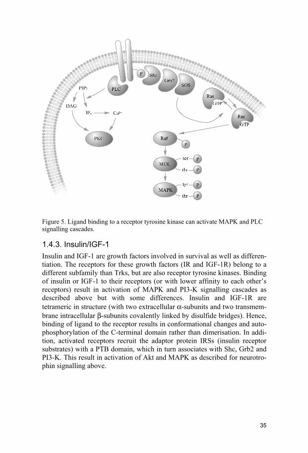
Figure 5. Ligand binding to a receptor tyrosine kinase can activate MAPK and PLC signalling cascades.
1.4.3. Insulin/IGF-1 Insulin and IGF-1 are growth factors involved in survival as well as differen-tiation. The receptors for these growth factors (IR and IGF-1R) belong to a different subfamily than Trks, but are also receptor tyrosine kinases. Binding of insulin or IGF-1 to their receptors (or with lower affinity to each other’s receptors) result in activation of MAPK and PI3-K signalling cascades as described above but with some differences. Insulin and IGF-1R are tetrameric in structure (with two extracellular α-subunits and two transmem-brane intracellular β-subunits covalently linked by disulfide bridges). Hence, binding of ligand to the receptor results in conformational changes and auto-phosphorylation of the C-terminal domain rather than dimerisation. In addi-tion, activated receptors recruit the adaptor protein IRSs (insulin receptor substrates) with a PTB domain, which in turn associates with Shc, Grb2 and PI3-K. This result in activation of Akt and MAPK as described for neurotro-phin signalling above.
35

2. AIMS OF THE STUDY
For a complete understanding of APP processing and to enable the develop-ment of good therapeutic agents that efficiently block the formation of Aβ-peptide, devoid of undesirable side-effects, we need more knowledge about the functions and processing of the two paralogues APLP1 and APLP2. Thus, our studies were focused on comparing the regulation of expression and processing of the APP family. The specific aims of the work in this the-sis were as follows:
• To analyse the expression and processing of the APP family during neuronal differentiation (papers I and II)
• To investigate the mechanism behind the RA-induced expression
and processing of the APP family (papers I-III)
• To examine the involvement of BDNF and TrkB in the synthesis and processing of the APP family (paper II)
• To determine the signalling pathways involved in IGF-1-induced
processing of the APP family (paper IV)
36

3. METHODOLOGICAL CONSIDERATIONS
3.1. Cell cultures Throughout this thesis, human SH-SY5Y neuroblastoma cells have been used to study the expression and proteolytic processing of the APP protein family. Immortalised cells are convenient to handle and experiments can be performed during continuous conditions in which biochemical processes easily can be studied. However, one should bear in mind that the nervous system include an enormous variety of cell types and cell contacts and that these factors are absent in an isolated cell line. 3.1.1. SH-SY5Y neuroblastoma cells SH-SY5Y is a neuroblast-like subclone of the SK-N-SH neuroblastoma cell line, originally derived from a metastatic tumour in the bone marrow. This cell line was established already in 1970 and has, since then, been widely used to study neuronal differentiation and neurodegeneration (Biedler et al. 1973; Biedler et al. 1978). Thus, by now, SH-SY5Y cells constitute a well defined cellular system. An additional advantage is the human origin. The parental cell line of SH-SY5Y cells comprise of both neuroblastic and sub-strate adherent cell-types. However, SH-SY5Y cells can be differentiated into a more neuronal-like phenotype with extended neurites using different neurotrophic factors like, RA, BDNF and IGF-1 (Påhlman et al. 1984; Påhlman et al. 1991; Kaplan et al. 1993; Encinas et al. 2000). In addition, SH-SY5Y cells endogenously express all the members of the APP protein family (Beckman and Iverfeldt 1997).
3.1.2. Treatments Treatments discussed below are illustrated in Fig. 6, to make the following section more comprehensible. In paper I and II, SH-SY5Y cells were differ-entiated with 10 μM RA during 6 days to achieve a fully differentiated neu-ronal-like population of cells. The effect of a PKC inhibitor, curcumin, on RA-differentiated cells was investigated. 10 μM curcumin was added during the last 24 h before harvesting or 2 μM curcumin was added concomitant with RA from the start. Two different protocols were used since 10 μM cur-cumin during longer periods was to harsh even for RA-differentiated cells. Metabolic labelling studies showed that the half life of the APP family in our
37

experiment during RA treatment was less than 90 minutes. Thus, using the two different protocols would have no impact on the expression levels of these proteins. When it comes to studying neurite outgrowth there is a differ-ence since RA treated cells are continuously extending their neurites. As a consequence adding 2 μM concomitantly with RA would resemble inhibi-tion of neurite outgrowth. Adding 10 μM curcumin during the last 24 h is rather equivalent to degeneration of neurites.
Figure 6. Treatments schematically illustrated. Arrows indicate change of medium.
In paper II, cells were first treated with RA during 3 days in order to induce expression of TrkB receptors and thereby responsiveness to BDNF (Kaplan et al. 1993). Subsequently two protocols were used, first BDNF were added concomitantly to RA for a 6 days period (to be compared with cells differen-tiated by RA alone for 6 days). However, even though differences in the mRNA expression of the APP family could be observed after 3 days, protein levels were significantly increased first after 6 days of RA treatment (in pa-
38

per I). Based on this finding, an additional protocol was used. Treatment of cells with RA was extended to in total 9 days and BDNF was added during the last 6 days together with RA (to be compared with cells treated with RA alone for 9 days). In paper IV, cells were grown for 6 days in serum-free medium with addition of N2 supplement (containing insulin, Bottenstein and Sato 1979). After this, treatments with insulin/IGF-1 with and without inhibitors to the major sig-nalling pathways were performed in N2 supplement devoid of insulin during the last 18 h. These studies were aimed at studying the processing of the APP family and not the synthesis. Thus, the time-period were chosen to be as short as possible. However, the APLPs are secreted into the medium in such small amounts that, in spite of concentrating the medium up to 20 times, 18 h was necessary for detection of these fragments. No significant differences in the steady-state levels of the full-length proteins were de-tected. Subsequently, we conclude that the observed increased levels of se-creted fragments is not a result of increased synthesis but rather an increase in the actual processing or alternatively, although less likely, increased sta-bility of the secreted fragments.
3.1.3. Cell viability and neurite outgrowth When analysing expression levels and processing of a protein in response to a certain treatment it is important to determine if the treatment in anyway affects cell growth or viability. To measure the number of viable cells (i.e., cell proliferation and/or survival) in response to various factors, a colorimet-ric assay (XTT, modified from MTT) was used. Cell viability is in this case measured as a function of mitochondrial activity. Mitochondrial dehydro-genase reduces MTT into a coloured derivative and the absorbance of the converted dye is then measured in a multi-well spectrophotometer. In paper I, we demonstrated that cell viability measured by MTT paralleled the total content of protein. If the number of cells is indeed affected by a treatment, it is especially important to compensate for this if the amount of a certain pro-tein that is released extracellular is determined. For example, if the number of cells decreases in response to a treatment then a larger volume of the me-dium should be analysed as compared to the untreated control cells. How-ever, a drawback of this way of compensation is that there is difficult to know at what time point during the treatment period the number of cells starts to differ and as a result less significant effects might be obtained. In paper I, the expression levels of the APP family was analysed in correla-tion to neuronal differentiation. Neuronal differentiation was demonstrated by neurite outgrowth in addition to expression of the synaptic proteins (growth-associated protein 43) GAP-43 and synapsin I. In order to estimate
39

neurite outgrowth both the number of long neurites as well as number of neurites per cell was calculated. The total neurite length was manually esti-mated as AU where 1 AU was defined as a standardised cell diameter. Only neurites longer than 1 AU were measured. In addition, the number of long neurites, with a length exceeding two times the average cell diameter, was counted.
3.2. Western blot analysis Western blot analysis, (also called immunoblotting), is a method that utilises primary antibodies raised against an epitope in order to selectively detect a protein of interest. In all papers, cells were harvested in a buffer containing a mixture of protease inhibitors in order to avoid proteolytic degradation of samples. Proteins were subsequently separated by their size using SDS-PAGE (sodium dodecyl sulfate polyacrylamide gel electrophoresis) and then transferred to a PVDF (polyvinylidene difluoride) membrane to enable de-tection by antibodies. Secondary antibodies are raised against the Fc region of primary antibodies and are coupled to an enzyme (HRP; horseradish per-oxidase) that catalyses the oxidation of a substrate into a product that emits light. This signal is detected on an autoradiography film and the relative abundance is quantified by densitometric analysis using computer software. Throughout this thesis the concentrations of antibodies were chosen to en-sure relative quantitative measurements. Since Western blotting is based on the separation of proteins by their size, the detection of different isoforms and posttranslational modifications of a particular protein is possible. Thus, Western blot analysis can give quantitative as well as qualitative data. It is a sensitive method and can be used for detection of proteins in the pn-ng range. However, it relies on the specificity of a certain antibody and if there is cross reactivity with other proteins the result can be difficult to interpret. Furthermore, this method only enables the detection of steady-state levels of a particular protein. The relative effect of RA-induced expression of APP and APLP1 is different between paper I and II. In paper I, cells were grown in Falcon primaria dishes and in paper II in Nunc dishes. Possibly this difference in culture conditions could have an impact on the expression levels in the non-treated cells. Additionally, the pattern of APLP1 in non-treated cells differs between paper I and paper II. Falcon primaria dishes seem to promote expression of the mature (i.e. N-glycosylated form) of APLP1. Moreover, even though both studies were performed with the same type of antibodies, in the first study we used CT11 antibodies kindly provided by Dr. Gopal Thinakaran, whereas in later studies these antibodies were purchased from a biotech company.
40

Deglycosylation of a protein can be performed using a chemical or enzy-matic approach. In paper I, we analysed the BDNF-induced changed APLP1 pattern. Cell lysate was harvested and subjected to peptide-N-glycosidase F. This endoglycosidase releases asparagine-linked oligosaccharides from gly-coproteins and glycopeptides by hydrolysing the innermost Asn-linked N-acetylglucosamine. Thus, deglycosylation, followed by SDS-PAGE and Western blot analysis, result in increased electrophoretic mobility compared to non-glycosylated control samples.
3.3. ELISA In our studies, endogenously expressed levels of Aβ could not be detected by the use of Western blot analysis or immunoprecipitation (section 3.4). ELISA (enzyme-linked immunosorbent assay) is an even more sensitive technique. Both methods are based on the binding of an antigen to specific antibodies. There are different types of ELISAs and in paper IV we used the sandwich technique to detect endogenously produced Aβ40 and Aβ42. In this assay, the capture antibody W0-2 (directed at the N-terminal 4-10 amino acids of Aβ, thus recognising both Aβ40 and Aβ42) was bound to a multi-well plate before the antigen (Aβ in cell medium or cell lysate) was added. Subsequently, different detection biotin-labelled antibodies, G2-10 (directed at the C-terminal 31-40 amino acids of Aβ, recognising only Aβ40) or G2-11 (directed at the C-terminal 33-42 amino acids of Aβ, recognising only Aβ42) were added to form an antibody-antigen-antibody complex (i.e. sand-wich). The detection of antigen is possible when a streptavidin-linked en-zyme binds to the biotin-linked detection antibody and subsequently con-verts a chromogen substrate into a chromophore. Emitted light is spectropho-tometrically measured and compared to a standard curve made of synthetic Aβ40 or Aβ42 making quantification possible. This protocol enabled detec-tion of Aβ concentrations as low as 25 pg/ml. Still this high-sensitive ELISA was not responsive enough to give absolutely accurate estimations of the levels of endogenously produced Aβ42. In addition, since we were primarily interested in effects of α- and β-cleavage and α- and β-secretases are be-lieved to compete for APP as a substrate and although the γ-secretase cleav-age differs; the initial β-secretase cleavage should be the same for Aβ40 and Aβ42. Indeed, we did observe similar effects for Aβ42 as for Aβ40. ELISA is a sensitive and convenient method allowing for analysis of small sample volumes in multi-well plates. However, in contrast to Western blotting only quantitative data can be obtained.
41

3.4. Metabolic labelling and immunoprecipitation In metabolic labelling studies, cells are incubated with medium containing radiolabelled amino acids. In paper II, we used the radioisotopes 35S-Met and 35S-Cys to study the synthesis and processing of the APP family in response to RA and BDNF. Radioisotopes are incorporated into the newly synthesised proteins and enable one to follow the biosynthesis and processing of a pro-tein with time. This is a major advantage, in contrast to using Western blot analysis that only allow for detection of steady-state levels. Metabolic label-ling can be used to determine the half-life of a protein. The protein of inter-est can then be immunoprecipitated i.e. captured with an antibody that in turn is immobilised on Protein G cross-linked sepharose beads, which enable separation of antigen from crude mixture of proteins. Both non-labelled and labelled proteins are pulled down by the antibody. Both antigen and antibody is recovered from the sepharose beads when incubated with sample buffer. Eluted antigen can then be separated by size using SDS-PAGE. Subsequent, autoradiography then enables detection of only labelled protein of interest. Immunoprecipitation can also be performed without metabolic labelling, followed by Western blot analysis. However, a major drawback is the high background caused by the presence of light and heavy chains, derived from the co-eluted antibody.
3.5. Northern blot analysis Although the levels of a particular protein does not always correspond to the level of its mRNA, studying mRNA and protein levels could give informa-tion on what level the regulation of the synthesis takes place. In paper I, the mRNA synthesis of the APP protein family during neuronal differentiation was investigated using a quantitative non-radioactive northern blot analysis. Total RNA was isolated and separated with respect to size by agarose-formaldehyde gel electrophoresis. The RNA was then transferred to a nylon membrane by capillary action. Subsequently, digoxigenin (DIG)-11-dUTP-labelled ssDNA or dsDNA probes were hybridised to their complementary target sequence. Probes were designed to recognise the mRNA sequence of highest diversity between the homologues. Alkaline phosphatase-coupled antibodies directed to DIG enabled detection of the probe and was detected with chemiluminescence, from a converted substrate. The emitted signal was visualised by autoradiography and the relative abundance of mRNA was quantitated by densitometric analyses of the films and compared to a stan-dard curve of known amounts of total RNA. House-keeping genes, GAPDH (glyceraldehyd 3-phosphate dehydrogenase) and actin, were used as an in-ternal control. Northern blot analysis allows for both quantitative and quali-tative data since both the relative abundance and length of transcript may be
42
obtained. Limitations for northern blot analysis, includes low sensitivity when the transcript of interest exists in low abundance. Advantages using a non-radioactive labelling include stability of the generated probes as well as safety during handling.
3.6. Statistical analysis Throughout this thesis one-way ANOVA (analysis of variance) has been conducted. One-way ANOVA tells us if the variance between the different treatments is significantly greater than expected by chance. A subsequent post-hoc test, provide information about which treatments that are statisti-cally different from each other. Statistically more correct would be to use two-way ANOVA that would also answer the question if the treatments dif-fer between repeated experiments. However, this is a much more compli-cated analysis and the treatments did not differ between experiments. Thus, one-way ANOVA is the method of our choice. Limitations with ANOVA (and all other parametric tests) include that it assumes that the data are sam-pled from populations with identical standard deviations. It should be kept in mind that this is rarely the case when comparing control with treatments that result in high effects, since the standard deviation is directly related to the average value. All statistical analysis were performed using Graph Pad In-Stat 3.05.
43

4. RESULTS AND DISCUSSION
4.1. Neuronal differentiation 4.1.1. RA induces neuronal differentiation of SH-SY5Y cells
(papers I and II) Human SH-SY5Y neuroblastoma cells were exposed to the potent morpho-gen RA. RA is known to induce neuronal differentiation of a number of neuroblastoma cell lines which have been used extensively to study neurite outgrowth (reviewed in Clagett-Dame et al. 2006). In accordance with pre-vious studies, we observed distinct effects on SH-SY5Y cells in response to RA. Typically, a decreased proliferation rate, morphological changes includ-ing more elongated cell bodies in parallel with induced length as well as an amplified number of neurites per cell, were observed. In addition, the cell bodies appeared more evenly distributed after RA treatment, indicating a migratory role for this morphogen on neuroblastoma cells. In parallel with analysis of neurite outgrowth, we investigated the effects of RA on the neuronal markers GAP-43 and synapsin I. GAP-43 is a presynap-tic protein, located in growth cones, developmentally regulated and is fre-quently used as a marker for sprouting (reviewed in Oestreicher et al. 1997). Synapsin I is another presynaptic protein, also located in the growth cone and involved in the elongation of axons and in synapse formation (reviewed in Ferreira and Rapoport 2002). We observed an increase in the levels of GAP-43 and synapsin I (Fig. 7) in response to RA, demonstrating the valid-ity of differentiated SH-SY5Y cells as a suitable model for studying events (i.e. expression of the APP family) correlated to neuronal differentiation.
Figure 7. RA induces protein expression of the presynaptic protein synapsin I.
44

4.1.2. Curcumin counteracts the effect of RA on neurite outgrowth (paper I)
To further characterise the effects of RA we used the NFκB/AP-1 (nuclear factor κB and activating protein 1) and PKC inhibitor curcumin. Curcumin (diferuloylmethane) is a naturally occurring substance derived from the plant Curcuma longa. This polyphenol is a major component of curry and is widely used both as a food additive and as a traditional oriental medicine, particularly in India. Many studies have indicated that curcumin exerts anti-cancer, anti-oxidant and anti-inflammatory effects mediated via the down-regulation of the transcription factors NFκB and AP-1. Previous studies have shown that RA activated AP-1 (Jenab and Inturrisi 2002) and NFκB proteins were demonstrated to be required for differentiation by RA (Feng and Porter 1999). The effect of curcumin on neurite outgrowth after differentiation of cells with RA was investigated by addition of 10 μM curcumin during the last 24 h before harvesting or 2 μM curcumin concomitant with RA. Both protocols resulted in a seemingly less dense network of neurites. However, only the higher concentration resulted in significant effects. When adding curcumin during the last 24 h, the cells already displayed long neurites as part of an extensive network in response to RA and the addition of curcumin led to an almost complete retraction of the neurites. Neither concentration of curcu-min affected the viability of the differentiated cells as shown by XTT assay and total protein content from cell lysate (i.e. adherent cells). In contrast, the higher concentration was toxic to un-differentiated cells and was therefore only used for treatment of differentiated cells. As mentioned above, curcumin has been proposed to down-regulate NFκB and AP-1. However, other studies suggest that the effects of curcumin can be mediated by inhibition of PKC (Liu et al. 1993; Varadkar et al. 2001; Woo et al. 2005). In addition, the mechanism underlying curcumin-mediated inhibi-tion of AP-1 and NFκB can involve PKC and/or c-Jun N-terminal kinase (JNK, cf. Chen and Tan 1998). Curcumin was shown to block the inter-leukin-1β (IL-1β)-induced nuclear export of the RXRα (i.e. an RA-activated nuclear transcription factor) (Zimmerman et al. 2006). The nuclear export of RXRα in response to IL-1β was dependent on a serine residue at position 260, suggesting that JNK is implicated in the phosphorylation of RXRα which keep it inside the nucleus (cf. 1.4.1). It has previously been demon-strated that RA-induced neuritogenesis of SH-SY5Y cells is PKC dependent (Miloso et al. 2004), suggesting a possible mechanism for curcumin to in-hibit or induce retraction of neurites via effects on PKC. Another signalling pathway required for RA-induced differentiation of SH-SY5Y cells involves PI3-K (Lopez-Carballo et al. 2002).
45

4.2. Regulation of the synthesis of the APP protein family
4.2.1. RA induces expression of the APP protein family (paper I, II and III)
As already described in section 1.2.1, APP has been suggested to be in-volved in neuronal development. APLP1, APLP2 and APP are all develop-mentally regulated, with increased expression during embryogenesis (Lorent et al. 1995). To further investigate the regulation of the APP protein family during neuronal differentiation and neurite outgrowth, we used SH-SY5Y cells exposed to RA for 3, 6 or 9 days.
4.2.1.1. mRNA levels are up-regulated in response to RA Quantitative Northern blot analysis revealed that mRNA levels of APP, APLP1 and APLP2 were increased approximately two-fold in response to 3 days of RA treatment. This increase has previously been shown to be pro-longed, remaining detectable after 6 days (Beckman and Iverfeldt 1997). A previous study have shown up-regulation of APP mRNA levels in SH-SY5Y cells in response to RA within 2 hours of treatment, suggesting a direct regu-lation of the APP transcription by RA (König et al. 1990). However, pro-found effects were not obtained until after 4–9 days of treatment (König et al. 1990; Hung et al. 1992), indicating an indirect mechanism behind the regulation. The steady-state levels of mRNA can also increase as a result of increased stability of the transcript and our study does not rule out the possi-bility that RA has this effect on the APP transcript.
4.2.1.2. Protein levels are up-regulated in response to RA Increased mRNA levels do not necessarily result in elevated protein levels, since regulation can occur also at the post-transcriptional level. Thus, it is important to analyse also the protein levels of the protein of interest. Western blot analysis of cell lysate from SH-SY5Y with antibodies directed against APLP1 showed two bands of approximately 80 and 90 kDa, equivalent to immature and mature forms (i.e. not fully and fully glycosylated forms) (cf. Paliga et al. 1997). When using antibodies directed against APLP2, bands of 80 and 140–190 kDa could be detected, corresponding to unmodified and chondroitin sulphate glycosaminoglycan (CS GAG) chain modified APLP2, respectively (Thinakaran and Sisodia 1994). The pattern of APP bands dif-fered between the papers as a result of different conditions used during the SDS-PAGE. In any case, the bands correspond to mature and immature forms of the APP695, 751 and 770 isoforms (Fig. 8) (Weidemann et al. 1989; Buxbaum et al. 1990).
46
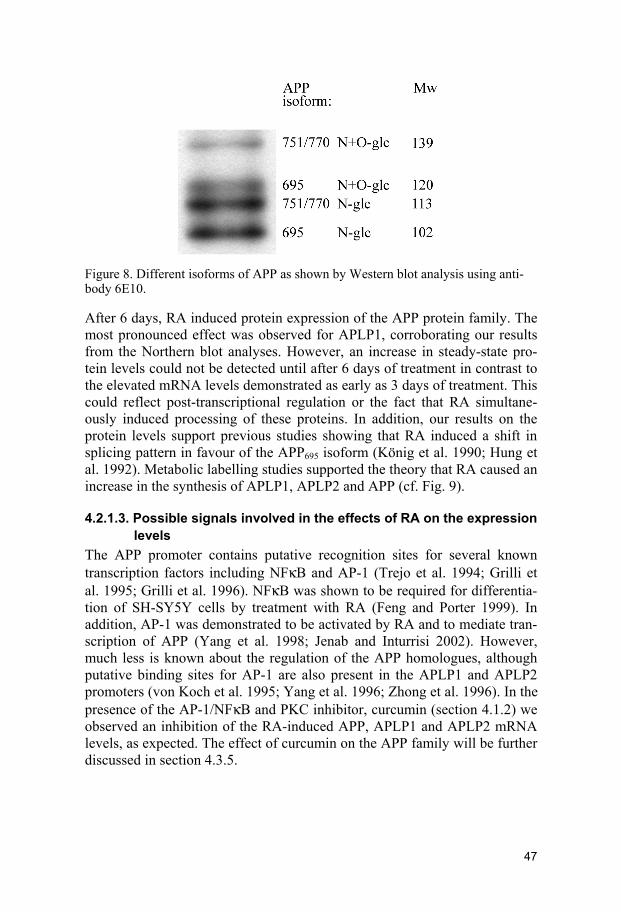
Figure 8. Different isoforms of APP as shown by Western blot analysis using anti-body 6E10.
After 6 days, RA induced protein expression of the APP protein family. The most pronounced effect was observed for APLP1, corroborating our results from the Northern blot analyses. However, an increase in steady-state pro-tein levels could not be detected until after 6 days of treatment in contrast to the elevated mRNA levels demonstrated as early as 3 days of treatment. This could reflect post-transcriptional regulation or the fact that RA simultane-ously induced processing of these proteins. In addition, our results on the protein levels support previous studies showing that RA induced a shift in splicing pattern in favour of the APP695 isoform (König et al. 1990; Hung et al. 1992). Metabolic labelling studies supported the theory that RA caused an increase in the synthesis of APLP1, APLP2 and APP (cf. Fig. 9).
4.2.1.3. Possible signals involved in the effects of RA on the expression levels
The APP promoter contains putative recognition sites for several known transcription factors including NFκB and AP-1 (Trejo et al. 1994; Grilli et al. 1995; Grilli et al. 1996). NFκB was shown to be required for differentia-tion of SH-SY5Y cells by treatment with RA (Feng and Porter 1999). In addition, AP-1 was demonstrated to be activated by RA and to mediate tran-scription of APP (Yang et al. 1998; Jenab and Inturrisi 2002). However, much less is known about the regulation of the APP homologues, although putative binding sites for AP-1 are also present in the APLP1 and APLP2 promoters (von Koch et al. 1995; Yang et al. 1996; Zhong et al. 1996). In the presence of the AP-1/NFκB and PKC inhibitor, curcumin (section 4.1.2) we observed an inhibition of the RA-induced APP, APLP1 and APLP2 mRNA levels, as expected. The effect of curcumin on the APP family will be further discussed in section 4.3.5.
47

4.2.2. BDNF together with RA selectively increases the protein levels of APP (paper II)
Besides RA, a number of factors have been reported to regulate APP gene expression; they include NGF, IL-1, phorbol esters, BDNF, basic fibroblast growth factor (bFGF) and epidermal growth factor (EGF) (Mobley et al. 1988; Goldgaber et al. 1989; Lahiri and Nall 1995; Ringheim et al. 1997; Ruiz-León and Pascual 2001; Villa et al. 2001). RA can also up-regulate the expression of TrkB in SH-SY5Y cells (Kaplan et al. 1993; Kobayashi et al. 1994; Encinas et al. 1999). Furthermore, in neuroblastoma cells transiently transfected with TrkB, BDNF was shown to induce APP promoter-driven expression of a reporter gene. We treated cells with BDNF in order to inves-tigate whether the previously demonstrated effects on APP were similar for all the members of the APP family. Previous studies found that detectable levels of functional TrkB were not observed until after 3 days of RA treat-ment in SH-SY5Y cells (Encinas et al. 1999). To assure TrkB expression and to allow the detection of differences in the levels of the APP protein in response to RA, two different protocols were used. Cells were grown in the presence of RA for 6 or 9 days. BDNF was added together with RA for 6 days, either from the onset of RA treatment or after 3 days of RA treatment. According to Western blot analysis, BDNF treatment had no effect on the APLP1 levels. Furthermore, the levels of APLP2 and CS GAG APLP2 rather decreased in the presence of BDNF. Conversely, APP levels were increased in response to BDNF. Different effects on the members of the APP family were observed, most likely as a result of different regulation of the processing in response to BDNF (section 4.3.2). It should be mentioned that treatment of undifferentiated cells (i.e. those not treated with RA) with BDNF had no effect on the steady-state levels of the APP family. Our results are in accordance with previous studies, where BDNF had no effect on the APP promoter activity in SH-SY5Y cells that were not transfected with TrkB (Ruiz-León and Pascual 2004). Thus, SH-SY5Y cells needs to be dif-ferentiated or transfected with TrkB in order to respond to BDNF. In order to investigate whether the effects of RA on the APP protein family could be mediated through the TrkB receptor, we used a TrkB chimera (Shelton et al. 1995) to block the effects of putative ligands. No significant effect on the RA-induced synthesis of the APP protein family was observed after co-treatment with the TrkB chimera. This suggests that the effect of RA was not mediated through BDNF as a ligand to the TrkB receptor. By using dominant-negative mutants, both the PI3K/Akt and Ras/MAPK pathways were shown to be involved in the regulation of APP promoter ac-tivity by BDNF (Ruiz-León and Pascual 2004). The same group also demon-strated the implication of Ras/MAPK signalling in APP promoter activity in PC12 cells after treatment with NGF, bFGF or EGF. However, the effects on
48

APLP1 and APLP2 promoter activity in response to various growth factors remain to be investigated.
4.3. Processing of the APP protein family Proteolytic processing of the APP family may be modulated by regulation of enzyme activity, or by specific post-translational modifications, or altered subcellular localisation of the substrate or the enzyme. A greater understand-ing of the factors that influence APP processing may assist in the design of effective therapeutic agents to inhibit Aβ formation and possibly halt the progression of AD. Regulated processing of APP can be induced by numer-ous different stimuli. This section will focus on our studies on growth factor mediated secretion of sAPPα. 4.3.1. RA increases proteolytic processing (papers II and III) Various growth factors involved in the synthesis of APP (section 4.2) are also reported to induce sAPP secretion. These include NGF, IL-1β, BDNF, platelet-derived growth factor (PDGF), bFGF and EGF (Refolo et al. 1989; Schubert et al. 1989; Buxbaum et al. 1994; Slack et al. 1997; Villa et al. 2001; Kim et al. 2002; Ma et al. 2005). PMA, a PKC activator, also induces secretion of sAPP (Ringheim et al. 1997; Kim et al. 2002). The complexity of the signalling pathways involved in regulating the secretion of APP in response to the various growth factors and PMA is illustrated in table 3.
Table 3. Signalling pathways involved in the regulated shedding of sAPPα.
sAPPα secretion Growth factor Dependent of Independent of Reference PDGF MAPK PKC (Kim et al. 2002)
IL-1β MAPK, JNK, PLC, PKC
PKC, PI3-K (Ma et al. 2005), (Buxbaum et al. 1994) EGF PKC PI3-K (Slack et al. 1997) NGF PKC, MAPK MAPK (Mills et al. 1997), (Villa et al. 2001), (Desdouits-Magnen et al. 1998)
PMA PKC, MAPK (Kim et al. 2002), (Ringheim et al. 1997)
BDNF Ras, Raf, MEK, MAPK (Ruiz-León and Pascual 2004)
insulin PI3-K PKC, MAPK (Solano et al. 2000) IGF-1 PI3-K, cdk5 MAPK paper IV
49

Unlike APP, the processing of APLP2 and its regulation has not been exten-sively studied. However, as for APP, there are a few studies reporting that NGF, EGF and PMA, induce secretion of sAPLP2 (Webster et al. 1995; Xu et al. 2001). 4.3.1.1. RA increases the levels of secreted APP and APLP2
ectodomains as well as the levels of the α-secretases ADAM10 and TACE
The ability of RA to induce synthesis of both mRNA and protein levels of the APP protein family has already been discussed in section 4.2.1. Elevated steady-state levels of the secreted fragments of sAPPα, as well as sAPLP2, were also observed after 6 days of RA treatment. Exposing cells to RA for a longer time periods than 6 days did not result in any further increase in the levels of the membrane-bound full-length proteins. However, the secretion of ectodomains of APP and APLP2 was further elevated when cells were subjected to RA for longer time periods. This could be interpreted as an in-crease in the rate of the actual processing or simply that higher levels of full-length proteins are accessible to processing enzymes leading to secretion of the ectodomain rather than intracellular degradation. Our results suggest that RA causes a shift in the processing in favour of the α-secretase pathway. α-secretase cleavage of APP occurs at the surface of neuronal cells (Parvathy et al. 1999). It is tempting to speculate that the lack of increased total mem-brane-bound full-length proteins (APP and APLP2), after 9 as compared to 6 days of RA treatment, is a result of increased shedding. Corroborating results from metabolic labelling studies showed an increase in newly synthesised APP (cf. Fig. 9) and together with the results from the Western blot analyses; this strongly supports the hypothesis that RA enhances both synthesis and processing after 9 days as compared to 6 days of treatment. To elucidate a possible regulatory mechanism of RA on APP and APLP2 ectodomain shedding, we analysed the expression levels of ADAM10 and TACE (section 1.2.5.1), since they are the most likely candidates for α-secretase (Buxbaum et al. 1998; Lammich et al. 1999). As expected, RA treatment resulted in higher levels of the active forms of both ADAM10 and TACE. This is in accordance with a previous study which reported that RA treatment of SH-SY5Y cells induced promoter activity and subsequently both mRNA and protein expression of ADAM10 (Endres et al. 2005). How-ever in contrast to our results, the same study also found that TACE levels were not affected by RA. The authors suggested that ADAM10 is responsi-ble for the increased α-secretase activity leading to a regulated increased release of sAPPα and sAPLP2. In previous studies, ADAM10 has been de-scribed as the enzyme responsible for constitutive and regulated shedding of APP (Lammich et al. 1999; Lopez-Perez et al. 2001; Asai et al. 2003), whereas TACE on the other hand has been implicated only in PKC-
50

stimulated and regulated shedding (Buxbaum et al. 1998; Parkin et al. 2002). In our system, the induced α-secretase cleavage could reflect the elevated levels of the active forms of either ADAM10 or TACE – or perhaps both. Additionally, we showed that the RA-induced expression of active ADAM10 was dependent on PI3-K signalling. Conversely, the levels of active TACE were neither affected by PI3-K nor MAPK inhibitors (cf. 4.3.4.1).
Figure 9. Autoradiography of immunoprecipitated APP and sAPPα. Cells were metabolically labelled and then chased 0, 0.5, 1, 2, 3 h (middle and lower panel). Cells were cultured in the absence (control) or presence of 10 μM RA for 6 days (RA6d). The relative abundance of APP and sAPPα was quantified by densitometric analysis and presented as % of total amount APP and sAPPα for each treatment (upper panel). Note that cells treated with RA6d contained approx. 3.5 times as much radio labelled APP compared to control. Data represent mean of 1-3 cultures derived from 1-3 independent experiments.
51

The involvement of PI3-K in RA-induced differentiation, APP synthesis, ADAM-10 activation and finally release of the neurotrophic sAPPα lends further support to the hypothesis that the APP family plays important roles in neuronal development and neuritogenesis.
4.3.1.2. RA affects the production of C-terminal fragments from APP and APLP1 as well as the levels of BACE1
Under the conditions used in this study, we were not able to detect ectodo-main secretion of the third member of the APP family, APLP1. We therefore turned our attention to the C-terminal fragments in an attempt to analyse the processing of APLP1. Indeed, after treating SH-SY5Y cells with RA, CTFs produced from endogenously expressed APLP1 and APP could be detected. These fragments have, to our knowledge, been previously reported only in studies using transfected cells that overexpress APLP1 or APP, or after co-transfection with stabilising proteins such as Fe65 (Scheinfeld et al. 2002; Eggert et al. 2004). When performing Western blot analysis of cell lysate using antibodies directed against the C-terminal 20 amino acids of APP, six different bands of approx. 19, 18, 15, 12, 11 and 10 kDa were identified. Since the apparent molecular weight of these APP fragments seemed higher than expected, their identity was confirmed using two additional antibodies (6E10 and 4G8 detecting C99 and C83, respectively). The APP CTFs of 18-19 kDa corresponded to C99, those of 15 kDa to C83 and those of 10-12 kDa to AICDs. After 9 days of RA treatment C99 decreases. This parallels the further increased levels of sAPPα and fits well with increased α-secretase processing. As previously mentioned, much less is known about the processing of APLP1. However, the antibody directed against the C-terminal 11 amino acids of APLP1 identified several bands of 18, 17, 15, 10, 9.5 and 8 kDa and the three latter fragments most likely correspond to ALID1s (cf. Eggert et al. 2004). RA exposure of cells for 6 days had different impact on the C-terminal stubs processed at the β-site from APP and APLP1. There was no increase of C99 as compared to control, further supporting the hypothesis that RA shifts the processing towards the α-secretase pathway. Conversely, the levels of the 15 kDa C-terminal stub generated from APLP1 increased, suggesting either different subcellular localisation of the corresponding full-length proteins, or that the fragments were not produced by the same en-zyme(s). Indeed, RA also resulted in elevated levels of activated BACE1, which, previously has been demonstrated to cleave APLP1 (Li and Südhof 2004). An increase in C-terminal stubs also indicates that, despite the ab-sence of released detectable sAPLP1, RA does in fact promote processing of APLP1. Our preliminary studies, where cells were exposed to RA in serum-free medium, show that sAPLP1 could actually be detected and increased in
52

response to RA. This give further support to the interpretation of our previ-ous results that RA increased the processing of APLP1. Since the levels of AICD and ALIDs were in direct correlation of full-length proteins, we suggest that γ-secretase activity was not affected. However, we can not rule out the possibility that the expression of stabilising proteins was increased.
Figure 10. RA induced secretion of sAPLP1 in serum-free medium (containing insu-lin). Note that there are also effects on the expression levels of full length APLP1, likely a result of the long (6 days) treatment time.
4.3.2. BDNF increases the proteolytic processing of APP and APLP2 in RA-differentiated cells (paper II and III)
Treatment with BDNF without RA had no effect on the processing or the synthesis of the APP protein family. However, BDNF together with RA increased the processing of APP and APLP2, since the levels of sAPPα, sAPLP2 and CS GAG sAPLP2 increased in parallel with decreased levels of C99 and the 15 kDa C-terminal stub generated from APLP1. This also dem-onstrated that BDNF clearly further enhanced the shift towards α-secretase processing of APP. However, when we analysed the steady-state levels of the active forms of the putative secretases ADAM10, TACE and BACE1, we found that they all decreased in response to BDNF. The BDNF-mediated decrease in BACE1 levels, corroborate our proposal that the 15 kDa C-terminal stub generated from APLP1 is produced by BACE1 (cf. Li and Südhof 2004).
Thus, our results suggest that the effects of BDNF on the processing are not due to enhanced enzyme activity since the levels of secretases decreased.
53
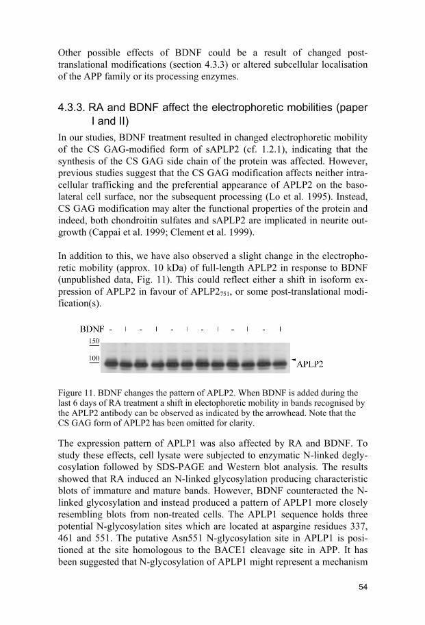
Other possible effects of BDNF could be a result of changed post-translational modifications (section 4.3.3) or altered subcellular localisation of the APP family or its processing enzymes.
4.3.3. RA and BDNF affect the electrophoretic mobilities (paper I and II)
In our studies, BDNF treatment resulted in changed electrophoretic mobility of the CS GAG-modified form of sAPLP2 (cf. 1.2.1), indicating that the synthesis of the CS GAG side chain of the protein was affected. However, previous studies suggest that the CS GAG modification affects neither intra-cellular trafficking and the preferential appearance of APLP2 on the baso-lateral cell surface, nor the subsequent processing (Lo et al. 1995). Instead, CS GAG modification may alter the functional properties of the protein and indeed, both chondroitin sulfates and sAPLP2 are implicated in neurite out-growth (Cappai et al. 1999; Clement et al. 1999). In addition to this, we have also observed a slight change in the electropho-retic mobility (approx. 10 kDa) of full-length APLP2 in response to BDNF (unpublished data, Fig. 11). This could reflect either a shift in isoform ex-pression of APLP2 in favour of APLP2751, or some post-translational modi-fication(s).
Figure 11. BDNF changes the pattern of APLP2. When BDNF is added during the last 6 days of RA treatment a shift in electophoretic mobility in bands recognised by the APLP2 antibody can be observed as indicated by the arrowhead. Note that the CS GAG form of APLP2 has been omitted for clarity.
The expression pattern of APLP1 was also affected by RA and BDNF. To study these effects, cell lysate were subjected to enzymatic N-linked degly-cosylation followed by SDS-PAGE and Western blot analysis. The results showed that RA induced an N-linked glycosylation producing characteristic blots of immature and mature bands. However, BDNF counteracted the N-linked glycosylation and instead produced a pattern of APLP1 more closely resembling blots from non-treated cells. The APLP1 sequence holds three potential N-glycosylation sites which are located at aspargine residues 337, 461 and 551. The putative Asn551 N-glycosylation site in APLP1 is posi-tioned at the site homologous to the BACE1 cleavage site in APP. It has been suggested that N-glycosylation of APLP1 might represent a mechanism
54

whereby proteolysis can be regulated (Eggert et al. 2004). This assumption was due to the observation that blocking N-glycosylation, either by an in-hibitor or by point mutation of Asn551, resulted in additional secreted frag-ments sensitive to inhibition of α-secretase. In addition, a fragment not sen-sitive to inhibition of α-secretase (i.e. putatively processed by BACE1) was decreased when N-glycosylation was blocked. In addition to changed levels of BACE1, this could explain our results on the 15 kDa C-terminal stub gen-erated from APLP1. RA increased N-glycosylation of APLP1 in parallel with increased 15 kDa CTFs, whereas BDNF conversely decreased N-glycosylation as well as fragments. Indeed, RA and BDNF resulted in changed electrophoretic mobilities, possi-bly reflecting post-translational modifications of the APP protein family. This phenomen might contribute to the complex mechanism behind the pro-teolytic processing of these proteins.
4.3.4. Insulin and IGF-1 increase proteolytic processing (paper IV)
Insulin is another growth factor implicated in the processing of APP (Solano et al. 2000). Recent studies have changed the view of insulin and its role is now considered to be broader than just regulation of peripheral glucose ho-meostasis. Insulin and also IGF-1 have been implicated to play a role in the CNS in complex cognitive processes such as memory and learning. In addi-tion, insulin is believed to be involved in neuroprotection (reviewed in van der Heide et al. 2006; Åberg et al. 2006; Cole and Frautschy 2007). We demonstrated that sAPPα release was stimulated approx. 10-fold by high concentrations of insulin. In addition, we showed that sAPLP1 secretion is increased to the same extent. To determine whether the effects of insulin were mediated through insulin or through IGF-1 receptors, we performed dose-response analysis of both insulin and IGF-1. Concentrations in the very low nanomolar range of IGF-1 had the same impact on APP and APLP1 processing as high concentrations of insulin. Somewhat lower effect on ectodomain shedding of APLP2 was observed in response to IGF-1. No ef-fects on steady-state levels of the membrane-bound full-length proteins could be observed and so the increased levels of the secreted ectodomains most probably reflect an increase in proteolytic processing rather than in-creased synthesis. Thus, our result suggests an increased processing in re-sponse to either IGF-1 or insulin mediated by activation of IGF-1R rather than IR. Interestingly, APP and sAPPα have previously been shown to en-hance the neurotrophic and neuroprotective activity of NGF through the insulin signalling pathway (Wallace et al. 1997; Luo et al. 2001a). Again, a
55
connection between APP and neuronal differentiation was established, this time in concert with the insulin signalling pathway.
4.3.4.1. Signalling cascades involved in IGF-1-induced secretion of the
APP family Activation of the IGF-1 receptor activates two major signalling pathways, the PI3-K and MAPK cascades (see Figs. 4 and 5 in section 1.4.2). Previous studies have demonstrated the involvement of MAPK and PI3-K in the ef-fects on the secretion of sAPPα in response to various growth factors (see table 3 in section 4.3.1). Interestingly, activation of MAPKs and PI3-K has been shown to be essential for neuronal differentiation of neuroblastoma cells induced by IGF-1 or RA (Kim et al. 1997; Kim et al. 1998; Lopez-Carballo et al. 2002; Lee and Kim 2004). In turn, MAPK has been shown to be responsible for the RA-induced cyclin-dependent kinase 5 (cdk5) and p35 expression, which is essential for neuronal differentiation (Lee and Kim 2004). Cdk5 has also been implicated in APP processing when overexpres-sion of the cdk5 activator p35/p25 increased the phosphorylation of APP (Thr668, referring to the 695 isoform numbering) as well as the secretion of Aβ, sAPPβ and sAPPα (Liu et al. 2003). It is tempting to speculate that ef-fects on sAPPα secretion and neurite outgrowth are mediated partly by the same signalling pathways. However, to focus on the processing and in order to investigate the downstream signalling pathways leading to IGF-1-induced secretion of the APP family, we used the inhibitors LY 294002, roscovitin and PD 98059 for PI3-K, cdk5 and MAPK activity, respectively. We showed that PI3-K and cdk5, but not MAPKs, are involved in IGF-1-induced secre-tion of sAPPα. This is in accordance with a previous study showing that insulin-induced sAPPα was dependent on PI3-K, but not on MAPK, activity (Solano et al. 2000). However, we also demonstrated differing regulation of the shedding of the APP family in response to IGF-1. For IGF-1-induced secretion of sAPLP1, PI3-K, MAPK and cdk5 activation is required, whereas for ectodomain shedding of APLP2 neither PI3-K nor MAPKs were involved (the data regarding the effect of cdk5 inhibition on sAPLP2 were inconclusive and are therefore omitted). In contrast, a previous study re-ported involvement of MAPK in the PMA- as well as EGF-induced secretion of sAPLP2 (Xu et al. 2001). Even though our studies were not performed under the same conditions (serum vs. serum free, RA and BDNF vs. IGF-1), it is interesting that IGF-1-induced secretion of sAPLP2 was not dependent on either PI3-K or MAPKs which was also true for RA-induced activation of TACE or BACE1 (section 4.3.1.1, cf. Endres et al. 2005). This may indicate that regulated shedding of sAPLP2 occurs through TACE or BACE1 rather than ADAM-10. However, TACE activation induced by PMA was previ-ously demonstrated to involve phosphorylation at Thr735 by MAPK (Diaz-
56

Rodriguez et al. 2002), suggesting that several different mechanisms might be involved.
4.3.5. Effects of curcumin on the protein levels (paper I) Curcumin counteracted the RA-induced neuronal differentiation of SH-SY5Y cells (section 4.1.2). In parallel, curcumin completely blocked the RA-induced mRNA expression of the APP family. This supports the role of NFκB and AP-1 in regulating RA-induced mRNA expression of the APP family (section 4.2.1.3). The RA-induced steady-state protein level of APLP1 was also blocked by curcumin, probably as a result of decreased synthesis. In undifferentiated cells, curcumin decreased the steady-state levels of mem-brane bound full-length APLP1. This could be interpreted as an effect on the basal promoter activity of APLP1, even though curcumin has previously been shown not to modulate the APP basal promoter activity (Yang et al. 1998).
Surprisingly, and in contrast to APLP1 protein levels and APP family mRNA levels, APLP2 full-length proteins accumulated in response to cur-cumin. In addition, APP protein levels were sustained. Since RA induced processing of both APP and APLP2, this could be a result of inhibited prote-olytic processing and hence, accumulation of the membrane-bound forms. As previously discussed and illustrated in table 3, PKC plays an important role in the regulated processing of APP as well as APLP2.
57

5. CONCLUSIONS
Although APP has been studied by researchers around the world for decades, the physiological function of this protein and its paralogues has not yet been determined. However, regulation of APP synthesis and processing is closely coupled to developmental events of the nervous system and has also been implicated in neurite outgrowth and survival. Since the phenotypes induced by the deletion of APP family members can be rescued by the other paralogues, there seems to be functional redundancy between these proteins. In our studies we have demonstrated that mRNA as well as protein levels of all the members of the APP family increased in parallel with neuronal differ-entiation induced by RA. Hence, our results add to previous knowledge that also indicates a function of the APP family during neuronal development. RA not only induced increased synthesis, but also increased the processing of the APP family in concert with increased levels of active forms of ADAM10 and TACE and increased the expression levels of BACE1. Proc-essing of APP was shifted towards α-secretase in response to RA, most likely a result of higher levels of activated α-secretases. We also concluded that the effect of RA, on either the synthesis or processing of the APP fam-ily, was not mediated through the TrkB receptor. However, BDNF concomi-tantly with RA had a different impact on the APP family members; the levels of APP were elevated, those of APLP1 sustained and those of APLP2 de-creased. BDNF increased the processing of APLP2 and even further shifted the processing of APP towards α-secretase processing, despite decreased levels of ADAM10 and TACE. Thus we concluded that BDNF changed the prerequisite for processing, possibly by altering the distribution of substrate and enzyme within the cell or by changing the affinity or efficiency of prote-olytic cleavage through post-translational modifications. Indeed, BDNF im-pinged upon the post-translational modifications of the APP family. BDNF counteracted the RA-induced N-glycosylation of APLP1 and also changed the appearance of the CS GAG side chain of APLP2. At the moment the effects of these types of post-translational modifications are not known. It would not be too far-fetched to speculate that processing and hence possibly the function of these proteins can be regulated in this way. We suggest that APLP1 is preferentially processed by BACE1, leaving the 15 kDa fragment of APLP1 in the plasma membrane to be further processed by the γ-secretase complex. Hence, this fragment decreases as a result of decreased BACE1
58

levels and possibly due to decreased N-glycosylation of APLP1 in response to BDNF. Many growth factors are involved in the regulation of the synthesis and processing of APP. In addition to RA and BDNF, we analysed the effects of insulin and IGF-1 on the processing of the APP family. In serum-free me-dium, we were also able to detect the shedded ectodomain of APLP1. Insulin and IGF-1 increased the proteolytic processing of the APP family through the IGF-1R. However, the signalling pathways involved in this event dif-fered between the paralogues. PI3-K and cdk5, but not MAPK, were shown to be involved in IGF-1-induced secretion of sAPPα. Although our different studies were performed under different conditions (serum vs serum-free) it is interesting to note that the RA-induced activation of ADAM10, but not of TACE, was also dependent on PI3-K. Hence, we speculate that ADAM10 might be the α-secretase responsible for IGF-1 induced processing of APP as well. The processing of APLP1 induced by IGF-1 was dependent on PI3-K, cdk5 and MAPK. Here the data from the RA and secretase study are not as straightforward, since the level of BACE1 was not affected by PI3-K, cdk5 or MAPK. However, the activity of BACE1 could still be regulated in a different way more closely resembling the signalling pathways behind APLP1 processing, since active and inactive forms of BACE1 were not dis-tinguished in our analysis. The IGF-1-induced processing of sAPLP2 was not dependent on either PI3-K or MAPKs. Again, the data on RA-induced activation of the α-secretases are of interest, since TACE was also shown not to be dependent on either PI3-K or MAPKs for activation. This led us to speculate that under stimulated conditions APLP2 might be preferentially cleaved by TACE rather than by ADAM10. In addition, curcumin blocked the RA-induced mRNA expression of the APP family. However, only APLP1 protein levels were decreased in response to curcumin. APP levels were sustained, but even more interestingly, APLP2 levels increased. We speculate that the processing of APLP2 was decreased as a result of inhibited PKC activation and that this is the main signalling pathway generating sAPLP2. The processing of APP per se is complex involving several secretases com-peting for the same substrate. Moreover, the effect of several different types of post-translational modifications makes it more complex. Also, it is not yet fully clear how the processing enzymes are activated and come in contact with the substrate in the cellular compartment where proteolysis takes place. The picture becomes even more intricate when considering the possible processing of two additional and homologuos proteins. Great effort has been made in order to understand the processing of APP, but yet more knowledge is needed. However, our studies have opened up the possibility that the APP family might be differentially regulated. In the future this knowledge might
59

be useful for the design of therapeutic agents that can block the production of Aβ, without affecting the processing and any vital but as yet unknown function(s), of APLP1 and APLP2.
Figure 12. Proposed signalling pathways involved in RA-, BDNF- or IGF-1-mediated secretion of the APP family.
60

6. ACKNOWLEDGEMENTS
Just i denna stund slog det mig, att ha skrivit denna avhandlingen innebär att det är dags att ta farväl! Lämna det som jag under de senaste 8 (jaaa, det är så länge) åren kämpat med och för! Många har skrivit att detta är de roligaste sidorna att skriva, jag tror de blir de svåraste. Jag vill börja med att tacka Ülo för att han introducerade mig för Kerstin för alla dessa år sedan när jag skulle göra mitt examensarbete. Kerstin, jag vet inte hur jag ska kunna tacka dig! Du är en underbar och enormt skicklig handledare och jag respekterar dig djupt för din oerhörda kunskap! Jag är glad för all frihet och tålamod du har gett mig under alla dessa år. Du har alltid funnits där när jag har behövt dig! En del av mig vill inte lämna dig ännu, det finns mycket du fortfarande skulle kunna lära mig! Jag vill också tacka institutionens andra lärare Anders, Mattias, Tiit och Coco för det oerhörda jobb ni lägger/la ner för att bevara vår institution och för att ni alltid hjälper till när man behöver det. Särskilt vill jag tacka dig Anna F, för att du när jag en gång började tog mig under din grupps vingar! Jag tänker mycket på dig nu och beundrar dig för din styrka! Tack Birgitta för all hjälp och för ditt tålamod med alla strulande fakturor! Tack Ulla för att du med din kunskap och nogrannhet alltid hjälper oss alla! Siv, jag har sagt det så många gånger, din arbetsinsats är sannerligen underskattad! Du är den fulla betydelsen av oumbärlig! Tack för all hjälp men också för alla samtal, uppmuntrande ord och för att du delat med dig av dina egna erfarenheter i livet – dina ord har hjälp mig mycket! Under alla dessa år är det många doktorander som har passerat och jag vet att ni vet vilka ni är. Tack för all hjälp, alla råd, allt delat slit i korridorerna och samarbetet i rådet för att tillsammans förbättra vår situation. Tack för att det är så självklart att kunna be vem som helst om hjälp och få den och för att det inte är konkurrens mellan grupperna. Tack för alla skratt och trevliga samtal under lunchen och i kontorsrummet. Tack till er som lärde känna mig när jag mådde som sämst, men som ändå har gett mig en andra chans att visa vem jag egentligen är! Ursel, thank you for giving me your trust and for sharing your knowledge with me.
61

Jag vill också tacka min grupp för allt gott samarbete, hjälp och trevligt umgänge! Henrietta, you are the vicest most humble person I have ever known. I am extremely grateful for having you as my mentor when I first started! Marie, du har varit och är fortfarande en källa till inspiration och kunskap! Veronica, du sitter på ett guldprojekt – Nature nästa! Malin, det är snart din tur – ta det lugnt, du kommer fixa det galant! Linda F, du vet att dessa rader aldrig hade skrivits utan dig! Jag hade aldrig kommit igenom allt slit utan dig vid min sida. Ingen har som du backat upp mig och du är sannerligen en av mina bästa vänner! Sofia – det finns inte ord. Du är en oerhört varm person som alltid sätter andra före dig själv. Jag hoppas du förstår vad du betyder för mig när jag skriver: jag litar på dig, jag litar på dig – fullständigt! Tack till alla er som läst och för era ovärdeliga kommentarer till manuset av denna avhandling! Jag vill tacka mina vänner, utan er vore jag intet! Mattias – du har alltid sagt att jag kan bli vad jag vill. Tack för att du alltid trott på mig och för att du gett mig perspektiv på saker när jag har behövt det mest! Maria – för att du alltid dragit iväg mig på äventyr och för att du inte tappar hoppet om mig! Karin och Ulrika – för att ni fortfarande finns i mitt liv! Jag vill tacka min familj även om tack känns som ett för litet ord i sammanhanget. Walle – för att du tagit Sara till ditt hjärta och för att du ställer upp på henne och oss i tid och otid! Bitta – tack för allt stöd och alla dina uppoffringar under årens lopp och för att du gett Sara en fristad under denna hektiska tid! I min öron är både svärmor och farmor vackra ord! Mårten – tack för all hjälp med program och bilder och för att du är den bästa farbror Sara kunde ha fått! Pappa – tack för att du stött oss ekonomiskt under alla dessa år och möjliggjorde resorna till och från Uppsala där det hela började en gång i tiden. Tack också för att du fått mig att tro på att precis vad som helst är möjligt och att jag kan lyckas med vad som helst om jag bara tror på det tillräckligt mycket. Tack för att du gjort mig till den jag är! Jag behöver dig fortfarande! Mamma – för att du alltid gjorde allt för att svara på mina frågor när jag var liten och för att du uppmuntrade mig att hela tiden vilja veta mer! Tack för allt tålamod du ger mig! Mormor – du är min förebild i livet! Anna, min kära lillasyster – tack för att du alltid ställer upp, hur galna idéer jag än har, för att du alltid skrattar åt och får mina sämsta sidor att framstå som goda. Jag är så glad över att du finns! När livet är lätt gör du det roligare, när livet är svårt gör du det lättare! Jag beundar dig! Sara – min skatt i livet. När du föddes förändrades min världsbild och nu vet jag vad som betyder något. Ditt leende är mitt allt! Jonas – jag skulle behöva skriva en bok till. Det finns ingen som du och jag älskar dig av hela mitt hjärta!
62

7. REFERENCES
Alzheimer, A. (1907). Uber einen eigenartige Erkranung der Hirnrinde. Allgemeine Zeitschrift fur Psychiatrie und Psychisch-Gerichtisch Medizin 64, 146–148.
Alzheimer A., Stelzmann R. A., Schnitzlein H. N. and Murtagh F. R. (1995) An English translation of Alzheimer's 1907 paper, "Uber eine eigenar-tige Erkankung der Hirnrinde". Clin Anat 8, 429-431.
Anders A., Gilbert S., Garten W., Postina R. and Fahrenholz F. (2001) Regu-lation of the α-secretase ADAM10 by its prodomain and proprotein convertases. Faseb J 15, 1837-1839.
Anliker B. and Müller U. (2006) The functions of mammalian amyloid pre-cursor protein and related amyloid precursor-like proteins. Neurode-gener Dis 3, 239-246.
Araki W., Kitaguchi N., Tokushima Y., Ishii K., Aratake H., Shimohama S., Nakamura S. and Kimura J. (1991) Trophic effect of β-amyloid pre-cursor protein on cerebral cortical neurons in culture. Biochem Bio-phys Res Commun 181, 265-271.
Asai M., Hattori C., Szabó B., Sasagawa N., Maruyama K., Tanuma S. and Ishiura S. (2003) Putative function of ADAM9, ADAM10, and ADAM17 as APP α-secretase. Biochem Biophys Res Commun 301, 231-235.
Bales K. R., Verina T., Dodel R. C., Du Y., Altstiel L., Bender M., Hyslop P., Johnstone E. M., Little S. P., Cummins D. J., Piccardo P., Ghetti B. and Paul S. M. (1997) Lack of apolipoprotein E dramatically re-duces amyloid β-peptide deposition. Nat Genet 17, 263-264.
Bard F., Cannon C., Barbour R., Burke R. L., Games D., Grajeda H., Guido T., Hu K., Huang J., Johnson-Wood K., Khan K., Kholodenko D., Lee M., Lieberburg I., Motter R., Nguyen M., Soriano F., Vasquez N., Weiss K., Welch B., Seubert P., Schenk D. and Yednock T. (2000) Peripherally administered antibodies against amyloid β-peptide enter the central nervous system and reduce pathology in a mouse model of Alzheimer disease. Nat Med 6, 916-919.
Beckman M. and Iverfeldt K. (1997) Increased gene expression of β-amyloid precursor protein and its homologues APLP1 and APLP2 in human neuroblastoma cells in response to retinoic acid. Neurosci Lett 221, 73-76.
63

Beffert U., Cohn J. S., Petit-Turcotte C., Tremblay M., Aumont N., Ramas-samy C., Davignon J. and Poirier J. (1999) Apolipoprotein E and β-amyloid levels in the hippocampus and frontal cortex of Alzheimer's disease subjects are disease-related and apolipoprotein E genotype dependent. Brain Res 843, 87-94.
Benjannet S., Elagoz A., Wickham L., Mamarbachi M., Munzer J. S., Basak A., Lazure C., Cromlish J. A., Sisodia S., Checler F., Chrétien M. and Seidah N. G. (2001) Post-translational processing of β-secretase (β-amyloid-converting enzyme) and its ectodomain shedding. The pro- and transmembrane/cytosolic domains affect its cellular activity and amyloid-β production. J Biol Chem 276, 10879-10887.
Bennett B. D., Babu-Khan S., Loeloff R., Louis J. C., Curran E., Citron M. and Vassar R. (2000a) Expression analysis of BACE2 in brain and pe-ripheral tissues. J Biol Chem 275, 20647-20651.
Bennett B. D., Denis P., Haniu M., Teplow D. B., Kahn S., Louis J. C., Cit-ron M. and Vassar R. (2000b) A furin-like convertase mediates propeptide cleavage of BACE, the Alzheimer's β-secretase. J Biol Chem 275, 37712-37717.
Bertram L. and Tanzi R. E. (2004) The current status of Alzheimer's disease genetics: what do we tell the patients? Pharmacol Res 50, 385-396.
Biedler J. L., Helson L. and Spengler B. A. (1973) Morphology and growth, tumorigenicity, and cytogenetics of human neuroblastoma cells in continuous culture. Cancer Res 33, 2643-2652.
Biedler J. L., Roffler-Tarlov S., Schachner M. and Freedman L. S. (1978) Multiple neurotransmitter synthesis by human neuroblastoma cell lines and clones. Cancer Res 38, 3751-3757.
Black R. A., Rauch C. T., Kozlosky C. J., Peschon J. J., Slack J. L., Wolfson M. F., Castner B. J., Stocking K. L., Reddy P., Srinivasan S., Nelson N., Boiani N., Schooley K. A., Gerhart M., Davis R., Fitzner J. N., Johnson R. S., Paxton R. J., March C. J. and Cerretti D. P. (1997) A metalloproteinase disintegrin that releases tumour-necrosis factor-α from cells. Nature 385, 729-733.
Blacker D., Haines J. L., Rodes L., Terwedow H., Go R. C., Harrell L. E., Perry R. T., Bassett S. S., Chase G., Meyers D., Albert M. S. and Tanzi R. (1997) ApoE-4 and age at onset of Alzheimer's disease: the NIMH genetics initiative. Neurology 48, 139-147.
Blacker M., Noe M. C., Carty T. J., Goodyer C. G. and LeBlanc A. C. (2002) Effect of tumor necrosis factor-α converting enzyme (TACE) and metalloprotease inhibitor on amyloid precursor protein metabo-lism in human neurons. J Neurochem 83, 1349-1357.
Bottenstein J. E. and Sato G. H. (1979) Growth of a rat neuroblastoma cell line in serum-free supplemented medium. Proc Natl Acad Sci U S A 76, 514-517.
64

Burdick D., Soreghan B., Kwon M., Kosmoski J., Knauer M., Henschen A., Yates J., Cotman C. and Glabe C. (1992) Assembly and aggregation properties of synthetic Alzheimer's A4/β amyloid peptide analogs. J Biol Chem 267, 546-554.
Buxbaum J. D., Ruefli A. A., Parker C. A., Cypess A. M. and Greengard P. (1994) Calcium regulates processing of the Alzheimer amyloid pro-tein precursor in a protein kinase C-independent manner. Proc Natl Acad Sci U S A 91, 4489-4493.
Buxbaum J. D., Gandy S. E., Cicchetti P., Ehrlich M. E., Czernik A. J., Fra-casso R. P., Ramabhadran T. V., Unterbeck A. J. and Greengard P. (1990) Processing of Alzheimer βA4 amyloid precursor protein: modulation by agents that regulate protein phosphorylation. Proc Natl Acad Sci U S A 87, 6003-6006.
Buxbaum J. D., Liu K. N., Luo Y., Slack J. L., Stocking K. L., Peschon J. J., Johnson R. S., Castner B. J., Cerretti D. P. and Black R. A. (1998) Evidence that tumor necrosis factor α converting enzyme is involved in regulated α-secretase cleavage of the Alzheimer amyloid protein precursor. J Biol Chem 273, 27765-27767.
Cai D., Leem J. Y., Greenfield J. P., Wang P., Kim B. S., Wang R., Lopes K. O., Kim S. H., Zheng H., Greengard P., Sisodia S. S., Thinakaran G. and Xu H. (2003) Presenilin-1 regulates intracellular trafficking and cell surface delivery of β-amyloid precursor protein. J Biol Chem 278, 3446-3454.
Caille I., Allinquant B., Dupont E., Bouillot C., Langer A., Muller U. and Prochiantz A. (2004) Soluble form of amyloid precursor protein regu-lates proliferation of progenitors in the adult subventricular zone. De-velopment 131, 2173-2181.
Cao X. and Südhof T. C. (2001) A transcriptionally active complex of APP with Fe65 and histone acetyltransferase Tip60. Science 293, 115-120.
Capell A., Grünberg J., Pesold B., Diehlmann A., Citron M., Nixon R., Beyreuther K., Selkoe D. J. and Haass C. (1998) The proteolytic fragments of the Alzheimer's disease-associated presenilin-1 form heterodimers and occur as a 100-150-kDa molecular mass complex. J Biol Chem 273, 3205-3211.
Cappai R., Mok S. S., Galatis D., Tucker D. F., Henry A., Beyreuther K., Small D. H. and Masters C. L. (1999) Recombinant human amyloid precursor-like protein 2 (APLP2) expressed in the yeast Pichia pas-toris can stimulate neurite outgrowth. FEBS Lett 442, 95-98.
Charlwood J., Dingwall C., Matico R., Hussain I., Johanson K., Moore S., Powell D. J., Skehel J. M., Ratcliffe S., Clarke B., Trill J., Sweitzer S. and Camilleri P. (2001) Characterization of the glycosylation profiles of Alzheimer's β-secretase protein Asp-2 expressed in a variety of cell lines. J Biol Chem 276, 16739-16748.
65

Chen Y. R. and Tan T. H. (1998) Inhibition of the c-Jun N-terminal kinase (JNK) signaling pathway by curcumin. Oncogene 17, 173-178.
Cissé M. A., Sunyach C., Lefranc-Jullien S., Postina R., Vincent B. and Checler F. (2005) The disintegrin ADAM9 indirectly contributes to the physiological processing of cellular prion by modulating ADAM10 activity. J Biol Chem 280, 40624-40631.
Citron M. (2004) β-secretase inhibition for the treatment of Alzheimer's disease--promise and challenge. Trends Pharmacol Sci 25, 92-97.
Citron M., Oltersdorf T., Haass C., McConlogue L., Hung A. Y., Seubert P., Vigo-Pelfrey C., Lieberburg I. and Selkoe D. J. (1992) Mutation of the β-amyloid precursor protein in familial Alzheimer's disease in-creases β-protein production. Nature 360, 672-674.
Clagett-Dame M., McNeill E. M. and Muley P. D. (2006) Role of all-trans retinoic acid in neurite outgrowth and axonal elongation. J Neurobiol 66, 739-756.
Clement A. M., Sugahara K. and Faissner A. (1999) Chondroitin sulfate E promotes neurite outgrowth of rat embryonic day 18 hippocampal neurons. Neurosci Lett 269, 125-128.
Colciaghi F., Borroni B., Pastorino L., Marcello E., Zimmermann M., Catta-beni F., Padovani A. and Di Luca M. (2002) α-Secretase ADAM10 as well as αAPPs is reduced in platelets and CSF of Alzheimer disease patients. Mol Med 8, 67-74.
Cole G. M. and Frautschy S. A. (2007) The role of insulin and neurotrophic factor signaling in brain aging and Alzheimer's Disease. Exp Gerontol 42, 10-21.
Corder E. H., Saunders A. M., Strittmatter W. J., Schmechel D. E., Gaskell P. C., Small G. W., Roses A. D., Haines J. L. and Pericak-Vance M. A. (1993) Gene dose of apolipoprotein E type 4 allele and the risk of Alzheimer's disease in late onset families. Science 261, 921-923.
Cordy J. M., Hooper N. M. and Turner A. J. (2006) The involvement of lipid rafts in Alzheimer's disease. Mol Membr Biol 23, 111-122.
Cupers P., Orlans I., Craessaerts K., Annaert W. and De Strooper B. (2001) The amyloid precursor protein (APP)-cytoplasmic fragment generated by γ-secretase is rapidly degraded but distributes partially in a nuclear fraction of neurones in culture. J Neurochem 78, 1168-1178.
Daigle I. and Li C. (1993) apl-1, a Caenorhabditis elegans gene encoding a protein related to the human β-amyloid protein precursor. Proc Natl Acad Sci U S A 90, 12045-12049.
De Strooper B., Saftig P., Craessaerts K., Vanderstichele H., Guhde G., An-naert W., Von Figura K. and Van Leuven F. (1998) Deficiency of presenilin-1 inhibits the normal cleavage of amyloid precursor pro-tein. Nature 391, 387-390.
Desdouits-Magnen J., Desdouits F., Takeda S., Syu L. J., Saltiel A. R., Bux-baum J. D., Czernik A. J., Nairn A. C. and Greengard P. (1998) Regu-
66

lation of secretion of Alzheimer amyloid precursor protein by the mi-togen-activated protein kinase cascade. J Neurochem 70, 524-530.
Diaz-Rodriguez E., Montero J. C., Esparis-Ogando A., Yuste L. and Pandi-ella A. (2002) Extracellular signal-regulated kinase phosphorylates tumor necrosis factor alpha-converting enzyme at threonine 735: a po-tential role in regulated shedding. Mol Biol Cell 13, 2031-2044.
Doan A., Thinakaran G., Borchelt D. R., Slunt H. H., Ratovitsky T., Pod-lisny M., Selkoe D. J., Seeger M., Gandy S. E., Price D. L. and Siso-dia S. S. (1996) Protein topology of presenilin 1. Neuron 17, 1023-1030.
Doyle E., Bruce M. T., Breen K. C., Smith D. C., Anderton B. and Regan C. M. (1990) Intraventricular infusions of antibodies to amyloid-beta-protein precursor impair the acquisition of a passive avoidance re-sponse in the rat. Neurosci Lett 115, 97-102.
Dyrks T., Weidemann A., Multhaup G., Salbaum J. M., Lemaire H. G., Kang J., Müller-Hill B., Masters C. L. and Beyreuther K. (1988) Iden-tification, transmembrane orientation and biogenesis of the amyloid A4 precursor of Alzheimer's disease. EMBO J 7, 949-957.
Edbauer D., Winkler E., Regula J. T., Pesold B., Steiner H. and Haass C. (2003) Reconstitution of γ-secretase activity. Nat Cell Biol 5, 486-488.
Eggert S., Paliga K., Soba P., Evin G., Masters C. L., Weidemann A. and Beyreuther K. (2004) The proteolytic processing of the amyloid pre-cursor protein gene family members APLP-1 and APLP-2 involves α-, β-, γ-, and ε-like cleavages: modulation of APLP-1 processing by N-glycosylation. J Biol Chem 279, 18146-18156.
Encinas M., Iglesias M., Llecha N. and Comella J. X. (1999) Extracellular-regulated kinases and phosphatidylinositol 3-kinase are involved in brain-derived neurotrophic factor-mediated survival and neuritogene-sis of the neuroblastoma cell line SH-SY5Y. J Neurochem 73, 1409-1421.
Encinas M., Iglesias M., Liu Y., Wang H., Muhaisen A., Ceña V., Gallego C. and Comella J. X. (2000) Sequential treatment of SH-SY5Y cells with retinoic acid and brain-derived neurotrophic factor gives rise to fully differentiated, neurotrophic factor-dependent, human neuron-like cells. J Neurochem 75, 991-1003.
Endres K., Postina R., Schroeder A., Mueller U. and Fahrenholz F. (2005) Shedding of the amyloid precursor protein-like protein APLP2 by dis-integrin-metalloproteinases. FEBS J 272, 5808-5820.
Endres K., Anders A., Kojro E., Gilbert S., Fahrenholz F. and Postina R. (2003) Tumor necrosis factor-α converting enzyme is processed by proprotein-convertases to its mature form which is degraded upon phorbol ester stimulation. Eur J Biochem 270, 2386-2393.
67

Esch F. S., Keim P. S., Beattie E. C., Blacher R. W., Culwell A. R., Olters-dorf T., McClure D. and Ward P. J. (1990) Cleavage of amyloid-β peptide during constitutive processing of its precursor. Science 248, 1122-1124.
Esler W. P. and Wolfe M. S. (2001) A portrait of Alzheimer secretases - new features and familiar faces. Science 293, 1449-1454.
Esler W. P., Kimberly W. T., Ostaszewski B. L., Ye W., Diehl T. S., Selkoe D. J. and Wolfe M. S. (2002) Activity-dependent isolation of the pre-senilin-γ-secretase complex reveals nicastrin and a γ substrate. Proc Natl Acad Sci U S A 99, 2720-2725.
Esler W. P., Kimberly W. T., Ostaszewski B. L., Diehl T. S., Moore C. L., Tsai J. Y., Rahmati T., Xia W., Selkoe D. J. and Wolfe M. S. (2000) Transition-state analogue inhibitors of γ-secretase bind directly to pre-senilin-1. Nat Cell Biol 2, 428-434.
Evin G., Sernee M. F. and Masters C. L. (2006) Inhibition of γ-secretase as a therapeutic intervention for Alzheimer's disease: prospects, limitations and strategies. CNS Drugs 20, 351-372.
Farzan M., Schnitzler C. E., Vasilieva N., Leung D. and Choe H. (2000) BACE2, a β-secretase homolog, cleaves at the β site and within the amyloid-β region of the amyloid-β precursor protein. Proc Natl Acad Sci U S A 97, 9712-9717.
Fazeli M. S., Breen K., Errington M. L. and Bliss T. V. (1994) Increase in extracellular NCAM and amyloid precursor protein following induc-tion of long-term potentiation in the dentate gyrus of anaesthetized rats. Neurosci Lett 169, 77-80.
Feng Z. and Porter A. G. (1999) NF-kappaB/Rel proteins are required for neuronal differentiation of SH-SY5Y neuroblastoma cells. J Biol Chem 274, 30341-30344.
Ferreira A. and Rapoport M. (2002) The synapsins: beyond the regulation of neurotransmitter release. Cell Mol Life Sci 59, 589-595.
Francis R., McGrath G., Zhang J., Ruddy D. A., Sym M., Apfeld J., Nicoll M., Maxwell M., Hai B., Ellis M. C., Parks A. L., Xu W., Li J., Gur-ney M., Myers R. L., Himes C. S., Hiebsch R., Ruble C., Nye J. S. and Curtis D. (2002) aph-1 and pen-2 are required for Notch pathway signaling, gamma-secretase cleavage of betaAPP, and presenilin pro-tein accumulation. Dev Cell 3, 85-97.
Furukawa K., Barger S. W., Blalock E. M. and Mattson M. P. (1996a) Acti-vation of K+ channels and suppression of neuronal activity by se-creted β-amyloid-precursor protein. Nature 379, 74-78.
Furukawa K., Sopher B. L., Rydel R. E., Begley J. G., Pham D. G., Martin G. M., Fox M. and Mattson M. P. (1996b) Increased activity-regulating and neuroprotective efficacy of α-secretase-derived se-creted amyloid precursor protein conferred by a C-terminal heparin-binding domain. J Neurochem 67, 1882-1896.
68

Glenner G. G. and Wong C. W. (1984) Alzheimer's disease: initial report of the purification and characterization of a novel cerebrovascular amy-loid protein. Biochem Biophys Res Commun 120, 885-890.
Glenner G. G., Wong C. W., Quaranta V. and Eanes E. D. (1984) The amy-loid deposits in Alzheimer's disease: their nature and pathogenesis. Appl Pathol 2, 357-369.
Goate A., Chartier-Harlin M. C., Mullan M., Brown J., Crawford F., Fidani L., Giuffra L., Haynes A., Irving N., James L. and et al. (1991) Segre-gation of a missense mutation in the amyloid precursor protein gene with familial Alzheimer's disease. Nature 349, 704-706.
Goedert M., Wischik C. M., Crowther R. A., Walker J. E. and Klug A. (1988) Cloning and sequencing of the cDNA encoding a core protein of the paired helical filament of Alzheimer disease: identification as the microtubule-associated protein tau. Proc Natl Acad Sci U S A 85, 4051-4055.
Goldgaber D., Lerman M. I., McBride O. W., Saffiotti U. and Gajdusek D. C. (1987) Characterization and chromosomal localization of a cDNA encoding brain amyloid of Alzheimer's disease. Science 235, 877-880.
Goldgaber D., Harris H. W., Hla T., Maciag T., Donnelly R. J., Jacobsen J. S., Vitek M. P. and Gajdusek D. C. (1989) Interleukin 1 regulates synthesis of amyloid beta-protein precursor mRNA in human endo-thelial cells. Proc Natl Acad Sci U S A 86, 7606-7610.
Goutte C., Hepler W., Mickey K. M. and Priess J. R. (2000) aph-2 encodes a novel extracellular protein required for GLP-1-mediated signaling. Development 127, 2481-2492.
Grilli M., Goffi F., Memo M. and Spano P. (1996) Interleukin-1beta and glutamate activate the NF-kappaB/Rel binding site from the regula-tory region of the amyloid precursor protein gene in primary neuronal cultures. J Biol Chem 271, 15002-15007.
Grilli M., Ribola M., Alberici A., Valerio A., Memo M. and Spano P. (1995) Identification and characterization of a kappa B/Rel binding site in the regulatory region of the amyloid precursor protein gene. J Biol Chem 270, 26774-26777.
Grimm M. O., Grimm H. S., Patzold A. J., Zinser E. G., Halonen R., Duer-ing M., Tschape J. A., De Strooper B., Muller U., Shen J. and Hart-mann T. (2005) Regulation of cholesterol and sphingomyelin metabo-lism by amyloid-β and presenilin. Nat Cell Biol 7, 1118-1123.
Grundke-Iqbal I., Iqbal K., Quinlan M., Tung Y. C., Zaidi M. S. and Wisniewski H. M. (1986a) Microtubule-associated protein tau. A component of Alzheimer paired helical filaments. J Biol Chem 261, 6084-6089.
Grundke-Iqbal I., Iqbal K., Tung Y. C., Quinlan M., Wisniewski H. M. and Binder L. I. (1986b) Abnormal phosphorylation of the microtubule-
69

associated protein tau (tau) in Alzheimer cytoskeletal pathology. Proc Natl Acad Sci U S A 83, 4913-4917.
Gu Y., Misonou H., Sato T., Dohmae N., Takio K. and Ihara Y. (2001) Dis-tinct intramembrane cleavage of the β-amyloid precursor protein fam-ily resembling γ-secretase-like cleavage of Notch. J Biol Chem 276, 35235-35238.
Haass C. and Steiner H. (2002) Alzheimer disease γ-secretase: a complex story of GxGD-type presenilin proteases. Trends Cell Biol 12, 556-562.
Hardy J. (2006) A hundred years of Alzheimer's disease research. Neuron 52, 3-13.
Hardy J., Duff K., Hardy K. G., Perez-Tur J. and Hutton M. (1998) Genetic dissection of Alzheimer's disease and related dementias: amyloid and its relationship to tau. Nat Neurosci 1, 355-358.
Hardy J. A. and Higgins G. A. (1992) Alzheimer's disease: the amyloid cas-cade hypothesis. Science 256, 184-185.
Hartmann D., de Strooper B., Serneels L., Craessaerts K., Herreman A., Annaert W., Umans L., Lübke T., Lena Illert A., von Figura K. and Saftig P. (2002) The disintegrin/metalloprotease ADAM 10 is essen-tial for Notch signalling but not for α-secretase activity in fibroblasts. Hum Mol Genet 11, 2615-2624.
Heber S., Herms J., Gajic V., Hainfellner J., Aguzzi A., Rülicke T., von Kretzschmar H., von Koch C., Sisodia S., Tremml P., Lipp H. P., Wolfer D. P. and Müller U. (2000) Mice with combined gene knock-outs reveal essential and partially redundant functions of amyloid pre-cursor protein family members. J Neurosci 20, 7951-7963.
Hérard A. S., Besret L., Dubois A., Dauguet J., Delzescaux T., Hantraye P., Bonvento G. and Moya K. L. (2006) siRNA targeted against amyloid precursor protein impairs synaptic activity in vivo. Neurobiol Aging 27, 1740-1750.
Herms J., Anliker B., Heber S., Ring S., Fuhrmann M., Kretzschmar H., Sisodia S. and Müller U. (2004) Cortical dysplasia resembling human type 2 lissencephaly in mice lacking all three APP family members. Embo J 23, 4106-4115.
Hilbich C., Kisters-Woike B., Reed J., Masters C. L. and Beyreuther K. (1991) Aggregation and secondary structure of synthetic amyloid βA4 peptides of Alzheimer's disease. J Mol Biol 218, 149-163.
Hotoda N., Koike H., Sasagawa N. and Ishiura S. (2002) A secreted form of human ADAM9 has an α-secretase activity for APP. Biochem Bio-phys Res Commun 293, 800-805.
Howard L., Lu X., Mitchell S., Griffiths S. and Glynn P. (1996) Molecular cloning of MADM: a catalytically active mammalian disintegrin-metalloprotease expressed in various cell types. Biochem J 317 ( Pt 1), 45-50.
70

Huber G., Martin J. R., Loffler J. and Moreau J. L. (1993) Involvement of amyloid precursor protein in memory formation in the rat: an indirect antibody approach. Brain Res 603, 348-352.
Hung A. Y. and Selkoe D. J. (1994) Selective ectodomain phosphorylation and regulated cleavage of β-amyloid precursor protein. Embo J 13, 534-542.
Hung A. Y., Koo E. H., Haass C. and Selkoe D. J. (1992) Increased expres-sion of beta-amyloid precursor protein during neuronal differentiation is not accompanied by secretory cleavage. Proc Natl Acad Sci U S A 89, 9439-9443.
Huse J. T., Pijak D. S., Leslie G. J., Lee V. M. and Doms R. W. (2000) Maturation and endosomal targeting of β-site amyloid precursor pro-tein-cleaving enzyme. The Alzheimer's disease beta-secretase. J Biol Chem 275, 33729-33737.
Hussain I., Powell D., Howlett D. R., Tew D. G., Meek T. D., Chapman C., Gloger I. S., Murphy K. E., Southan C. D., Ryan D. M., Smith T. S., Simmons D. L., Walsh F. S., Dingwall C. and Christie G. (1999) Identification of a novel aspartic protease (Asp 2) as β-secretase. Mol Cell Neurosci 14, 419-427.
Hutton M., Lendon C. L., Rizzu P., Baker M., Froelich S., Houlden H., Pickering-Brown S., Chakraverty S., Isaacs A., Grover A., Hackett J., Adamson J., Lincoln S., Dickson D., Davies P., Petersen R. C., Ste-vens M., de Graaff E., Wauters E., van Baren J., Hillebrand M., Joosse M., Kwon J. M., Nowotny P., Che L. K., Norton J., Morris J. C., Reed L. A., Trojanowski J., Basun H., Lannfelt L., Neystat M., Fahn S., Dark F., Tannenberg T., Dodd P. R., Hayward N., Kwok J. B., Schofield P. R., Andreadis A., Snowden J., Craufurd D., Neary D., Owen F., Oostra B. A., Hardy J., Goate A., van Swieten J., Mann D., Lynch T. and Heutink P. (1998) Association of missense and 5'-splice-site mutations in tau with the inherited dementia FTDP-17. Na-ture 393, 702-705.
Jarrett J. T., Berger E. P. and Lansbury P. T., Jr. (1993) The carboxy termi-nus of the β amyloid protein is critical for the seeding of amyloid formation: implications for the pathogenesis of Alzheimer's disease. Biochemistry 32, 4693-4697.
Jellinger K., Braak H., Braak E. and Fischer P. (1991) Alzheimer lesions in the entorhinal region and isocortex in Parkinson's and Alzheimer's diseases. Ann N Y Acad Sci 640, 203-209.
Jenab S. and Inturrisi C. E. (2002) Retinoic acid regulation of mu opioid receptor and c-fos mRNAs and AP-1 DNA binding in SH-SY5Y neuroblastoma cells. Brain Res Mol Brain Res 99, 34-39.
Johnston J. A., Liu W. W., Todd S. A., Coulson D. T., Murphy S., Irvine G. B. and Passmore A. P. (2005) Expression and activity of β-site amy-
71

loid precursor protein cleaving enzyme in Alzheimer's disease. Bio-chem Soc Trans 33, 1096-1100.
Kaether C., Haass C. and Steiner H. (2006a) Assembly, trafficking and func-tion of γ-secretase. Neurodegener Dis 3, 275-283.
Kaether C., Schmitt S., Willem M. and Haass C. (2006b) Amyloid precursor protein and Notch intracellular domains are generated after transport of their precursors to the cell surface. Traffic 7, 408-415.
Kamino K., Orr H. T., Payami H., Wijsman E. M., Alonso M. E., Pulst S. M., Anderson L., O'Dahl S., Nemens E., White J. A. and et al. (1992) Linkage and mutational analysis of familial Alzheimer disease kin-dreds for the APP gene region. Am J Hum Genet 51, 998-1014.
Kang J., Lemaire H. G., Unterbeck A., Salbaum J. M., Masters C. L., Grzeschik K. H., Multhaup G., Beyreuther K. and Müller-Hill B. (1987) The precursor of Alzheimer's disease amyloid A4 protein re-sembles a cell-surface receptor. Nature 325, 733-736.
Kaplan D. R., Matsumoto K., Lucarelli E. and Thiele C. J. (1993) Induction of TrkB by retinoic acid mediates biologic responsiveness to BDNF and differentiation of human neuroblastoma cells. Eukaryotic Signal Transduction Group. Neuron 11, 321-331.
Katzman R. (1976) Editorial: The prevalence and malignancy of Alzheimer disease. A major killer. Arch Neurol 33, 217-218.
Kidd M. (1963) Paired helical filaments in electron microscopy of Alz-heimer's disease. Nature 197, 192-193.
Kim B., Leventhal P. S., Saltiel A. R. and Feldman E. L. (1997) Insulin-like growth factor-I-mediated neurite outgrowth in vitro requires mitogen-activated protein kinase activation. J Biol Chem 272, 21268-21273.
Kim B., Leventhal P. S., White M. F. and Feldman E. L. (1998) Differential regulation of insulin receptor substrate-2 and mitogen-activated pro-tein kinase tyrosine phosphorylation by phosphatidylinositol 3-kinase inhibitors in SH-SY5Y human neuroblastoma cells. Endocrinology 139, 4881-4889.
Kim C., Jang C. H., Bang J. H., Jung M. W., Joo I., Kim S. U. and Mook-Jung I. (2002) Amyloid precursor protein processing is separately regulated by protein kinase C and tyrosine kinase in human astro-cytes. Neurosci Lett 324, 185-188.
Kim T. W., Wu K., Xu J. L., McAuliffe G., Tanzi R. E., Wasco W. and Black I. B. (1995) Selective localization of amyloid precursor-like protein 1 in the cerebral cortex postsynaptic density. Brain Res Mol Brain Res 32, 36-44.
Kimberly W. T., Zheng J. B., Guenétte S. Y. and Selkoe D. J. (2001) The intracellular domain of the β-amyloid precursor protein is stabilized by Fe65 and translocates to the nucleus in a notch-like manner. J Biol Chem 276, 40288-40292.
72

King G. D. and Scott Turner R. (2004) Adaptor protein interactions: modula-tors of amyloid precursor protein metabolism and Alzheimer's disease risk? Exp Neurol 185, 208-219.
Kitaguchi N., Takahashi Y., Tokushima Y., Shiojiri S. and Ito H. (1988) Novel precursor of Alzheimer's disease amyloid protein shows prote-ase inhibitory activity. Nature 331, 530-532.
Kobayashi M., Kurihara K. and Matsuoka I. (1994) Retinoic acid induces BDNF responsiveness of sympathetic neurons by alteration of Trk neurotrophin receptor expression. FEBS Lett 356, 60-65.
Koike H., Tomioka S., Sorimachi H., Saido T. C., Maruyama K., Okuyama A., Fujisawa-Sehara A., Ohno S., Suzuki K. and Ishiura S. (1999) Membrane-anchored metalloprotease MDC9 has an alpha-secretase activity responsible for processing the amyloid precursor protein. Bio-chem J 343, 371-375.
Kojro E., Gimpl G., Lammich S., März W. and Fahrenholz F. (2001) Low cholesterol stimulates the nonamyloidogenic pathway by its effect on the α -secretase ADAM 10. Proc Natl Acad Sci U S A 98, 5815-5820.
Koo E. H., Sisodia S. S., Archer D. R., Martin L. J., Weidemann A., Beyreuther K., Fischer P., Masters C. L. and Price D. L. (1990) Pre-cursor of amyloid protein in Alzheimer disease undergoes fast antero-grade axonal transport. Proc Natl Acad Sci U S A 87, 1561-1565.
Kärkkäinen I., Rybnikova E., Pelto-Huikko M. and Huovila A. P. (2000) Metalloprotease-disintegrin (ADAM) genes are widely and differen-tially expressed in the adult CNS. Mol Cell Neurosci 15, 547-560.
König G., Masters C. L. and Beyreuther K. (1990) Retinoic acid induced differentiated neuroblastoma cells show increased expression of the beta A4 amyloid gene of Alzheimer's disease and an altered splicing pattern. FEBS Lett 269, 305-310.
Lahiri D. K. and Nall C. (1995) Promoter activity of the gene encoding the beta-amyloid precursor protein is up-regulated by growth factors, phorbol ester, retinoic acid and interleukin-1. Brain Res Mol Brain Res 32, 233-240.
Lammich S., Kojro E., Postina R., Gilbert S., Pfeiffer R., Jasionowski M., Haass C. and Fahrenholz F. (1999) Constitutive and regulated α-secretase cleavage of Alzheimer's amyloid precursor protein by a dis-integrin metalloprotease. Proc Natl Acad Sci U S A 96, 3922-3927.
Laudon H., Hansson E. M., Melén K., Bergman A., Farmery M. R., Winblad B., Lendahl U., von Heijne G. and Näslund J. (2005) A nine-transmembrane domain topology for presenilin 1. J Biol Chem 280, 35352-35360.
LeBlanc A. C., Chen H. Y., Autilio-Gambetti L. and Gambetti P. (1991) Differential APP gene expression in rat cerebral cortex, meninges, and primary astroglial, microglial and neuronal cultures. FEBS Lett 292, 171-178.
73

Lee J. H. and Kim K. T. (2004) Induction of cyclin-dependent kinase 5 and its activator p35 through the extracellular-signal-regulated kinase and protein kinase A pathways during retinoic-acid mediated neuronal dif-ferentiation in human neuroblastoma SK-N-BE(2)C cells. J Neuro-chem 91, 634-647.
Leem J. Y., Saura C. A., Pietrzik C., Christianson J., Wanamaker C., King L. T., Veselits M. L., Tomita T., Gasparini L., Iwatsubo T., Xu H., Green W. N., Koo E. H. and Thinakaran G. (2002) A role for preseni-lin 1 in regulating the delivery of amyloid precursor protein to the cell surface. Neurobiol Dis 11, 64-82.
Lemere C. A., Blusztajn J. K., Yamaguchi H., Wisniewski T., Saido T. C. and Selkoe D. J. (1996) Sequence of deposition of heterogeneous amyloid β-peptides and APO E in Down syndrome: implications for initial events in amyloid plaque formation. Neurobiol Dis 3, 16-32.
Lewis J., Dickson D. W., Lin W. L., Chisholm L., Corral A., Jones G., Yen S. H., Sahara N., Skipper L., Yager D., Eckman C., Hardy J., Hutton M. and McGowan E. (2001) Enhanced neurofibrillary degeneration in transgenic mice expressing mutant tau and APP. Science 293, 1487-1491.
Levy-Lahad E., Wijsman E. M., Nemens E., Anderson L., Goddard K. A., Weber J. L., Bird T. D. and Schellenberg G. D. (1995a) A familial Alzheimer's disease locus on chromosome 1. Science 269, 970-973.
Levy-Lahad E., Wasco W., Poorkaj P., Romano D. M., Oshima J., Pettingell W. H., Yu C. E., Jondro P. D., Schmidt S. D., Wang K. and et al. (1995b) Candidate gene for the chromosome 1 familial Alzheimer's disease locus. Science 269, 973-977.
Leyssen M., Ayaz D., Hebert S. S., Reeve S., De Strooper B. and Hassan B. A. (2005) Amyloid precursor protein promotes post-developmental neurite arborization in the Drosophila brain. Embo J 24, 2944-2955.
Li Q. and Südhof T. C. (2004) Cleavage of amyloid-β precursor protein and amyloid-β precursor-like protein by BACE 1. J Biol Chem 279, 10542-10550.
Li X. and Greenwald I. (1996) Membrane topology of the C. elegans SEL-12 presenilin. Neuron 17, 1015-1021.
Li Y. M., Xu M., Lai M. T., Huang Q., Castro J. L., DiMuzio-Mower J., Harrison T., Lellis C., Nadin A., Neduvelil J. G., Register R. B., Sar-dana M. K., Shearman M. S., Smith A. L., Shi X. P., Yin K. C., Shafer J. A. and Gardell S. J. (2000) Photoactivated γ-secretase in-hibitors directed to the active site covalently label presenilin 1. Nature 405, 689-694.
Lin X., Koelsch G., Wu S., Downs D., Dashti A. and Tang J. (2000) Human aspartic protease memapsin 2 cleaves the beta-secretase site of beta-amyloid precursor protein. Proc Natl Acad Sci U S A 97, 1456-1460.
74

Liu F., Su Y., Li B., Zhou Y., Ryder J., Gonzalez-DeWhitt P., May P. C. and Ni B. (2003) Regulation of amyloid precursor protein (APP) phos-phorylation and processing by p35/Cdk5 and p25/Cdk5. FEBS Lett 547, 193-196.
Liu J. Y., Lin S. J. and Lin J. K. (1993) Inhibitory effects of curcumin on protein kinase C activity induced by 12-O-tetradecanoyl-phorbol-13-acetate in NIH 3T3 cells. Carcinogenesis 14, 857-861.
Lo A. C., Thinakaran G., Slunt H. H. and Sisodia S. S. (1995) Metabolism of the amyloid precursor-like protein 2 in MDCK cells. Polarized traf-ficking occurs independent of the chondroitin sulfate glycosami-noglycan chain. J Biol Chem 270, 12641-12645.
Lopez-Carballo G., Moreno L., Masia S., Perez P. and Barettino D. (2002) Activation of the phosphatidylinositol 3-kinase/Akt signaling pathway by retinoic acid is required for neural differentiation of SH-SY5Y human neuroblastoma cells. J Biol Chem 277, 25297-25304.
Lopez-Perez E., Zhang Y., Frank S. J., Creemers J., Seidah N. and Checler F. (2001) Constitutive alpha-secretase cleavage of the beta-amyloid precursor protein in the furin-deficient LoVo cell line: involvement of the pro-hormone convertase 7 and the disintegrin metalloprotease ADAM10. J Neurochem 76, 1532-1539.
Lorent K., Overbergh L., Moechars D., De Strooper B., Van Leuven F. and Van den Berghe H. (1995) Expression in mouse embryos and in adult mouse brain of three members of the amyloid precursor protein fam-ily, of the α2-macroglobulin receptor/low density lipoprotein recep-tor-related protein and of its ligands apolipoprotein E, lipoprotein li-pase, α2-macroglobulin and the 40,000 molecular weight receptor-associated protein. Neuroscience 65, 1009-1025.
Luo J. J., Wallace M. S., Hawver D. B., Kusiak J. W. and Wallace W. C. (2001a) Characterization of the neurotrophic interaction between nerve growth factor and secreted alpha-amyloid precursor protein. J Neurosci Res 63, 410-420.
Luo Y., Bolon B., Kahn S., Bennett B. D., Babu-Khan S., Denis P., Fan W., Kha H., Zhang J., Gong Y., Martin L., Louis J. C., Yan Q., Richards W. G., Citron M. and Vassar R. (2001b) Mice deficient in BACE1, the Alzheimer's β-secretase, have normal phenotype and abolished β-amyloid generation. Nat Neurosci 4, 231-232.
Lyckman A. W., Confaloni A. M., Thinakaran G., Sisodia S. S. and Moya K. L. (1998) Post-translational processing and turnover kinetics of pre-synaptically targeted amyloid precursor superfamily proteins in the central nervous system. J Biol Chem 273, 11100-11106.
Ma G., Chen S., Wang X., Ba M., Yang H. and Lu G. (2005) Short-term interleukin-1(beta) increases the release of secreted APP(alpha) via MEK1/2-dependent and JNK-dependent alpha-secretase cleavage in neuroglioma U251 cells. J Neurosci Res 80, 683-692.
75

Mann D. M., Yates P. O. and Marcyniuk B. (1984) Alzheimer's presenile dementia, senile dementia of Alzheimer type and Down's syndrome in middle age form an age related continuum of pathological changes. Neuropathol Appl Neurobiol 10, 185-207.
Marcinkiewicz M. and Seidah N. G. (2000) Coordinated expression of β-amyloid precursor protein and the putative β-secretase BACE and α-secretase ADAM10 in mouse and human brain. J Neurochem 75, 2133-2143.
Masliah E., Westland C. E., Rockenstein E. M., Abraham C. R., Mallory M., Veinberg I., Sheldon E. and Mucke L. (1997) Amyloid precursor pro-teins protect neurons of transgenic mice against acute and chronic ex-citotoxic injuries in vivo. Neuroscience 78, 135-146.
Masliah E., Hansen L., Adame A., Crews L., Bard F., Lee C., Seubert P., Games D., Kirby L. and Schenk D. (2005) Aβ vaccination effects on plaque pathology in the absence of encephalitis in Alzheimer disease. Neurology 64, 129-131.
Masters C. L., Simms G., Weinman N. A., Multhaup G., McDonald B. L. and Beyreuther K. (1985) Amyloid plaque core protein in Alzheimer disease and Down syndrome. Proc Natl Acad Sci U S A 82, 4245-4249.
Mattson M. P. (1997) Cellular actions of β-amyloid precursor protein and its soluble and fibrillogenic derivatives. Physiol Rev 77, 1081-1132.
Mattson M. P., Cheng B., Culwell A. R., Esch F. S., Lieberburg I. and Rydel R. E. (1993) Evidence for excitoprotective and intraneuronal calcium-regulating roles for secreted forms of the β-amyloid precursor protein. Neuron 10, 243-254.
Mayeux R. (2003) Epidemiology of neurodegeneration. Annu Rev Neurosci 26, 81-104.
Merlos-Suárez A., Fernández-Larrea J., Reddy P., Baselga J. and Arribas J. (1998) Pro-tumor necrosis factor-α processing activity is tightly con-trolled by a component that does not affect notch processing. J Biol Chem 273, 24955-24962.
Mi K. and Johnson G. V. (2006) The role of tau phosphorylation in the pathogenesis of Alzheimer's disease. Curr Alzheimer Res 3, 449-463.
Mills J., Laurent Charest D., Lam F., Beyreuther K., Ida N., Pelech S. L. and Reiner P. B. (1997) Regulation of amyloid precursor protein catabo-lism involves the mitogen-activated protein kinase signal transduction pathway. J Neurosci 17, 9415-9422.
Miloso M., Villa D., Crimi M., Galbiati S., Donzelli E., Nicolini G. and Tredici G. (2004) Retinoic acid-induced neuritogenesis of human neuroblastoma SH-SY5Y cells is ERK independent and PKC depend-ent. J Neurosci Res 75, 241-252.
Mobley W. C., Neve R. L., Prusiner S. B. and McKinley M. P. (1988) Nerve growth factor increases mRNA levels for the prion protein and the
76

beta-amyloid protein precursor in developing hamster brain. Proc Natl Acad Sci U S A 85, 9811-9815.
Mohan M. J., Seaton T., Mitchell J., Howe A., Blackburn K., Burkhart W., Moyer M., Patel I., Waitt G. M., Becherer J. D., Moss M. L. and Milla M. E. (2002) The tumor necrosis factor-α converting enzyme (TACE): a unique metalloproteinase with highly defined substrate se-lectivity. Biochemistry 41, 9462-9469.
Moss M. L., Jin S. L., Milla M. E., Bickett D. M., Burkhart W., Carter H. L., Chen W. J., Clay W. C., Didsbury J. R., Hassler D., Hoffman C. R., Kost T. A., Lambert M. H., Leesnitzer M. A., McCauley P., McGee-han G., Mitchell J., Moyer M., Pahel G., Rocque W., Overton L. K., Schoenen F., Seaton T., Su J. L., Becherer J. D. and et al. (1997) Cloning of a disintegrin metalloproteinase that processes precursor tumour-necrosis factor-α. Nature 385, 733-736.
Moya K. L., Benowitz L. I., Schneider G. E. and Allinquant B. (1994) The amyloid precursor protein is developmentally regulated and correlated with synaptogenesis. Dev Biol 161, 597-603.
Mullan M., Crawford F., Axelman K., Houlden H., Lilius L., Winblad B. and Lannfelt L. (1992) A pathogenic mutation for probable Alz-heimer's disease in the APP gene at the N-terminus of β-amyloid. Nat Genet 1, 345-347.
Naruse S., Thinakaran G., Luo J. J., Kusiak J. W., Tomita T., Iwatsubo T., Qian X., Ginty D. D., Price D. L., Borchelt D. R., Wong P. C. and Sisodia S. S. (1998) Effects of PS1 deficiency on membrane protein trafficking in neurons. Neuron 21, 1213-1221.
Nicoll J. A., Wilkinson D., Holmes C., Steart P., Markham H. and Weller R. O. (2003) Neuropathology of human Alzheimer disease after immuni-zation with amyloid-β peptide: a case report. Nat Med 9, 448-452.
Nilsberth C., Westlind-Danielsson A., Eckman C. B., Condron M. M., Ax-elman K., Forsell C., Stenh C., Luthman J., Teplow D. B., Younkin S. G., Näslund J. and Lannfelt L. (2001) The 'Arctic' APP mutation (E693G) causes Alzheimer's disease by enhanced Aβ protofibril for-mation. Nat Neurosci 4, 887-893.
Nitsch R. M., Deng M., Tennis M., Schoenfeld D. and Growdon J. H. (2000) The selective muscarinic M1 agonist AF102B decreases levels of total Aβ in cerebrospinal fluid of patients with Alzheimer's disease. Ann Neurol 48, 913-918.
Obregon D. F., Rezai-Zadeh K., Bai Y., Sun N., Hou H., Ehrhart J., Zeng J., Mori T., Arendash G. W., Shytle D., Town T. and Tan J. (2006) ADAM10 activation is required for green tea (-)-epigallocatechin-3-gallate-induced α-secretase cleavage of amyloid precursor protein. J Biol Chem 281, 16419-16427.
Oddo S., Billings L., Kesslak J. P., Cribbs D. H. and LaFerla F. M. (2004) Aβ immunotherapy leads to clearance of early, but not late, hyper-
77
phosphorylated tau aggregates via the proteasome. Neuron 43, 321-332.
Oddo S., Caccamo A., Shepherd J. D., Murphy M. P., Golde T. E., Kayed R., Metherate R., Mattson M. P., Akbari Y. and LaFerla F. M. (2003) Triple-transgenic model of Alzheimer's disease with plaques and tan-gles: intracellular Aβ and synaptic dysfunction. Neuron 39, 409-421.
Oestreicher A. B., De Graan P. N., Gispen W. H., Verhaagen J. and Schrama L. H. (1997) B-50, the growth associated protein-43: modulation of cell morphology and communication in the nervous system. Prog Neurobiol 53, 627-686.
Oh Y. S. and Turner R. J. (2005) Topology of the C-terminal fragment of human presenilin 1. Biochemistry 44, 11821-11828.
Orgogozo J. M., Gilman S., Dartigues J. F., Laurent B., Puel M., Kirby L. C., Jouanny P., Dubois B., Eisner L., Flitman S., Michel B. F., Boada M., Frank A. and Hock C. (2003) Subacute meningoencephalitis in a subset of patients with AD after Aβ42 immunization. Neurology 61, 46-54.
Paganetti P., Calanca V., Galli C., Stefani M. and Molinari M. (2005) β-site specific intrabodies to decrease and prevent generation of Alzheimer's Aβ peptide. J Cell Biol 168, 863-868.
Paliga K., Peraus G., Kreger S., Dürrwang U., Hesse L., Multhaup G., Mas-ters C. L., Beyreuther K. and Weidemann A. (1997) Human amyloid precursor-like protein 1 - cDNA cloning, ectopic expression in COS-7 cells and identification of soluble forms in the cerebrospinal fluid. Eur J Biochem 250, 354-363.
Palotas A. and Kalman J. (2006) Candidate susceptibility genes in Alz-heimer's disease are at high risk for being forgotten-- they don't give peace of mind. Curr Drug Metab 7, 273-293.
Pangalos M. N., Shioi J. and Robakis N. K. (1995a) Expression of the chon-droitin sulfate proteoglycans of amyloid precursor (appican) and amy-loid precursor-like protein 2. J Neurochem 65, 762-769.
Pangalos M. N., Efthimiopoulos S., Shioi J. and Robakis N. K. (1995b) The chondroitin sulfate attachment site of appican is formed by splicing out exon 15 of the amyloid precursor gene. J Biol Chem 270, 10388-10391.
Parkin E. T., Trew A., Christie G., Faller A., Mayer R., Turner A. J. and Hooper N. M. (2002) Structure-activity relationship of hydroxamate-based inhibitors on the secretases that cleave the amyloid precursor protein, angiotensin converting enzyme, CD23, and pro-tumor necro-sis factor-α. Biochemistry 41, 4972-4981.
Parvathy S., Hussain I., Karran E. H., Turner A. J. and Hooper N. M. (1999) Cleavage of Alzheimer's amyloid precursor protein by β-secretase oc-curs at the surface of neuronal cells. Biochemistry 38, 9728-9734.
78

Pericak-Vance M. A., Bebout J. L., Gaskell P. C., Jr., Yamaoka L. H., Hung W. Y., Alberts M. J., Walker A. P., Bartlett R. J., Haynes C. A., Welsh K. A. and et al. (1991) Linkage studies in familial Alzheimer disease: evidence for chromosome 19 linkage. Am J Hum Genet 48, 1034-1050.
Poirier J. (2000) Apolipoprotein E and Alzheimer's disease. A role in amy-loid catabolism. Ann N Y Acad Sci 924, 81-90.
Poirier J., Davignon J., Bouthillier D., Kogan S., Bertrand P. and Gauthier S. (1993) Apolipoprotein E polymorphism and Alzheimer's disease. Lancet 342, 697-699.
Ponte P., Gonzalez-DeWhitt P., Schilling J., Miller J., Hsu D., Greenberg B., Davis K., Wallace W., Lieberburg I. and Fuller F. (1988) A new A4 amyloid mRNA contains a domain homologous to serine proteinase inhibitors. Nature 331, 525-527.
Postina R., Schroeder A., Dewachter I., Bohl J., Schmitt U., Kojro E., Prinzen C., Endres K., Hiemke C., Blessing M., Flamez P., Dequenne A., Godaux E., van Leuven F. and Fahrenholz F. (2004) A disin-tegrin-metalloproteinase prevents amyloid plaque formation and hip-pocampal defects in an Alzheimer disease mouse model. J Clin Invest 113, 1456-1464.
Prince J. A., Zetterberg H., Andreasen N., Marcusson J. and Blennow K. (2004) APOE ε4 allele is associated with reduced cerebrospinal fluid levels of Aβ42. Neurology 62, 2116-2118.
Påhlman S., Ruusala A. I., Abrahamsson L., Mattsson M. E. and Esscher T. (1984) Retinoic acid-induced differentiation of cultured human neuroblastoma cells: a comparison with phorbolester-induced differ-entiation. Cell Differ 14, 135-144.
Påhlman S., Meyerson G., Lindgren E., Schalling M. and Johansson I. (1991) Insulin-like growth factor I shifts from promoting cell division to potentiating maturation during neuronal differentiation. Proc Natl Acad Sci U S A 88, 9994-9998.
Refolo L. M., Salton S. R., Anderson J. P., Mehta P. and Robakis N. K. (1989) Nerve and epidermal growth factors induce the release of the Alzheimer amyloid precursor from PC 12 cell cultures. Biochem Bio-phys Res Commun 164, 664-670.
Reinhard C., Hebert S. S. and De Strooper B. (2005) The amyloid-β precur-sor protein: integrating structure with biological function. Embo J 24, 3996-4006.
Ringheim G. E., Aschmies S. and Petko W. (1997) Additive effects of basic fibroblast growth factor and phorbol ester on beta-amyloid precursor protein expression and secretion. Neurochem Int 30, 475-481.
Roberds S. L., Anderson J., Basi G., Bienkowski M. J., Branstetter D. G., Chen K. S., Freedman S. B., Frigon N. L., Games D., Hu K., Johnson-Wood K., Kappenman K. E., Kawabe T. T., Kola I., Kuehn R., Lee
79

M., Liu W., Motter R., Nichols N. F., Power M., Robertson D. W., Schenk D., Schoor M., Shopp G. M., Shuck M. E., Sinha S., Svensson K. A., Tatsuno G., Tintrup H., Wijsman J., Wright S. and McConlogue L. (2001) BACE knockout mice are healthy despite lacking the primary beta-secretase activity in brain: implications for Alzheimer's disease therapeutics. Hum Mol Genet 10, 1317-1324.
Roberts S. B., Ripellino J. A., Ingalls K. M., Robakis N. K. and Felsenstein K. M. (1994) Non-amyloidogenic cleavage of the β-amyloid precur-sor protein by an integral membrane metalloendopeptidase. J Biol Chem 269, 3111-3116.
Roch J. M., Masliah E., Roch-Levecq A. C., Sundsmo M. P., Otero D. A., Veinbergs I. and Saitoh T. (1994) Increase of synaptic density and memory retention by a peptide representing the trophic domain of the amyloid β/A4 protein precursor. Proc Natl Acad Sci U S A 91, 7450-7454.
Rogaev E. I., Sherrington R., Rogaeva E. A., Levesque G., Ikeda M., Liang Y., Chi H., Lin C., Holman K., Tsuda T. and et al. (1995) Familial Alzheimer's disease in kindreds with missense mutations in a gene on chromosome 1 related to the Alzheimer's disease type 3 gene. Nature 376, 775-778.
Roghani M., Becherer J. D., Moss M. L., Atherton R. E., Erdjument-Bromage H., Arribas J., Blackburn R. K., Weskamp G., Tempst P. and Blobel C. P. (1999) Metalloprotease-disintegrin MDC9: intracel-lular maturation and catalytic activity. J Biol Chem 274, 3531-3540.
Rosen D. R., Martin-Morris L., Luo L. Q. and White K. (1989) A Drosophila gene encoding a protein resembling the human β-amyloid protein pre-cursor. Proc Natl Acad Sci U S A 86, 2478-2482.
Ruiz-León Y. and Pascual A. (2001) Brain-derived neurotrophic factor stimulates β-amyloid gene promoter activity by a Ras-dependent/AP-1-independent mechanism in SH-SY5Y neuroblastoma cells. J Neu-rochem 79, 278-285.
Ruiz-León Y. and Pascual A. (2004) Regulation of β-amyloid precursor protein expression by brain-derived neurotrophic factor involves acti-vation of both the Ras and phosphatidylinositide 3-kinase signalling pathways. J Neurochem 88, 1010-1018.
Sakai T. and Hohjoh H. (2006) Gene silencing analyses against amyloid precursor protein (APP) gene family by RNA interference. Cell Biol Int.
Sambamurti K., Shioi J., Anderson J. P., Pappolla M. A. and Robakis N. K. (1992) Evidence for intracellular cleavage of the Alzheimer's amyloid precursor in PC12 cells. J Neurosci Res 33, 319-329.
Sandbrink R., Masters C. L. and Beyreuther K. (1994) βA4-amyloid protein precursor mRNA isoforms without exon 15 are ubiquitously ex-
80
pressed in rat tissues including brain, but not in neurons. J Biol Chem 269, 1510-1517.
Sandbrink R., Masters C. L. and Beyreuther K. (1996) APP gene family. Alternative splicing generates functionally related isoforms. Ann N Y Acad Sci 777, 281-287.
Sastre M., Steiner H., Fuchs K., Capell A., Multhaup G., Condron M. M., Teplow D. B. and Haass C. (2001) Presenilin-dependent γ-secretase processing of β-amyloid precursor protein at a site corresponding to the S3 cleavage of Notch. EMBO Rep 2, 835-841.
Saunders A. M., Strittmatter W. J., Schmechel D., George-Hyslop P. H., Pericak-Vance M. A., Joo S. H., Rosi B. L., Gusella J. F., Crapper-MacLachlan D. R., Alberts M. J. and et al. (1993) Association of apolipoprotein E allele epsilon 4 with late-onset familial and sporadic Alzheimer's disease. Neurology 43, 1467-1472.
Scheinfeld M. H., Ghersi E., Laky K., Fowlkes B. J. and D'Adamio L. (2002) Processing of β-amyloid precursor-like protein-1 and -2 by γ-secretase regulates transcription. J Biol Chem 277, 44195-44201.
Schellenberg G. D., Bird T. D., Wijsman E. M., Orr H. T., Anderson L., Nemens E., White J. A., Bonnycastle L., Weber J. L., Alonso M. E. and et al. (1992) Genetic linkage evidence for a familial Alzheimer's disease locus on chromosome 14. Science 258, 668-671.
Schenk D., Barbour R., Dunn W., Gordon G., Grajeda H., Guido T., Hu K., Huang J., Johnson-Wood K., Khan K., Kholodenko D., Lee M., Liao Z., Lieberburg I., Motter R., Mutter L., Soriano F., Shopp G., Vasquez N., Vandevert C., Walker S., Wogulis M., Yednock T., Games D. and Seubert P. (1999) Immunization with amyloid-β at-tenuates Alzheimer-disease-like pathology in the PDAPP mouse. Na-ture 400, 173-177.
Schubert D. and Behl C. (1993) The expression of amyloid-β protein precur-sor protects nerve cells from β-amyloid and glutamate toxicity and al-ters their interaction with the extracellular matrix. Brain Res 629, 275-282.
Schubert D., Jin L. W., Saitoh T. and Cole G. (1989) The regulation of amy-loid beta protein precursor secretion and its modulatory role in cell adhesion. Neuron 3, 689-694.
Seeger M., Nordstedt C., Petanceska S., Kovacs D. M., Gouras G. K., Hahne S., Fraser P., Levesque L., Czernik A. J., George-Hyslop P. S., Siso-dia S. S., Thinakaran G., Tanzi R. E., Greengard P. and Gandy S. (1997) Evidence for phosphorylation and oligomeric assembly of pre-senilin 1. Proc Natl Acad Sci U S A 94, 5090-5094.
Seiffert D., Bradley J. D., Rominger C. M., Rominger D. H., Yang F., Mere-dith J. E., Jr., Wang Q., Roach A. H., Thompson L. A., Spitz S. M., Higaki J. N., Prakash S. R., Combs A. P., Copeland R. A., Arneric S. P., Hartig P. R., Robertson D. W., Cordell B., Stern A. M., Olson R.
81

E. and Zaczek R. (2000) Presenilin-1 and -2 are molecular targets for γ-secretase inhibitors. J Biol Chem 275, 34086-34091.
Selkoe D. J. (1991) The molecular pathology of Alzheimer's disease. Neuron 6, 487-498.
Selkoe D. J. (2002) Deciphering the genesis and fate of amyloid beta-protein yields novel therapies for Alzheimer disease. J Clin Invest 110, 1375-1381.
Shelton D. L., Sutherland J., Gripp J., Camerato T., Armanini M. P., Phillips H. S., Carroll K., Spencer S. D. and Levinson A. D. (1995) Human trks: molecular cloning, tissue distribution, and expression of ex-tracellular domain immunoadhesins. J Neurosci 15, 477-491.
Sherrington R., Rogaev E. I., Liang Y., Rogaeva E. A., Levesque G., Ikeda M., Chi H., Lin C., Li G., Holman K. and et al. (1995) Cloning of a gene bearing missense mutations in early-onset familial Alzheimer's disease. Nature 375, 754-760.
Shi X. P., Chen E., Yin K. C., Na S., Garsky V. M., Lai M. T., Li Y. M., Platchek M., Register R. B., Sardana M. K., Tang M. J., Thiebeau J., Wood T., Shafer J. A. and Gardell S. J. (2001) The pro domain of β-secretase does not confer strict zymogen-like properties but does as-sist proper folding of the protease domain. J Biol Chem 276, 10366-10373.
Sinha S., Anderson J. P., Barbour R., Basi G. S., Caccavello R., Davis D., Doan M., Dovey H. F., Frigon N., Hong J., Jacobson-Croak K., Jew-ett N., Keim P., Knops J., Lieberburg I., Power M., Tan H., Tatsuno G., Tung J., Schenk D., Seubert P., Suomensaari S. M., Wang S., Walker D., Zhao J., McConlogue L. and John V. (1999) Purification and cloning of amyloid precursor protein β-secretase from human brain. Nature 402, 537-540.
Sisodia S. S., Koo E. H., Beyreuther K., Unterbeck A. and Price D. L. (1990) Evidence that β-amyloid protein in Alzheimer's disease is not derived by normal processing. Science 248, 492-495.
Slack B. E., Ma L. K. and Seah C. C. (2001) Constitutive shedding of the amyloid precursor protein ectodomain is up-regulated by tumour ne-crosis factor-α converting enzyme. Biochem J 357, 787-794.
Slack B. E., Breu J., Muchnicki L. and Wurtman R. J. (1997) Rapid stimula-tion of amyloid precursor protein release by epidermal growth factor: role of protein kinase C. Biochem J 327 ( Pt 1), 245-249.
Slunt H. H., Thinakaran G., Von Koch C., Lo A. C., Tanzi R. E. and Sisodia S. S. (1994) Expression of a ubiquitous, cross-reactive homologue of the mouse β-amyloid precursor protein (APP). J Biol Chem 269, 2637-2644.
Smith M. J., Kwok J. B., McLean C. A., Kril J. J., Broe G. A., Nicholson G. A., Cappai R., Hallupp M., Cotton R. G., Masters C. L., Schofield P.
82

R. and Brooks W. S. (2001) Variable phenotype of Alzheimer's dis-ease with spastic paraparesis. Ann Neurol 49, 125-129.
Soba P., Eggert S., Wagner K., Zentgraf H., Siehl K., Kreger S., Löwer A., Langer A., Merdes G., Paro R., Masters C. L., Müller U., Kins S. and Beyreuther K. (2005) Homo- and heterodimerization of APP family members promotes intercellular adhesion. Embo J 24, 3624-3634.
Solano D. C., Sironi M., Bonfini C., Solerte S. B., Govoni S. and Racchi M. (2000) Insulin regulates soluble amyloid precursor protein release via phosphatidyl inositol 3 kinase-dependent pathway. FASEB J 14, 1015-1022.
Spillantini M. G., Crowther R. A. and Goedert M. (1996) Comparison of the neurofibrillary pathology in Alzheimer's disease and familial presenile dementia with tangles. Acta Neuropathol (Berl) 92, 42-48.
Spillantini M. G., Bird T. D. and Ghetti B. (1998) Frontotemporal dementia and Parkinsonism linked to chromosome 17: a new group of tau-opathies. Brain Pathol 8, 387-402.
Sprecher C. A., Grant F. J., Grimm G., O'Hara P. J., Norris F., Norris K. and Foster D. C. (1993) Molecular cloning of the cDNA for a human amyloid precursor protein homolog: evidence for a multigene family. Biochemistry 32, 4481-4486.
Steiner H., Kostka M., Romig H., Basset G., Pesold B., Hardy J., Capell A., Meyn L., Grim M. L., Baumeister R., Fechteler K. and Haass C. (2000) Glycine 384 is required for presenilin-1 function and is con-served in bacterial polytopic aspartyl proteases. Nat Cell Biol 2, 848-851.
Steiner H., Duff K., Capell A., Romig H., Grim M. G., Lincoln S., Hardy J., Yu X., Picciano M., Fechteler K., Citron M., Kopan R., Pesold B., Keck S., Baader M., Tomita T., Iwatsubo T., Baumeister R. and Haass C. (1999) A loss of function mutation of presenilin-2 interferes with amyloid β-peptide production and notch signaling. J Biol Chem 274, 28669-28673.
Strittmatter W. J., Saunders A. M., Schmechel D., Pericak-Vance M., Enghild J., Salvesen G. S. and Roses A. D. (1993) Apolipoprotein E: high-avidity binding to β-amyloid and increased frequency of type 4 allele in late-onset familial Alzheimer disease. Proc Natl Acad Sci U S A 90, 1977-1981.
Suzuki N., Cheung T. T., Cai X. D., Odaka A., Otvos L., Jr., Eckman C., Golde T. E. and Younkin S. G. (1994a) An increased percentage of long amyloid beta protein secreted by familial amyloid beta protein precursor (beta APP717) mutants. Science 264, 1336-1340.
Suzuki T., Oishi M., Marshak D. R., Czernik A. J., Nairn A. C. and Green-gard P. (1994b) Cell cycle-dependent regulation of the phosphoryla-tion and metabolism of the Alzheimer amyloid precursor protein. EMBO J 13, 1114-1122.
83

Suzuki T., Ando K., Isohara T., Oishi M., Lim G. S., Satoh Y., Wasco W., Tanzi R. E., Nairn A. C., Greengard P., Gandy S. E. and Kirino Y. (1997) Phosphorylation of Alzheimer β-amyloid precursor-like pro-teins. Biochemistry 36, 4643-4649.
Tanzi R. E., Gusella J. F., Watkins P. C., Bruns G. A., St George-Hyslop P., Van Keuren M. L., Patterson D., Pagan S., Kurnit D. M. and Neve R. L. (1987) Amyloid β protein gene: cDNA, mRNA distribution, and genetic linkage near the Alzheimer locus. Science 235, 880-884.
Thinakaran G. and Sisodia S. S. (1994) Amyloid precursor-like protein 2 (APLP2) is modified by the addition of chondroitin sulfate glycosa-minoglycan at a single site. J Biol Chem 269, 22099-22104.
Thinakaran G., Slunt H. H. and Sisodia S. S. (1995a) Novel regulation of chondroitin sulfate glycosaminoglycan modification of amyloid pre-cursor protein and its homologue, APLP2. J Biol Chem 270, 16522-16525.
Thinakaran G., Harris C. L., Ratovitski T., Davenport F., Slunt H. H., Price D. L., Borchelt D. R. and Sisodia S. S. (1997) Evidence that levels of presenilins (PS1 and PS2) are coordinately regulated by competition for limiting cellular factors. J Biol Chem 272, 28415-28422.
Thinakaran G., Kitt C. A., Roskams A. J., Slunt H. H., Masliah E., von Koch C., Ginsberg S. D., Ronnett G. V., Reed R. R., Price D. L. and et al. (1995b) Distribution of an APP homolog, APLP2, in the mouse olfac-tory system: a potential role for APLP2 in axogenesis. J Neurosci 15, 6314-6326.
Thinakaran G., Borchelt D. R., Lee M. K., Slunt H. H., Spitzer L., Kim G., Ratovitsky T., Davenport F., Nordstedt C., Seeger M., Hardy J., Levey A. I., Gandy S. E., Jenkins N. A., Copeland N. G., Price D. L. and Sisodia S. S. (1996) Endoproteolysis of presenilin 1 and accumu-lation of processed derivatives in vivo. Neuron 17, 181-190.
Thompson L. A., Bronson J. J. and Zusi F. C. (2005) Progress in the discov-ery of BACE inhibitors. Curr Pharm Des 11, 3383-3404.
Trejo J., Massamiri T., Deng T., Dewji N. N., Bayney R. M. and Brown J. H. (1994) A direct role for protein kinase C and the transcription fac-tor Jun/AP-1 in the regulation of the Alzheimer's beta-amyloid pre-cursor protein gene. J Biol Chem 269, 21682-21690.
Turner P. R., O'Connor K., Tate W. P. and Abraham W. C. (2003) Roles of amyloid precursor protein and its fragments in regulating neural activ-ity, plasticity and memory. Prog Neurobiol 70, 1-32.
Tyler S. J., Dawbarn D., Wilcock G. K. and Allen S. J. (2002) α- and β-secretase: profound changes in Alzheimer's disease. Biochem Biophys Res Commun 299, 373-376.
Wallace W. C., Akar C. A., Lyons W. E., Kole H. K., Egan J. M. and Wo-lozin B. (1997) Amyloid precursor protein requires the insulin signal-
84

ing pathway for neurotrophic activity. Brain Res Mol Brain Res 52, 213-227.
Walsh D. M. and Selkoe D. J. (2007) Aβ Oligomers - a decade of discovery. J Neurochem.
Walsh D. M., Fadeeva J. V., LaVoie M. J., Paliga K., Eggert S., Kimberly W. T., Wasco W. and Selkoe D. J. (2003) γ-Secretase cleavage and binding to FE65 regulate the nuclear translocation of the intracellular C-terminal domain (ICD) of the APP family of proteins. Biochemistry 42, 6664-6673.
Walter J., Capell A., Hung A. Y., Langen H., Schnölzer M., Thinakaran G., Sisodia S. S., Selkoe D. J. and Haass C. (1997) Ectodomain phos-phorylation of β-amyloid precursor protein at two distinct cellular lo-cations. J Biol Chem 272, 1896-1903.
Walter J., Fluhrer R., Hartung B., Willem M., Kaether C., Capell A., Lam-mich S., Multhaup G. and Haass C. (2001) Phosphorylation regulates intracellular trafficking of beta-secretase. J Biol Chem 276, 14634-14641.
van der Heide L. P., Ramakers G. M. and Smidt M. P. (2006) Insulin signal-ing in the central nervous system: learning to survive. Prog Neurobiol 79, 205-221.
Vance J. E., Hayashi H. and Karten B. (2005) Cholesterol homeostasis in neurons and glial cells. Semin Cell Dev Biol 16, 193-212.
Wang P., Yang G., Mosier D. R., Chang P., Zaidi T., Gong Y. D., Zhao N. M., Dominguez B., Lee K. F., Gan W. B. and Zheng H. (2005) Defec-tive neuromuscular synapses in mice lacking amyloid precursor pro-tein (APP) and APP-Like protein 2. J Neurosci 25, 1219-1225.
Varadkar P., Dubey P., Krishna M. and Verma N. (2001) Modulation of radiation-induced protein kinase C activity by phenolics. J Radiol Prot 21, 361-370.
Wasco W., Bupp K., Magendantz M., Gusella J. F., Tanzi R. E. and Solo-mon F. (1992) Identification of a mouse brain cDNA that encodes a protein related to the Alzheimer disease-associated amyloid beta pro-tein precursor. Proc Natl Acad Sci U S A 89, 10758-10762.
Wasco W., Gurubhagavatula S., Paradis M. D., Romano D. M., Sisodia S. S., Hyman B. T., Neve R. L. and Tanzi R. E. (1993) Isolation and characterization of APLP2 encoding a homologue of the Alzheimer's associated amyloid beta protein precursor. Nat Genet 5, 95-100.
Vassar R., Bennett B. D., Babu-Khan S., Kahn S., Mendiaz E. A., Denis P., Teplow D. B., Ross S., Amarante P., Loeloff R., Luo Y., Fisher S., Fuller J., Edenson S., Lile J., Jarosinski M. A., Biere A. L., Curran E., Burgess T., Louis J. C., Collins F., Treanor J., Rogers G. and Citron M. (1999) β-secretase cleavage of Alzheimer's amyloid precursor pro-tein by the transmembrane aspartic protease BACE. Science 286, 735-741.
85

Webster M. T., Groome N., Francis P. T., Pearce B. R., Sherriff F. E., Thi-nakaran G., Felsenstein K. M., Wasco W., Tanzi R. E. and Bowen D. M. (1995) A novel protein, amyloid precursor-like protein 2, is pre-sent in human brain, cerebrospinal fluid and conditioned media. Bio-chem J 310, 95-99.
Weggen S., Eriksen J. L., Das P., Sagi S. A., Wang R., Pietrzik C. U., Findlay K. A., Smith T. E., Murphy M. P., Bulter T., Kang D. E., Marquez-Sterling N., Golde T. E. and Koo E. H. (2001) A subset of NSAIDs lower amyloidogenic Aβ42 independently of cyclooxy-genase activity. Nature 414, 212-216.
Weidemann A., König G. r., Bunke D., Fischer P., Salbaum J. M., Masters C. L. and Beyreuther K. (1989) Identification, biogenesis, and local-ization of precursors of Alzheimer's disease A4 amyloid protein. Cell 57, 115-126.
Weidemann A., Eggert S., Reinhard F. B., Vogel M., Paliga K., Baier G., Masters C. L., Beyreuther K. and Evin G. (2002) A novel ε-cleavage within the transmembrane domain of the Alzheimer amyloid precur-sor protein demonstrates homology with Notch processing. Biochem-istry 41, 2825-2835.
Weihofen A., Binns K., Lemberg M. K., Ashman K. and Martoglio B. (2002) Identification of signal peptide peptidase, a presenilin-type as-partic protease. Science 296, 2215-2218.
Verdile G., Gandy S. E. and Martins R. N. (2006) The Role of Presenilin and its Interacting Proteins in the Biogenesis of Alzheimer's Beta Amy-loid. Neurochem Res.
Westerman M. A., Cooper-Blacketer D., Mariash A., Kotilinek L., Kawara-bayashi T., Younkin L. H., Carlson G. A., Younkin S. G. and Ashe K. H. (2002) The relationship between Aβ and memory in the Tg2576 mouse model of Alzheimer's disease. J Neurosci 22, 1858-1867.
Vetrivel K. S., Zhang Y. W., Xu H. and Thinakaran G. (2006) Pathological and physiological functions of presenilins. Mol Neurodegener 1, 4.
Villa A., Latasa M. J. and Pascual A. (2001) Nerve growth factor modulates the expression and secretion of beta-amyloid precursor protein through different mechanisms in PC12 cells. J Neurochem 77, 1077-1084.
Wischik C. M., Novak M., Edwards P. C., Klug A., Tichelaar W. and Crow-ther R. A. (1988a) Structural characterization of the core of the paired helical filament of Alzheimer disease. Proc Natl Acad Sci U S A 85, 4884-4888.
Wischik C. M., Novak M., Thøgersen H. C., Edwards P. C., Runswick M. J., Jakes R., Walker J. E., Milstein C., Roth M. and Klug A. (1988b) Iso-lation of a fragment of tau derived from the core of the paired helical filament of Alzheimer disease. Proc Natl Acad Sci U S A 85, 4506-4510.
86
Wisniewski K., Howe J., Williams D. G. and Wisniewski H. M. (1978) Pre-cocious aging and dementia in patients with Down's syndrome. Biol Psychiatry 13, 619-627.
Wolfe M. S. (2006) The γ-secretase complex: membrane-embedded prote-olytic ensemble. Biochemistry 45, 7931-7939.
Wolfe M. S., Xia W., Ostaszewski B. L., Diehl T. S., Kimberly W. T. and Selkoe D. J. (1999) Two transmembrane aspartates in presenilin-1 re-quired for presenilin endoproteolysis and γ-secretase activity. Nature 398, 513-517.
von Koch C. S., Lahiri D. K., Mammen A. L., Copeland N. G., Gilbert D. J., Jenkins N. A. and Sisodia S. (1995) The mouse APLP2 gene. Chro-mosomal localization and promoter characterization. J Biol Chem 270, 25475-25480.
von Koch C. S., Zheng H., Chen H., Trumbauer M., Thinakaran G., van der Ploeg L. H., Price D. L. and Sisodia S. S. (1997) Generation of APLP2 KO mice and early postnatal lethality in APLP2/APP double KO mice. Neurobiol Aging 18, 661-669.
von Rotz R. C., Kohli B. M., Bosset J., Meier M., Suzuki T., Nitsch R. M. and Konietzko U. (2004) The APP intracellular domain forms nuclear multiprotein complexes and regulates the transcription of its own pre-cursor. J Cell Sci 117, 4435-4448.
Woo M. S., Jung S. H., Kim S. Y., Hyun J. W., Ko K. H., Kim W. K. and Kim H. S. (2005) Curcumin suppresses phorbol ester-induced matrix metalloproteinase-9 expression by inhibiting the PKC to MAPK sig-naling pathways in human astroglioma cells. Biochem Biophys Res Commun 335, 1017-1025.
Xu K. P., Zoukhri D., Zieske J. D., Dartt D. A., Sergheraert C., Loing E. and Yu F. S. (2001) A role for MAP kinase in regulating ectodomain shedding of APLP2 in corneal epithelial cells. Am J Physiol Cell Physiol 281, 603-614.
Yamazaki T., Selkoe D. J. and Koo E. H. (1995) Trafficking of cell surface β-amyloid precursor protein: retrograde and transcytotic transport in cultured neurons. J Cell Biol 129, 431-442.
Yan R., Munzner J. B., Shuck M. E. and Bienkowski M. J. (2001) BACE2 functions as an alternative α-secretase in cells. J Biol Chem 276, 34019-34027.
Yan R., Bienkowski M. J., Shuck M. E., Miao H., Tory M. C., Pauley A. M., Brashier J. R., Stratman N. C., Mathews W. R., Buhl A. E., Carter D. B., Tomasselli A. G., Parodi L. A., Heinrikson R. L. and Gurney M. E. (1999) Membrane-anchored aspartyl protease with Alzheimer's disease β-secretase activity. Nature 402, 533-537.
Yang G., Gong Y. D., Gong K., Jiang W. L., Kwon E., Wang P., Zheng H., Zhang X. F., Gan W. B. and Zhao N. M. (2005) Reduced synaptic
87

vesicle density and active zone size in mice lacking amyloid precursor protein (APP) and APP-like protein 2. Neurosci Lett 384, 66-71.
Yang Y., Quitschke W. W. and Brewer G. J. (1998) Upregulation of amyloid precursor protein gene promoter in rat primary hippocampal neurons by phorbol ester, IL-1 and retinoic acid, but not by reactive oxygen species. Brain Res Mol Brain Res 60, 40-49.
Yang Y., Martin L., Cuzin F., Mattei M. G. and Rassoulzadegan M. (1996) Genomic structure and chromosomal localization of the mouse CDEI-binding protein CDEBP (APLP2) gene and promoter sequences. Ge-nomics 35, 24-29.
Yu C., Kim S. H., Ikeuchi T., Xu H., Gasparini L., Wang R. and Sisodia S. S. (2001) Characterization of a presenilin-mediated amyloid precursor protein carboxyl-terminal fragment γ. Evidence for distinct mecha-nisms involved in γ-secretase processing of the APP and Notch1 transmembrane domains. J Biol Chem 276, 43756-43760.
Yu G., Chen F., Levesque G., Nishimura M., Zhang D. M., Levesque L., Rogaeva E., Xu D., Liang Y., Duthie M., St George-Hyslop P. H. and Fraser P. E. (1998) The presenilin 1 protein is a component of a high molecular weight intracellular complex that contains beta-catenin. J Biol Chem 273, 16470-16475.
Yu G., Nishimura M., Arawaka S., Levitan D., Zhang L., Tandon A., Song Y. Q., Rogaeva E., Chen F., Kawarai T., Supala A., Levesque L., Yu H., Yang D. S., Holmes E., Milman P., Liang Y., Zhang D. M., Xu D. H., Sato C., Rogaev E., Smith M., Janus C., Zhang Y., Aebersold R., Farrer L. S., Sorbi S., Bruni A., Fraser P. and St George-Hyslop P. (2000) Nicastrin modulates presenilin-mediated notch/glp-1 signal transduction and βAPP processing. Nature 407, 48-54.
Zhao G., Mao G., Tan J., Dong Y., Cui M. Z., Kim S. H. and Xu X. (2004) Identification of a new presenilin-dependent ζ-cleavage site within the transmembrane domain of amyloid precursor protein. J Biol Chem 279, 50647-50650.
Zheng H. and Koo E. H. (2006) The amyloid precursor protein: beyond amy-loid. Mol Neurodegener 1, 5.
Zheng H., Jiang M., Trumbauer M. E., Sirinathsinghji D. J., Hopkins R., Smith D. W., Heavens R. P., Dawson G. R., Boyce S., Conner M. W., Stevens K. A., Slunt H. H., Sisoda S. S., Chen H. Y. and Van der Ploeg L. H. (1995) β-Amyloid precursor protein-deficient mice show reactive gliosis and decreased locomotor activity. Cell 81, 525-531.
Zhong S., Wu K., Black I. B. and Schaar D. G. (1996) Characterization of the genomic structure of the mouse APLP1 gene. Genomics 32, 159-162.
Zimmerman T. L., Thevananther S., Ghose R., Burns A. R. and Karpen S. J. (2006) Nuclear export of retinoid X receptor alpha in response to in-
88

terleukin-1beta-mediated cell signaling: roles for JNK and SER260. J Biol Chem 281, 15434-15440.
Zimmermann M., Gardoni F., Marcello E., Colciaghi F., Borroni B., Pado-vani A., Cattabeni F. and Di Luca M. (2004) Acetylcholinesterase in-hibitors increase ADAM10 activity by promoting its trafficking in neuroblastoma cell lines. J Neurochem 90, 1489-1499.
Åberg N. D., Brywe K. G. and Isgaard J. (2006) Aspects of growth hormone and insulin-like growth factor-I related to neuroprotection, regenera-tion, and functional plasticity in the adult brain. ScientificWorldJour-nal 6, 53-80
89