prognostic and mechanistic potential of …prognostic and mechanistic potential of progesterone...
TRANSCRIPT
Prognostic and Mechanistic Potentialof Progesterone Sulfates inIntrahepatic Cholestasis of Pregnancyand Pruritus GravidarumShadi Abu-Hayyeh,1* Caroline Ovadia,1* TinaMarie Lieu,2 Dane D. Jensen,2 Jenny Chambers,1,3 Peter H. Dixon,1,3
Anita L€ovgren-Sandblom,4 Ruth Bolier,5 Dagmar Tolenaars,5 Andreas E. Kremer,5,6 Argyro Syngelaki,7 Muna Noori,3
David Williams,8 Jose J.G. Marin,9 Maria J. Monte,9 Kypros H. Nicolaides,7 Ulrich Beuers,5 Ronald Oude-Elferink,5
Paul T. Seed,1 Lucy Chappell,1 Hanns-Ulrich Marschall,10 Nigel W. Bunnett,2,11 and Catherine Williamson1,3
A challenge in obstetrics is to distinguish pathological symptoms from those associated with normal changes of preg-
nancy, typified by the need to differentiate whether gestational pruritus of the skin is an early symptom of intrahepatic
cholestasis of pregnancy (ICP) or due to benign pruritus gravidarum. ICP is characterized by raised serum bile acids
and complicated by spontaneous preterm labor and stillbirth. A biomarker for ICP would be invaluable for early diag-
nosis and treatment and to enable its differentiation from other maternal diseases. Three progesterone sulfate com-
pounds, whose concentrations have not previously been studied, were newly synthesized and assayed in the serum of
three groups of ICP patients and found to be significantly higher in ICP at 9-15 weeks of gestation and prior to symp-
tom onset (group 1 cases/samples: ICP n 5 35/80, uncomplicated pregnancy 5 29/100), demonstrating that all three
progesterone sulfates are prognostic for ICP. Concentrations of progesterone sulfates were associated with itch severity
and, in combination with autotaxin, distinguished pregnant women with itch that would subsequently develop ICP
from pruritus gravidarum (group 2: ICP n 5 41, pruritus gravidarum n 5 14). In a third group of first-trimester sam-
ples all progesterone sulfates were significantly elevated in serum from low-risk asymptomatic women who subsequently
developed ICP (ICP/uncomplicated pregnancy n 5 54/51). Finally, we show mechanistically that progesterone sulfates
mediate itch by evoking a Tgr5-dependent scratch response in mice. Conclusion: Our discovery that sulfated progester-
one metabolites are a prognostic indicator for ICP will help predict onset of ICP and distinguish it from benign pruri-
tus gravidarum, enabling targeted obstetric care to a high-risk population. Delineation of a progesterone sulfate-TGR5
pruritus axis identifies a therapeutic target for itch management in ICP. (HEPATOLOGY 2016;63:1287-1298)
SEE EDITORIAL ON PAGE 1080
Amajor challenge for obstetricians is to distin-guish serious disorders associated withincreased maternal and fetal mortality from
low-risk gestational changes. Currently, the presentingsymptoms of many obstetric syndromes are nonspecificwith few early biomarkers of serious maternal disease.We aimed to address this problem for intrahepatic
cholestasis of pregnancy (ICP), the commonest liver-specific disorder of pregnancy.(1) ICP is complicatedby spontaneous preterm labor, fetal distress, and intra-uterine death.(1,2) Early recognition of ICP is impor-tant to enable prompt treatment and appropriatepregnancy surveillance. The presenting symptom ofICP is pruritus (skin), and diagnosis is confirmed bydemonstration of raised total serum bile acids. How-ever, maternal pruritus without hepatic impairment or
Abbreviations: cAMP, cyclic adenosine monophosphate; CAMYEL, cAMP sensor using YFP-Epac-RLuc; cDNA, complementary DNA; CI, confidence
interval; FXR, farnesoid X receptor; ICP, intrahepatic cholestasis of pregnancy; KO, knockout; MeOH, methanol; OR, odds ratio; PG, pruritus gravi-
darum; PM2DiS, 5a-pregnan-3a,-20a-diol-3,20-disulfate; PM3DiS, 5b-pregnan-3a,-20a-diol-3,20-disulfate; PM3S, 5b-pregnan-3a,-20a-diol-3-sulfate; UDCA, ursodeoxycholic acid; WT, wild type.
Received July 15, 2015; accepted September 28, 2015.Additional Supporting Information may be found at onlinelibrary.wiley.com/doi/10.1002/hep.28265/suppinfo.
1287
HEPATOLOGY, VOL. 63, NO. 4, 2016 AUTOIMMUNE, CHOLESTATIC AND BILIARY DISEASE
AHE STUDY OF LIVER D I S E ASESTMERICAN ASSOCIATION FOR

dermatological disorder (i.e., pruritus gravidarum[PG]) affects up to 25% of pregnant women,(3,4) whileICP is much less common. It has a variable geographicprevalence: In the United Kingdom ICP affects 0.7%of pregnant women but is twice as common in womenof Indian or Pakistani origin,(5) while in Chile it affectsup to 4% of pregnant women.(6)
The etiology of gestational pruritus (both benign andin ICP) is not established. Several endogenous com-pounds have been proposed as biochemical mediatorsof pruritus in ICP, including lysophosphatidic acid, aneuronal activator that can act as a pruritogen, the for-mation of which is catalyzed by the enzyme auto-taxin.(7,8) These molecules are raised in the serum ofwomen with ICP after disease onset.(8) The secondarybile acids deoxycholic acid and lithocholic acid can acti-vate the G protein-coupled receptor TGR5 on sensorynerves to stimulate release of itch-selective neuropepti-des in the spinal cord and evoke a Tgr5-dependent itchresponse in mice.(9) These results indicate that bile acidsmay induce pruritus but require further evaluation inICP as secondary bile acids are not typically raised in
the condition and concentrations of total maternalserum bile acids do not correlate with pruritus sever-ity.(10) Although studies of urine samples from ICPcases implicate progesterone sulfates as pruritogens,(11)
the precise structures of the compounds and theircapacity to cause pruritus remain to be determined.Sulfated progesterone metabolites contribute to the
etiology of ICP; they are partial agonists of the bile acidreceptor farnesoid X receptor (FXR)(12) and competi-tively inhibit hepatic bile acid uptake(13) and efflux,(14)
resulting in cholestasis and hypercholanemia.(12) Serumconcentrations of progesterone sulfates are elevated inwomen with ICP at 35-41 weeks of gestation,(12,15) typi-cally after diagnosis. We hypothesized that progesteronesulfates are raised in early pregnancy prior to the onset ofICP and thus are potential early biomarkers that can dis-tinguish ICP from benign PG. We also hypothesizedthat they signal through TGR5 to mediate pruritus.This study used three groups of ICP cases and
pregnant controls to establish whether progesteronesulfates are biomarker candidates for ICP diagnosisprior to biochemical derangement and to evaluate
*These authors contributed equally to this work.Supported by the Wellcome Trust (grant P30874); the National Institute of Health Research Biomedical Research Centre at Guy’s and St Thomas NHS
Foundation Trust and King’s College London, National Health and Medical Research Council (grants 63303, 1049682, 1031886); the AustralianResearch Council; and Monash University. The views expressed are those of the author(s) and not necessarily those of the NHS, NIHR or the Departmentof Health.Copyright VC 2015 The Authors. HEPATOLOGY published by Wiley Periodicals, Inc., on behalf of the American Association for the Study of Liver
Diseases. This is an open access article under the terms of the Creative Commons Attribution License, which permits use, distribution and
reproduction in any medium, provided the original work is properly cited.
View this article online at wileyonlinelibrary.com.
DOI 10.1002/hep.28265
Potential conflict of interest: Dr. Bunnett received grants from Takeda.
ARTICLE INFORMATION:
From the 1Women’s Health Academic Centre, King’s College London, London, United Kingdom; 2Monash Institute of Pharmaceutical
Sciences and Australian Research Council Centre of Excellence in Convergent Bio-Nano Science and Technology, Monash University,
Parkville, Victoria, Australia; 3Institute of Reproductive and Developmental Biology, Imperial College London, London, United Kingdom;4Department of Clinical Chemistry, Karolinska University Hospital Huddinge, Stockholm, Sweden; 5Tytgat Institute for Liver and Intesti-
nal Research, Academic Medical Centre, Amsterdam, The Netherlands; 6Department of Medicine 1, Friedrich-Alexander-University of
Erlangen-Nuremberg, Erlangen, Germany; 7Harris Birthright Research Centre for Fetal Medicine, King’s College Hospital, London,
United Kingdom; 8Institute for Women’s Health, University College London Hospitals, London, United Kingdom; 9Laboratory of Experi-
mental Hepatology and Drug Targeting (HEVEFARM), Biomedical Research Institute of Salamanca (IBSAL), University of Salamanca,
National Institute for the Study of Liver and Gastrointestinal Diseases (CIBERehd), Salamanca, Spain; 10Institute of Medicine, Depart-
ment of Molecular and Clinical Medicine, University of Gothenburg, Gothenburg, Sweden; 11Department of Pharmacology, University of
Melbourne, Parkville, Victoria, Australia
ADDRESS CORRESPONDENCE AND REPRINT REQUESTS TO:
Catherine Williamson, M.D., F.R.C.P.
Maternal and Fetal Disease Group
Hodgkin Building, Guy’s Campus
London, SE1 1UL, UK.
Email: [email protected]
Tel: 144(0)2078486350
ABU-HAYYEH, OVADIA, ET AL. HEPATOLOGY, April 2016
1288

their role and potential mechanism of action as pruri-togens using in vitro and in vivo approaches. Ourresults reveal a key role for the progesterone sulfate-TGR5 axis in ICP.
Materials and Methods
STUDY APPROVAL
This study conformed to the 1975 Declaration ofHelsinki guidelines; permission was obtained from theethics committees of Hammersmith Hospitals NHSTrust, London (97/5197 and 08/H0707/21), andKing’s College Hospitals NHS Trust, London(03WH06). Written informed consent was receivedfrom participants prior to inclusion in the study.Murine studies were approved by the Monash Univer-sity Animal Ethics Committee.
HUMAN SERUM SAMPLES
Serial blood samples were collected from three pro-spectively recruited groups of women with ICP, PG,or controls with uncomplicated pregnancies at intervalsdependent upon gestation and patient attendance.Sample preparation was as described.(16) Three sepa-rate patient groups were used, to ensure that resultscould be replicated.Group 1 comprised 64 women: 35 opportunistically
recruited “high-risk” ICP cases with a history of cho-lestasis in a previous pregnancy and 29 with uncompli-cated pregnancies. Women with ICP commencedursodeoxycholic acid (UDCA) treatment per personaland practitioner preference following diagnosis (sevenwere untreated, six were treated with UDCA, and 22were recruited untreated and subsequently UDCA-treated). Group 2 (the pruritus group) comprised 55women with skin pruritus in pregnancy, 41 of whomhad pregnancies complicated by ICP (23 with previousICP) and 14 of whom had normal pregnancies (ninewith previous ICP); of the women who subsequentlydeveloped ICP, 14 provided serum samples prior tothe onset of hypercholanemia (raised bile acids) butafter the onset of pruritus. Women in groups 1 and 2were recruited while undergoing antenatal care at thetertiary hospitals of Imperial College London orthrough the ICP Support charity. Cases were recruitedbetween 2007 and 2014 and selected to include allcases where longitudinal samples were available;20% of ICP cases were tertiary referrals (of this group71% were referred from specialists in different UK
regions and 29% were referred from the UK charityICP Support).To evaluate whether progesterone sulfate concen-
trations reduce after delivery, we identified postnatalserum samples from a subgroup of 12 ICP cases(due to the limited number of postnatal samplescollected) and compared the concentration of pro-gesterone sulfates with the third-trimester serumsample.Group 3 comprised 105 asymptomatic women at
11-14 weeks’ gestation, 54 of whom later developedICP and 51 of whom subsequently had normalpregnancies. Women were recruited at aneuploidyscreening at King’s College Hospital, serum sam-ples were taken, and clinical follow-up by aresearch midwife identified women who developedICP; the next sample taken from a woman with anormal pregnancy was then used as a control(serum analyses were incomplete due to technicalerror resulting in exclusion of three women withnormal pregnancies).All cases of ICP were confirmed by demonstration
of serum bile acids �10 lmol/L, and some cases alsohad raised liver transaminases in association with pru-ritus and no additional identifiable cause for their liverdysfunction. Exclusion criteria were other causes ofhepatic dysfunction, including preeclampsia; hemoly-sis, elevated liver enzymes, and low platelets (HELLP)syndrome; acute fatty liver of pregnancy; primary bili-ary cirrhosis; active viral hepatitis; any ultrasoundabnormality that may result in biliary obstruction; andmultifetal pregnancy.
BIOPHYSICAL PROFILING OFPARTICIPANTS
Details of the participants’ relevant previous medi-cal history, family history, ethnicity, results of inves-tigations, pregnancy, and delivery were takenthroughout their attendance. Birth weight centilewas calculated according to gestational age andweight at delivery(17) using GROW software (http://www.gestation.net/cc/about.htm). At the time ofserum sampling, patients with pruritus used a hori-zontal visual analogue score(18) (0-100 mm) to quan-tify in millimeters the worst itch symptomsexperienced over the previous 24 hours. The markedpoint was measured, and the distance in millimetersfrom 0 (no itch) was converted to an itch score from0 to 100. The itch analogue score quantified severity
HEPATOLOGY, Vol. 63, No. 4, 2016 ABU-HAYYEH, OVADIA, ET AL.
1289

of pruritus but did not specify the physical locationand extent of the itch.Prior to analysis, participants were grouped accord-
ing to retrospective assessment of their diagnosis ofICP at any point during the pregnancy.
SERUM BILE ACID ANDPROGESTERONE SULFATEANALYSIS BY HIGH-PERFORMANCE LIQUIDCHROMATOGRAPHY-TANDEMMASS SPECTROMETRY
Internal standards (100 ng of d4-glycholic acid, d4-glycochenodeoxycholic acid, d4-glycodeoxycholic acid,d4-glyco-UDCA, d4-glycolithocholic acid, d4-UDCA, d4-lithocholic acid (all from Qmx Laborato-ries, Essex, UK), d5-cholic acid (Toronto ResearchChemicals, Toronto, Canada), and d4-taurocholic acid(TLC PharmaChem, Vaughan, Canada), dissolved in40 lL methanol [MeOH]) were added to 100 lL ofserum and vortexed. Acetonitrile (800 lL) was addedto precipitate proteins. After vortexing and centrifuga-tion, the supernatant was dried in a stream of nitrogenand then first taken up in 125 lL MeOH, followed by125 lL of an aqueous solution containing 40%MeOH, 0.02% formic acid, and 10 mmol/L ammo-nium acetate. Before injection 75 lL of the sample wastransferred to new vials and 80 lL of the followingmix was added: three parts of MeOH and one part ofan aqueous solution containing 40% MeOH, 0.02%formic acid, and 10 mmol/L ammonium acetate.Ten microliters of this mixture was analyzed on a
high-performance liquid chromatography Alliance2695 system coupled to a Xevo TQ mass spectrometer(Waters, Manchester, UK) using a SunFire C18 (4.63 100 mm, 3.5 lm) column (Waters) and gradientelution with 0.01% formic acid and 5 mmol/L ammo-nium acetate in water along with 0.01% formic acid 1
5 mmol/L ammonium acetate in MeOH as the mobilephase. Cone voltage was 60 V and collision energy 18eV for unconjugated bile acids, 60 V and 29-43 eV forglycine conjugates, and 88 V and 56-65 eV for taurineconjugates, respectively. Analytes were detected usingselected ion monitoring and quantified by internalstandard methods. The desolvation temperature was6508C, and the source temperature was 1508C.Selected reaction monitoring was used with dwelltimes of 100 ms. Analytes were quantified using deu-terized internal standards except for progesterone sul-
fates for which d4-glyco-UDCA was used. Resultswere calculated as response (area analyte/areainternal std).Retention times and response curves of bile acids listed(Supporting Table S1) were evaluated from referencecompounds obtained from Sigma; 5b-pregnan-3b-ol,20-one,3-sulfate (pregnandiol-3-sulfate), 5a-pregnan-3a-ol,20-one,3-sulfate (allopregnandiol-3-sulfate), and5a-pregnan-3b-ol,20-one,3-sulfate (epiallopregnandiol-3-sulfate) were obtained from Steraloids, USA; 5b-pregnan-3a,20a-diol-3-sulfate, 5b-pregnan-3a,20a-diol-disulfate, and 5a-pregnan-3a,20a-diol-disulfate werefrom Sai Advantium, India. 5a-Pregnan-3b,20a-diol-disulfate was tentatively identified as the remaining iso-mer from its retention times and mass spectrum. 5b-Pregnan-3a,20a-diol-disulfate and 5a-pregnan-3a,20a-diol-disulfate coeluted at all of the conditions tested.Using this system, we observed less than 10% intra-assayvariability when rerunning the same sample. Theseassays were performed in the Department of Molecularand Clinical Medicine, University of Gothenburg,Gothenburg, Sweden.
MEASUREMENT OF SERUMAUTOTAXIN ACTIVITY
Autotaxin activity was measured as described.(7)
Serum was incubated with 1 mmol/L lysophosphati-dylcholine 14:0, 500 mmol/L NaCl, 5 mmol/LMgCl2, 100 mmol/L Tris (pH 9.0), and 0.05% Tri-ton X-100 for 60 minutes at 37oC. Liberated cholinewas detected using choline oxidase (2 U/mL), horse-radish peroxidase (1.6 U/mL), and homovanillic acid,with emitted fluorescence recorded using a NOVOs-tar analyzer. Using this system, we observed less than10% inter-assay and intra-assay variance. The auto-taxin assay was performed at the Academic MedicalCentre, Amsterdam, The Netherlands.
CYCLIC ADENOSINEMONOPHOSPHATEBIOLUMINESCENCE RESONANCEENERGY TRANSFER CAMYELASSAY
The bioluminescence resonance energy transferCAMYEL (cAMP sensor using YFP-Epac-RLuc)cyclic adenosine monophosphate (cAMP) sensorpermits quantification of intracellular cAMP concen-trations with high sensitivity and a broad dynamicrange(19) and has been used previously to measure
ABU-HAYYEH, OVADIA, ET AL. HEPATOLOGY, April 2016
1290

TGR5 signaling in cells.(20) HEK293 cells that stablyexpress the TGR5 receptor were generated using theFLP-In system (Invitrogen). The characterization ofthese cells has been described.(21) HEK293 andHEK-HA-TGR5 cells (4 3 106 per 10-cm plate)were transfected with 4 lg of complementary DNA(cDNA) encoding the CAMYEL sensor. Cells weretransfected using polyethylenimine with a 6:1 polye-thylenimine:cDNA ratio in 500 lL of 0.15 M NaCl.The polyethylenimine:cDNA mixture was added tothe cells in the 10-cm plate, and cells were incubatedovernight in 5% CO2 at 378C in Dulbecco’s modifiedEagle’s medium supplemented with 10% fetal bovineserum. Cells were washed in phosphate-bufferedsaline and incubated in 1 mL of versene for 10minutes. Cells were suspended in Dulbecco’s modifiedEagle’s medium and 10% fetal bovine serum, platedonto poly-D-lysine-treated 96-well plates, and incu-bated overnight in 5% CO2 at 378C. To test cAMPproduction, cells were washed in prewarmed Hank’sbalanced salt solution and then incubated in 80 lLHank’s balanced salt solution for 30 minutes at 378C.Coelenterazine (NanoLight Technology, Pinetop,AZ; 10 lL of 10 lmol/L in Hank’s balanced salt solu-tion) was added, and cells were incubated in the darkfor 10 minutes at 378C. Luminescence for RLuc8(480 nm) and YFP (530 nm) was measured using amicroplate reader (PHERAstar Omega; BMG Lab-tech, Mornington, Australia). A 2-minute baselinewas established before addition of the agonists. cAMPproduction was measured for 10 minutes followingaddition of the agonists, forskolin (10 lmol/L), orvehicle. Baseline and vehicle control values were sub-tracted, and the bioluminescence resonance energytransfer signal was normalized as a percentage of theforskolin response. This assay was performed at Mon-ash University, Parkville, Australia.
SCRATCHING BEHAVIOR
Scratching behavior was studied in mice (C57BL/6(wild-type [WT]), Tgr5 knockout [KO], male andfemale, 6-10 weeks) as described.(9) The fur at the baseof the neck was shaved, and mice were placed in indi-vidual cylinders on a glass shelf. Mice were acclimatizedto the experimental room, restraint apparatus, andinvestigator for 2-hour periods on 2 successive daysbefore experiments. After acclimatization, 20 lL of 100lmol/L 5b-pregnan-3a-20a-diol-sulfate (PM3S) orvehicle (1% dimethyl sulfoxide) was injected intrader-mally at the nape of the neck (vehicle WT n 5 4,
PM3S WT n 5 5, PM3S Tgr5-KO n 5 4). Hindlimb scratching to the injection site was video-recordedfor 120 minutes. Two observers unaware of test agentsor genotypes quantified scratching behavior. Onescratch was defined as lifting the hind limb to the injec-tion site and then placing the paw on the floor, regard-less of the number of strokes. If counts differed by morethan three scratches over a 30-minute period, bothobservers reevaluated the record. Results are expressedas scratching events during 60 minutes of observation.
TRANSACTIVATION ASSAYS
Huh7 cells seeded into 96-well plates were trans-fected with 10.4 ng plasmid circular DNA (pcDNA)-retinoid X receptor, 10.4 ng pcDNA-FXRa2/pcDNA3.1 together with 10.4 ng and 40 ng pGL3-IBAP-Luc and pcDNA3.1-green fluorescent proteinusing Fugene 6 transfection reagent (Promega) at a3:1 ratio. Twenty-four hours later, cells were washedand treated with 0 or 50 lM compound 6 0.5 lMGW4064 (Sigma-Aldrich). After 24 hours, green flu-orescent protein activity was measured (internal con-trol for normalization) followed by the addition ofSteadylite plus (PerkinElmer) to determine luciferaseactivity, both of which were measured in a PheraStarFS (BMG) plate reader. Transfection experimentswere performed three times, and the results are shownas mean values of triplicates and standard deviations.
STATISTICS
For group 1, log transformations of data wereundertaken and results are presented as ratios of thegeometric mean values between groups and over time.Results were corrected for multiple measures and mul-tiple markers being analyzed. Interval regression wasused for each assay.Trend tests were performed by analyzing the
random-effects interval regression on the logged con-centrations, with interactions between patient groupsand linear effects of time.For group 2, patient demographic group results
and visual analogue itch scores were compared usingthe Mann-Whitney U test, and serial serum concen-trations of progesterone metabolites using unpairedStudent t test (Prism 6; Graphpad Software Inc.).Progesterone metabolite concentrations were log-transformed prior to analysis due to nonnormally dis-tributed data. Longitudinal comparisons between dis-ease groups of pruritus scores with biochemical
HEPATOLOGY, Vol. 63, No. 4, 2016 ABU-HAYYEH, OVADIA, ET AL.
1291
markers were performed using Stata software (version
11; StataCorp, College Station, TX). Confounding
based on multiple measures and gestational effects
was accounted for, and subsequent linear and logistic
regression analyses were performed.For the HEK-HA-TGR5 cAMP assays and murine
scratching assays, results are expressed as mean 6
standard error of the mean. Data were compared statis-tically using Graphpad Prism 6 for multiple groupsanalysis of variance and Tukey-Kramer post hoc test.P< 0.05 was considered significant.
Results
PROGESTERONE SULFATES AREPROGNOSTIC INDICATORS OF ICP
To establish whether progesterone sulfates can predictwomen at risk of ICP in early pregnancy before symp-tom onset, a group of ICP cases and uncomplicatedpregnancy controls was used to establish gestational
profiles of three sulfated progesterone metabolites(group 1; Table 1). We obtained the following proges-terone sulfate standards, which were previously impli-cated in ICP based on analysis of gas chromatograhic/mass spectrometric spectra(22,23) and all of which weresynthesized de novo: 5a-pregnan-3a,-20a-diol-3,20-disulfate (PM2DiS), 5b-pregnan-3a,-20a-diol-3-sulfate(PM3S), and 5b-pregnan-3a,-20a-diol-3,20-disulfate(PM3DiS) (Supporting Fig. S1).A comparison of geometric means across all gesta-
tional weeks for PM2DiS, PM3S, and PM3DiSrevealed, respectively, 4.5-fold, 2.0-fold, and 12.2-fold significant increases in serum concentrations inuntreated ICP cases compared to pregnant controls(P < 0.001) (Fig. 1). Importantly, concentrations ofPM2DiS, PM3S, and PM3DiS were supraphysiologi-cally raised compared to normal pregnant controls by10.6-fold, 1.7-fold, and 24.3-fold, respectively, atweeks 9-15 (P < 0.05), when 91% of these partici-pants were asymptomatic, indicating their potential aspredictive biomarkers for ICP. Concentrations ofPM3S in ICP steadily increased at a constant rate
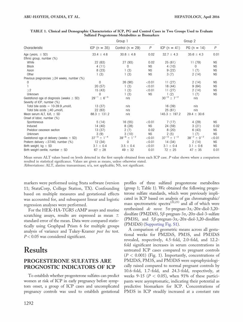
TABLE 1. Clinical and Demographic Characteristics of ICP, PG and Control Cases in Two Groups Used to EvaluateSulfated Progesterone Metabolites as Biomarkers
Characteristic
Group 1 Group 2
ICP (n 5 35) Control (n 5 29) P ICP (n 5 41) PG (n 5 14) P
Age (years, 6 SD) 33.4 6 4.6 30.8 6 4.8 0.02 32.7 6 4.3 35.6 6 4.3 0.01Ethnic group, number (%)
White 22 (63) 27 (93) 0.02 25 (61) 11 (79) NSBlack 4 (11) 0 NS 4 (10) 0 NSAsian 8 (23) 1 (3) NS 9 (22) 1 (7) NSOther 1 (3) 1 (3) NS 3 (7) 2 (14) NS
Previous pregnancies �24 weeks, number (%)0 0 26 (90) <0.01 11 (27) 2 (14) NS1 20 (57) 1 (3) <0.01 18 (44) 9 (64) NS�2 15 (43) 1 (3) <0.01 11 (27) 2 (14) NSUnknown 0 1 (3) NS 1 (2) 1 (7) NS
Gestational age at diagnosis (weeks 6 SD) 2911 6 613 n/a 3010 6 712 n/aSeverity of ICP, number (%)
Total bile acids 5 10-39.9 lmol/L 13 (37) n/a 16 (39) n/aTotal bile acids �40 lmol/L 22 (63) n/a 25 (61) n/a
Mean serum ALT, IU/L 6 SD 88.3 6 131.2 n/a 145.3 6 197.2 29.4 6 30.6Onset of labor, number (%)
Spontaneous 5 (14) 16 (55) <0.01 7 (17) 4 (29) NSInduced 14 (40) 8 (28) NS 24 (59) 3 (21) 0.02Prelabor cesarean section 13 (37) 2 (7) 0.02 8 (20) 6 (43) NSUnknown 3 (9) 3 (10) NS 2 (5) 1 (7) NS
Gestational age at delivery (weeks 6 SD) 3710 6 114 3916 6 112 <0.01 3710 6 113 3812 6 012 <0.01Preterm delivery <37/40, number (%) 12 (34) 0 <0.01 14 (34) 2 (14) NSBirth weight, kg 6 SD 3.1 6 0.4 3.5 6 0.4 <0.01 3.1 6 0.4 3.1 6 0.6 NSBirth weight centile, number 6 SD 67 6 28 49 6 32 0.01 72 6 25 47 6 35 0.01
Mean serum ALT values based on levels detected in the first sample obtained from each ICP case. P value shown where a comparisonresulted in statistical significance. Values are given as means, unless otherwise stated.Abbreviations: ALT, alanine transaminase; n/a, not applicable; NS, not significant.
ABU-HAYYEH, OVADIA, ET AL. HEPATOLOGY, April 2016
1292

from 9 to 41 weeks, whereas concentrations ofPM3DiS and PM2DiS increased steeply from 24 to41 weeks for the ICP group compared to controls (P< 0.05) (Fig. 1).UDCA treatment improves maternal pruritus and
biochemical derangements in ICP. UDCA signifi-cantly reduced PM2DiS and PM3DiS concentra-tions relative to untreated ICP women throughoutthe last trimester of pregnancy (P < 0.05). A trendanalysis showed a significant change in the trend ofPM3DiS with UDCA treatment in the third trimes-ter of ICP compared to the untreated ICP group(P< 0.05), becoming similar to the pregnant controlgroup trend (Fig. 1).
PROGESTERONE SULFATECONCENTRATIONS RAPIDLYRESOLVE IN ICP SERUMFOLLOWING PARTURITION
To establish whether progesterone sulfate concen-trations persist following parturition in ICP, concen-trations of progesterone metabolites in the lastsample in ICP cases prior to parturition and postnatalsamples collected thereafter were assayed in a sub-group of patients. Concentrations of PM2DiS,PM3S, and PM3DiS decreased rapidly followingbirth and normalized to almost undetectable levels asearly as 12 days postpartum (Table 2).
PROGESTERONE SULFATES AREASSOCIATED WITH SEVERITY OFITCH IN ICP AND CAN PREDICTITS SUBSEQUENT ONSET
To assess the involvement of progesterone sulfatesin pruritus, we investigated the relationship betweenpruritus severity and serum concentrations of proges-terone metabolites in women with pregnancy-
� � � � � � � � � � � � � � � � � � � � � � � � � � � � � � � � � � � � � � � � � � � � � � � � � � � � � � � � � � � � � � � � � �
FIG. 1. Gestational serum profiles of PM2DiS, PM3S, andPM3DiS in group 1. Panels A, B, and C show the mean con-centrations of PM2DiS, PM3S, and PM3DiS, respectively, forserum samples obtained at different gestational time points fromwomen with uncomplicated pregnancies (control, closed squares),untreated ICP (closed circles), and UDCA-treated ICP (closedtriangles). Error bars represent 6 standard error of the mean. Pvalues for gestational week category comparison of untreated ICPversus controls were determined by Student t test.
� � � � � � � � � � � � � � � � � � � � � � � � � � � � � � � � � � � � � � � � � � � � � � � � � � � � � � � � � � � � � � � � � �
TABLE 2. Maternal Concentrations of Progesterone Sulfatesin the Last Serum Sample Prior to Parturition and in
Subsequent Postnatal Serum Samples in ICP
CaseGestational
Day/Postnatal DayPM3S
(lmol/L)PM3DiS(lmol/L)
PM2DiS(lmol/L)
1 GD 266 8.03 0.62 1.48PN16 2.24 0.09 0.49
2 GD 241 2.75 1.28 1.27PN166 0.00 0.00 0.00
3 GD 256 12.44 1.88 3.89PN134 0.03 0.01 0.00
4 GD 255 7.22 2.19 3.30PN112 0.04 0.03 0.09
5 GD 232 21.07 6.71 3.31PN121 0.00 0.00 0.00
6 GD 269 2.28 0.52 1.25PN140 0.00 0.00 0.00
7 GD 237 47.36 11.81 1.81PN11 19.99 9.45 0.94PN142 0.00 0.00 0.00
8 GD 261 9.85 4.76 5.94PN140 0.02 0.00 0.00
9 GD 248 11.10 14.41 8.97PN142 0.02 0.00 0.00
10 GD 252 20.04 5.31 4.90PN113 3.74 3.44 1.72
11 GD 268 23.72 3.88 4.28PN156 0.06 0.00 0.07
12 GD 255 15.64 5.60 6.04PN11 9.29 5.15 6.00
Abbreviations: GD, gestational day; PN, postnatal.
HEPATOLOGY, Vol. 63, No. 4, 2016 ABU-HAYYEH, OVADIA, ET AL.
1293

associated pruritus (Table 1). Serum PM2DiS, PM3S,and PM3DiS concentrations all differentiated womenwith ICP from PG (P < 0.05) (Table 3). Serum con-centrations of PM3S (odds ratio [OR] 5 6.1, 95%confidence interval [CI] 0.6-11.5, P < 0.05) and auto-taxin activity (OR 5 1.4, 95% CI 0.3-2.4, P < 0.05)were significantly associated with itch severity in ICP.To determine whether progesterone sulfates could
predict subsequent ICP, logistic regression was per-formed on PM2DiS, PM3S, and PM3DiS concentra-tions and autotaxin activity, using the first serumsample from women at presentation with pruritus andnormal serum biochemistry (Table 4). PM2DiS andPM3DiS differentiated between the women whowould subsequently develop ICP (P < 0.05, OR 5
2.8, 95% CI 1.5-5.2, and OR 5 2.5, 95% CI 1.2-5.4,respectively).To refine a prediction algorithm, we evaluated
whether a combination of markers could more reliablypredict disease. PM2DiS, PM3DiS, and autotaxin incombination resulted in an improved area under thereceiver operating characteristic (ROC) curve of 0.91(95% CI 0.80-1.00) in contrast to autotaxin (0.73,95% CI 0.52-0.94), PM2DiS (0.72, 95% CI 0.52-0.92), or PM3DiS (0.74, 95% CI 0.55-0.94) alone(Fig. 2A). Plotting this combination as a predictivescore for the first serum sample from women present-ing with PG enabled clear differentiation between
those who would subsequently develop ICP and thosewho continue to have benign PG (Fig. 2B).
PROGESTERONE SULFATES ARESUPRAPHYSIOLOGICALLY RAISEDIN EARLY GESTATION IN LOW-RISK ICP CASES
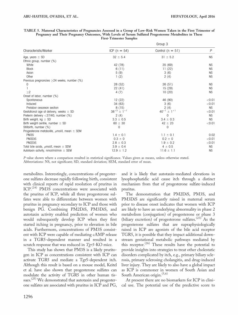
We evaluated this predictive algorithm in a thirdgroup of asymptomatic pregnant women who gaveserum samples at 11-14 gestational weeks for a studyof serum biomarkers to predict adverse pregnancy out-come (Table 5). Fifty-four women from this groupdeveloped ICP in later pregnancy, and their progester-one sulfate concentrations were compared to those of51 women with uncomplicated pregnancies. PM2DiS,PM3S, and PM3DiS concentrations were significantlyraised in women with subsequent ICP (Table 5).Autotaxin did not predict ICP at this early gestation(area under the curve 5 0.55, 95% CI 0.43-0.66),while PM3DiS and PM2DiS in combination showedsome predictive ability (area under the curve 5 0.68,95% CI 0.58-0.78) (Supporting Fig. S2).
PROGESTERONE SULFATESSIGNAL THROUGH TGR5 TOMEDIATE ITCH
Activation of the G protein-coupled receptor Tgr5elicits an itch response in mice.(9) We thereforehypothesized that progesterone metabolites associatedwith itch in ICP can activate TGR5 in vitro. HEKcells stably transfected with TGR5 or empty vectorcontrol cells were transfected with the CAMYELcAMP sensor and treated with the cAMP inducer for-skolin or increasing concentrations of PM2DiS,PM3S, and PM3DiS. In HEK-TGR5 cells, PM3Selicited a cAMP response at concentrations of �1
TABLE 3. Associations Between Biochemical Markers and Pruritus Scores and Their Ability to Differentiate ICP From PG
Biomarker
Association With Pruritus
ICP PG Ability to Identify ICP
Change in VAS (95% CI) P Change in VAS (95% CI) P OR (95% CI) P
PM3S 6.1 (0.6-11.5) 0.03 0.9 (24.6–6.3) NS 1.7 (1.1-2.4) 0.01PM3DiS 2.2 (21.6–6) NS 22.5 (26.2–1.2) NS 2.1 (1.4-3.4) <0.01PM2DiS 20.3 (25.3–4.6) NS 28.0 (214.9–1.2) 0.03 1.7 (1.2-2.5) 0.01Autotaxin 1.4 (0.3-2.4) 0.01 2.0 (20.1–4.1) NS 2.3 (2.1-2.6) <0.01
Linear regression results showing the effect of doubling biochemical markers and change in visual analogue score for ICP and PG andORs for developing ICP. P value shown where a comparison resulted in statistical significance.Abbreviations: NS, not significant; VAS, visual analogue score.
TABLE 4. Autotaxin, PM2DiS, and PM3DiS All Have theAbility to Predict ICP When Measured at the Time of Onset
of Gestational Pruritus
ICP MarkerOR of Future
ICP (95% CI) PArea UnderROC Curve
PM3S 1.70 (0.97-3.01) 0.07 0.45 (0.23-0.68)PM3DiS 2.77 (1.48-5.19) <0.01 0.74 (0.55-0.94)PM2DiS 2.54 (1.18-5.44) 0.02 0.72 (0.52-0.92)Autotaxin 2.22 (0.99-4.86) 0.07 0.73 (0.52-0.94)
Abbreviation: ROC, receiver operating characteristic.
ABU-HAYYEH, OVADIA, ET AL. HEPATOLOGY, April 2016
1294

lmol/L, whereas there was no cAMP response in con-trol cells. PM3S stimulated a concentration-dependentformation of cAMP with a 50% effective concentrationof 5.5 lmol/L (Fig. 3A; and Supporting Fig. S3).Notably, these PM3S concentrations were demon-strated from 20 weeks’ gestation in women with ICP(Fig. 1) and significantly associated with pruritusseverity (Table 3). In contrast, PM2DiS and PM3DiSstimulated cAMP formation at extremely high concen-trations. The temporal profile for the �1 lmol/LPM3S-mediated cAMP response was consistent withthe rapid actions of an activated G protein-coupledreceptor (Fig. 3B). We also excluded FXR as a possiblemediator of the progesterone sulfate signal as all threeprogesterone sulfates were unable to either transacti-vate FXR or inhibit GW4064-mediated FXR transac-tivity in an FXR-reporter assay system (SupportingFig. S4).Because PM3S activates TGR5 in HEK-TGR5
cells, we examined whether it could evoke Tgr5-mediated scratching in mice. PM3S or vehicle (con-trol) was intradermally injected into the nape of theneck of WT and Tgr5-KO mice, and scratching
behavior was measured for 60 minutes. In WT mice,PM3S stimulated a robust scratching response in thefirst 30 minutes, which was 16-fold higher than thatevoked by vehicle (P < 0.05) (Fig. 3C) and signifi-cantly blunted by three-fold in Tgr5-KO mice com-pared to WT mice (P < 0.05) (Fig. 3C). PM3Scontinued to stimulate scratching in WT mice from 30to 60 minutes, whereas the response in Tgr5-KO micewas attenuated after 30 minutes. Cumulatively over thewhole hour, there was a 21-fold increase in observedscratches in the PM3S-challenged WT mice(P < 0.05), which was significantly abrogated in theTgr5-KO mice (P < 0.05).
DiscussionOur results show that the sulfated progesterone
metabolites PM2DiS, PM3S, and PM3DiS are prog-nostic for ICP as their concentrations are elevated dur-ing early gestation when patients are asymptomatic.Furthermore, UDCA treatment reduces the ICP-associated elevation of disulfated progesterone
� � � � � � � � � � � � � � � � � � � � � � � � � � � � � � � � � � � � � � � � � � � � � � � � � � � � � � � � � � � � � � � � � � � � � � � � � � � � � � � � � � � � � � � � � � � � � � � � � � � � � � � � � � � � � � � � � � � � � � � � � � � � � � � � � � � � � � �
FIG. 2. Progesterone sulfates and autotaxin can predict subsequent onset of ICP in pregnant women with pruritus. The receiver oper-ating curves (A) improved toward an optimal area under the curve of 1.0 when biomarkers were evaluated in combination: PM2DiS1 PM3DiS (complete line), autotaxin (dashed line), and PM2DiS 1 PM3DiS 1 autotaxin (dotted and dashed line). (B) A com-bined predictive score (PM2DiS 1 PM3DiS 1 autotaxin) of greater than 0.25 for individual samples plotted against the gestationalday of sampling reliably predicted all ICP cases. Women who developed ICP (n 5 14, closed circles) and PG (n 5 14, open trian-gles) were reliably distinguished by this score; dashed line represents demarcation between the two groups. Abbreviation: AUC, areaunder the curve.
� � � � � � � � � � � � � � � � � � � � � � � � � � � � � � � � � � � � � � � � � � � � � � � � � � � � � � � � � � � � � � � � � � � � � � � � � � � � � � � � � � � � � � � � � � � � � � � � � � � � � � � � � � � � � � � � � � � � � � � � � � � � � � � � � � � � � � �
HEPATOLOGY, Vol. 63, No. 4, 2016 ABU-HAYYEH, OVADIA, ET AL.
1295
metabolites. Interestingly, concentrations of progester-one sulfates decrease rapidly following birth, consistentwith clinical reports of rapid resolution of pruritus inICP.(24) PM3S concentrations were associated withthe pruritus of ICP, while all three progesterone sul-fates were able to differentiate between women withpruritus in pregnancy secondary to ICP and those withbenign PG. Combining PM2DiS, PM3DiS, andautotaxin activity enabled prediction of women whowould subsequently develop ICP when they firststarted itching in pregnancy, prior to elevation in bileacids. Furthermore, concentrations of PM3S consist-ent with ICP were capable of mediating cAMP releasein a TGR5-dependent manner and resulted in ascratch response that was reduced in Tgr5-KO mice.This study has shown that PM3S is a likely prurito-
gen in ICP as concentrations consistent with ICP canactivate TGR5 and mediate a Tgr5-dependent itch.Although this result is based on a mouse model, Keitelet al. have also shown that progesterone sulfates canmodulate the activity of TGR5 in other human tis-sues.(25) We demonstrated that autotaxin and progester-one sulfates are associated with pruritus in ICP and PG,
and it is likely that autotaxin-mediated elevations inlysophosphatidic acid cause itch through a distinctmechanism from that of progesterone sulfate-inducedpruritus.The demonstration that PM2DiS, PM3S, and
PM3DiS are significantly raised in maternal serumprior to disease onset indicates that women with ICPare likely to have an underlying abnormality in phase 2metabolism (conjugation) of progesterone or phase 3(biliary excretion) of progesterone sulfates.(23) As theprogesterone sulfates that are supraphysiologicallyraised in ICP are agonists of the bile acid receptorTGR5, it is possible that they impact additional down-stream gestational metabolic pathways mediated bythis receptor.(26) These results have the potential toprovide insights into strategies to treat other cholestaticdisorders complicated by itch, e.g., primary biliary scle-rosis, primary sclerosing cholangitis, and drug-inducedliver injury. They are likely to also have a global impactas ICP is commoner in women of South Asian andSouth American origin.(5,6)
At present there are no biomarkers for ICP in clini-cal use. The potential use of the predictive score to
TABLE 5. Maternal Characteristics of Pregnancies Assessed in a Group of Low-Risk Women Taken in the First Trimester ofPregnancy and Their Pregnancy Outcomes, With Levels of Serum Sulfated Progesterone Metabolites in These
First-Trimester Samples
Characteristic/Marker
Group 3
ICP (n 5 54) Control (n 5 51) P
Age, years 6 SD 32 6 5.4 31 6 5.2 NSEthnic group, number (%)
White 42 (78) 35 (69) NSBlack 6 (11) 11 (22) NSAsian 5 (9) 3 (6) NSOther 1 (2) 2 (4) NS
Previous pregnancies �24 weeks, number (%)0 28 (52) 26 (51) NS1 22 (41) 15 (29) NS�2 4 (7) 10 (20) NS
Onset of labor, number (%)Spontaneous 12 (22) 46 (90) <0.01Induced 34 (63) 3 (6) <0.01Prelabor cesarean section 8 (15) 2 (4) NS
Gestational age at delivery, weeks 6 SD 3813 6 111 4011 6 111 <0.01Preterm delivery <37/40, number (%) 2 (4) 0 NSBirth weight, kg 6 SD 3.3 6 0.5 3.4 6 0.3 NSBirth weight centile, number 6 SD 60 6 30 42 6 23 <0.01Stillbirth, number (%) 0 0Progesterone metabolite, lmol/L mean 6 SEM
PM3S 1.4 6 0.1 1.1 6 0.1 0.02PM3DiS 0.3 6 0 0.2 6 0 <0.01PM2DiS 2.8 6 0.3 1.9 6 0.2 <0.01
Total bile acids, lmol/L mean 6 SEM 3.9 6 0.4 4 6 0.5 NSAutotaxin activity, nmol/ml/min 6 SEM 12.9 6 1.2 11.6 6 1.1 NS
P value shown where a comparison resulted in statistical significance. Values given as means, unless otherwise stated.Abbreviations: NS, not significant; SD, standard deviation; SEM, standard error of mean.
ABU-HAYYEH, OVADIA, ET AL. HEPATOLOGY, April 2016
1296

establish whether pregnant women with pruritus willdevelop ICP is enticing and should be evaluated infuture prospective, well-powered studies. This isimportant as the patient groups in the current studywere all managed in a single specialist center and thismay have introduced population bias. If the results areconfirmed in different populations, a feasible exten-sion to this study would be to assay concentrations ofurinary progesterone sulfates (Glantz et al.(11)) toidentify a predictive score that can be used in earlypregnancy to establish whether a woman with prurituswill develop this high-risk disease. This could havewider clinical application with the development ofhigh-throughput urinary assays for progesterone sul-fates or similar laboratory tests for serum levels of pro-
gesterone sulfates and autotaxin. This will enableobstetricians to refer women for hospital care in ahigh-risk setting or alternatively to reassure them thattheir pruritus is unlikely to have pathologicalconsequences.In conclusion, this study describes the mechanism
of action of pruritogens that are prognostic for ICP,which has the potential to enable obstetricians todiagnose ICP, a common metabolic disorder of preg-nancy, prior to onset of symptoms or biochemicalderangements.
Acknowledgment: We thank Dr. Graeme Hogarth forassistance with chemical structures and Mr. Bilal forhelpful discussion.
� � � � � � � � � � � � � � � � � � � � � � � � � � � � � � � � � � � � � � � � � � � � � � � � � � � � � � � � � � � � � � � � � � � � � � � � � � � � � � � � � � � � � � � � � � � � � � � � � � � � � � � � � � � � � � � � � � � � � � � � � � � � � � � � � � � � � � �
FIG. 3. Progesterone metabolites can activate TGR5 and elicit a Tgr5-mediated itch response in mice. cAMP formation was moni-tored over time in HEK293 cells expressing TGR5 or control cells that were treated with 10 lmol/L of forskolin or increasing con-centrations of PM3S, PM3DiS, or PM2DiS. Data are presented as a dose response for all three compounds in both cell types (A) ortime course for PM3S in TGR5-expressing cells (B). Values represent mean 6 standard deviation of n 5 3. (C) Wild-type or Tgr5-KO mice were intradermally injected with vehicle or 20 lL of 100 lmol/L PM3S, and scratching events were counted for the indi-cated time periods. Values represent mean 6 standard error of the mean of n � 4. *P < 0.05 for vehicle versus PM3S-administeredmice scratch comparison; #P < 0.05 for PM3S WT versus PM3S Tgr5-KO scratch comparison as determined by one-way analysis ofvariance. Abbreviation: BRET, bioluminescence resonance energy transfer.
� � � � � � � � � � � � � � � � � � � � � � � � � � � � � � � � � � � � � � � � � � � � � � � � � � � � � � � � � � � � � � � � � � � � � � � � � � � � � � � � � � � � � � � � � � � � � � � � � � � � � � � � � � � � � � � � � � � � � � � � � � � � � � � � � � � � � � �
HEPATOLOGY, Vol. 63, No. 4, 2016 ABU-HAYYEH, OVADIA, ET AL.
1297

REFERENCES
1) Williamson C, Geenes V. Intrahepatic cholestasis of pregnancy.
Obstet Gynecol 2014;124:120-133.
2) Glantz A, Marschall HU, Mattsson LA. Intrahepatic cholestasis
of pregnancy: relationships between bile acid levels and fetal
complication rates. HEPATOLOGY 2004;40:467-474.
3) Geenes V, Chappell LC, Seed PT, Steer PJ, Knight M,
Williamson C. Association of severe intrahepatic cholestasis of
pregnancy with adverse pregnancy outcomes: a prospective
population-based case-control study. HEPATOLOGY 2014;59:
1482-1491.
4) Kenyon AP, Tribe RM, Nelson-Piercy C, Girling JC,
Williamson C, Seed PT, et al. Pruritus in pregnancy: a study of
anatomical distribution and prevalence in relation to the develop-
ment of obstetric cholestasis. Obstet Med 2010;3:25-29.
5) Abedin P, Weaver JB, Egginton E. Intrahepatic cholestasis of
pregnancy: prevalence and ethnic distribution. Ethn Health
1999;4:35-37.
6) Reyes H. Sex hormones and bile acids in intrahepatic cholestasis
of pregnancy. HEPATOLOGY 2008;47:376-379.
7) Kremer AE, Martens JJ, Kulik W, Rueff F, Kuiper EM, van
Buuren HR, et al. Lysophosphatidic acid is a potential mediator
of cholestatic pruritus. Gastroenterology 2010;139:1008-1018.
8) Kremer AE, Bolier R, Dixon PH, Geenes V, Chambers J,
Tolenaars D, et al. Autotaxin activity has a high accuracy to
diagnose intrahepatic cholestasis of pregnancy. J Hepatol 2015;
62:897-904.
9) Alemi F, Kwon E, Poole DP, Lieu T, Lyo V, Cattaruzza F,
et al. The TGR5 receptor mediates bile acid-induced itch and
analgesia. J Clin Invest 2013;123:1513-1530.
10) Heikkinen J, Maentausta O, Ylostalo P, Janne O. Serum bile acid
levels in intrahepatic cholestasis of pregnancy during treatment
with phenobarbital or cholestyramine. Eur J Obstet Gynecol
Reprod Biol 1982;14:153-162.
11) Glantz A, Reilly SJ, Benthin L, Lammert F, Mattsson LA,
Marschall HU. Intrahepatic cholestasis of pregnancy: ameliora-
tion of pruritus by UDCA is associated with decreased progester-
one disulphates in urine. HEPATOLOGY 2008;47:544-551.
12) Abu-Hayyeh S, Papacleovoulou G, Lovgren-Sandblom A, Tahir
M, Oduwole O, Jamaludin NA, et al. Intrahepatic cholestasis of
pregnancy levels of sulfated progesterone metabolites inhibit far-
nesoid X receptor resulting in a cholestatic phenotype. HEPATO-
LOGY 2013;57:716-726.
13) Abu-Hayyeh S, Martinez-Becerra P, Sheikh Abdul Kadir SH,
Selden C, Romero MR, Rees M, et al. Inhibition of Na1-tauro-
cholate co-transporting polypeptide-mediated bile acid transport
by cholestatic sulfated progesterone metabolites. J Biol Chem
2010;285:16504-16512.
14) Vallejo M, Briz O, Serrano MA, Monte MJ, Marin JJ. Potential
role of trans-inhibition of the bile salt export pump by progester-
one metabolites in the etiopathogenesis of intrahepatic cholestasis
of pregnancy. J Hepatol 2006;44:1150-1157.
15) Meng LJ, Reyes H, Palma J, Hernandez I, Ribalta J, Sjovall J.
Profiles of bile acids and progesterone metabolites in the urine
and serum of women with intrahepatic cholestasis of pregnancy.
J Hepatol 1997;27:346-357.
16) Geenes V, Lovgren-Sandblom A, Benthin L, Lawrance D,
Chambers J, Gurung V, et al. The reversed feto-maternal bile
acid gradient in intrahepatic cholestasis of pregnancy is corrected
by ursodeoxycholic acid. PLoS One 2014;9:e83828.
17) Moser K, Stanfield KM, Leon DA. Birthweight and gestational
age by ethnic group, England and Wales 2005: introducing new
data on births. Health Stat Q 2008;(39):22-55.
18) Reich A, Heisig M, Phan NQ, Taneda K, Takamori K,
Takeuchi S, et al. Visual analogue scale: evaluation of the instru-
ment for the assessment of pruritus. Acta Derm Venereol 2012;
92:497-501.
19) Jiang LI, Collins J, Davis R, Lin KM, DeCamp D, Roach T,
et al. Use of a cAMP BRET sensor to characterize a novel regu-
lation of cAMP by the sphingosine 1-phosphate/G13 pathway.
J Biol Chem 2007;282:10576-10584.
20) Jensen DD, Godfrey CB, Niklas C, Canals M, Kocan M, Poole
DP, et al. The bile acid receptor TGR5 does not interact with
beta-arrestins or traffic to endosomes but transmits sustained sig-
nals from plasma membrane rafts. J Biol Chem 2013;288:22942-
22960.
21) Poole DP, Godfrey C, Cattaruzza F, Cottrell GS, Kirkland JG,
Pelayo JC, et al. Expression and function of the bile acid receptor
GpBAR1 (TGR5) in the murine enteric nervous system. Neuro-
gastroenterol Motil 2010;22:814-818.
22) Sjovall J, Sjovall K. Steroid sulphates in plasma from pregnant
women with pruritus and elevated plasma bile acid levels. Ann
Clin Res 1970;2:321-337.
23) Reyes H, Sjovall J. Bile acids and progesterone metabolites in
intrahepatic cholestasis of pregnancy. Ann Med 2000;32:94-106.
24) Chappell LC, Gurung V, Seed PT, Chambers J, Williamson C,
Thornton JG. Ursodeoxycholic acid versus placebo, and early
term delivery versus expectant management, in women with
intrahepatic cholestasis of pregnancy: semifactorial randomised
clinical trial. BMJ 2012;344:e3799.
25) Keitel V, Spomer L, Marin JJ, Williamson C, Geenes V, Kubitz
R, et al. Effect of maternal cholestasis on TGR5 expression in
human and rat placenta at term. Placenta 2013;34:810-816.
26) Watanabe M, Houten SM, Mataki C, Christoffolete MA, Kim
BW, Sato H, et al. Bile acids induce energy expenditure by pro-
moting intracellular thyroid hormone activation. Nature 2006;
439:484-489.
Author names in bold designate shared co-firstauthorship.
Supporting InformationAdditional Supporting Information may be found at
onlinelibrary.wiley.com/doi/10.1002/hep.28265/suppinfo.
ABU-HAYYEH, OVADIA, ET AL. HEPATOLOGY, April 2016
1298