programmed cell death in plant disease: the purpose and promise of
TRANSCRIPT
P1: PSA/ARY P2: PSA/VKS QC: PSA/bs T1: PSA
July 8, 1998 22:24 Annual Reviews AR061-17
Annu. Rev. Phytopathol. 1998. 36:393–414Copyright c© 1998 by Annual Reviews. All rights reserved
PROGRAMMED CELL DEATHIN PLANT DISEASE: The Purposeand Promise of Cellular Suicide
D. G. GilchristDepartment of Plant Pathology and the NSF Center for Engineering Plants forResistance Against Pathogens (CEPRAP), University of California, Davis,California 95616; e-mail [email protected]
KEY WORDS: apoptosis, lesion mimics, hypersensitive reaction, ceramide, cell cycle
Truth is seldom pure and never simple.Oscar Wilde
ABSTRACT
The interaction of pathogens with plants leads to a disruption in cellular homeosta-sis, often leading to cell death, in both compatible and incompatible relationships.The mechanistic basis of this cellular disruption and consequent death is complexand poorly characterized, but it is established that host responses to pathogens aredependent on gene expression, involve signal transduction, and require energy.Recent data suggest that in animals, a genetically regulated, signal transduction–dependent programmed cell death process, commonly referred to as apoptosis, isconserved over a wide range of phyla. The basic function of apoptosis is to directthe selective elimination of certain cells during development, but it also is a mas-ter template that is involved in host responses to many pathogens. Programmedcell death in plants, while widely observed, has not been studied extensively ateither the biochemical or genetic level. Current data suggest that activation orsuppression of programmed cell death may underlie diseases in plants as it doesin animals. This review describes some of the fundamental characteristics ofapoptosis in animals and points to a number of connections to programmed celldeath in plants that may lead to both a better understanding of disease processesand novel strategies for engineering disease resistance in plants.
3930066-4286/98/0901-0393$08.00

P1: PSA/ARY P2: PSA/VKS QC: PSA/bs T1: PSA
July 8, 1998 22:24 Annual Reviews AR061-17
394 GILCHRIST
INTRODUCTION
Cellular SuicideSuicide, in human terms, is intellectually difficult to accept and generally viewedas impulsive or irrational and inconsistent with balanced behavior. Suicide incellular terms, however, is exactly the converse: it is pervasive, organized,rational, and leads to organismal balance, both in development and in responseto stress. To achieve and maintain homeostasis, cells in multicellular organismsself-destruct when they are no longer needed or if they are damaged (3). Thisis accomplished by activation of genetically regulated cell suicide machinerythat requires the active participation of the cell in a suicide process known asprogrammed cell death (PCD) (6, 62, 78, 104). In contrast to PCD, necrosisis most commonly defined as cell death that results from exposure to highlytoxic compounds, severe cold or heat stress, or traumatic injury that leads toimmediate damage to membranes or cellular organelles (19). The key distinctionis that necrosis does not require the active participation of the cell in its owndemise. In retrospect, the lethal logic of PCD has long been recognized, butonly recently has extensive evidence for a conserved genetic template appearedthat responds to a range of inter- and intracellular factors.
Moreover, PCD is now recognized as a common cellular default mechanismin that almost all cells appear to possess the machinery necessary to carry outthe terminal steps of death. Evidence for this includes the constitutive presenceof many proteins required for PCD in nucleated animal cells beginning withthe zygote (106, 117). In fact, the nucleus does not appear to be required insome cases as anucleate cytoplasts undergo PCD when treated with the proteinkinase inhibitor staurosporine (61). The death program is triggered during de-velopmental transitions in situations that lead to sculpting of structures, deletingunneeded structures, controlling cell numbers, eliminating abnormal or harmfulcells, and producing differentiated cells without organelles (61). For example,PCD eliminates cells between tadpole tails (66) and between developing digitssuch as fingers and toes (62). It occurs where epithelial sheets invaginate andpinch off to form tubes or vesicles like the vertebrate neural tube or lens (62).In the vertebrate nervous system, up to half of new nerve cells die by PCD,apparently to match their numbers to the number of target cells they innervate(91). These are but a few examples among many that underscore the preva-lence, diversity, and importance of programmed cell death in the developmentof organized tissues in eukaryotic organisms.
Conversely, the existence of such a default death program represents a point ofvulnerability for the organism if it is inappropriately triggered or inappropriatelysuppressed during development or in disease situations (30, 31, 102, 107, 110,119). It is this potential for co-option by pathogens that renders the consideration

P1: PSA/ARY P2: PSA/VKS QC: PSA/bs T1: PSA
July 8, 1998 22:24 Annual Reviews AR061-17
APOPTOSIS IN PLANT DISEASE 395
of programmed cell death a significant issue for plant and animal pathologists.Examples such as those indicated above make both the genes that define the PCDprogram and the mechanisms that keep it from occurring at random compellingresearch targets for cell biologists and geneticists (62). The need for cells tomanage a default programmed cell death pathway(s) suggests a number offundamental questions for cell biologists that are currently under intensivestudy in many laboratories. Why do cells die when and where they do duringdevelopment? How are the PCD genes regulated to ensure transcription oractivation of the gene product at the appropriate or necessary time and place?What are the PCD signals, how are they propagated, and what limits the spreadof these signals?
PROGRAMMED CELL DEATHIN ANIMALS: APOPTOSIS
Characteristics of ApoptosisOne reason why research had not focused on programmed death templates untilrecently was the lack of evidence for a common mechanism, highly conservedand widely distributed, that regulated cellular homeostasis through the selectiveelimination of certain cells. The origin of active study of PCD began in 1972with the publication of a landmark paper that introduced the term apoptosis (68)and described fundamental characteristics of apoptosis as “a basic biologicalphenomenon with wide-ranging implications in tissue kinetics” (68). The termapoptosis was derived from two Greek roots: apo (away) and ptosis (to fall, alsoa medical term referring to the drooping of eyelids) that was used to describepetals falling from flowers or leaves from trees. Kerr and colleagues describedseveral epithelial cell types that underwent orderly death during development.These included cells that died owing to withdrawal of survival signals and cellsthat had been exposed to toxins. The creative synthesis of their observationsled to the conclusion that in each of these cases, the dying cells exhibitedconsistent morphological features during death and that cell deletion playeda complementary but opposite role to mitosis in the regulation of animal cellpopulations (67, 68).
Later, other scientists recognized the existence of a series of morphologicaland biochemical hallmarks of apoptosis in animals (108, 109). Animal cellsundergoing apoptosis exhibit shrinkage, loss of cell-to-cell contact in organizedtissues, and the orderly fragmentation of nuclear DNA at internucleosomalsites, which is then organized into sharply defined membrane-bound apoptoticbodies. In animal cells, these apoptotic bodies are taken up by adjacent cellsand degraded within minutes to hours (9). Hence, the function of the apoptoticbodies is thought to be the protection of neighboring cells from damage due to
P1: PSA/ARY P2: PSA/VKS QC: PSA/bs T1: PSA
July 8, 1998 22:24 Annual Reviews AR061-17
396 GILCHRIST
leakage of toxic cellular contents of the dying cell until resorption is completed.Internucleosomal cleavage of DNA is catalyzed by an endogenous calcium-dependent endonuclease that generates 180-bp multiples of these fragments(Figure 1). Multiples of these units can be resolved by gel electrophoresisas a laddered pattern of DNA that corresponds to the nucleosomal fragments(108). Fragmentation of DNA during apoptosis also can be detected in situby reagents that react with the exposed 3′ hydroxyls on the nucleosomal units(39). The assay procedure, commonly referred to as the TUNEL assay, involvesend labeling of the nucleosomal DNA fragments by terminal deoxynucleotidyltransferase with UTP conjugated to a detectable marker (44). The TUNELassay is capable of detecting limited amounts of DNA fragmentation duringapoptosis (75). However, this assay must be used with caution since DNAdegraded during nonapoptotic cell death and during the fixing of tissue alsomay give positive reactions (45, 115).
Evolution of Research on ApoptosisWhat has made this topic so compelling to so many today? Recognition of thefundamental importance of apoptosis was not immediate. In fact, it took nearlytwo decades after the landmark publication by Kerr and colleagues (68) for theterm apoptosis and its biological ramifications to be fully appreciated. Since1972, however, the number of publications related to apoptosis has increasedexponentially every year (93). Today, apoptosis is not only accepted as a uni-versal mechanism regulating cellular homeostasis in animals, it is also drivinga veritable biological revolution in basic research in cell biology and medicine.This has occurred for several reasons. First, there has been an explosion ofdiscoveries in laboratories worldwide in resolving the structure and function ofgenes, signal molecules, and signal transduction pathways regulating apoptosis.Second, it has been recognized that the functional machinery of apoptosis, bothgenes and signaling events, is highly conserved from nematodes to insects tohumans, albeit with subtle differences in the actual mechanisms. Third, it hasnow become clear that the dysregulation of apoptosis is the basis of many humandiseases (Table 1). The convergence of these pieces of scientific informationhas now placed both the fundamental and the practical study of apoptosis in theforefront of biological and pharmaceutical research (25, 62, 93, 111, 123).
Role of Apoptosis in Animal DiseaseIn what may seem at first to be a paradox, apoptosis plays a key role in both de-generative and proliferative diseases (Table 1). Elucidation of controlling genesand signaling pathways has provided evidence for an evolutionarily conservedlink between maladies ranging from Alzheimer’s disease (112) to cancer (107).It is perhaps intuitive that any process that is highly conserved to maintain

P1: PSA/ARY P2: PSA/VKS QC: PSA/bs T1: PSA
July 8, 1998 22:24 Annual Reviews AR061-17
APOPTOSIS IN PLANT DISEASE 397
Fig
ure
1R
epre
sent
ativ
ege
nes,
sign
alin
gpa
thw
ays,
and
othe
rde
ath-
depe
nden
tch
ange
sas
soci
ated
with
prog
ram
med
cell
deat
hin
anim
als.
See
text
for
deta
ils.

P1: PSA/ARY P2: PSA/VKS QC: PSA/bs T1: PSA
July 8, 1998 22:24 Annual Reviews AR061-17
398 GILCHRIST
Table 1 Examples of human diseases where dysregulation of apoptosis is believed to playan important role in the syndrome
Immune system Autoimmune diseases; Lupus, Graves disease, rheumatoid arthritis,AIDS, Type I diabetes
Cancer related Lymphoma, leukemia, colon cancer, solid tumors
Neurological system Alzheimer’s disease, Lou Gehrig’s disease, Parkinson’s disease,Huntington’s disease, multiple sclerosis
Viral diseases Cowpox virus, Adenovirus, Epstein-Bar virus
order can result in harsh consequences for an organism when circumstancesbeyond the control of the organism disrupt that order. For example, excessivecell death can lead to impaired development and degenerative diseases (99),whereas insufficient gene-directed cell death can lead to cancer (35), permitviral infections (17, 102), and contribute to autoimmune diseases (88). HelperT cells undergo apoptosis in AIDS, and neurons die by apoptosis in Alzheimer’sdisease, Parkinson’s disease, Huntington’s disease, and Lou Gehrig’s disease(31). In disease situations where immortalization or extensive proliferation of acell line occurs, such as in cancer or virus infections, the expression of variousproto-oncogenes and virus-encoded genes (Figure 1; adenovirus E1A, SV40Large T antigen) that actively suppress apoptosis appears to be widespread(12, 16, 17, 76). Hence, the genes and signal molecules involved in apoptosis(2, 123) have become widely sought targets for therapeutic manipulation in bothdegenerative and proliferative diseases of animals and humans (20, 25, 30, 107),including diseases of the immune system (22).
Regulation and Propagation of the Apoptotic SignalA central question in the field of PCD is whether an irreversible commitment todeath leads from a common biochemical pathway to the morphological featuresof apoptosis (47). The general feeling at this time seems to favor the view thatthere are several pathways capable of being invoked in a cell-specific fashionwith different final morphologies. This means some pathways may lead to thetypical morphology of apoptosis whereas others may have a different final mor-phology (47), yet the outcome is the same. A number of factors are involvedin the triggering and propagation of apoptosis in animals (Figure 1). Amongthese are cell surface receptors, transmembrane domains, intracellular proteinsinvolved in propagation of death signals (death domains), second messengersincluding inositol triphosphate and ceramides, Ca2+ fluxes, reactive oxygenspecies, cell cycle regulating factors (cyclins and coupled cdc kinases), andproteins that act as either suppressors (e.g. Bcl2, iap) or activators (e.g. Bax) ofcell death (101, 104, 105) (Figure 1). There seem to be two central connectionsto most forms of PCD: an arrest of the cell cycle and the activation of a protease
P1: PSA/ARY P2: PSA/VKS QC: PSA/bs T1: PSA
July 8, 1998 22:24 Annual Reviews AR061-17
APOPTOSIS IN PLANT DISEASE 399
cascade. Members of the family of cysteine proteases related to the interleukin1-β converting enzyme (ICE) have been shown to be necessary for programmedcell death in a number of biological systems (82). The activation of these spe-cific proteases, now referred to as caspases, from proenzymes (ProICE), is nowrecognized as a key biochemical marker of apoptosis, often detected by theiraction on specific substrates (94). The fact that a number of resistance genescloned recently from plants, including the N gene from tobacco, have sequencesimilarity to the Toll gene or interleukin receptor suggests a possible connectionto signal response in plants to those involved in PCD in animals (118) (Figure 1).
Role of the Cell Cycle in Programmed Cell DeathMorphological, biochemical, and gene-expression studies have suggested thatapoptosis may represent a disordered type of cell cycle progression (Figure 1).Several lines of reasoning have led to this conclusion (65). Cells undergoingmitosis or apoptosis lose cell volume, condense their chromatin, and disassem-ble the nuclear lamina in a morphologically similar fashion. Manipulation ofthe cell cycle may either prevent or induce apoptosis (34). The ability (suscep-tibility) of cells to undergo apoptosis following reception of a “death” signaloften depends on the stage of the cell cycle or the presence of specific cell cy-cle checkpoint–regulating compounds, including cyclins and cyclin-dependentkinases (cdk) (69). Phosphorylation and solubilization of nuclear lamins havebeen observed in some apoptotic animal models. The induction of the cellcycle regulating p34cdc2kinase activity during apoptosis and a dependency onthe activity of p34cdc2 kinase have been reported (103). It is well establishedthat p34cdc2activity is elevated as cells traverse the G2/M transition and that thisprotein is a universal regulator of mitosis in eukaryotic cells (47) (Figure 1).
Arrest or disruption of the cell cycle is a common feature of cells that eventu-ally undergo PCD (14, 15, 79). Arrest at the G1/S checkpoint appears to be a keypoint (14). The signals, receptors, and signal transduction pathways appear tobe functionally conserved from bacteria to fungi to plants and animals (3, 110).Hence, any cell in the path of the signaling wave may receive and respondto that signal by undergoing arrest and, upon failing escape the impact of thesignal, will be triggered to undergo PCD. A recently described example of auniversally conserved gene that appears to play a key role in regulation of thecell cycle, cell death and that appears to play a role in the suppression of celldeath by virus encoded factors is the retinoblastoma gene (Rb) (65) (Figure 1).Rb activity is regulated by phosphorlyation and dephosphorylation reactionsthat are mediated by various signals including expression of other genes andsecond messengers like ceramide (see below). The E1A and E7 genes, encodedby animal viruses, bind to Rb, alter regulation of the cell cycle and lead to thesuppression of PCD during viral replication. Rb has recently been identifiedin plants and the RepA protein, encoded by plant gemini viruses also can bind

P1: PSA/ARY P2: PSA/VKS QC: PSA/bs T1: PSA
July 8, 1998 22:24 Annual Reviews AR061-17
400 GILCHRIST
to Rb. This connection between plant virus genes and Rb suggests that thevirus replication scenario, at least in these viruses, may act in a fashion similarto animal viruses although this has not been established. If the signals andsignaling pathways that arrest animal and plant cells at specific stages in thecell cycle originated with single cell organisms, then the signals and receptorsthat lead to cell death could conceivably be conserved with cross-kingdom re-sponsiveness from bacteria to fungi to animals and to plants. Using the samelogic, it also may be true that an invading pathogen could send signals to hostcells that trigger cell cycle arrest and thus lead to the induction of cell deathduring disease.
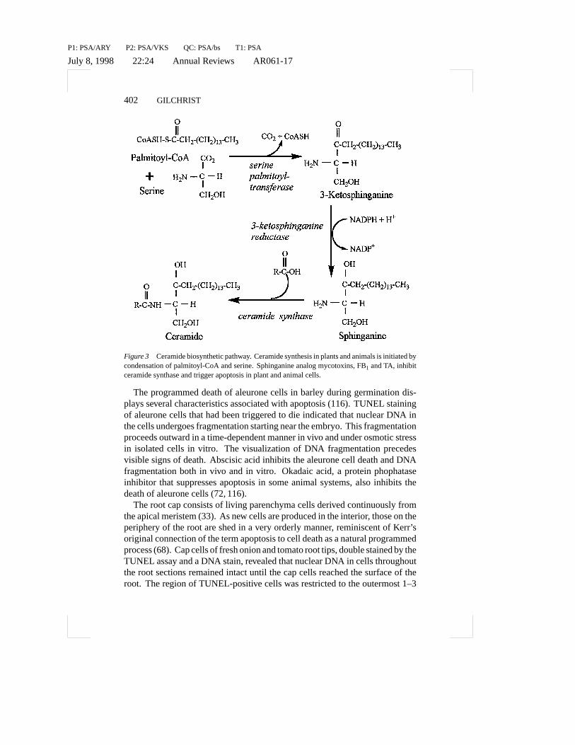
Ceramide Signaling and ApoptosisThe signals that regulate apoptosis in animals are a topic of extreme interest.Several signals were noted earlier but among the most intriguing is the re-cent and novel involvement of ceramide-related compounds in affecting bothdevelopment and apoptosis. The linkage of ceramide signaling to apoptosis,cancer, and degenerative disease in animals is now driving a rapidly emergingnovel area in signal transduction in animal biology (13, 54, 55, 80, 90), whereinceramide-linked signaling pathways are studied as critical second-messengersystems (54, 70) (Figure 2). For example, sphingolipids have been implicatedas playing a direct role in cell contact, growth, and differentiation as well asin influencing the proliferative potential of mammalian cells by induction orsuppression of apoptosis (reviewed in 54). Sphingolipids also have been linkedto increased superoxide formation and calcium influx in neutrophils (122).Fumonisin B1 (FB1), a sphinganine-analog mycotoxin (Figure 3), alters cellmorphology, cell-cell interactions, the behavior of cell surface proteins, pro-tein kinase activity, and cell growth and viability (as reviewed 80), in additionto inducing apoptosis in animal cells (115) (discussed later). Results from ourlaboratory and others suggest that plants and animals may share a heretofore un-realized connection to apoptosis through ceramide-based signaling pathways.If ceramide signaling is widely conserved, it could provide a new biochemicallink between programmed cell death and disease responses in plants.
PROGRAMMED CELL DEATH IN PLANTS
Programmed Cell Death in Plant DevelopmentAlthough apoptosis in animals was likened to senescence of plant leaves (68),evidence of genetic controls and signaling molecules in plants undergoing PCD,analogous to those now widely studied in animals, has emerged only recently(40, 97). This is in spite of the recognition that programmed cell death regimesoccur in numerous situations in plants (see reviews by 7, 37, 38 46, 92). Exam-ples of programmed cell death in plant development include the timely death of

P1: PSA/ARY P2: PSA/VKS QC: PSA/bs T1: PSA
July 8, 1998 22:24 Annual Reviews AR061-17
APOPTOSIS IN PLANT DISEASE 401
Figure 2 Structural relationships between sphinganine, ceramide, and the sphinganine analogmycotoxins, Fumonisin B1 and AAL toxin TA.
petals after fertilization, during diploid parthenogenesis (56), the developmentof tracheary elements (37, 38, 51, 84, 115), and senescence of leaves (8). Thecell death associated with tracheary element formation serves as a model sys-tem for analysis of specific cells clearly targeted for death but not destruction(36–38).
One of the major difficulties in detecting morphological markers of apoptosisin cells, or in those cells that are preparing to undergo PCD, is that the process islocalized, generally to a few cells, and is not synchronized. Given the speed ofthe process in animals (minutes to hours) (75), observation of the various stepsin the process in plants is limited by the ability to predict which cells are destinedto undergo PCD and when it will begin. These hurdles may be overcome byexamining tissues in which the process is ongoing continuously. Situationswhere cell death is semisynchronized and where sufficient numbers of cellsregularly undergo a form of PCD include root cap cells and aleurone cells.
P1: PSA/ARY P2: PSA/VKS QC: PSA/bs T1: PSA
July 8, 1998 22:24 Annual Reviews AR061-17
402 GILCHRIST
Figure 3 Ceramide biosynthetic pathway. Ceramide synthesis in plants and animals is initiated bycondensation of palmitoyl-CoA and serine. Sphinganine analog mycotoxins, FB1 and TA, inhibitceramide synthase and trigger apoptosis in plant and animal cells.
The programmed death of aleurone cells in barley during germination dis-plays several characteristics associated with apoptosis (116). TUNEL stainingof aleurone cells that had been triggered to die indicated that nuclear DNA inthe cells undergoes fragmentation starting near the embryo. This fragmentationproceeds outward in a time-dependent manner in vivo and under osmotic stressin isolated cells in vitro. The visualization of DNA fragmentation precedesvisible signs of death. Abscisic acid inhibits the aleurone cell death and DNAfragmentation both in vivo and in vitro. Okadaic acid, a protein phophataseinhibitor that suppresses apoptosis in some animal systems, also inhibits thedeath of aleurone cells (72, 116).
The root cap consists of living parenchyma cells derived continuously fromthe apical meristem (33). As new cells are produced in the interior, those on theperiphery of the root are shed in a very orderly manner, reminiscent of Kerr’soriginal connection of the term apoptosis to cell death as a natural programmedprocess (68). Cap cells of fresh onion and tomato root tips, double stained by theTUNEL assay and a DNA stain, revealed that nuclear DNA in cells throughoutthe root sections remained intact until the cap cells reached the surface of theroot. The region of TUNEL-positive cells was restricted to the outermost 1–3
P1: PSA/ARY P2: PSA/VKS QC: PSA/bs T1: PSA
July 8, 1998 22:24 Annual Reviews AR061-17
APOPTOSIS IN PLANT DISEASE 403
cells of the root cap (115). The appearance of nuclear DNA changed from acompact circular form to elongated TUNEL-positive structures that eventuallytook on the form of numerous membrane-bound circular bodies that migratedto the periphery of the dying cell. The formation of these bodies appeared to bethe final step reached before a cell is shed from the root cap. The morphologyof PCD in aleurone and root cap cells confirmed the conservation of at least theterminal phases of the apoptotic process in plants.
Although there are recent examples where many of the stereotypical hall-marks of apoptosis have been observed in plants, they have clearly not beendetected in all cases where programmed death occurs (7, 37, 38, 52). Severalexplanations are plausible. First is that the death of specific cells, while pro-grammed, occurs by mechanisms that do not invoke an apoptotic template andtherefore the markers will not be present. Second is the fact that programmedcell death in plant tissues, as in animal tissues, is initiated in a few cells andthe wave of death that may spread into surrounding cells is asynchronous. Thisresults in immense difficulty in detecting common controlling events and a se-rious dilution of key signal molecules or other markers when extracts are madefrom a mix of responding and nonresponding cells. Notice also is taken of thefact that all programmed cell death in animals does not proceed by a strictlyapoptotic process, nor do all the typical hallmarks appear in all cases whereapoptosis is involved (21, 100).
Programmed Cell Death in Plant DiseaseCell death and disease in plants are intimately connected in several ways. As inanimals, localized cell death in plants appears to play opposite roles in disease,where cell death is both a symptom of susceptibility (115) and of resistance,at least in the case of the hypersensitive resistance response (HR) where celldeath is a characteristic phenotype (43). A number of recent reviews have dealtwith possible connections between plant disease and PCD associated with theHR (23, 24, 40, 48, 84).
There is recent evidence of morphological markers of apoptosis associatedwith cell death in plant disease–related circumstances. The markers includeTUNEL-positive cells, DNA ladders, and apoptotic-like bodies, as have beenreported in plant disease–associated death and in response to arachidonic acid,which is an elicitor of the HR (115). Positive TUNEL reactions and DNAladders were reported by Ryerson & Heath (98) in cowpea leaf cells exhibitingHR resistance toUromyces vignae. Studies by Lam and coworkers detectedthe presence of TUNEL-positive cells in tissues expressing HR in response toinfection by tobacco mosaic virus and following the transgenic expression ofa bacterial proton pump (84–86). The significance of the activation of protonpump ATPase may lie in the fact that it has also been shown to be part of earlydefense responses of plants to pathogens (5).

P1: PSA/ARY P2: PSA/VKS QC: PSA/bs T1: PSA
July 8, 1998 22:24 Annual Reviews AR061-17
404 GILCHRIST
Mycotoxins Connect Apoptotic Morphologyand Ceramide SignalingA connection between apoptosis and ceramide signaling in plants and ani-mals was revealed in concurrent studies with animal and plant cells exposedto sphinganine analog mycotoxins (SAMs) (114, 115). The SAMs include thefumonisins and AAL toxins, a recently discovered class of mycotoxins thatare synthesized byFusarium moniliformeand strains ofAlternaria alternata(11, 89), respectively. The AAL toxins consist of a family of disease-dependenthost-specific toxins (11) associated with the stem canker disease of tomatocaused byA. alternataf. sp. lycopersici(18, 41, 120). Fumonisin B1 (FB1) in-duces a variety of responses in the challenged animals including neuro-, renal-,or has been reported to be a virulence determinant in plant disease, and alsoinduces a variety of responses in challenged animals, including heptatoxicosisand neoplasms as well as cell death (26, 32, 80). AAL toxin (TA) has beenreported to have similar effects where tested in animal cells (120).
The induction of cell death in both animal and plant cells by FB1 and TAoccurred at similar toxin concentrations (10–50 nm) and followed similar timeframes (12–24 h). Morphological markers, characteristic of apoptosis, wereobserved in both tomato and green African monkey kidney (CV-1) cells. Thekey markers included TUNEL-positive cells, DNA ladders, Ca2+-activated nu-cleosomal DNA cleavage that was inhibited by Zn2+, and the formation ofapoptotic-like bodies (114, 115).
With respect to mode of toxin action, ceramide signaling, and PCD, severalreports have now established that exposure of animals and cultured animal cellsto SAMs leads to altered sphingolipid metabolism, as evidenced by rapid anddramatic changes in sphingoid bases and ceramide (1; reviewed in 80). Thestructural relationship of SAMs to sphinganine, an intermediate in the biosyn-thesis of ceramides, sphingomyelin cerebrosides, gangliosides, and sulfatides,is the basis for current models linking mode of action to alterations in thesynthesis of sphingoid bases (Figure 2). One site of metabolic disruption isthe reaction catalyzed by sphingosine N-acetyltransferase (ceramide synthase),wherein FB1 was first reported to be a potent competitive inhibitor of this en-zyme from liver and brain microsomes, as well as in several mammalian celllines (81, 113) (Figure 3).
An increase in sphinganine, consistent with inhibition of ceramide synthase invivo, is commonly observed following exposure to SAMs. Physiological stud-ies have confirmed that animals fed fumonisins at toxic concentrations showchanges in levels of sphinganine and sphingosine consistent with in vivo inhi-bition of ceramide synthase (96, 113). Similar observations have been reportedfor toxin-treated plant cells (1). Later it was shown that both FB1 and TA inhibitceramide synthase in rat hepatocytes (81), and in microsomal preparations from

P1: PSA/ARY P2: PSA/VKS QC: PSA/bs T1: PSA
July 8, 1998 22:24 Annual Reviews AR061-17
APOPTOSIS IN PLANT DISEASE 405
green tomato fruit (41). In tomato tissues, there is a significant and equivalentinhibition of the enzyme at 20 nM, with an I50 in the range of 35–40 nM forboth AAL-TA and FB1 (41). Taken together, the data suggest that the inductionof apoptotic-like cell death by the SAMs in plant and animal cells is caused byalterations in ceramide metabolism.
The death induced by SAMs appears to show one additional linkage to char-acteristics associated with cells undergoing an apoptotic form of cell death. Asindicated earlier, disruption of cell cycle progression is frequently associatedwith cells undergoing apoptosis (69, 79). Treatment of CV-1 cells with FB1resulted in G1 arrest. In contrast, CV-1 cells transformed by the simian virus40 (SV40) large T-antigen (COS-7) are not sensitive to the same levels of FB1or AAL-TA that kill CV-1 cells (114). Since large T-antigen has pleiotropiceffects on cell cycle regulation and induction of apoptosis, it may overcome theapoptotic properties of FB1 by preventing cell cycle arrest (Figure 1). Takentogether, the current data suggest that the action of the SAMs in inducing celldeath is consistent with an apoptotic process that involves ceramide signalingand disruption of the cell cycle.
There are many reports in the literature indicating that ceramides and sphingo-sine derivatives are potent second messengers that trigger apoptosis and regulatedevelopmental processes in animals (reviewed in 54). Sphingolipid synthesisin plants occurs by the same pathway as in animals but, by comparison, little isknown about the enzymes involved, their kinetic properties, regulatory mech-anisms, or intracellular location in plants (reviewed in 77). Even less is knownabout ceramides in plants, although one report indicated that 17% of the lipidsin the tonoplast of mung bean consists of ceramide monohexoside (124). Itappears now that plants also may utilize ceramide-linked signaling systems, atleast in some stress responses, as noted above. While the complexity of cel-lular responses observed and the direct connections between toxicity of thesemycotoxins and ceramide-mediated responses are unresolved, they should beuseful tools for deciphering the role of ceramides in development and disease.If ceramide signaling is conserved in plants as in animals, the role of ceramidesecond messengers in plant disease deserves further study in relation to boththe induction and the suppression of PCD in plant disease.
Death in the Hypersensitive Resistance Response:Cause or Consequence?As indicated earlier, cell death in plant tissues responding to pathogens occursnot only in susceptible reactions but also in resistant responses. In particu-lar, cell death has been assumed to play a role is the previously mentionedHR that is characteristic of several incompatible plant-microbe interactions.HR-linked cell death requires active plant metabolism and depends on the ac-tivity of host transcriptional machinery (57). Included among the resistance

P1: PSA/ARY P2: PSA/VKS QC: PSA/bs T1: PSA
July 8, 1998 22:24 Annual Reviews AR061-17
406 GILCHRIST
markers expressed in cells undergoing an HR cell death are the expression ofso-called pathogenesis-related proteins, generation of reactive oxygen interme-diates, rapid influx of calcium, production of phytoalexins, and the cross-linkingof components of the cell wall (29, 53, 64). In several studies of inducible hostresponses to infection by incompatible strains of bacterial and fungal pathogenswith the HR phenotype, the affected host cells show dramatic changes in cal-cium influx and generation of an oxidative burst. Both hydrogen peroxide andsuperoxide anion have been suggested as factors important in the execution ofinfected cells by the HR (5, 60, 74), although the active oxygen response alonemay not be sufficient to cause the HR (42). Many of these same changes areassociated with the induction of apoptosis in animal cells (69).
The sum of extensive research in this area is that, although these chemicalfactors have been shown to be toxic to certain pathogens in vitro and theirinduction is correlated with resistance in vivo, the exact mechanistic role thatany or all of them play in resistance is not known. With respect to the associationof cell death as a common phenotype of the HR, it was noted earlier in thisreview that morphological markers of apoptosis have been reported in lesionsassociated with the HR (84–86, 98, 115). This suggests that activation of PCDpathways may be involved in the resistance response. Regardless of whetherPCD pathways are co-opted in the HR, it remains to be proven that cell deathhas a determinative role in resistance. The unresolved question is whether thefailure of the pathogen to extend invasion beyond the point where cell deathis observed is due to host cell death or whether the spread of the pathogen isarrested before the affected host cells die and death is an anticlimax (58, 87).Stated differently, even if PCD pathways are activated in the HR, it remainsto be proven that cell death has a determinative role in resistance. Resolutionof this issue will require isolation and characterization of genes regulating celldeath in the HR. Only then will it be possible to assess the functional role inresistance of cell death and the biochemical changes that are associated but notdefinitely proven to be required for resistance.
Lesion Mimic Mutations, Programmed Cell Death,and Plant DiseasePlants express cell death–initiating mutations at defined genetic loci in manyspecies of commercial plants, including tomato, maize, and barley (121), as wellasArabidopsis thaliana(27, 50). These genetic lesions are controlled by singlegenetic loci and have been designated lesion mimic mutations. Similarities be-tween the phenotypes associated with these examples of genetically regulatedcell death in plants and the apoptotic process in animals have been noted by sev-eral authors (4, 27, 46, 49, 50, 73, 86). In the case of several of these genes, resis-tance, at least to some pathogens, seems to be enhanced in plants capable of ex-pressing genetic lesions, and this resembles a wild-type resistance response (87).
P1: PSA/ARY P2: PSA/VKS QC: PSA/bs T1: PSA
July 8, 1998 22:24 Annual Reviews AR061-17
APOPTOSIS IN PLANT DISEASE 407
For example, the Arabidopsis lesion mimic mutantlsd1shows both enhancedexpression of disease-resistance markers and increased resistance to bacterialand oomycete pathogens when lesions are present (27). A second mutation inArabidopsis,acd2, developed spontaneous lesions in the absence of pathogensand expressed an HR phenotype in otherwise healthy areas of the plant whendistal tissues were inoculated with either virulent (cause disease) or aviru-lent (trigger HR) bacterial pathogens (50). In contrast, wild-type plants lackingthese recessive mutations express the HR phenotype only when inoculated withavirulent strains of the pathogen. Barley plants expressing the recessiveml-oalleles that condition broad-spectrum resistance to all known races ofErysiphegraminusf. sp.hordeiencode a putative transmembrane protein (10) that plays acritical role in the host response. In the absence of the pathogen and at low tem-peratures, themlo plants sporadically show spontaneous formation of lesions(121). It is speculated that in themlo plants there is either a hypersensitizedsensor of cell homeostasis responding to unknown environmental stimuli or thepathway is prematurely activated to trigger cell death in a fashion related tosenescence. Interestingly, the cell death is localized, occurring in only a fewcells, and does not spread over time.
Recently, Kosslak et al (71) reported a soybean root necrosis (rn) mutationthat causes a progressive browning of the root soon after germination. Theappearance of cell death in thern mutants is associated with accumulation ofpathogenesis-related (PR) factors, including the phytoalexin glyceollin and thegroup 2 anionic peroxidases. Homozygousrn plants also showed resistanceto root infection byPhytophthora sojaethat correlated with the onset of thebrowning reaction (71). Interestingly, the resistance expressed in the roots wasnot systemically transferred to hypocotyl tissue, which remained susceptibleto P. sojae, unlike several of the aforementioned Arabidopsis lesion mimicmutants.
The recent map-based cloning of theLSD1gene adds another dimension tothe death process initiated by lesion mimic genes in relation to the involvementof reactive oxygen in cell death and plant response to disease (28). In thiscase, reactive oxygen intermediates may play a role in mediating the responseassociated with the alleles of the lesion mimic mutation.LSD1appears linkedto a superoxide-dependent pathway and negatively regulates a plant cell deathpathway at the level of transcription. In the plants carrying thelsd1mutation,the presence of superoxide is a necessary and sufficient signal for cell death(28, 87). The mechanism by which theLSD1gene functions could involve eitherthe repression of a prodeath pathway or the activation of an antideath pathway.Whether the mutations that give rise to these lesions are in pathways that de-termine cell death in the HR response or in lesions occurring in compatibleplant-microbe interactions is not known. In the case of thelsd andacd mu-tations in Arabidopsis, there is a corresponding increase in the expression of

P1: PSA/ARY P2: PSA/VKS QC: PSA/bs T1: PSA
July 8, 1998 22:24 Annual Reviews AR061-17
408 GILCHRIST
a number of defense-related genes in plants expressing the lesion mimic phe-notype and an increased resistance to some pathogens (27, 63, 87). Still, thedata are not conclusive on the matter of direct biochemical and morphologicallinks between cell death associated with lesion mimics and disease-related celldeath. Additional biochemical and signal transduction linkages will need to beresolved before this interesting association can be defined at a functional level.
PCD Targeted Therapies: The Next Big Thingin BiotechnologyTherapeutic modification of gene expression or signaling pathways involvedin apoptosis is a rapidly expanding area in animal and human health research.However, the selective therapeutic regulation of PCD for purposes of diseasemodulation presents numerous challenges while offering tremendous potentialrewards (28, 83). Do similar promises and challenges also exist for plant biol-ogists and plant pathologists? Current information would suggest that they dohave tremendous potential, but, in truth, translation into practical applicationsmay be neither pure nor simple. Clearly the complete suppression of PCD inplants or animals would be developmentally unacceptable. Hence, whatevermodification is imposed on PCD pathways to alter development or the pro-gression of disease, it will have to be limited or site specific. In the case ofplant diseases, the effect of either induction or suppression of PCD would beexpected to be different for obligate and nonobligate parasites. Facultative par-asites able to use the contents of dead cells as a nutrient source would benefitfrom inappropriate host cell death but could be thwarted if death is suppressed.Early death of host cells before an obligate pathogen is established could serveto limit infection, in contrast to the suppression of cell death during infectionthat might favor them.
In the case of nonobligate parasites, the general result of infection is deathof tissues during colonization. Both bacterial and fungal pathogens that arenot obligately parasitic secrete toxic substances that are either necessary forinfection or facilitate infection by causing cell death in advance of the growthof the pathogen through the affected tissue. Physiological changes in the toxin-stimulated cells could trigger default genetic programs leading to cell death.Except for the fumonisins and AAL toxins, no studies have been reported thatdirectly test this hypothesis.
The active suppression of default programs to eliminate cells undergoingpathogen-induced stress (suppression of apoptosis) is a formal possibility inplant-microbe interactions involving compatible obligate parasites, includingviruses, fungi, and plant parasitic nematodes, as well as symbiotic interactionsas with Rhizobia and endomychorrizal fungi. Similarly the existence and roleof both exogenous and endogenous inhibitors of apoptosis (IAPs) are welldocumented in animals (102). For example, many animal viruses encode IAPs
P1: PSA/ARY P2: PSA/VKS QC: PSA/bs T1: PSA
July 8, 1998 22:24 Annual Reviews AR061-17
APOPTOSIS IN PLANT DISEASE 409
such as p35 from baculovirus and CrmA from cowpox virus that serve to blockapoptosis during viral replication (17, 95). No evidence has been reported forIAPs in plants or pathogens but their existence is logical and warrants specificstudy.
Given such scenarios, site-specific suppression (necrotrophs) or induction(biotrophs) of PCD pathways by either genetic or chemical tactics present un-explored opportunities to alter host response to pathogens. The major challengelies in the characterization of the genetic and biochemical components of theprocess. Analogous to animals, the confirmation, characterization, and manip-ulation of a gene expression–dependent cell death process in plants will requirelinkage to specific genes, sets of interacting genes, or gene products that re-spond to the signals transduced by a range of stimuli. Directed alterations inany of these genes and focused expression at sites of infection will be aided bycoupling to pathogen-induced promoters.
Many laboratories currently are using both genetic and biochemical ap-proaches to identify genes and pathways involved in PCD in plants. Cloningof genes in plants by DNA homology may not be easy even if major portionsof the apoptotic process is conserved in plants. For example, where functionalhomology is conserved in animals, the homology at the DNA level betweengenes regulating complementary functions is often less than 20%. Identifica-tion by differential expression is problematic because small subpopulations ofasynchronously responding cells must be analyzed against a large backgroundof nonresponding cells. Furthermore, the number of cells that actually completethe stereotypical steps of apoptosis prior to metabolic collapse may representonly a fraction of the total and thus difficult to detect. Regardless of the difficul-ties, the information value of characterizing the genes and signal transductionpathways is very high for understanding and modifying plant development andplant disease.
CONCLUSIONS
The molecular and biochemical events that occur at the sites of infection inplants include a plethora of changes that are triggered by both host and pathogengenes (40, 48, 58, 87). The biochemical range of plant response to pathogensexhibits similarities to those associated with apoptosis in animals. With respectto the nutritional and growth requirements of the pathogen, the consequencesof an apoptosis-like induction or suppression of host cell death during theearly phases of infection could determine the eventual outcome (41, 115) ofdisease for both obligate and nonobligate parasites. Hence, PCD represents apotentially vulnerable target for pathogen signals that may act to either induceor suppress death by altered gene expression or by interfering directly with keysignal transduction pathways.
P1: PSA/ARY P2: PSA/VKS QC: PSA/bs T1: PSA
July 8, 1998 22:24 Annual Reviews AR061-17
410 GILCHRIST
The current information on PCD in plants indicates that the morphologicalfeatures of apoptosis can be found in contexts analogous to its appearance inanimals. This suggests that at least the terminal steps in the apoptotic process,as characterized in animals, are conserved in plants. This does not confirmthat the signaling pathways are equally conserved. However, triggers of celldeath like the sphinganine analog mycotoxins and arachidonic acid that induceapoptosis in animal cells lead to expression of the same apoptotic markersin sensitive plant cells. This suggests that, at least in these cases, commonsignaling pathways and regulatory genes are conserved in both animals andplants. The opposing effects of PCD in development and disease also appearto exist in a functional context.
However, the extent to which additional signaling pathways and functionalhomologs of the controlling genetic elements of programmed cell death regimesare conserved in plants is unresolved. Yet, since the final morphological char-acteristics of apoptosis have been observed in plants and complementary signalresponses appear also to be conserved in specific situations, it is likely thatmany of the functional elements will be similar. It is also highly likely thatplant-specific pathways and their corresponding genes will have diverged toaccount for the differences in form and function of individuals within the twokingdoms. Regardless of the extent of similarity, the genes and signal moleculesthat define programmed cell death in plants will become important targets forboth understanding and manipulating this process in development and disease.
Given the complexity and the diametric consequences of ordered death indevelopment and disease, the admonition of Oscar Wilde is relevant to the dialogand discovery of the role of PCD in plant development and plant disease.
ACKNOWLEDGMENTS
Appreciation is expressed to Jim Lincoln, Bert Overduin Hong Wang, andRichard Bostock, for helpful discussions. Portions of the research discussedherein were supported by the NSF Center for Engineering Plants for ResistanceAgainst Pathogens (CEPRAP).
Visit the Annual Reviews home pageathttp://www.AnnualReviews.org.
Literature Cited
1. Abbas HK, Tanaka T, Duke SO, PorterJK, Wray EM, et al. 1994. Fumonisinand AAL-toxin induced disruption of sph-ingolipid metabolism with accumulationof free sphingoid bases.Plant Physiol.106:1085–93
2. Alison MR, Sarraf CE. 1995. Apoptosis:regulation and relevance to toxicology.Hum. Exp. Toxicol.14:234–47
3. Ameisen JC. 1996. The origin of pro-grammed cell death.Science272:1278–79

P1: PSA/ARY P2: PSA/VKS QC: PSA/bs T1: PSA
July 8, 1998 22:24 Annual Reviews AR061-17
APOPTOSIS IN PLANT DISEASE 411
4. Bachmair A, Becker F, Masterson RV,Schell J. 1990. Perturbation of the ubiq-uitin system causes leaf curling, vasculartissue alterations and necrotic lesions in ahigher plant.EMBO J.9:4543–49
5. Baker CJ, Orlandi EW. 1995. Active oxy-gen in plant pathogenesisAnnu. Rev. Phy-topathol.33:299–322
6. Bar PR. 1996. Apoptosis—the cell’ssilent exit.Life Sci.59:369–78
7. Beers EP. 1997. Programmed cell deathduring plant growth and development.Cell Death Differ.5:649–61
8. Buchanan-Wollaston V. 1997. The molec-ular biology of leaf senescence.J. Exp.Bot.48:181–99
9. Bursch W, Paffe S, Putz B, Barthel G,Schulte-Hermann R. 1990. Determina-tion of the length of the histological stagesof apoptosis in the normal liver and in al-tered hepatic foci of rats.Carcinogenesis11:847–53
10. Buschages R, Hollricher K, Panstruga R,Simons G, Wolter M, et al. 1997. The bar-ley Mlo gene: a novel control element ofplant pathogen resistance.Cell 88:695–705
11. Caldas ED, Jones AD, Ward B, Win-ter CK, Gilchrist DG. 1994. Structuralcharacterization of three new AAL toxinsproduced byAlternaria alternataf. sp.ly-copersici. J. Agric. Food Chem.42:327–33
12. Cartier JL, Hershberger PA, Friesen PD.1994. Suppression of apoptosis in in-sect cells stably transfected with bac-ulovirus p35: dominant interference byN-terminal sequencesp351–76. J. Virol.68:7728–37
13. Chao MV. 1995. Ceramide: a potentialsecond messenger in the nervous system.Mol. Cell. Neurosci.6:91–96
14. Chiarugi V, Magnelli L, Cinelli M, Basi G.1994. Apoptosis and the cell cycle.Cell.Mol. Biol. Res.40:603–12
15. Choisy-Rossl C, Yonish-Rouach E. 1998.Apoptosis and the cell cycle: the p53 con-nection.Cell Death Differ.5:129–31
16. Clem RJ, Fechheimer M, Miller LK.1991. Prevention of apoptosis by a bac-ulovirus gene during infection of insectcells.Science254:1388–90
17. Clem RJ, Miller LK. 1994. Control of pro-grammed cell death by the baculovirusgenes p35 and iap. Mol. Cell. Biol.14:5212–22
18. Clouse SD, Gilchrist DG. 1987. Interac-tion of the Asc locus in F8 paired linesof tomato withAlternaria alternataf. sp.lycopersiciand AAL-toxin.Phytopathol-ogy77:80–82
19. Cohen JJ. 1993. Apoptosis.Immunol. To-day14:126–30
20. Cohen JJ, Al-Rubeai M. 1995. Apoptosis-targeted therapies: the ‘next big thing’in biotechnology? Trends Biotechnol.13:281–83
21. Columbano A. 1995. Cell death: currentdifficulties in discriminating apoptosisfrom necrosis in the context of the patho-logical process in vivo.J. Cell. Biochem.58:181–90
22. Critchfield JM, Lenardo MJ. 1995. Anti-gen-induced programmed T cell death asa new approach to immune therapy.Clin.Immunol. Immunopathol.75:13–19
23. Dangl JL. 1995. Pi`ece de r´esistance:novel classes of plant disease resistancegenes.Cell 80:363–66
24. Dangl JL, Dietrich RA, Richberg MH.1996. Death don’t have no mercy: celldeath programs in plant-microbe interac-tions.Plant Cell8:1793–807
25. Danheiser SL. 1995. Apoptosis modula-tion gains drug company interest as criti-cal therapeutic strategy.Genet. Eng. News15:22–23
26. Diaz GJ, Boermans HJ. 1994. Fumonisintoxicosis in domestic animals: a review.Vet. Human Toxicol.36:548–55
27. Dietrich RA, Delaney TP, Uknes SJ, WardER, Ryals JA, Dangl JL. 1994. Arabidop-sis mutants simulating disease resistanceresponse.Cell 77:565–77
28. Dietrich RA, Richberg MH, Schmidt R,Dean C, Dangl JL. 1997. A novel zinc fin-ger protein is encoded by the ArabidopsisLSD1 gene and functions as a negativeregulator of plant cell death.Cell88:685–94
29. Dixon RA, Harrison MJ, Lamb CJ.1994. Early events in the activation ofplant-defense responses.Annu. Rev. Phy-topathol.32:479–96
30. Dixon SC, Soriano BJ, Lush RM, BornerMM, Figg WD. 1997. Apoptosis: its rolein the development of malignancies andits potential as a novel therapeutic target.Ann. Pharmacol.31:76–82
31. Duke RC, Ojcius DM, Young JD. 1996.Cell suicide in health and disease.Sci. Am.275:80–87
32. Dutton ME. 1996. Fumonisins, mycotox-ins of increasing importance: their na-ture and their effects.Pharmacol. Ther.70:137–61
33. Esau K. 1977.Seed Plant Anatomy. NewYork: Academic
34. Evan GI, Brown L, Whyte M, Harring-ton E. 1995. Apoptosis and the cell cycle.Curr. Opin. Cell Biol.7:825–34
35. Fisher DE. 1994. Apoptosis in cancer
P1: PSA/ARY P2: PSA/VKS QC: PSA/bs T1: PSA
July 8, 1998 22:24 Annual Reviews AR061-17
412 GILCHRIST
therapy: crossing the threshhold.Cell78:539–42
36. Fukuda H. 1996. Xylogenesis: initiation,progression and cell death.Annu. Rev.Plant Physiol. Plant Mol. Biol.47:299–325
37. Fukuda H. 1997. Tracheary element dif-ferentiation.Plant Cell9:1147–56
38. Fukuda H. 1997. Programmed cell deathduring vascular system formation.CellDeath Differ.5:684–88
39. Gavrieli Y, Sherman Y, Ben-Sasson SA.1992. Identification of programmed celldeath in situ via specific labeling of nu-clear DNA fragmentation.J. Cell Biol.119:493–501
40. Gilchrist DG. 1997. Mycotoxins revealconnections between plants and animalsin apoptosis and ceramide signaling.CellDeath Differ.4:1312–17
41. Gilchrist DG, Wang H, Bostock RM.1995. Sphingosine related-mycotoxins inplant and animal diseases.Can. J. Bot.73(Suppl. 1):S459–67
42. Glazener JA, Orlandi EQ, Baker CJ. 1996.The active oxygen response of cell sus-pensions to incompatible bacteria is notsufficient to cause hypersensitive celldeath.Plant Physiol.110:759–63
43. Goodman RN, Novacky AJ. 1994.TheHypersensitive Reaction in Plants toPathogens. A Resistance Phenomena. St.Paul, MN: APS Press
44. Gorczya W, Gong J, Darzynkiewicz Z.1993. Detection of DNA strand breaks inindividual apoptotic cells by thein situter-minal deoxynucleotidyl transferase andnick translation assays.Cancer Res.53:1945–51
45. Grasl-Kraupp B, Rullkay-Nedecky B,Koudelka H, Bukowska K, Bursch W,Schulte-Hermann R. 1995.In situdectionof fragmented DNA (TUNEL assay) failsto discriminate among apoptosis, necro-sis, and autolytic cell death: a cautionarynote.Hepatology21:1465–68
46. Gray J, Johal GS. 1997. Programmedcell death in plants. InArabidopsis, ed.JA Roberts, M Anderson. Sheffield:Sheffield Acad. Press. In press
47. Greenberg AH, Litchfield DW. 1995.Granzymes and apoptosis: targeting thecell cycle.Curr. Top. Microbiol. Immunol.198:95–119
48. Greenberg JT. 1997. Programmed celldeath in plant-microbe interactions.Annu.Rev. Plant Physiol. Plant Mol. Biol.48:525–45
49. Greenberg JT, Ausubel F. 1993. Ara-bidopsis mutants compromised for thecontrol of cellular damage during pa-
thogenesis and aging.Plant J. 4:327–42
50. Greenberg JT, Guo A, Klessig DF,Ausubel FM. 1994. Programmed celldeath in plants: a pathogen-triggered re-sponse activated coordinately with mul-tiple defense functions.Cell 77:551–63
51. Groover A, Dewitt N, Heidel A, Jones A.1997. Programmed cell death of plant tra-cheary elements differentiating in vitro.Protoplasma196:197–211
52. Hadfield KA, Bennett AB. 1997. Pro-grammed senescence of plant organs.CellDeath Differ.4:662–70
53. Hammond-Kosack KM, Jones JDG.1996. Inducible plant defense mecha-nisms and resistance gene function.PlantCell 8:1773–91
54. Hannun YA. 1996. Functions of ce-ramide in coordinating cellular responsesto stress.Science274:1855–59
55. Hannun A, Obeid LM. 1995. Ceramide:an intracellular signal for apoptosis.Trends Biochem. Sci.20:73–77
56. Havel L, Durzan D. 1996. Apoptosis inplants.Bot. Acta109:261–340
57. He SY, Bauer DW, Collmer A, Beer SV.1994. Hypersensitive response elicited byErwinia amylovoraharpin requires activeplant metabolism.Mol. Plant-Microbe In-teract.7:289–92
58. Heath MC. 1998. Apoptosis, pro-grammed cell death and the hypersensi-tive response.Eur. J. Plant Pathol.In press
59. Huang C, Dickman M, Henderson G,Jones C. 1995. Repression of protein ki-nase C and stimulation of cyclic AMPresponse elements by fumonisin, a fun-gal encoded toxin which is a carcinogen.Cancer Res.55:1655–59
60. Jabs T, Dietrich RA, Dangl JL. 1996. Ini-tiation of runaway cell death in an Ara-bidopsis mutant by extracellular superox-ide.Science273:1853–56
61. Jacobson MD, Bume JF, Raff MC. 1994.Programmed cell death and Bcl-2 protec-tion in the absence of a nucleus.EMBO J.13:1899–910
62. Jacobson MD, Weil M, Raff MC. 1997.Programmed cell death in animal devel-opment.Cell 88:347–54
63. Jones AL, Dangl JL. 1996. Logjam at theStyx: programmed cell death in plants.Trends Plant Sci.1:114–19
64. Jones JDG. 1994. Paranoid plants havetheir genes examined.Curr. Biol. 4:749–51
65. Kasten MM, Giordano A. 1998. PRb andthe Cdks in apopotosis and the cell cycle.Cell Death Differ.5:132–40

P1: PSA/ARY P2: PSA/VKS QC: PSA/bs T1: PSA
July 8, 1998 22:24 Annual Reviews AR061-17
APOPTOSIS IN PLANT DISEASE 413
66. Kerr JF, Harmon B, Searle J. 1974. Anelectron-microscope study of cell deletionin the anuran tadpole tail during sponta-neous metamorphosis with special refer-ence to apoptosis of striated muscle fibers.J. Cell Sci.14:571–85
67. Kerr JFR, Harmon BV. 1991. Definitionand incidence of apoptosis. See Ref. 108,pp. 5–29
68. Kerr JFR, Wyllie AH, Currie AR. 1972.Apoptosis: a basic biological pheno-menon with wide-ranging implications intissue kinetics.Br. J. Cancer26:239–57
69. King KL, Cidlowski JA. 1995. Cell cycleand apoptosis: common pathways to lifeand death.J. Cell. Biochem.58:175–80
70. Kolesnick R, Fuks Z. 1995. Ceramide: asignal for apoptosis or mitogenesis?J.Exp. Med.181:1949–52
71. Kosslak RM, Chamberlin MA, PalmerRG, Bowen BA. 1997. Programmed celldeath in the root cortex of soybean rootnecrosis mutants.Plant J.11:729–45
72. Kuo A, Cappelluti S, Cervantes MC, Ro-driguez M, Bush DS. 1996. Okadaic acid,a protein phosphatase inhibitor, blockscalcium changes, gene expression, andcell death induced by gibberellin in wheataleurone cells.Plant Physiol. 8:259–69
73. Lamb CJ. 1994. Plant disease resistancegenes in signal perception and transduc-tion. Cell 75:419–22
74. Levine A, Tenhaken R, Dixon R, Lamb C.1994. H2O2 from the oxidative burst or-chestrates the plant hypersensitive diseaseresistance response.Cell 79:583–93
75. Li Y, Chopp M, Jiang N, Zhang ZG, Za-loga C. 1995. Induction of DNA fragmen-tation after 10 to 120 minutes of focalcerebral ischemia in rats.Stroke26:1252–58
76. Li Y, Miller LK. 1995. Expression andfunctional analysis of a baculovirus geneencoding a truncated protein kinase ho-molog.Virology206:314–23
77. Lynch D. 1993. Sphingolipids. InLipidMetabolism in Plants, ed. TS Moore, pp.279–302. Boca Raton, FL: CRC Press
78. Martin SJ. 1993. Apoptosis: suicide, ex-ecution or murder? Trends Cell Biol.3:141–44
79. Meikrantz W, Schlegel R. 1995. Apopto-sis and the cell cycle.J. Cell. Biochem.58:160–74
80. Merrill AH, Liotta DC, Riley RT. 1996.Fumonisins: fungal toxins that shed lighton sphingolipid function.Trends CellBiol. 6:218–23
81. Merrill AH, Wang E, Gilchrist DG, RileyRT. 1993. Fumonisins and other inhibitors
of de novosphingolipid biosynthesis.Adv.Lipid Res.26:215–34
82. Miller DK, Myerson J, Becker JW. 1997.The interleukin-1β converting enzymefamily of cysteine proteases.J. Cell. Bio-chem.64:2–10
83. Milligan CE, Prevette D, Yaginuma H,Homma S, Cardwell C, et al. 1995. Pep-tide inhibitors of the ICE protease familyarrest programmed cell death of motoneu-rons in vivo and in vitro.Neuron15:385–93
84. Mittler R, Lam E. 1995.In situ detectionof nDNA fragmentation during differen-tiation of tracheary elements in higherplants.Plant Physiol.108:489–93
85. Mittler R, Lam E. 1996. Sacrifice inthe face of foes: pathogen-induced pro-grammed cell death in plants.Trends Mi-crobiol. 4:10–15
86. Mittler R, Shulaev V, Lam E. 1995. Co-ordinated activation of programmed celldeath and defense mechanisms in trans-genic tobacco plants expressing a bacte-rial proton pump.Plant Cell7:29–42
87. Morel JB, Dangl JL. 1997. The hypersen-sitive response and the induction of celldeath in plants.Cell Death Differ.4:1318–28
88. Mountz FD, Zhou T, Su D, Wu J, Cheng J.1996. The role of programmed cell deathas an emerging new concept for the patho-genesis of autoimmune diseases.Clin. Im-munol. Immunopathol.80:S2–S14
89. Nelson PE, Desjardins AE, Plattner RD.1993. Fumonisins, mycotoxins producedby Fusariumspecies: biology, chemistry,and significance.Annu. Rev. Phytopathol.31:233–52
90. Obeid LM, Hannun YA. 1995. Ceramide:a stress signal and mediator of growth sup-pression and apoptosis.J. Cell. Biochem.58:191–98
91. Oppenheim RW. 1991. Cell death dur-ing development of the nervous system.Annu. Rev. Neurosci.14:453–501
92. Pennell RJ, Lamb CJ. 1997. Programmedcell death in plants.Plant Cell9:1157–68
93. Peter ME, Heufelder AE, HengartnerMO. 1997. Advances in apoptosis.Proc.Natl. Acad. Sci. USA94:12736–37
94. Porter AG, Ng P, Janicke RU. 1997. Deathsubstrates come alive.BioEssays19:501–7
95. Ray CA, Pickup DJ. 1996. The mode ofdeath of pig kidney cells infected by cow-pox virus is governed by the expressionof the CrmA gene.Virology217:384–91
96. Riley RT, Wang E, Schroeder JJ, SmithER, Plattner RD, et al. 1996. Evidence fora disruption of sphingolipid metabolism

P1: PSA/ARY P2: PSA/VKS QC: PSA/bs T1: PSA
July 8, 1998 22:24 Annual Reviews AR061-17
414 GILCHRIST
as a contributing factor in the toxicity andcarcinogenicity of fumonisins.Nat. Tox-ins4:3–15
97. Rubinstein B, Osborne BA. 1997. Dyingfor a living: plants do it too.Cell DeathDiffer. 4:647–48
98. Ryerson DE, Heath MC. 1996. Cleavageof nuclear DNA into oligonucleosomalfragments during cell death induced byfungal infection or by abiotic treatments.Plant Cell8:393–402
99. Savill J. 1994. Apoptosis and disease.Eur.J. Clin. Invest.24:715–23
100. Schwartz LM, Smith SW, Jones MEE, Os-borne BA. 1993. Do all programmed celldeaths occur via apoptosis?Proc. Natl.Acad. Sci. USA90:980–84
101. Schwartzman RA, Cidlowski JA. 1993.Apoptosis: the biochemistry and molec-ular biology of programmed cell death.Endocrine Rev.14:133–51
102. Shen Y, Shenk TE. 1995. Viruses andapoptosis.Curr. Biol. 5:105–11
103. Shi L, Nishioka WK, Th’ng J, BradburyEM, Litchfield DW, Greenberg AH. 1994.Premature P34CDC2 activation requiredfor apoptosis.Science263:1143–45
104. Steller H. 1995. Mechanisms and genesof cellular suicide.Science267:1445–49
105. Stewart BW. 1994. Mechanisms of apop-tosis: integration of genetic, biochemi-cal and cellular indicators.J. Natl. CancerInst.86:1286–96
106. Tamaoki T, Nakano T. 1990. Potent andspecific inhibitors of protein kinase C ofmicrobial origin. BioTechnology8:732–35
107. Thompson CB. 1995. Apoptosis in thepathogenesis and treatment of disease.Science267:1456–62
108. Tomei LD, Cope FO, eds. 1991.Apopto-sis: The Molecular Basis of Cell Death.Curr. Commun. Cell Mol. Biol. ColdSpring Harbor, NY: Cold Spring HarborLab. Press. 246 pp.
109. Tomei LD, Cope FO, eds. 1994.ApoptosisII: The Molecular Basis of Apoptosis inDisease. Curr. Commun. Cell Mol. Biol.Cold Spring Harbor, NY: Cold SpringHarbor Lab. 430 pp.
110. Vaux DL, Haecker G, Strasser A. 1994.An evolutionary perspective on apopto-sis.Cell 76:777–79
111. Vaux DL, Strasser A. 1996. The molecu-lar biology of apoptosis.Proc. Natl. Acad.Sci. USA93:2239–44
112. Vito P, Lacan´a E, D’Adamio L. 1996. In-terfering with apoptosis: Ca2+-binding
protein ALG-2 and Alzheimer’s diseasegeneALG-3. Science271:521–25
113. Wang E, Norred WP, Bacon CW, RileyRT, Merrill AH. 1991. Inhibition of sph-ingolipid biosynthesis by fumonisins.J.Biol. Chem.266:14486–90
114. Wang H, Jones C, Ciacci-Zannella J, HoltT, Gilchrist DG, Dickman M. 1996. Sph-inganine analog mycotoxins induce apop-tosis in monkey kidney cells.Proc. Natl.Acad. Sci. USA93:3461–65
115. Wang H, Li J, Bostock RM, Gilchrist DG.1996. Apoptosis: a functional paradigmfor programmed plant cell death inducedby a host-selective phytotoxin and in-voked during development.Plant Cell8:375–91
116. Wang M, Oppedijk BJ, Lu X, Van DuijnB, Schilperoort RA. 1996. Apoptosis inbarley aleurone during germination andits inhibition by abscisic acid.Plant Mol.Biol. 32:1125–34
117. Weil M, Jacobson MD, Coles HSR,Davies TJ, Gardener RL, Raff KD, RaffMC. 1996. Constitutive expression of themachinery for programmed cell death.J.Cell. Biol.133:1053–59
118. Whitham S, Dinesh-Kumar SP, Choi D,Hehl R, Corr C, Baker B. 1994. The prod-uct of the tobacco mosaic virus resis-tance gene N: similarity to the toll and theinterleukin-1 receptor.Cell 78:1101–15
119. Williams GT. 1994. Programmed celldeath: a fundamental protective responseto pathogens.Trends Microbiol.2:463–64
120. Winter C, Gilchrist D, Dickman M, JonesC. 1996. Chemistry and biological ac-tivity of AAL-toxins. In Fumonisins andFood, ed. L Jackson, J DeVries, L Buller-man, pp. 307–16. New York: Plenum
121. Wolter M, Hollricher K, Lalamini F,Schulze-lefert P. 1993. Themlo resis-tance alleles to powdery mildew infectionin barley trigger a developmentally con-trolled defence mimic phenotype.Mol.Gen. Genet.239:122–28
122. Wong K, Li X-B, Hunchuk N. 1995.N-acetylsphingosine (C2-ceramide) in-hibited neutrophil superoxide forma-tion and calcium influx.J. Biol Chem.270:3056–62
123. Wyllie AH. 1995. The genetic regulationof apoptosis.Curr. Opin. Gen. Dev.5:97–104
124. Yoshida S, Uemura M. 1986. Lipid com-position of plasma membranes and tono-plasts isolated from etiolated seedlingsof mung bean (Vigna radiataL.). PlantPhysiol.82:807–12