progressivebiparietal atrophy: atypical presentation of ... · mackenzieross, graham,stuart-green,...
TRANSCRIPT

38ournal ofNeurology, Neurosurgery, and Psychiatry 1996;61:388-395
Progressive biparietal atrophy: an atypicalpresentation of Alzheimer's disease
Sarah J Mackenzie Ross, Naida Graham, Lindsay Stuart-Green, Miriam Prins,John Xuereb, Karalyn Patterson, John R Hodges
University NeurologyUnit, University ofCambridge,Cambridge, UKS J Mackenzie RossN GrahamL Stuart-GreenJ R HodgesDepartment ofMentalHealth for the Elderly,HinchingbrookeHospital, Huntingdon,UKM Prins
University Departmentof Pathology,Addenbrooke'sHospital, Cambridge,UKJ XuerebMRC AppliedPsychology Unit,Cambridge, UKK PattersonCorrespondence to:Dr John R Hodges,Department of Neurology,University of CambridgeClinical School,Addenbrooke's Hospital,Hills Road, CambridgeCB2 2QQ.Received 21 February 1996and in revised form20 May 1996Accepted 3 June 1996
AbstractObjectives-To define the clinical, neu-ropsychological, and radiological featuresofbilateral parietal lobe atrophy.Methods-Four patients underwent acomprehensive longitudinal neuropsy-chological assessment, as well as MRI andHMPAO-SPECT.Results-The consistent findings in thepatients were early visuospatial problems,agraphia of a predominantly peripheral(or apraxic) type, and difficulty withbimanual tasks, all of which outweigheddeficits in memory and language untillater in the course of the illness. As thedisease progressed, impairments in thephonological aspects of language and inauditory-verbal short term memory wereoften striking, perhaps reflecting spreadfrom the parietal lobe to perisylvian lan-guage areas. Three patients went on todevelop a global dementia and fulfilledthe criteria for a clinical diagnosis ofprobable Alzheimer's disease; the fourthpatient has only recently been identified.Neuroimaging disclosed bilateral parietallobe atrophy (MRI) and hypoperfusion(SPECT), which was out of proportion tothat seen elsewhere in the brain. Onepatient has died and had pathologicallyconfirmed Alzheimer's disease with par-ticular concentration in both superiorparietal lobes.Conclusions-Bilateral biparietal atrophyis a recognisable clinical syndrome whichcan be the presenting feature ofAlzheimer's disease. Although the label"posterior cortical atrophy" has beenapplied to such cases, review of the med-ical literature suggests that this broadrubric actually consists of two main clini-cal syndromes with features reflectinginvolvement of the occipitotemporal(ventral) and biparietal (dorsal) corticalareas respectively.
(J Neurol Neurosurg Psychiatry 1996;61:388-395)
Keywords: Alzheimer's disease; dementia; apraxia;agraphia
It is now well established that, in most patientswith Alzheimer's disease, impairment inanterograde episodic memory is the earliestneuropsychological deficit.'-3 The profounddeficit in new learning reflects the fact that theinitial acquisition of plaques and tangles
occurs in the transentorhinal region whicheffectively deafferentates the hippocampalcomplex.4 In a previous study, we have sug-gested that the breakdown of semantic mem-ory, which in some patients begins not longafter the episodic deficit, reflects spread of dis-ease into more lateral neocortical regionswhich are critical for the representation of longterm knowledge.2 Involvement of the posteriorassociation cortices is also common as the dis-ease progresses, resulting in deficits in visu-ospatial, perceptual, and praxic abilities.5
Against this background of the typicalamnestic presentation, it is important, particu-larly with the fast approaching advent of dis-ease modifying drugs, to know whetherAlzheimer's disease can present with otherfocal cognitive syndromes. Primary progres-sive aphasia usually occurs in the context ofvarious non-Alzheimer forms of dementia5-8but there are now several reports of patientswith pathologically verified Alzheimer's dis-ease presenting with progressive aphasia ofeither a fluent or a non-fluent type.8-"
In addition to progressive aphasia, there arealso a growing number of reports of patientswith progressive occipitoparietal atrophy-so-called posterior cortical atrophy; such patientspresent with either visual agnosias (alexia,achromatopsia, apperceptual object agnosia),features of Balint's syndrome (visual disorien-tation, optic apraxia, and simultanagnosia), orapraxic disorders.5 12-23 In most cases, patho-logical confirmation of the diagnosis has beenunavailable: a very few cases of histologicallyestablished Alzheimer's disease have shownpredominantly occipitoparietal neurofibrillarytangles and neuritic plaques, but no behav-ioural or neuropsychological data, other thanthe presence of Balint's syndrome, werereported.524 To date, there have been onlythree reported patients with histologically con-firmed Alzheimer's disease presenting withposterior cortical atrophy, all of whom hadstriking deficits in visuoperceptual processingsuggestive of the occipitotemporal, rather thanthe parietal variant of this syndrome.'2 18 21 Weargue in the discussion that posterior corticalatrophy comprises two fairly distinct clinicalsyndromes which present with symptomsreflecting involvement of the occipitotemporal(ventral) and occipitoparietal (dorsal) streamsof visuoperceptual ("what") and visuomotor("where") processing respectively.2526
This report describes four patients with pro-gressive cerebral dysfunction associated withbilateral parietal lobe atrophy. Initially, all fourpatients presented with striking deficits in
388
on 30 March 2019 by guest. P
rotected by copyright.http://jnnp.bm
j.com/
J Neurol N
eurosurg Psychiatry: first published as 10.1136/jnnp.61.4.388 on 1 O
ctober 1996. Dow
nloaded from

Progressive biparietal atrophy: an atypical presentation ofAlzheimer's disease
writing, hand coordination, and general visu-ospatial skills which overshadowed deficits inlanguage and memory. Three showed rapidlyprogressive deterioration to a state of gener-alised dementia. Postmortem examination ofone of these patients provided histopathologi-cal confirmation of Alzheimer's disease.
Patients and methodsPATIENTSThe patients, all of whom presented to thememory and cognitive disorders clinic atAddenbrooke's Hospital, Cambridge, weretwo men and two women with an age rangefrom 54 to 73 years. All patients received astandard neurological examination and anextensive neuropsychological evaluation.
NEUROPSYCHOLOGICAL TESTSThe followings tests were given to all fourpatients whenever possible.
Global measures(a) Mini mental state examination (MMSE);(b) dementia rating scale (DRS).27
Language tests(a) National adult reading test (NART); (b)letter fluency for words beginning with F, A,and S; (c) test for reception of grammar;28 (d)reading of 126 pairs of monosyllabic regularand exception words varying in frequency,plus 40 non-words;29 (e) writing and oralspelling of 36 words;29(f) phoneme segmenta-tion and blending tasks which require thepatient to delete the first sound of a single syl-lable spoken stimulus and say what remained,and add a single phoneme onset to the "rime"of a single syllable spoken stimulus and say theresult. Forty eight real and non-word stimuliwere used. A rhyme production task requiredthe patient to produce a rhyme in response toeach of 24 target words.30
Memory tests(a) digit span; (b) logical memory (immediateand delayed story recall); (c) recall of the Reycomplex figure; (d) the recognition memorytest.3'
Perceptual testsBenton's judgement of line orientation test;32an object matching (unusual views) test.33
Semantic knowledge tests(a) the Pyramids and palm trees test;34 (b) thesemantic battery of Hodges and coworkers635which employs one consistent set of stimulusitems and assesses input and output from cen-tral representational knowledge about thesame group of items via different sensorymodalities. It contains 48 items representingthree categories of animals (land animals, seaanimals, and birds) and three categories ofmanmade items (household items, vehicles,and musical instruments) matched for cate-gory prototypicality and word frequency. Ofthe seven subtests in the battery the followingfour were used in this study: (a) category flu-
ency for each of the main categories plus twolower order categories (breeds of dog andtypes of boat), (b) naming of all 48 pictureswithout cues, (c) naming in response to a verbaldescription (for example "what do we call alarge grey animal with a trunk?"), for half ofthe 48 items, (d) semantic features question-naire consisting of 192 (eight per item) yes-noquestions half of which explore knowledge ofphysical features (size, shape, colour, etc) andhalf tap knowledge of non-perceptual attrib-utes (habitat, diet, uses, etc).
Case reportsCASE 1A 56 year old right handed woman who hadworked as a teacher, presented in 1992 with atwo year history of dyspraxic difficulties,deficits in writing, and visuoperceptual prob-lems. She was unable to write because of diffi-culty manipulating a pen but her oral spellingwas relatively well preserved and she was ableto read. Her husband reported that until veryrecently her memory for day to day events wasgood; she was able to provide details concern-ing recent family events and to keep track ofappointments. More recently, she had devel-oped problems with dressing and would puther clothes on the wrong way round and insideout. By the time she was assessed by us therewere features of severe visual disorientation,such that she was unable to locate and pick upa knife and fork. Although her visual acuity(6/6 bilaterally), colour vision (as measured byIshihara charts), and visual fields were intact,she had difficulty reaching in space andshowed a degree of simultanagnosia; she couldrecognise and identify small objects and partsof large pictures but she had difficulty synthe-sising together the parts of scenes. Clinically,her language was well articulated and fluentwith mild anomia and preserved single wordrepetition. Comprehension of nominal termsseemed intact but she showed deficits whenattempting to follow sequential commands.
Neuropsychological examination disclosedwidespread cognitive impairment (table) Sheperformed extremely poorly on tests of visu-ospatial ability, obtaining a score of zero onthe line orientation test and a chance levelscore on the unusual views (object matching)test. She was virtually unable to copy the Reycomplex figure. Her ability to perform bothtransitive and intransitive hand gestures wasextremely poor even when they were demon-strated by the examiner.
Language and semantic testing showeddeficits in both comprehension and verbal out-put. She performed poorly on all of the seman-tic memory measures and her understandingof syntax was also severely impaired as evi-denced from her score on the test for receptionof grammar.
She attempted the written condition of the36 word spelling test but the test had to beabandoned after 12 items. All of her responseswere illegible although the occasional letterwas appropriately formed. By contrast, whengiven the oral spelling condition of the same
389 on 30 M
arch 2019 by guest. Protected by copyright.
http://jnnp.bmj.com
/J N
eurol Neurosurg P
sychiatry: first published as 10.1136/jnnp.61.4.388 on 1 October 1996. D
ownloaded from

Mackenzie Ross, Graham, Stuart-Green, Prins, Xuereb, Patterson, Hodges
Performance on neuropsychological test battery (maximum scores in parentheses)
Case I Case 2 Case 3 Case 4Dec 92 Dec 92 Aug 92 Feb 95
MMSE (30) 14 10 7 26DRS (144) 79 68 56 142Semantics:
Category fluency (eight categories) 42 19 18 107Naming (48) 28 32 16 36Naming to description (24) 16 10 6 22Word picture matching (48)* 40 45 45 45Sem feature questions (172)4 159 160 160Pyramids and palm trees (52)t 36 39 38 49
Language:Letter fluency (FAS) 13 10 2 36Comprehension, TROG (80)t 45 54 44 76Reading, regular words (126) 124 106 110 125
Exception words (126) 120 96 94 122Non-words (40) 29 23 15 38
Oral spelling (36) 27 10 1 35Visuospatial skills:
Line orientation (30) 0 0 0 23Object matching (40)4 23 26 27 35Reyfigure copy (36) 1 5 0 7-5
Memory and attention:Digit span forward/ back 3/2 4/2 3/2 6/3Recognition, words (50)4 33 26 23 49
Faces (50)t 34 39 38 38Stories, immediate/delayed recall (25) 3/1 2 5/0 4 5/3 5 6 5/6
Chance levels of performance *0-125; tO-25; ::05.
test she succeeded in correctly spelling 27 outof 36 items. Although her performance on thistest was poorer than that of control subjects(who correctly spell an average of 35 of 36words), it was clearly better than her perfor-mance on the written condition of this task.She made equal numbers of errors on regularand exception words and her error types con-
sisted of letter omissions or substitutions.Although she attempted a letter copying taskshe was completely unable to copy in eitherupper or lower case. This profile of resultssuggests that the dysgraphia was of a periph-eral (apraxic) rather than central (linguistic)type. By contrast with her performance on
writing tasks her reading ability was well pre-served.
There was also evidence that her verbal andnon-verbal episodic memory were impairedand her immediate memory span was reducedto three digits forwards and two backwards.
Brain SPECT showed greatly decreasedperfusion in the superior parietal lobes bilater-ally.
After 1992 there was a rapid and globaldecline in cognitive function, particularly on
tasks requiring hand coordination or motorfunction, such that she became totally unableto dress, feed, or toilet herself. Her languageabilities also declined dramatically, withlargely phonological errors both in sponta-neous conversation and on naming tasks; shealso performed poorly on tests of rhyme pro-duction (12/24) and phoneme blending(10/48). Her oral spelling had dramaticallydeteriorated and she was only able to spell fourout of 36 words correctly. Her errors consistedof perseverations and bizarre letter strings.
She died in December 1994. Neuro-pathological examination was carried outaccording to the CERAD protocol. The brainshowed a moderate degree of generalised cere-
bral gyral atrophy, with a particular emphasison both superior parietal lobules (especiallythe right). The medial occipital lobe showedsome thinning of the visual cortex, and thewhite matter beneath it was reduced.
Microscopic examination found severe nervecell loss in the parietal cortex and a smallerloss in the medial temporal areas. There wasalso extensive neuritic plaque formation andnumerous neurofibrillary tangles in archicor-tex and cerebral neocortex, with particularlysevere parietal involvement. The neuropatho-logical changes fulfilled the CERAD diagnos-tic criteria for definite Alzheimer's disease.
In summary, the course of her disease wasatypical with the onset characterised by severedifficulties with writing, hand coordination,and visuospatial skills.
CASE 2A 54 year old right handed man who leftschool with three A levels and most recentlyworked as a garden designer, presented in1992 with a three year history of difficulty per-forming everyday tasks requiring manual orvisuospatial abilities. He had developed prob-lems in dressing, and his difficulty in drawingand following plans had led him to abandonhis hobbies. Twelve months before being seenby us he had apparently lost the ability to writebut had continued to be an avid book reader.Both the patient and his family reportedpreservation of day to day memory and per-sonality.As with patient 1, although he presented
with symptoms suggesting a focal form ofdementia, he too rapidly progressed to a stageof more global dementia. He performedextremely poorly on all tests of visuospatialability and was completely unable to attemptthe block design subtest from the Wechsleradult intelligence scale revised.On language tests he showed considerable
word finding difficulties and made frequentphonological paraphasic errors. His compre-hension of simple commands was good but hehad difficulty with more complex tasks. On thewritten version of the 36 item spelling test heobtained a score of 8, but by contrast withpatient 1, his oral spelling was not much better(10/36). Although he often produced wellformed letters at the beginning of a word, the
390 on 30 M
arch 2019 by guest. Protected by copyright.
http://jnnp.bmj.com
/J N
eurol Neurosurg P
sychiatry: first published as 10.1136/jnnp.61.4.388 on 1 October 1996. D
ownloaded from

Progressive biparietal atrophy: an atypical presentation ofAlzheimer's disease
rest of the word was usually illegible. His per-formances on regular and exception words wereequivalent. He was unable to copy single lettersor words. This profile suggests a combined cen-tral and peripheral dysgraphic disorder. Likepatient 1 he performed poorly on a test ofrhyme production (15/24) and was unable tocomplete the phoneme blending task.
It was difficult to assess his memory in viewof his language and spatial problems but therewas evidence that this too was impaired. Forexample, his orientation was poor, his digitspan was reduced, and his performance on asimplified version of the recognition memorytest was impaired.
Brain SPECT in 1992 showed focal hypo-perfusion of the left temporoparietal regionwith less reduction in the right parietal region.
Since 1992 he has shown a steady andglobal decline in cognitive function including aconsiderable impairment on tests of semanticknowledge. His spontaneous language outputis now largely unintelligible due to hesitations,phonological approximations, and neologisms.By contrast with this picture of universalimpairment, his reading of both words andnon-words remains well preserved.
Brain MRI in 1994 showed a mild degree ofgeneralised atrophy with pronounced atrophyin the parietal lobes. Of note was the preserva-tion of the medial temporal complex. As withpatient 1, he fulfilled the criteria for a clinicaldiagnosis of probable Alzheimer's disease, butonce again, the onset of his disease was atypicalwith prominent apraxia and visuospatial prob-lems.
CASE 3A 68 year old right handed man who had lec-tured in an art college, complained in 1987 ofdifficulty performing manual tasks and aninability to write. In particular, he was unableto tie his shoelaces, had changed to wearingclothes with no buttons, and could no longerperform simple bimanual tasks such as wash-ing up, peeling vegetables, or gardening. Hiswriting had deteriorated to such an extent thathe could only sign his initials and was other-wise unable to write. His memory at this stagewas reported by the patient and his family tobe normal and there was no change in person-ality.
Although he initially presented with focalsymptoms suggestive of biparietal pathology,he progressed over the next five years to a stageof more global dementia. In 1992, when firstassessed in Cambridge, neuropsychologicalexamination found evidence of generalisedimpairment. As in the two previous patients,his performance on all tests of visuospatial abil-ity was extremely impaired and he showedgross impairment on tests of limb praxis involv-ing both transitive and intransitive gestures.On language assessment, he showed consid-
erable impairment of syntactic comprehen-sion, and there was also evidence that hissemantic knowledge was fundamentallyimpaired on both verbal and non-verbal tests.He was completely unable to write and couldspell orally only one out of 36 words correctly,
indicating a combined central and peripheralagraphia. He performed poorly on the rhymeproduction task (12/24) and could not manageto do phoneme blending at all.
Since 1992 his language output has becomeprogressively more dysfluent and dominatedby phonological errors. His reading skills havealso declined, whereas his performance onsemantic tests which do not require speech hasstayed about the same. Brain SPECT showedbiparietal reduction in perfusion and MRI in1994 disclosed notable asymmetry in thedegree of cerebral atrophy with left parietaland temporal lobes being particularlyinvolved. He fulfils the criteria for a clinicaldiagnosis of probable Alzheimer's diseasealthough the pathological basis of his diseaseremains a little uncertain. The presentation ofhis disease was, however, very atypical, beingdominated by difficulties with praxis, writingand somewhat later, spoken language.
CASE 4A 73 year old right handed woman whoworked as a special needs teacher presented in1994 with a four year history of writing diffi-culties and problems reaching for things inspace. Having been a keen letter writer shehad progressively lost the ability to write usingjoined up (lower case) script; the process ofwriting had become slow and laborious, andthe product was difficult to read because it wasshaky and characterised by incomplete lettersand letter omissions. She had compensated byprinting in upper case letters. More recently,she had experienced difficulty dressing andfound that she often knocked over objectswhen reaching, and described that when shewas looking at things in space "they seem todisappear". Reading had become difficult asthe lines seemed "jumbled" and she com-plained of difficulty scanning across lines.Neurological examination showed normalvisual acuity (6/6), colour vision, and visualfields.By contrast with the other three patients,
neuropsychological examination did not findevidence of global impairment. She showedmild impairment on some tests of visuospatialability such as the line orientation test and anobject matching test. However, she performedvery poorly on the Rey figure and failed all ofthe subtests from the visual object and spaceperception battery36 except for the initialscreening test (figure-ground discrimination)and the number location subtest.Her scores on tests of language comprehen-
sion and reading were within normal limits butshe showed mild impairments in language out-put, particularly on picture naming tests.Performance on rhyme and phoneme blendingtasks was normal at presentation. As notedabove, her writing was difficult to read whenshe used joined up script, but when asked towrite to dictation using separate lower orupper case letters, she could write legibly. Shewas also able to copy upper and lower case let-ters but made two errors when asked to tran-scribe upper case into lower case letters. Giventhe written version of the 36 item spelling test
391 on 30 M
arch 2019 by guest. Protected by copyright.
http://jnnp.bmj.com
/J N
eurol Neurosurg P
sychiatry: first published as 10.1136/jnnp.61.4.388 on 1 October 1996. D
ownloaded from

Mackenzie Ross, Graham, Stuart-Green, Prins, Xuereb, Patterson, Hodges
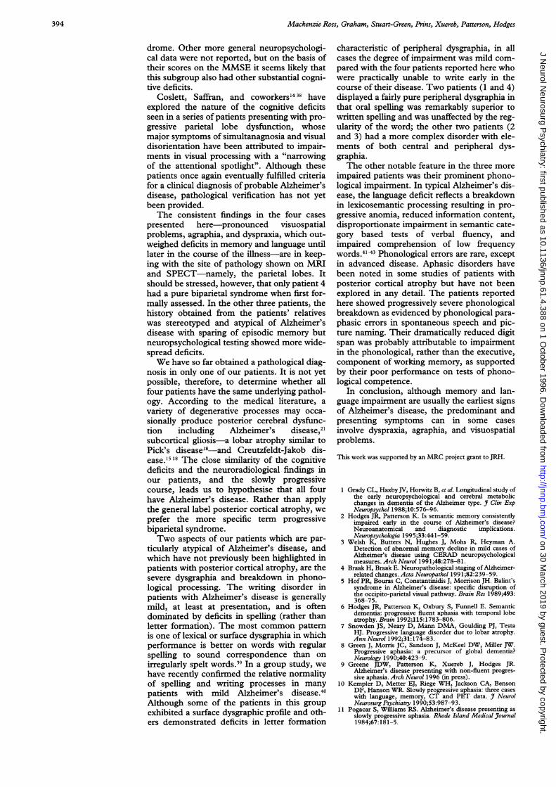
Brain MRIfor patient 4:coronally oriented Tlweighted images throughthe anterior (A) andposterior temporal region(B) showing mild globalatrophy but well preservedhippocampal complex.Slices through the posteriorparietal region (C and D)show dramatic bilateralsuperior parietal atrophywith compensatory dilationof the lateral ventricles.
and allowed to print her responses, she madeonly one error, indicating that her spelling waswell preserved and suggesting that her dys-graphia is peripheral rather than central.On memory tests, her verbal recognition
memory was intact but her recognition mem-ory for faces was impaired. She also performedpoorly on the logical memory test but withoutthe accelerated forgetting which is typicallyseen in patients with Alzheimer's disease.
Brain SPECT in 1994 showed pronouncedbilateral hypoperfusion in the posterior pari-etal lobes and mild bilateral reduction in thetemporal lobes. Brain MRI in 1995 (figure)showed striking bilateral atrophy in the supe-rior parietal lobes with compensatory dilata-tion of the lateral ventricles and a fewscattered periventricular lucencies of doubtfulsignificance. By contrast, the temporal lobes,particularly the hippocampus and relatedstructures, seemed normal.
Follow up neuropsychological testing sixmonths later disclosed a generally stable per-formance. Her dyspraxic difficulties had, how-ever, worsened and were causing her somedistress. For example, she was finding it so dif-ficult to use a knife and fork that she was tooembarrassed to eat out in restaurants. Herdressing dyspraxia had worsened, and she wasfinding it difficult to tell the time on her non-digital watch.
In conclusion, patient 4 presented withwriting deficits and visuospatial problemswhich overshadow more subtle deficits in lan-guage output and memory.
DiscussionWe have described four patients whose illness
began with features of progressive biparietalpathology. The onset of their illness was char-acterised by visuospatial problems, dyspraxia,and dysgraphia. In three of the patients, othercognitive deficits have become apparent, par-ticularly in the phonological aspects of lan-guage and in auditory-verbal short termmemory. This pattern may indicate spread ofthe disease from parietal areas to the perisyl-vian language area, particularly the superiortemporal gyrus. The fourth patient does notshow evidence of global intellectual declinebut has selective cognitive deficits involvingvisuospatial functions and writing. Brain MRIshowed particularly notable bilateral parietallobe atrophy in three of the patients and post-mortem examination of patient 1 showedAlzheimer's disease with particular involve-ment of the parietal lobes. The clinical fea-tures conform to what has been termedposterior cortical atrophy although, for rea-sons outlined below, we prefer the term pro-gressive biparietal atrophy.The term posterior cortical atrophy was first
used by Benson et al'3 to describe five patientspresenting with slowly progressive disorders ofhigher visual function without evidence ofbasic sensorimotor dysfunction or impairedvisual acuity. All five patients met the criteriafor a diagnosis of probable Alzheimer's diseasebut confirmatory pathology was not available.The patients initially complained of difficultyreading, visual distortion, problems withobject recognition, and a tendency to get losteven in familiar surroundings. All five wereunable to copy simple drawings. Their insightand memory seemed relatively well preserveduntil later in the course of the illness. Over athree to five year period, three patients devel-
392 on 30 M
arch 2019 by guest. Protected by copyright.
http://jnnp.bmj.com
/J N
eurol Neurosurg P
sychiatry: first published as 10.1136/jnnp.61.4.388 on 1 October 1996. D
ownloaded from

Progressive biparietal atrophy: an atypical presentation ofAlzheimer's disease
oped a full blown Balint's syndrome (visualdisorientation, optic apraxia, and simultanag-nosia) implying bilateral parietal-occipitalinvolvement. All five patients eventually devel-oped the four components of Gerstmann'ssyndrome (finger agnosia, right/left disorienta-tion, agraphia, and acalculia) suggestive ofspread of the pathology to the angular gyrusarea. Radiological findings (CT and MRI) inthree patients showed cortical degenerationwith particular involvement of the posteriorcerebral hemispheres. On the basis of thesefindings, Benson et all' suggested that theirpatients' disease initially involved occipital andparietal association cortices then progressed tothe angular gyrus or its connections bilaterally.
Since this initial description by Benson etal,'3 several groups have reported similarpatients.5 12 14-21 24 Although all of these patientshave been grouped under one general class, afiner grained analysis of this and earlier rele-vant medical literature in fact suggests twobroad presentations of posterior cortical atro-phy which reflect the predominant site ofpathology-that is, occipitotemporal andbiparietal-although in many patients featuresof both eventually develop.Those in the occipitotemporal group pre-
sent with complaints of visual distortion, diffi-culty with object recognition, topographicalagnosia, and alexia. Examination typicallyshows restricted visual fields or unilateralextinction on bilateral stimulation, impairedcolour vision and stereopsis, deficits in objectrecognition, prosopagnosia, and alexia (eitherletter by letter reading or attentional alexia). Inthe original study by Benson et al,"3 three ofthe five patients presented in this way. Similarpatients were reported in the ophthalmologicalliterature by Coganl9 and by Kiyosawa andcolleagues.22 The second group described fivepatients with prominent visual symptoms; fourhad difficulty reading because they could notstay on the correct line of print and com-plained of visual distortion. As the disease pro-gressed other prominent symptoms includedenvironmental agnosia and simultanagnosia,and three patients developed mild to moderateexpressive dysphasia. Most showed atrophy ofthe posterior cerebral hemispheres on MRI,and PET studies showed significant decreasesin glucose uptake in occipital areas with spar-ing of the primary motor and sensory cortex.Very similar cases of "slowly progressive visualagnosia" with features of apperceptualagnosia, prosopagnosia, and alexia werereported by De Renzi.23The nature of the reading disorder associ-
ated with this form of posterior cortical atro-phy was described by Freedman et al20 in areport of a 54 year old man who was found tohave the characteristics of letter by letter read-ing (pure alexia) and subsequently developedprofound constructional problems, agraphia, acomplete Gerstmann's syndrome, and severeaphasia with compromised naming and com-prehension. Brain PET showed symmetricbilateral occipitotemporal hypoperfusion butpathological confirmation of the diagnosis wasnot available.
More recently, three pathologically verifiedcases of Alzheimer's disease presenting withprogressive visual loss and other features ofoccipital pathology have been reported.'2 1821In a remarkable 12 year longitudinal study,Levine and colleagues2' reported a 59 year oldman who presented with what they termed"the visual variant of Alzheimer's disease". Hecomplained of difficulty in reading, driving,and locating items by sight and was found tohave constricted visual fields, widespreadvisuoperceptual deficits, and an attentionaldyslexia. Intellect and memory were initiallypreserved but declined along with the visualdeficits over a 12 year period. Towards theend of his illness he developed aphasic difficul-ties with word finding problems and occa-sional phonological paraphasias. Postmortemexamination confirmed Alzheimer's diseasewith particular involvement of the posteriorcingulate cortex (area 23), primary visual cortex(area 17), and visual association cortex (area18 and 20). In terms of the suggesteddichotomy, this occipitotemporal subgroupmanifest either impairment in basic visual abil-ities, reflecting involvement of primary visualcortex, or disruption of the ventral stream ofhigher order visual processing which is vital forobject, face, and written word identification.26The second major category of patients with
posterior cortical atrophy is that with featuresof parietal lobe dysfunction which is usuallybilateral, although often asymmetric. Unlikethose with early occipitotemporal involve-ment, such patients initially complain of visu-ospatial problems, agraphia, and dyspraxia.With progression they may manifest a fullblown Balint's syndrome but show preserva-tion of visual fields, basic perceptual abilities,object recognition, and reading. These deficitsreflect interruption of the dorsal stream ofvisuomotor processing which is critical forobject location and visually guided move-ments,25 and damage to areas of the parietallobe which are involved with general motorprogramming and writing.
Features of parietal lobe pathology are wellrecognised in the context of Alzheimer's dis-ease, and indeed most patients with moder-ately severe dementia show such signs37; butthe fact that Alzheimer's disease may occa-sionally present in this way has not beenclearly established. In a series of elegant neu-ropathological studies Hof and colleagues524showed that patients with Alzheimer's diseasewho had prominent features of Balint's syn-drome showed involvement of the dorsaloccipitoparietal pathways, which are known tobe critical for visuospatial and perceptual-motor processing; because clinical details ofthe cases were not included, it is unclearwhether these patients presented with Balint'ssyndrome or merely developed such featuresduring the course of their disease. A similarproblem exists in interpreting the findings ofMendez et al'7 who investigated visuospatialand perceptual abilities in 30 mildly to moder-ately demented patients with probableAlzheimer's disease. They identified sixpatients who met the criteria for Balint's syn-
393
on 30 March 2019 by guest. P
rotected by copyright.http://jnnp.bm
j.com/
J Neurol N
eurosurg Psychiatry: first published as 10.1136/jnnp.61.4.388 on 1 O
ctober 1996. Dow
nloaded from
Mackenzie Ross, Graham, Stuart-Green, Prins, Xuereb, Patterson, Hodges
drome. Other more general neuropsychologi-cal data were not reported, but on the basis oftheir scores on the MMSE it seems likely thatthis subgroup also had other substantial cogni-tive deficits.
Coslett, Saffran, and coworkers"4 38 haveexplored the nature of the cognitive deficitsseen in a series of patients presenting with pro-gressive parietal lobe dysfunction, whosemajor symptoms of simultanagnosia and visualdisorientation have been attributed to impair-ments in visual processing with a "narrowingof the attentional spotlight". Although thesepatients once again eventually fulfilled criteriafor a clinical diagnosis of probable Alzheimer'sdisease, pathological verification has not yetbeen provided.The consistent findings in the four cases
presented here pronounced visuospatialproblems, agraphia, and dyspraxia, which out-weighed deficits in memory and language untillater in the course of the illness-are in keep-ing with the site of pathology shown on MRIand SPECT-namely, the parietal lobes. Itshould be stressed, however, that only patient 4had a pure biparietal syndrome when first for-mally assessed. In the other three patients, thehistory obtained from the patients' relativeswas stereotyped and atypical of Alzheimer'sdisease with sparing of episodic memory butneuropsychological testing showed more wide-spread deficits.We have so far obtained a pathological diag-
nosis in only one of our patients. It is not yetpossible, therefore, to determine whether allfour patients have the same underlying pathol-ogy. According to the medical literature, avariety of degenerative processes may occa-sionally produce posterior cerebral dysfunc-tion including Alzheimer's disease,2'subcortical gliosis-a lobar atrophy similar toPick's disease'8 and Creutzfeldt-Jakob dis-ease.'18 The close similarity of the cognitivedeficits and the neuroradiological findings inour patients, and the slowly progressivecourse, leads us to hypothesise that all fourhave Alzheimer's disease. Rather than applythe general label posterior cortical atrophy, weprefer the more specific term progressivebiparietal syndrome.Two aspects of our patients which are par-
ticularly atypical of Alzheimer's disease, andwhich have not previously been highlighted inpatients with posterior cortical atrophy, are thesevere dysgraphia and breakdown in phono-logical processing. The writing disorder inpatients with Alzheimer's disease is generallymild, at least at presentation, and is oftendominated by deficits in spelling (rather thanletter formation). The most common patternis one of lexical or surface dysgraphia in whichperformance is better on words with regularspelling to sound correspondence than onirregularly spelt words.3" In a group study, wehave recently confirmed the relative normalityof spelling and writing processes in manypatients with mild Alzheimer's disease.40Although some of the patients in this groupexhibited a surface dysgraphic profile and oth-ers demonstrated deficits in letter formation
characteristic of peripheral dysgraphia, in allcases the degree of impairment was mild com-pared with the four patients reported here whowere practically unable to write early in thecourse of their disease. Two patients (1 and 4)displayed a fairly pure peripheral dysgraphia inthat oral spelling was remarkably superior towritten spelling and was unaffected by the reg-ularity of the word; the other two patients (2and 3) had a more complex disorder with ele-ments of both central and peripheral dys-graphia.The other notable feature in the three more
impaired patients was their prominent phono-logical impairment. In typical Alzheimer's dis-ease, the language deficit reflects a breakdownin lexicosemantic processing resulting in pro-gressive anomia, reduced information content,disproportionate impairment in semantic cate-gory based tests of verbal fluency, andimpaired comprehension of low frequencywords.4' 43 Phonological errors are rare, exceptin advanced disease. Aphasic disorders havebeen noted in some studies of patients withposterior cortical atrophy but have not beenexplored in any detail. The patients reportedhere showed progressively severe phonologicalbreakdown as evidenced by phonological para-phasic errors in spontaneous speech and pic-ture naming. Their dramatically reduced digitspan was probably attributable to impairmentin the phonological, rather than the executive,component of working memory, as supportedby their poor performance on tests of phono-logical competence.
In conclusion, althoagh memory and lan-guage impairment are usually the earliest signsof Alzheimer's disease, the predominant andpresenting symptoms can in some casesinvolve dyspraxia, agraphia, and visuospatialproblems.
This work was supported by an MRC project grant to JRH.
1 Grady CL, Haxby JV, Horwitz B, et al. Longitudinal study ofthe early neuropsychological and cerebral metabolicchanges in dementia of the Alzheimer type. 3 Clin ExpNeuropsychol 1988;1O:576-96.
2 Hodges JR, Patterson K. Is semantic memory consistentlyimpaired early in the course of Alzheimer's disease?Neuroanatomical and diagnostic implications.Neuropsychologia 1995;33:441-59.
3 Welsh K, Butters N, Hughes J, Mohs R, Heyman A.Detection of abnormal memory decline in mild cases ofAlzheimer's disease using CERAD neuropsychologicalmeasures. Arch Neurol 1991 ;48:278-8 1.
4 Braak H, Braak E. Neuropathological staging of Alzheimer-related changes. Acta Neuropathol 1991;82:239-59.
5 Hof PR, Bouras C, Constantinidis J, Morrison JH. Balint'ssyndrome in Alzheimer's disease: specific disruption ofthe occipito-parietal visual pathway. Brain Res 1989;493:368-75.
6 Hodges JR, Patterson K, Oxbury S, Funnell E. Semanticdementia: progressive fluent aphasia with temporal lobeatrophy. Brain 1992;115:1783-806.
7 Snowden JS, Neary D, Mann DMA, Goulding PJ, TestaHJ. Progressive language disorder due to lobar atrophy.Ann Neurol 1992;31:174-83.
8 Green J, Morris JC, Sandson J, McKeel DW, Miller JW.Progressive aphasia: a precursor of global dementia?Neurology 1990;40:423-9.
9 Greene JDW, Patterson K, Xuereb J, Hodges JR.Alzheimer's disease presenting with non-fluent progres-sive aphasia. Arch Neurol 1996 (in press).
10 Kempler D, Metter EJ, Riege WH, Jackson CA, BensonDF, Hanson WR. Slowly progressive aphasia: three caseswith language, memory, CT and PET data. _7 NeurolNeurosurg Psychiatry 1990;53:987-93.
11 Pogacar S, Williams RS. Alzheimer's disease presenting asslowly progressive aphasia. Rhode Island Medical Journal1984;67:181-5.
394 on 30 M
arch 2019 by guest. Protected by copyright.
http://jnnp.bmj.com
/J N
eurol Neurosurg P
sychiatry: first published as 10.1136/jnnp.61.4.388 on 1 October 1996. D
ownloaded from

Progressive biparietal atrophy: an atypical presentation ofAlzheimer's disease
12 Berthier ML, Leiguarda R, Starkstein SE, Sevlever G,Taratuto AL. Alzheimer's disease in a patient with poste-rior cortical atrophy. Jf Neurol Neurosurg Psychiatry 199 1;54:1110-1.
13 Benson DF, Davis RJ, Snyder BD. Posterior cortical atro-phy. Arch Neurol 1988;45:789-93.
14 Coslett HB, Stark M, Rajaram S, Saffran EM. Narrowingthe spotlight: a visual attentional disorder in presumedAD. Neurocase 1995;1:305-19.
15 Pantel J, De Bleser R, Schwartz M, Weiller C. Rapidly pro-gressive form of posterior cerebral dysfunction withapperceptive agnosia in a 34 year old man. Neurocase1995;1:319-31.
16 Graff-Radford NR, Bolling JP, Earnest F, Shuster EA,Caselli RJ, Brazis PW. Simultanagnosia as the initial signof degenerative dementia. Mayo Clin Proc 1993;68:955-64.
17 Mendez MF, Mendez MA, Martin R, Smyth KA,Whitehouse PJ. Complex visual disturbances inAlzheimer's disease. Neurology 1990;40:439-43.
18 Victoroff J, Webster Ross G, Benson F, Anthony Verity M,Vinters HV. Posterior cortical atrophy: neuropathologiccorrelations. Arch Neurol 1994;51:269-74.
19 Cogan DC. Visual disturbances with focal progressivedementing disease. Am Y Ophthalmol 1985;100:68-72.
20 Freedman L, Selchen DH, Black SE, R. K, Garnett ES,Nahmias C. Posterior cortical dementia with alexia: neu-robehavioural, MRI, and PET findings. J NeurolNeurosurg Psychiatry 199 1;54:443-8.
21 Levine DN, Lee JM, Fisher CM. The visual variant ofAlzheimer's disease: a clinicopathologic case study.Neurology 1993;43:305-13.
22 Kiyosawa M, Bosley TM, Chawluk J, et al. Alzheimer's dis-ease with prominent visual symptoms: clinical and meta-bolic evaluation. Ophthalmology 1989;96: 1077-86.
23 De Renzi E. Slowly progressive visual agnosia or apraxiawithout dementia. Cortex 1986;22:171-80.
24 Hof PR, Bouras C, Constantinidis J, Morrison JH.Selective disconnection of specific visual associationpathways in cases of Alzheimer's disease presenting withBalint's syndrome. J Neuropathol Exp Neurol 1990;49:168-84.
25 Goodale MA, Milner AD. Separate visual pathways forperception and action. TINS 1992;15:20-5.
26 Ungerleider LG, Mishkin M. Two cortical visual systems.In: Ingle DJ, Goodale MA, Mansfield RJW, eds. Analysisof visual behaviour. Cambridge, MA: MIT Press, 1982:549-86.
27 Mattis S. Dementia rating scale. Windsor: NFER-Nelson,1988.
28 Bishop DVM. Test for the reception of grammar. 2nd ed.London: Medical Research Council, 1989.
29 Patterson K, Hodges JR. Deterioration of word meaning:implications for reading. Neuropsychologia 1992;30:1025-40.
30 Patterson K, Marcel A. Phonological ALEXIA orPHONOLOGICAL alexia? In: Alegria J, Holender D,Junca de Morais J, Radeau M, eds. Analytic approaches tohuman cognition. Amsterdam: North Holland, 1992.
31 Warrington EK. Recognition memory test. Windsor: NFER-Nelson, 1984.
32 Benton AL, deS Hamsher K, Varney NR, Spreen 0.Judgement of line orientation. New York: OxfordUniversity Press, 1983.
33 Humphreys GW, Riddoch MJ. Routes to object constancy:implications for neurological impairment of object con-stancy. QJ ExpPsychol 1984;36:384-415.
34 Howard D, Patterson K. Pyramids and palm trees: a test ofsemantic access from pictures and words. Bury St Edmunds,Suffolk: Thames Valley Test Company, 1992.
35 Hodges JR, Salmon DP, Butters N. Semantic memoryimpairment in Alzheimer's disease: failure of access ordegraded knowledge? Neuropsychologia 1992;30(4):310-4.
36 Warrington EK, James M. The visual object and space percep-tion battery. Bury St Edmunds: Thames Valley TestCompany, 1991.
37 Kaskie B, Storandt M. Visuospatial deficit in dementia ofthe Alzheimer type. Arch Neurol 1995;52:422-5.
38 Saffran E, Fitzpatrick-DeSalme E, Coslett HB. Visual dis-turbances in dementia. In: Schwartz MF, ed. Modulardeficits in Alzheimer-type dementia. London: The MITPress 1990:297-328.
39 Rapcsak SZ, Arthur SA, Bliklen DA, Rubens AB. Lexicalagraphia in Alzheimer's disease. Arch Neurol 1989;46:65-8.
40 Hughes JC, Graham N, Patterson K, Hodges JR.Dysgraphia in mild dementia of Alzheimer's type.Neuropsychologia 1996 (in press).
41 Bayles KA, Tolmoeda DK. Confrontational namingimpairment in dementia. Brain Lang 1983;19:98-114.
42 Hart S. Language and dementia: a review. Psychol Med1988;18:99-112.
43 Giles E, Patterson K, Hodges JR. Performance on theBoston cookie theft picture description task in patientswith early dementia of the Alzheimer's type: missinginformation. Aphasiology 1996;10:395-408.
395
on 30 March 2019 by guest. P
rotected by copyright.http://jnnp.bm
j.com/
J Neurol N
eurosurg Psychiatry: first published as 10.1136/jnnp.61.4.388 on 1 O
ctober 1996. Dow
nloaded from