prostaglandin e signals monocyte/macrophage survival to ... · prostaglandin e 2 signals...
TRANSCRIPT

of November 12, 2018.This information is current as
Driven by 5-Hydroxyeicosatetraenoic Acid-Dependent PathwayαProtein Kinase C
Converging in Bad Phosphorylation with the Peroxynitrite via Protein Kinase AMonocyte/Macrophage Survival to
Signals2Prostaglandin E
Orazio CantoniIlaria Tommasini, Liana Cerioni, Letizia Palomba and
http://www.jimmunol.org/content/181/8/5637doi: 10.4049/jimmunol.181.8.5637
2008; 181:5637-5645; ;J Immunol
Referenceshttp://www.jimmunol.org/content/181/8/5637.full#ref-list-1
, 11 of which you can access for free at: cites 37 articlesThis article
average*
4 weeks from acceptance to publicationFast Publication! •
Every submission reviewed by practicing scientistsNo Triage! •
from submission to initial decisionRapid Reviews! 30 days* •
Submit online. ?The JIWhy
Subscriptionhttp://jimmunol.org/subscription
is online at: The Journal of ImmunologyInformation about subscribing to
Permissionshttp://www.aai.org/About/Publications/JI/copyright.htmlSubmit copyright permission requests at:
Email Alertshttp://jimmunol.org/alertsReceive free email-alerts when new articles cite this article. Sign up at:
Print ISSN: 0022-1767 Online ISSN: 1550-6606. Immunologists All rights reserved.Copyright © 2008 by The American Association of1451 Rockville Pike, Suite 650, Rockville, MD 20852The American Association of Immunologists, Inc.,
is published twice each month byThe Journal of Immunology
by guest on Novem
ber 12, 2018http://w
ww
.jimm
unol.org/D
ownloaded from
by guest on N
ovember 12, 2018
http://ww
w.jim
munol.org/
Dow
nloaded from

Prostaglandin E2 Signals Monocyte/Macrophage Survivalto Peroxynitrite via Protein Kinase A Converging in BadPhosphorylation with the Protein Kinase C�-DependentPathway Driven by 5-Hydroxyeicosatetraenoic Acid1
Ilaria Tommasini,2 Liana Cerioni,2 Letizia Palomba, and Orazio Cantoni3
Monocytes/macrophages committed to death by peroxynitrite nevertheless survive with a signaling response promoting Badphosphorylation, as well as its cytosolic localization, via upstream activation of cytosolic phospholipase A2, 5-lipoxygenase, andprotein kinase C�. We now report evidence for an alternative mechanism converging in Bad phosphorylation when the expression/activity of the above enzymes are suppressed. Under these conditions, also associated with peroxynitrite-dependent severe inhi-bition of Akt, an additional Bad kinase, Bad dephosphorylation promoted its accumulation in the mitochondria and a promptlethal response. PGE2 prevented toxicity via EP2 receptor-mediated protein kinase A-dependent Bad phosphorylation. This notionwas established in U937 cells by the following criteria: 1) there was a strong correlation between survival and cAMP accumulation,both in the absence and presence of phosphodiesterase inhibitors; 2) direct activation of adenylyl cyclase afforded cytoprotection;and 3) PGE2 promoted loss of mitochondrial Bad and cytoprotection, mimicked by EP2 receptor agonists, and prevented by EP2
receptor antagonists or protein kinase A inhibitors. Finally, selected experiments performed in human monocytes/macrophagesand in rat peritoneal macrophages indicated that the above cytoprotective pathway is a general response of cells belonging to themonocyte/macrophage lineage to both exogenous and endogenous peroxynitrite. The notion that two different pathways mediatedby downstream products of arachidonic acid metabolism converge in Bad phosphorylation emphasizes the relevance of thisstrategy for the regulation of macrophage survival to peroxynitrite at the inflammatory sites. The Journal of Immunology, 2008,181: 5637–5645.
P eroxynitrite, the coupling product of superoxide and NO,is produced by macrophages and other inflammatory cellsin response to a variety of proinflammatory stimuli. This
species is extremely reactive (1), promotes direct and indirect dam-age in different biomolecules (1, 2), and is heavily implicated intissue damage associated with inflammatory conditions (3).
The above information is documented by an extensive literature,but much less is known about the effects of peroxynitrite in theperspective of peroxynitrite-producing cells. It is indeed reason-able to assume that these cells will be affected by peroxynitrite inthe first place, when the oxidant is generated either within the cellor in its close vicinity. Our work performed in cells belonging tothe monocyte/macrophage lineage provided evidence for an inge-nious survival strategy based on the ability of these cells to re-spond to the oxidant with the triggering of signaling events thatallow survival regardless of the damage accumulated (4–11).
We found that these cells, as for many other cell types, areseverely affected and committed to death by low concentrations of
the oxidant. Unlike other cell types, however, monocytes/macro-phages did not die since they were able to respond to moleculeslargely available at the inflammatory sites with a signaling re-sponse leading to survival.
Macrophages use arachidonic acid (ARA)4 (4, 5) and productsof 5-lipoxygenase (5-LO) (9–11), as 5-hydroxyeicosatetraenoicacid (5-HETE), to promote downstream events associated withprevention of mitochondrial permeability transition (MPT)-depen-dent toxicity. We detected significant levels of Bad in the mito-chondria of untreated cells and found that the above signalingleads to Bad phosphorylation, and thus to its cytosolic accumula-tion (7, 8). Phosphorylation inhibits binding of Bad to Bcl-2 orBcl-xL and promotes its translocation to the cytosol, thus enablingBcl-2 or Bcl-xL to exert its anti-MPT functions (12). Upstreaminhibition of the survival signaling promotes the mitochondrial ac-cumulation of Bad and the rapid onset of MPT-dependent toxicitytaking place soon after the mitochondrial translocation of Bax (8).Bax can also dimerize with Bcl-2 or Bcl-xL, and it is provided witha well-defined pore-forming activity (13). Thus, the cytosolic ac-cumulation of Bad creates optimal conditions for the anti-MPTfunctions of Bcl-2 and Bcl-xL.
Istituto di Farmacologia e Farmacognosia, Universita degli Studi di Urbino “CarloBo”, Urbino, Italy
Received for publication February 26, 2008. Accepted for publication August 14,2008.
The costs of publication of this article were defrayed in part by the payment of pagecharges. This article must therefore be hereby marked advertisement in accordancewith 18 U.S.C. Section 1734 solely to indicate this fact.1 This work was supported by a grant from the Associazione Italiana per la Ricercasul Cancro (to O.C.).2 I.T. and L.C. contributed equally to the work described in this paper.3 Address correspondence and reprint requests to Dr. Orazio Cantoni, Istituto diFarmacologia e Farmacognosia, Universita degli Studi di Urbino “Carlo Bo”, viaSanta Chiara 27, 61029 Urbino (PU), Italy. E-mail address: [email protected]
4 Abbreviations used in this paper: ARA, arachidonic acid; AACOCF3, arachidonyltrifluoromethyl ketone; AS-ON, antisense oligonucleotide; cPLA2, cytosolic phos-pholipase A2; FLAP, 5-LO-activating protein; 5-HETE, 5-hydroxyeicosatetraenoicacid; IBMX, 3-isobutyl-1-methylxanthine; L-NAME, NG-nitro-L-arginine methyl es-ter; 5-LO, 5-lipoxygenase; MPT, mitochondrial permeability transition; NS-ON, non-sense oligonucleotide; ON, oligonucleotide; PKA, protein kinase A; PKC�, proteinkinase C�.
Copyright © 2008 by The American Association of Immunologists, Inc. 0022-1767/08/$2.00
The Journal of Immunology
www.jimmunol.org
by guest on Novem
ber 12, 2018http://w
ww
.jimm
unol.org/D
ownloaded from

In short, the survival strategy adopted by macrophages is sim-ple, yet very effective: while committed to MPT-dependent toxic-ity, these cells nevertheless survive by promoting Bad phosphor-ylation via a pathway triggered by molecules they produce and/orare largely available in the extracellular inflammatory milieu.
In our experiments, phosphorylation of Bad was mediated by5-HETE-dependent mitochondrial translocation of protein kinaseC� (PKC�) (6, 8, 9). Indeed, Bad phosphorylation may result fromPKC-dependent pathways triggering a p90RSK-mediated phos-phorylation at serine 112 (14), and this event obviously requiresthe mitochondrial localization of PKC�. An extensive literaturedocuments that Bad phosphorylation is often mediated by proteinkinase B (Akt), a serine-threonine protein kinase (15). It is difficultto establish whether this pathway is relevant in peroxynitrite sig-naling since few reports describe stimulation of Akt (16, 17), al-though others rather describe inhibition, most likely attributable totyrosine nitration, in a variety of cell types (18–21), includingmacrophages (22).
There is, however, a third pathway that may lead to Bad phos-phorylation and survival in macrophages under inflammatory con-ditions. This pathway is dependent on phosphorylation mediatedby protein kinase A (PKA), which is stimulated by receptor-me-diated signaling driven by a variety of agonists, including PGs, thatare largely released in the extracellular inflammatory milieu byboth constitutive and inducible cyclooxygenases (23).
The present study provides results in line with the notion thatAkt is severely inhibited by peroxynitrite, thereby ruling out thecontribution of this pathway in the regulation of monocyte/mac-rophage survival. In remarkable contrast, PGs effectively signalsurvival through EP2 receptors by promoting Bad phosphorylation,as previously shown for the 5-LO-dependent pathway (7–9).Hence, ARA appears to be critical for macrophage survival toperoxynitrite in that it is a substrate for both 5-LO, directly (in-tracellular mechanism) promoting survival via PKC�-dependentBad phosphorylation, and cyclooxygenases, thereby contributingto the definition of extracellular levels of PGs triggering PKA-dependent Bad phosphorylation.
Materials and MethodsCell culture and treatment conditions
Human promonocytic tumor U937 cells were cultured as previously de-scribed (4). Human peripheral mononuclear cells were isolated by Ficollgradient centrifugation and monocytes were purified by adherence on aplastic substrate in RPMI 1640 medium. Macrophages were obtained byculturing monocytes (1 � 106 cells/ml) for 8–10 days in RPMI 1640 me-dium. Macrophages were scraped with PBS/EDTA, centrifuged, and uti-lized for experiments. Rat peritoneal exudate macrophages were collected4 days after intraperitoneal injection of 4% thioglycolate medium (Sigma-Aldrich) to 25- or 30-day-old Sprague-Dawley rats (Charles River Labo-ratories). The cells were plated on 35-mm culture dishes (Falcon, BD La-bware) in DMEM supplemented with 2 mM glutamine, 25 mM HEPES,penicillin (100 U/ml), streptomycin (100 �g/ml), and 10% FBS (HyCloneLaboratories) for 24 h. Nonadherent cells were removed with warmDMEM. At the treatment stage, rat peritoneal macrophages were between4.5 and 5.0 � 105 cells/dish.
Peroxynitrite was synthesized by the reaction of nitrite with acidifiedH2O2, as described previously (24). For the different cell types and exper-iments, treatments were performed in 2 ml of prewarmed saline A (8.182g/L NaCl, 0.372 g/L KCl, 0.336 g/L NaHCO3 and 0.9 g/L glucose) con-taining 4.5–5 � 105 cells. The same concentration of peroxynitrite (i.e.,100 �M) was used throughout the study. Arachidonyl trifluoromethyl ke-tone (AACOCF3), MK886, PGE2, ARA, butaprost, sulprostone, andGo6850 were given to the cultures 3 min after peroxynitrite. Forskolin,KT5720, AH6809, and AH23848 were added to the cultures 30 min beforeperoxynitrite; 3-isobutyl-1-methylxanthine (IBMX) (5 or 30 min) andwortmannin (15 min) were also added to cultures before peroxynitrite.Formation of endogenous peroxynitrite was induced in rat peritoneal mac-rophages, as recently described (25). Briefly, the cells were treated with 10
�g/ml LPS (serotype 0127:B8; Sigma-Aldrich) plus 10 ng/ml IFN-� (2 �107 IU/mg of protein; R&D Systems, Space) and, after 2 h, also exposedto 2.5 �g/ml PMA (Sigma-Aldrich), with or without PGE2. Cytotoxicity,nitrite concentrations, or nitrotyrosine immunoreattivity was assessed aftera further 9 h of growth. L-NAME, L-methionine, or apocynin was added tothe cultures 10 min before addition of PMA.
Cytotoxicity assay
Cytotoxicity was determined with the trypan blue exclusion assay. Briefly,an aliquot of the cell suspension was diluted 1/1 (v/v) with 0.4% trypanblue and the viable cells were counted with a hemocytometer.
Reduction of 3-(4,5-dimethylthiazol-2-yl)-2,5-diphenyltetrazolium bro-mide (MTT) was used to measure viability of rat peritoneal macrophages.MTT (25 �g/ml) was added directly to culture medium 1 h before the endof the incubation period. The medium was then removed and replaced with1 ml of DMSO. Cellular MTT reductase activity was determined by mea-suring the absorbance of the DMSO extracts at 570 nm. Results are ex-pressed as the percentage of MTT reducing activity of treated vs untreatedcells.
Transfection with cytosolic phospholipase A2 (cPLA2),5-LO-activating protein (FLAP), or PKC� antisense (AS)and nonsense (NS) oligonucleotides (ONs)
The human AS-ON for cPLA2 (5�-GTA AGG ATC TAT AAA TGA CAT-3�), for FLAP (5�-TTC TTG ATC CAT GTT TGC TT-3�), and for PKC�(5�-GTT CTC GCT GGT GAG TTT CA-3�) were directed against theinitiation site (cPLA2), the start codon of mRNA (FLAP), or against the 3�region of the mRNA (PKC�). The NS-ON for cPLA2 (5�-AGT AGA TTGAAT AGA CAC TAT-3�), for FLAP (5�-CTT GTT TAA TCC CCT TTTGT-3�), and for PKC� (5�-TGG GCC GGG CAA TTT TTT TC-3�) werea random sequence of the AS bases. The ONs were phosphorothioate mod-ified and synthesized by MWG Biotech. The phosphorothioate backboneprovides resistance to exonuclease and increases the stability of the ON inserum and within the cell. U937 cells were transfected with the above ONsas follows: cells were washed twice with serum-free medium and seeded(1 � 106/ml) in serum-free RPMI 1640 for 6 h in the absence or presenceof ONs (10 �M). A final concentration of 5% FBS was then added, thecells were cultured for an additional 24 h, and finally utilized for experi-ments. Using these conditions, we obtained good transfection efficienciesand thus avoided the use of transfection reagents, such as Lipofectamine.The effect of ON transfection on enzyme expression was tested by Westernblot analysis (see below). Occasionally, cPLA2 or 5-LO activities were alsoassessed by measuring ARA release and leukotriene B4 formation, respec-tively, in response to peroxynitrite. The outcome of these experiments wasin line with our previously published work (4, 9). We observed identicallevels of ARA release in untreated cells, whereas there was a significantincrease in ARA release only in the cells transfected with cPLA2 NS-ONs(typically 2-fold increase). Leukotriene B4 levels were also identical inuntreated cells transfected with FLAP NS-ONs or AS-ONs. Peroxynitriteenhanced leukotriene B4 formation (typically a 1.5-fold increase) in cellstransfected with the NS-ONs and had hardly any effect in the cells trans-fected with the AS-ONs.
Subcellular fractionation and Western blot analysis
After treatments, the cells were processed to obtain mitochondrial and cy-tosolic fractions, as described by Yu et al. (26), or the whole cell lysates,as described by Guidarelli et al. (6). Western blot analysis was next per-formed using Abs against cPLA2, FLAP, Akt, heat shock protein-60 (allobtained from Santa Cruz Biotechnology), pAkt, PKC�, Bad (all obtainedfrom BD Transduction Laboratories), and actin (Sigma-Aldich). Details onWestern blotting apparatus and conditions are reported elsewhere (6). Absagainst Akt, actin, and heat shock protein-60 were used to assess the equalloading of the lanes.
Measurement of intracellular cAMP production
Intracellular cAMP levels were determined in cell extracts (0.1 M HCl) byan ELISA kit (Cayman Chemical).
Nitrite measurement
Nitrite production was estimated using the Griess reaction. Briefly, 800 �lof samples was mixed with 60 �l sulfanilamide (12.5 mM) and 60 �l HCl(6 M). After 5 min, 60 �l N-(1-naphthyl)-ethylene diamine was added tothe mixture and the absorbance was read at 540 nm using a Uvicon 923spectrophotometer. Nitrite concentration was calculated from a standardcurve of NaNO2 in the medium.
5638 PGE2 SIGNALS MONOCYTES/MACROPHAGE SURVIVAL TO PEROXYNITRITE
by guest on Novem
ber 12, 2018http://w
ww
.jimm
unol.org/D
ownloaded from

Immunocytochemical detection of nitrotyrosine
Nitrotyrosine-containing proteins were detected using an immunocyto-chemical technique following the manufacturer’s instructions. Briefly,rat peritoneal macrophages were seeded on polylysine-coated cover-slips and treated as indicated above. The coverslips were then removedfrom the dishes, washed, and the cells fixed for 1 min with ethanol/acetic acid (95/5, v/v). These preparations were finally rinsed with PBSand blocked in PBS-containing BSA (2%, w/v). Rabbit polyclonal anti-nitrotyrosine (5 �g/ml in PBS containing 2% BSA) was used as a pri-mary Ab. After 18 h at 4°C, the cell monolayers were washed andexposed to FITC-conjugated secondary Ab for 2 h in the dark. Stainedcells were analyzed using a BX-51 fluorescence microscope (OlympusItalia) equipped with a SPOT-RT camera unit (Diagnostic Instruments,Delta Sistemi). The excitation and emission wavelengths were 495 and515 nm, respectively. Images were collected with exposure times of100 – 400 ms, digitally acquired, and processed for fluorescence deter-mination at the single cell level on a personal computer using ScionImage software. Mean fluorescence values were determined by averag-ing the fluorescence values of at least 50 cells per treatment conditionper experiment.
Statistical analysis
Statistical analysis of the data for multiple comparisons was performed byANOVA followed by a Dunnett’s test. For comparison between twogroups, Student’s unpaired t test was used.
ResultsPGE2 promotes survival in U937 cells exposed to peroxynitrite
Previous work from our laboratory showed that peroxynitrite-de-pendent activation of cPLA2, associated with ARA metabolism viathe 5-LO pathway, promotes a cascade of events leading to cyto-protection (4–11). Under these conditions, 5-LO translocated tothe nuclear membrane, an event critical for interaction with FLAPand 5-HETE formation (27, 28), and indeed was readily detectedafter peroxynitrite exposure (9). Thus, consistently with these find-ings, down-regulation of cPLA2 or FLAP expression with specificAS-ONs (Fig. 1A, inset) promoted toxicity after exposure to oth-erwise nontoxic concentrations of peroxynitrite (100 �M, Fig. 1A).Cells transfected with the respective NS-ONs did not show appre-ciable changes vs the nontransfected cells in terms of protein ex-pression (Fig. 1A, inset) or susceptibility to peroxynitrite (notshown). Pharmacological inhibitors of cPLA2 (AACOCF3) orFLAP (MK886), under conditions inhibiting ARA release and5-HETE formation (Refs. 4, 9 and Materials and Methods), re-spectively, promoted toxicity in nontransfected cells (Fig. 1B) and,with similar outcomes, in cells transfected with NS-ONs (notshown).
The results illustrated in Fig. 1, A and B, also indicate that ad-dition of PGE2 after peroxynitrite promotes cytoprotection underconditions in which cytotoxicity would arise as a consequence ofinhibition of the survival signaling, at the level of either cPLA2 or5-LO. Interestingly, lower concentrations of PGE2 were requiredfor maximal protection against inhibition of lipoxygenase activity.This observation implies a contribution of endogenous PGs in thatmore ARA becomes available for the cyclooxygenases upon inhi-bition of lipoxygenases.
These results are consistent with a signaling initiated by extra-cellular PGs, which may cooperate with the previously describedintracellular signaling driven by 5-LO products (9) to promote Badphosphorylation. A contribution of the Akt pathway, however, isunlikely (Fig. 1, C and D) since peroxynitrite suppressed Akt phos-phorylation as effectively as wortmannin, a PI3K inhibitor. Addi-tionally, wortmannin did not promote toxicity in cells exposed toperoxynitrite (Fig. 1C), as previously observed with the inhibitorsof either cPLA2 or 5-LO (Fig. 1B).
The survival signaling triggered by PGE2 leads to the cytosolicaccumulation of Bad via a PKC�-independent mechanism
The above results indicate that PGE2 rescues U937 cells commit-ted to death by peroxynitrite via upstream inhibition of the survivalsignaling. We therefore asked the question of whether the down-stream events of this signaling were the same as those previouslyidentified downstream to 5-LO activation, as the mitochondrialtranslocation of PKC� causally linked to the cytosolic accumula-tion of Bad (7–9).
As previously shown (8, 9), peroxynitrite indeed promoted themitochondrial accumulation of PKC�, and this response was sup-pressed by AACOCF3 (Fig. 2A). The observation that the effect ofthe PLA2 inhibitor was sensitive to ARA, but insensitive to PGE2,is therefore consistent with the notion that the survival responseevoked by PGE2 does not involve PKC�. This notion is furthersupported by the observation that PGE2, unlike ARA, preventedtoxicity mediated by peroxynitrite and Go6850, an inhibitor ofPKC (Fig. 2B). Additionally, down-regulation of PKC� expres-sion with specific AS-ONs (Fig. 2C, inset) promoted toxicity afterexposure to peroxynitrite via an ARA-insensitive albeit PGE2-sen-sitive mechanism (Fig. 2C). Finally, cells transfected with the NS-ONs did not show reduced PKC� expression (Fig. 2C) and wereresistant to peroxynitrite (not shown) (6).
The next question addressed was whether Bad is a target ofthe PGE2-dependent survival signaling. The results illustrated
FIGURE 1. PGE2 prevents the lethal response mediated by an otherwisenontoxic concentration of peroxynitrite in cells supplemented with eitherAS-ONs or pharmacological inhibitors of cPLA2 and FLAP. A, cPLA2 andFLAP AS-ON-transfected cells were exposed for 3 min to peroxynitrite(100 �M), for an additional 57 min to increasing concentration of PGE2,and finally analyzed with the trypan blue exclusion assay. Also shown isthe effect of peroxynitrite alone in cells transfected with cPLA2 NS-ONs(similar results were obtained in cells transfected with FLAP NS-ONs).The inset shows the levels of cPLA2 and FLAP expression in nontrans-fected cells as well as in NS-ON- and AS-ON-transfected cells. B, Non-transfected cells were treated as indicated above with the further additionof either AACOCF3 (50 �M) or MK886 (1 �M). Also shown is the effectof peroxynitrite alone. C, Cells, with or without prior addition of wort-mannin (300 nM), were treated for 60 min with peroxynitrite, AACOCF3,and PGE2 (3 �M) as indicated above (see Materials and Methods fordetails) and processed for the assessment of viability. D, Cells were ex-posed to wortmannin and/or peroxynitrite for 10 min, lysed, and finallyprocessed for Western blot analysis using Ab against pAkt and Akt. Blotsshown in D are representative of three separate experiments. Results rep-resent the means � SEM from three to five separate experiments. ��, p �0.01; �, p � 0.05 as compared with cells exposed to peroxynitrite alone (Aand C) or associated with AACOCF3 or MK886 (B) (ANOVA followed byDunnett’s test).
5639The Journal of Immunology
by guest on Novem
ber 12, 2018http://w
ww
.jimm
unol.org/D
ownloaded from

in Fig. 3A indicate that significant levels of Bad are located inthe mitochondria of untreated U937 cells and that peroxynitritepromotes the mitochondria to cytosol translocation of the pro-tein, sensitive to the PKC inhibitor Go6850, which indeedcaused a remarkable mitochondrial Bad accumulation. This ob-servation is in keeping with our previous findings indicatingthat, under the above conditions, Bad phosphorylation andcytosolic accumulation require activation and mitochondrialtranslocation of PKC� (7, 8). Interestingly, the process of mi-tochondrial accumulation of Bad mediated by peroxynitrite inPKC-inhibited cells was sensitive to PGE2 (Fig. 3A).
These results collectively indicate that PGE2 signals survival viaa PKC�-independent mechanism nevertheless leading to the cyto-solic accumulation of Bad.
The survival signaling triggered by PGE2 is mediated by cAMP
The results illustrated in Fig. 4A indicate that concentrationsof PGE2 �3 �M (10 min) are required to significantly enhanceU937 cell cAMP. The observation that a 5-min exposure tophosphodiesterase inhibitor (IBMX) promotes a leftward shiftof the above curve leading to appreciable cAMP levels afterexposure to �3 �M PGE2, however, provides an indication oftransient increases of the second messenger also under theseconditions. Cells were next treated with peroxynitrite/AACOCF3,manipulated to enhance cAMP as detailed above, and assayedfor survival. The results shown in Fig. 4, A and B, collectivelyprovide evidence for a good correlation between these param-eters, a notion further supported by the observation that longerexposure (30 min) to IBMX promotes even larger increasesin cAMP (Fig. 4A) and virtually afforded complete cytoprotec-tion in the absence of PGE2. As a final note, the effects of PGE2
FIGURE 3. PGE2 signaling leads to the cytosolic localization of Bad.Cells were treated for 10 min, as indicated in the figure (see Materials andMethods for details). KT5720 or AH6809 were used at 3 or 50 �M, re-spectively. After treatments, mitochondrial and cytosolic fractions wereisolated and processed for Western blot analysis using an Ab against Bad.Blots shown are representative of three separate experiments. The relativeamount of Bad was quantified by densitometric analysis and expressed asOD integration.
FIGURE 2. PGE2 signals survival via a PKC�-independent mechanism.A, Cells were treated for 10 min with peroxynitrite, AACOCF3, ARA (0.1�M), and PGE2, as indicated in the figure (see Materials and Methods fordetails). Western blot analysis for PKC� was performed in the mitochon-drial fraction. B, Cells were exposed for 60 min to peroxynitrite, Go6850(3 �M), ARA, PGE2, and butaprost (50 �M), as indicated, and finallyanalyzed with the trypan blue exclusion assay. C, PKC� AS-ON-trans-fected cells were treated as indicated in B (with the exception of Go6850,not employed in these experiments) and analyzed with the trypan blueexclusion assay. The inset shows the level of PKC� expression in non-transfected cells and in cells transfected with PKC� NS-ONs or AS-ONs.Blots shown in A and C are representative of three separate experiments.Results represent the means � SEM from three separate experiments. �, p �0.01 as compared with cells exposed to peroxynitrite (B) or to untreated cells(C) (ANOVA followed by Dunnett’s test). The relative amount of PKC�was quantified by densitometric analysis and expressed as ODintegration.
5640 PGE2 SIGNALS MONOCYTES/MACROPHAGE SURVIVAL TO PEROXYNITRITE
by guest on Novem
ber 12, 2018http://w
ww
.jimm
unol.org/D
ownloaded from

are mimicked by forskolin, an activator of adenylyl cyclase(Fig. 4C).
Additional experiments revealed that the effects of PGE2 areboth insensitive to wortmannin (Fig. 1C) and abolished by thePKA inhibitor KT5720 (Fig. 5A). The specificity of the latter re-sponse is emphasized by the observation that KT5720 promoted aconcentration-dependent lethal response in cells committed todeath by peroxynitrite/AACOCF3 and rescued with PGE2, whereasPKA inhibition had no significant effect on cytoprotection offeredby ARA. Additionally, KT5720 neither promoted toxicity in thepresence of peroxynitrite/absence of other treatments nor affectedthe lethal response mediated by peroxynitrite/AACOCF3. Fi-nally, KT5720 also caused toxicity in cells committed to deathby peroxynitrite/Go6850 but rescued with PGE2 (not shown)
and under the same conditions promoted a remarkable mito-chondrial Bad accumulation (Fig. 3B).
Collectively, these results further emphasize the involvement ofcAMP in the PGE2-dependent cytoprotective signaling and implya role for PKA.
PGE2 signals survival through EP2 receptors
The results illustrated above demonstrate a role for cAMP inPGE2-induced cytoprotection, thereby ruling out the involvementof EP1 receptors, resulting in the elevation of intracellular freeCa2� (29) and EP3 receptors, decreasing cAMP (30). This notionis also confirmed by the observed inability of sulprostone, an EP1–EP3 agonist, to promote cytoprotection after exposure to peroxyni-trite/AACOCF3 (Table I). Additionally, sulprostone promoted lossof viability in cells exposed to peroxynitrite alone. EP2 and EP4
receptors, stimulating adenylyl cyclase activity via a Gs protein(31), are therefore likely candidates for the PGE2-dependent cy-toprotective mechanism. The results illustrated in Fig. 5B would
FIGURE 4. PGE2 signals survival via cAMP. A, Cells, with or withoutprior exposure to IBMX (300 �M), were treated for 10 min with increasingconcentration of PGE2 and processed for the assessment of cAMP levels.B and C, Cells were treated for 60 min as indicated in the figure (seeMaterials and Methods for details) and analyzed with the trypan blue ex-clusion assay. Results represent the mean � SEM from three to five sep-arate experiments. ��, p � 0.01; �, p � 0.05 as compared with cells treatedin the absence of PGE2 (A and B) or forskolin (C) (ANOVA followed byDunnett’s test).
FIGURE 5. PGE2 signals survival via EP2 receptor-dependent PKA ac-tivation. Cells were treated for 60 min as indicated in the figure (see Ma-terials and Methods for details) and analyzed with the trypan blue exclu-sion assay. Results represent the means � SEM from three to five separateexperiments. ��, p � 0.01; �, p � 0.05 as compared with cells treated inthe absence of KT5720 (A), AH6809 (C), butaprost (D), or as comparedwith cells exposed to peroxynitrite alone (B). (ANOVA followed by Dun-nett’s test).
Table I. EP1 and EP3 receptors are not involved in the survivalsignaling mediated by PGE2
Treatmenta
Trypan Blue-Negative Cells(% of Control)
WithoutSulprostone
WithSulprostone
Untreated 98.3 � 0.9 92.0 � 1.0Peroxynitrite 94.8 � 0.8 75.8 � 7.4*�AACOCF3 53.5 � 4.1 51.7 � 6.0
a Cells were treated 3 min with peroxynitrite (100 �M) and for an additional 57min with AACOCF3 (50 �M) with or without sulprostone (50 �M). The number ofviable cells was then counted with the trypan blue exclusion assay. Results representthe means � SEM from three separate experiments. �, p � 0.01 compared to cellstreated without sulprostone (unpaired Student’s t test).
5641The Journal of Immunology
by guest on Novem
ber 12, 2018http://w
ww
.jimm
unol.org/D
ownloaded from

suggest the selective involvement of EP2 receptors since the cy-toprotective effects of PGE2, while insensitive to the EP4 antago-nist AH23848, were lost upon exposure to the EP2 antagonistAH6809. The specificity of the effects of AH6809 was establishedby using the same approach adopted for the PKA inhibitorKT5720. AH6809 promoted a concentration-dependent lethal re-sponse in cells committed to death by peroxynitrite/AACOCF3 andrescued with PGE2, whereas the EP2 receptor antagonist had nosignificant effect on cytoprotection offered by ARA (Fig. 5C). Ad-ditionally, AH6809 failed to promote toxicity in the absence ofother treatments and did not affect the lethal response mediated byperoxynitrite/AACOCF3. Importantly, loss of mitochondrial Badassociated with exposure to PGE2 was also inhibited by AH6809(Fig. 3B).
The involvement of EP2 receptors in the PGE2-dependent sur-vival signaling is also emphasized by the observation that butap-rost, an EP2 receptor agonist, prevents toxicity mediated by per-oxynitrite associated with either AACOCF3 (Fig. 5D) or Go6850(Fig. 2B). Additionally, butaprost promoted survival in cells trans-fected with PKC� AS-ONs after exposure to the sole peroxynitrite(Fig. 2C). Butaprost also mimicked the effects of PGE2 on themitochondrial accumulation of Bad promoted by peroxynitrite/Go6850 (Fig. 3B).
Collectively, the above results indicate that PGE2 signals U937cell survival through EP2 receptors, thereby triggering PKA-de-pendent inactivation of Bad.
PGE2 signals PKA-dependent cytoprotection via EP2 receptorsalso in human monocytes and macrophages
Our previous work showed that the cPLA2/5-LO/PKC�-dependentsurvival signaling is not restricted to U937 cells but, rather, is ageneral response of cells belonging to the monocyte/macrophagelineage (6, 9, 10). The results illustrated in Fig. 6 strongly suggestthat the same is true for the PGE2-dependent survival signalingdescribed in this study. Indeed, PGE2 was protective for bothmonocytes (Fig. 6A) and macrophages (Fig. 6B) exposed to an
otherwise nontoxic dose of peroxynitrite and AACOCF3. Addi-tionally, the survival signaling promoted by PGE2 was sensitive toeither KT5720 or AH6809 and mimicked by butaprost in bothmonocytes (Fig. 6C) and macrophages (Fig. 6D).
We therefore conclude that the PGE2-dependent survival sig-naling identified in U937 cells is also operative in human mono-cytes and macrophages.
PGE2 signals cytoprotection against endogenous peroxynitrite
Having established that PGE2 affords cytoprotection against exog-enous peroxynitrite in a human tumor pro-monocytic cell line aswell as in human monocytes/macrophages, we sought to determinewhether the same protective effects are mediated against endoge-nous peroxynitrite. The above cell types, however, do not producesignificant amounts of the oxidant in response to proinflammatorystimuli, and we therefore employed a recently published strategy,involving the sequential addition of LPS/IFN-� and PMA, toachieve significant levels of peroxynitrite formation in activatedrat peritoneal macrophages (25). As documented in Fig. 7, thistreatment (9 h) indeed promoted a L-NAME- (1 mM, a NO syn-thase inhibitor), L-methionine- (20 mM, a peroxynitrite scavenger),
FIGURE 6. PGE2 signals survival also in human monocytes and mac-rophages. Human monocytes (A and C) purified by adherence on a plasticsubstrate and macrophages (B and D) obtained from monocytes grown for10 days were treated (60 min) as indicated in the figure (see Materials andMethods for details) and analyzed with the trypan blue exclusion assay.Results represent the means � SEM from three to five separate experi-ments. ��, p � 0.01; �, p � 0.05 as compared with cells exposed toperoxynitrite associated with AACOCF3 (A and B) or to peroxynitrite alone(C and D) (ANOVA followed by Dunnett’s test).
FIGURE 7. PGE2 prevents the lethal response mediated by endogenousperoxynitrite in rat peritoneal macrophages exposed to proinflammatorystimuli. Cells were treated (9 h) as indicated in the figure (see Materialsand Methods for details) and analyzed for viability (A), nitrite concentra-tion (B), or nitrotyrosine immunoreactivity (C). In particular, PMA, andeventually PGE2 (2.5 �M), was given to the cultures 2 h after LPS/IFN-�,whereas L-NAME (1 mM), L-methionine (20 mM), or apocynine (10 �M)was given 10 min before PMA. Results represent the means � SEM fromthree to five separate experiments. �, p � 0.01 or #, p � 0.01 as comparedwith untreated or LPS/IFN-�/PMA-treated cells, respectively (ANOVAfollowed by Dunnett’s test).
5642 PGE2 SIGNALS MONOCYTES/MACROPHAGE SURVIVAL TO PEROXYNITRITE
by guest on Novem
ber 12, 2018http://w
ww
.jimm
unol.org/D
ownloaded from

and apocyanin- (10 �M, a NADPH oxidase inhibitor) sensitiveloss of MTT-reductase activity (Fig. 7A, an index of cytotoxicity)and induction of nitrotyrosine immunoreactivity (Fig. 7C, an indexof peroxynitrite formation). The proinflammatory stimuli also pro-moted nitrite accumulation (Fig. 7B, an index of NO formation),and this response was prevented only by L-NAME. These resultsprovide evidence for the specificity of the effects of the inhibitorsemployed and lead to the univocal conclusion that endogenousperoxynitrite mediates rat macrophage toxicity in response to theabove proinflammatory stimuli. Also note that in the absence ofPMA, LPS/IFN-� promoted formation of NO (Fig. 7B) but failedto generate peroxynitrite (Fig. 7C) and toxicity (Fig. 7A).
It was interesting to observe that PGE2, given 2 h after LPS/IFN-�(i.e., at the time of PMA addition) prevented loss of MTTstaining (Fig. 7A) under the same conditions in which hardly anyeffect was detected on nitrite release (Fig. 7B) or nitrotyrosineimmunoreactivity (Fig. 7C). Hence, macrophage deletion was pre-vented via a mechanism downstream to peroxynitrite formation.
We therefore conclude that PGE2 also promotes survival in ratperitoneal macrophages otherwise committed to death by endog-enous peroxynitrite.
DiscussionThe present work extends our previous findings on the mechanismwhereby monocytes/macrophages survive to peroxynitrite, a reac-tive species extensively produced by these and other cell types inresponse to proinflammatory stimuli. The use of inflammatoryproducts to promote survival is clearly an ingenious strategy forcells producing and sensing peroxynitrite at the inflammatory sites.Monocytes/macrophages indeed survive with a signaling responseinvolving the sequential activation of cPLA2 (4–6) and 5-LO (9–11), thereby promoting the mitochondrial translocation of PKC�(8), an event associated with the cytosolic accumulation of Badand Bax (7–9). Thus, this signaling optimizing the anti-MPT func-tions of Bcl-2/Bcl-xL (12) promotes monocyte/macrophage sur-vival even under harsh conditions of peroxynitrite exposure, re-gardless of the damage accumulated by the cells (4, 5). Obviouslythese cells may also use exogenous ARA, which can freely crossthe plasma membrane, to support the 5-LO-dependent pathwayleading to Bad phosphorylation.
The present study describes an additional ARA-dependent path-way leading to Bad phosphorylation in, and survival of, mono-cytes/macrophages otherwise driven to death via inhibition of the5-LO-dependent signaling. This pathway is triggered by PGs, asPGE2, via EP2 receptor activation leading to PKA-dependent Badphosphorylation. Hence, different pathways may converge in elic-iting Bad phosphorylation in macrophages at the inflammatorysites, since sustained ARA release mediated by different PLA2
isoforms (32) may stimulate both the 5-LO/PKC�- and the PGE2/PKA-dependent signaling pathways. Inflammation is indeed asso-ciated with enhanced PG formation, due to both constitutive andinducible cyclooxygenases (23). Additionally, each of the two dif-ferent pathways may successfully promote Bad phosphorylationand survival when the other is suppressed. Finally, the PI3K/Aktpathway, regulating Bad phosphorylation and promoting cytopro-tection in response to an array of survival factors in different tox-icity paradigms, is clearly not involved in the regulation of mono-cyte/macrophage survival to peroxynitrite. Exposure to nontoxicconcentrations of peroxynitrite, but promoting toxicity in cells inwhich cPLA2 or FLAP was either knocked down (Fig. 1A) orinhibited pharmacologically (Fig. 1B), was indeed associated withsuppression of Akt phosphorylation (Fig. 1D). Additionally, wort-mannin failed to promote toxicity in the presence of peroxynitrite
but nevertheless suppressed Akt phosphorylation alone or in com-bination with the oxidant (Fig. 1, C and D).
Toxicity observed under the above conditions was not only sen-sitive to supplementation of downstream products of the inhibitedsteps (e.g., ARA in cPLA2-inhibited cells and 5-HETE in bothcPLA2- and FLAP-inhibited cells), as previously documented (4,5, 9), but was also suppressed by micromolar concentrations ofPGE2 (Fig. 1, A and B). The requirement of lower concentrationsof PGs to rescue 5-LO-inhibited cells implies the contribution ofenforced formation of endogenous PGs in the PGE2-dependentsurvival signaling.
The cytoprotective effects of PGE2 were not mediated by acti-vation of the Akt pathway, as expected (Fig. 1C), and were notassociated with the mitochondrial translocation of PKC� (Fig. 2A),critically involved in Bad phosphorylation promoted via the 5-LO-dependent signaling (6–9). More generally, we can rule out theinvolvement of PKC� and other PKC isoforms, since PGE2 af-forded protection also in cells in which the 5-LO-dependent sig-naling was intercepted downstream with a general PKC inhibitor(Fig. 2B) or via down-regulated PKC� expression (Fig. 2C).
The protective effects of PGE2 were, however, associated withthe triggering of events promoting Bad phosphorylation, and itscytosolic accumulation (Fig. 3A), as normally occurs is response toperoxynitrite via the 5-LO/PKC�-dependent signaling (6–9).Hence, PGE2 must promote Bad phosphorylation via activation ofa Bad kinase different from Akt or PKC.
The biological actions of PGE2 are mediated by four distinctpharmacological classes of G protein-coupled receptors, EP1–EP4
(31). EP1 is not widely distributed and enhances phosphoinositolturnover, thereby resulting in the elevation of intracellular freeCa2� (29, 31). EP2 receptors are more widespread and increaseintracellular cAMP via adenylyl cyclase activation (31). An oppo-site response is mediated by EP3 receptors, the most ubiquitous EPreceptors, inhibiting adenylyl cyclase activity through Gi proteins(30, 31). Finally, EP4 receptors, like EP2, also enhance adenylylcyclase activity (31). However, the effects mediated by EP2 andEP4 receptors are primarily through PKA- and PI3K-dependentpathways, respectively (33, 34).
The above information strongly suggests that PGE2 promotesU937 cell survival via EP2 receptor signaling and that, under theseconditions, PKA mediates Bad phosphorylation. PKA may indeedinduce Bad phosphorylation on Ser112 and, more importantly, inSer155, an event promoting the dissociation of Bad from Bcl-2/Bcl-xL and its interaction with the 14-3-3 protein (35, 36). Thiseffect may contribute to the anti-apoptotic effects of cAMP-elevat-ing agents and more generally result in prevention of MPT asso-ciated with either necrotic or apoptotic death. MPT is indeed crit-ically involved in the lethal response mediated by peroxynitriteafter inhibition of the 5-LO-dependent survival signaling (7, 9).
The above mechanism of PGE2-mediated cytoprotection viaEP2 receptor-dependent PKA signaling was demonstrated by thefollowing criteria. First, PGE2 promoted cAMP accumulation, andthis response was significantly enhanced via phosphodiesterase in-hibition (Fig. 4A). There was a good correlation between cAMPaccumulation and survival (Fig. 4, A and B), and indeed adenylylcyclase activators reproduced cytoprotection mediated by PGE2
(Fig. 4C). Finally, PGE2 promoted loss of mitochondrial Bad andcytoprotection, mimicked by EP2 receptor agonists and preventedby EP2 receptor antagonists or PKA inhibitors (Figs. 2, 3, and 5).
These results therefore provide compelling evidence for an al-ternative mechanism, triggered by PGE2, promoting the cytosolicaccumulation of Bad and survival to peroxynitrite under conditionsin which the 5-LO-dependent survival pathway is inhibited. It wastherefore interesting to obtain results in line with these findings
5643The Journal of Immunology
by guest on Novem
ber 12, 2018http://w
ww
.jimm
unol.org/D
ownloaded from
when human monocytes/macrophages were used in the place ofU937 cells (Fig. 6). The outcome of our studies implies that theabove cytoprotective pathway is a general response of cells be-longing to the monocyte/macrophage lineage.
To further establish the biological relevance of our findings weinvestigated whether PGE2 affords cytoprotection also against en-dogenous peroxynitrite. For this purpose, we could not use humanmonocytes/macrophages, which ex vivo respond to proinflamma-tory stimuli with poor inducible NO synthase expression, despitethe recognition of the occurrence of this event in vivo in a varietyof human diseases involving inflammation (37). We therefore em-ployed a toxicity paradigm known to promote peroxynitrite-depen-dent toxicity (25) involving sequential exposure of rat peritonealmacrophages to LPS/IFN-� and PMA. Under the same conditionsin which survival was mediated by different treatments preventingperoxynitrite formation (i.e., via inhibition of NO or NADPH re-ductase activities) or scavenging of peroxynitrite (i.e., using L-methionine), PGE2 was found to afford cytoprotection via a mech-anism downstream to peroxynitrite formation (Fig. 7).
Hence, PGE2 signals macrophage survival against both endog-enous and exogenous peroxynitrite. The notion that monocytes/macrophages cope with peroxynitrite with the triggering of two
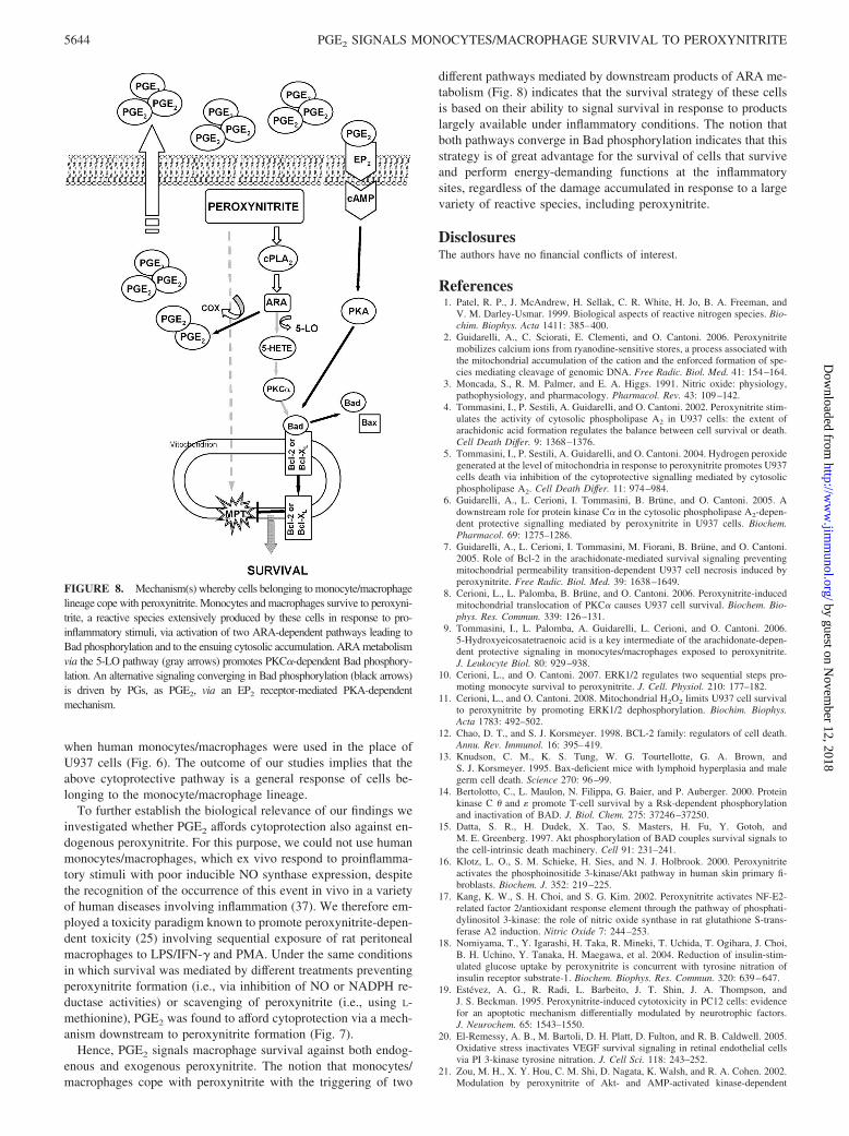
different pathways mediated by downstream products of ARA me-tabolism (Fig. 8) indicates that the survival strategy of these cellsis based on their ability to signal survival in response to productslargely available under inflammatory conditions. The notion thatboth pathways converge in Bad phosphorylation indicates that thisstrategy is of great advantage for the survival of cells that surviveand perform energy-demanding functions at the inflammatorysites, regardless of the damage accumulated in response to a largevariety of reactive species, including peroxynitrite.
DisclosuresThe authors have no financial conflicts of interest.
References1. Patel, R. P., J. McAndrew, H. Sellak, C. R. White, H. Jo, B. A. Freeman, and
V. M. Darley-Usmar. 1999. Biological aspects of reactive nitrogen species. Bio-chim. Biophys. Acta 1411: 385–400.
2. Guidarelli, A., C. Sciorati, E. Clementi, and O. Cantoni. 2006. Peroxynitritemobilizes calcium ions from ryanodine-sensitive stores, a process associated withthe mitochondrial accumulation of the cation and the enforced formation of spe-cies mediating cleavage of genomic DNA. Free Radic. Biol. Med. 41: 154–164.
3. Moncada, S., R. M. Palmer, and E. A. Higgs. 1991. Nitric oxide: physiology,pathophysiology, and pharmacology. Pharmacol. Rev. 43: 109–142.
4. Tommasini, I., P. Sestili, A. Guidarelli, and O. Cantoni. 2002. Peroxynitrite stim-ulates the activity of cytosolic phospholipase A2 in U937 cells: the extent ofarachidonic acid formation regulates the balance between cell survival or death.Cell Death Differ. 9: 1368–1376.
5. Tommasini, I., P. Sestili, A. Guidarelli, and O. Cantoni. 2004. Hydrogen peroxidegenerated at the level of mitochondria in response to peroxynitrite promotes U937cells death via inhibition of the cytoprotective signalling mediated by cytosolicphospholipase A2. Cell Death Differ. 11: 974–984.
6. Guidarelli, A., L. Cerioni, I. Tommasini, B. Brune, and O. Cantoni. 2005. Adownstream role for protein kinase C� in the cytosolic phospholipase A2-depen-dent protective signalling mediated by peroxynitrite in U937 cells. Biochem.Pharmacol. 69: 1275–1286.
7. Guidarelli, A., L. Cerioni, I. Tommasini, M. Fiorani, B. Brune, and O. Cantoni.2005. Role of Bcl-2 in the arachidonate-mediated survival signaling preventingmitochondrial permeability transition-dependent U937 cell necrosis induced byperoxynitrite. Free Radic. Biol. Med. 39: 1638–1649.
8. Cerioni, L., L. Palomba, B. Brune, and O. Cantoni. 2006. Peroxynitrite-inducedmitochondrial translocation of PKC� causes U937 cell survival. Biochem. Bio-phys. Res. Commun. 339: 126–131.
9. Tommasini, I., L. Palomba, A. Guidarelli, L. Cerioni, and O. Cantoni. 2006.5-Hydroxyeicosatetraenoic acid is a key intermediate of the arachidonate-depen-dent protective signaling in monocytes/macrophages exposed to peroxynitrite.J. Leukocyte Biol. 80: 929–938.
10. Cerioni, L., and O. Cantoni. 2007. ERK1/2 regulates two sequential steps pro-moting monocyte survival to peroxynitrite. J. Cell. Physiol. 210: 177–182.
11. Cerioni, L., and O. Cantoni. 2008. Mitochondrial H2O2 limits U937 cell survivalto peroxynitrite by promoting ERK1/2 dephosphorylation. Biochim. Biophys.Acta 1783: 492–502.
12. Chao, D. T., and S. J. Korsmeyer. 1998. BCL-2 family: regulators of cell death.Annu. Rev. Immunol. 16: 395–419.
13. Knudson, C. M., K. S. Tung, W. G. Tourtellotte, G. A. Brown, andS. J. Korsmeyer. 1995. Bax-deficient mice with lymphoid hyperplasia and malegerm cell death. Science 270: 96–99.
14. Bertolotto, C., L. Maulon, N. Filippa, G. Baier, and P. Auberger. 2000. Proteinkinase C � and � promote T-cell survival by a Rsk-dependent phosphorylationand inactivation of BAD. J. Biol. Chem. 275: 37246–37250.
15. Datta, S. R., H. Dudek, X. Tao, S. Masters, H. Fu, Y. Gotoh, andM. E. Greenberg. 1997. Akt phosphorylation of BAD couples survival signals tothe cell-intrinsic death machinery. Cell 91: 231–241.
16. Klotz, L. O., S. M. Schieke, H. Sies, and N. J. Holbrook. 2000. Peroxynitriteactivates the phosphoinositide 3-kinase/Akt pathway in human skin primary fi-broblasts. Biochem. J. 352: 219–225.
17. Kang, K. W., S. H. Choi, and S. G. Kim. 2002. Peroxynitrite activates NF-E2-related factor 2/antioxidant response element through the pathway of phosphati-dylinositol 3-kinase: the role of nitric oxide synthase in rat glutathione S-trans-ferase A2 induction. Nitric Oxide 7: 244–253.
18. Nomiyama, T., Y. Igarashi, H. Taka, R. Mineki, T. Uchida, T. Ogihara, J. Choi,B. H. Uchino, Y. Tanaka, H. Maegawa, et al. 2004. Reduction of insulin-stim-ulated glucose uptake by peroxynitrite is concurrent with tyrosine nitration ofinsulin receptor substrate-1. Biochem. Biophys. Res. Commun. 320: 639–647.
19. Estevez, A. G., R. Radi, L. Barbeito, J. T. Shin, J. A. Thompson, andJ. S. Beckman. 1995. Peroxynitrite-induced cytotoxicity in PC12 cells: evidencefor an apoptotic mechanism differentially modulated by neurotrophic factors.J. Neurochem. 65: 1543–1550.
20. El-Remessy, A. B., M. Bartoli, D. H. Platt, D. Fulton, and R. B. Caldwell. 2005.Oxidative stress inactivates VEGF survival signaling in retinal endothelial cellsvia PI 3-kinase tyrosine nitration. J. Cell Sci. 118: 243–252.
21. Zou, M. H., X. Y. Hou, C. M. Shi, D. Nagata, K. Walsh, and R. A. Cohen. 2002.Modulation by peroxynitrite of Akt- and AMP-activated kinase-dependent
FIGURE 8. Mechanism(s) whereby cells belonging to monocyte/macrophagelineage cope with peroxynitrite. Monocytes and macrophages survive to peroxyni-trite, a reactive species extensively produced by these cells in response to pro-inflammatory stimuli, via activation of two ARA-dependent pathways leading toBad phosphorylation and to the ensuing cytosolic accumulation. ARA metabolismvia the 5-LO pathway (gray arrows) promotes PKC�-dependent Bad phosphory-lation. An alternative signaling converging in Bad phosphorylation (black arrows)is driven by PGs, as PGE2, via an EP2 receptor-mediated PKA-dependentmechanism.
5644 PGE2 SIGNALS MONOCYTES/MACROPHAGE SURVIVAL TO PEROXYNITRITE
by guest on Novem
ber 12, 2018http://w
ww
.jimm
unol.org/D
ownloaded from

Ser1179 phosphorylation of endothelial nitric oxide synthase. J. Biol. Chem. 277:32552–32557.
22. Hellberg, C. B., S. E. Boggs, and E. G. Lapetina. 1998. Phosphatidylinositol3-kinase is a target for protein tyrosine nitration. Biochem. Biophys. Res. Com-mun. 252: 313–317.
23. Rocca, B., and G. A. FitzGerald. 2002. Cyclooxygenases and prostaglandins:shaping up the immune response. Int. Immunopharmacol. 2: 603–630.
24. Radi, R., J. S. Beckman, K. M. Bush, and B. A. Freeman. 1991. Peroxynitriteoxidation of sulfhydryls: the cytotoxic potential of superoxide and nitric oxide.J. Biol. Chem. 266: 4244–4250.
25. Borutaite, V., H. Hope, and G. C. Brown. 2006. Arachidonate and NADPH ox-idase synergise with iNOS to induce death in macrophages: mechanisms of in-flammatory degeneration. Pharmacol. Rep. 58: 96–102.
26. Yu, W., B. G. Sanders, and K. Kline. 2003. RRR-�-tocopheryl succinate-inducedapoptosis of human breast cancer cells involves Bax translocation to mitochon-dria. Cancer Res. 63: 2483–2491.
27. Peters-Golden, M., and T. G. Brock. 2000. Intracellular compartmentalization ofleukotriene biosynthesis. Am. J. Respir. Crit. Care Med. 161: S36–S40.
28. Dixon, R. A., R. E. Diehl, E. Opas, E. Rands, P. J. Vickers, J. F. Evans,J. W. Gillard, and D. K. Miller. 1990. Requirement of a 5-lipoxygenase-activat-ing protein for leukotriene synthesis. Nature 343: 282–284.
29. Watabe, A., Y. Sugimoto, A. Honda, A. Irie, T. Namba, M. Negishi, S. Ito,S. Narumiya, and A. Ichikawa. 1993. Cloning and expression of cDNA for amouse EP1 subtype of prostaglandin E receptor. J. Biol. Chem. 268:20175–20178.
30. Breyer, R. M., C. K. Bagdassarian, S. A. Myers, and M. D. Breyer. 2001. Pro-stanoid receptors: subtypes and signaling. Annu. Rev. Pharmacol. Toxicol. 41:661–690.
31. Coleman, R. A., W. L. Smith, and S. Narumiya. 1994. International Union ofPharmacology classification of prostanoid receptors: properties, distribution, andstructure of the receptors and their subtypes. Pharmacol. Rev. 46: 205–229.
32. Dennis, E. A. 1994. Diversity of group types, regulation, and function of phos-pholipase A2. J. Biol. Chem. 269: 13057–13060.
33. Fujino, H., K. A. West, and J. W. Regan. 2002. Phosphorylation of glycogensynthase kinase-3 and stimulation of T-cell factor signaling following activationof EP2 and EP4 prostanoid receptors by prostaglandin E2. J. Biol. Chem. 277:2614–2619.
34. Fujino, H., W. Xu, and J. W. Regan. 2003. Prostaglandin E2 induced functionalexpression of early growth response factor-1 by EP4, but not EP2, prostanoidreceptors via the phosphatidylinositol 3-kinase and extracellular signal-regulatedkinases. J. Biol. Chem. 278: 12151–12156.
35. Harada, H., B. Becknell, M. Wilm, M. Mann, L. J. Huang, S. S. Taylor,J. D. Scott, and S. J. Korsmeyer. 1999. Phosphorylation and inactivation of BADby mitochondria-anchored protein kinase A. Mol. Cell 3: 413–422.
36. Lizcano, J. M., N. Morrice, and P. Cohen. 2000. Regulation of BAD by cAMP-dependent protein kinase is mediated via phosphorylation of a novel site, Ser155.Biochem. J. 349: 547–557.
37. Pacher, P., J. S. Beckman, and L. Liaudet. 2007. Nitric oxide and peroxynitrite inhealth and disease. Physiol. Rev. 87: 315–424.
5645The Journal of Immunology
by guest on Novem
ber 12, 2018http://w
ww
.jimm
unol.org/D
ownloaded from