prosthetics and orthotics international - oandplibrary.com fileprosthetics and orthotics...
TRANSCRIPT
The Journal of the International Society for Prosthetics and Orthotics
Prosthetics and Orthotics International
April 1994, Vol 18, No. 1

Otto Bock 3R70 Modular, Poly centric Knee Joint.
Polycentric plus pneumatic for a natural gait.
Q44*<ftook O R T H O P Ä D I S C H E I N D U S T R I E
A company of the Otto Bock Gtonp P . O . Box 1 2 6 0 3 7 1 0 5 D u d e t s t a d t Telefon (0 55 27) 8 48-0 Telefax (0 55 27) 7 23 30
Your patient demands comfort, stability,
and easily controlled, natural movement
from his or her prosthesis. While
comfort depends primarily on your skill
as a certified practitioner, the 3R7()
provides features that will help meet
many of your patient's other expectations.
Stability during stance-phase is provided
by the kinematics of this four-bar
linkage knee.
A gear drive connected to the piston rod
of the pneumatic cylinder optimizes
P R O V E N Q U A L I T Y
movement duting swing-phase. The
extension assist as well as fesistance to
extension and flexion are all easy to
adjust.
Finally, variable knee stability, smooth
movement, a larger flexion angle and
minimal weight arc all features that will
please your patient-
P R A C T I C A L S O L U T I O N S
©Otto Bock 6 4 6 S 1 = 1 1 9 3 GB

Prosthetics and Orthotics International
Co-Editors: JOHN HUGHES
NORMAN A . JACOBS
Editorial Board: HANS ARENDZEN DAVID N . CONDIE
JOHN HUGHES
NORMAN A . JACOBS
THAMRONGRAT KEOKARN
HAROLD SHANGALI
Prosthetics and Orthotics International is published three times yearly by the International Society for Prosthetics and Orthotics (ISPO), Borgervaenget 5,2100 Copenhagen 0 , Denmark, (Tel. +45 31 20 72 60). The subscription rate for 1994 is GBP70 per annum, single numbers GBP24. The journal is provided free to Members of ISPO. Remittances should be made payable to ISPO.
Editorial correspondence, advertisement bookings and enquiries should be directed to Prosthetics and Orthotics International, National Centre for Training and Education in Prosthetics and Orthotics, University of Strathclyde, Curran Building, 131 St. James' Road, Glasgow G4 0LS, Scotland (Tel. +44 41 552 4049).
ISSN 0309-3646
Produced by the National Centre for Training and Education in Prosthetics and Orthotics, University of Strathclyde, Glasgow
Printed by David J. Clark Limited, Glasgow

pneumatic swing phase control technology
Nu l ind tu «Her nail 10 compensait
for different speed*
Simple ;iiosi . i r , i phi I ; procédures
tti meet iπdhidnal needs
Adjusts automatically Id diflcrenl
speeds «fier programming
All IIk' advantages of hydraulic control
»ilh less effort and no « vi um penally
Accepts » wide ranee of foot and
ankle configuration1*
Comprehensive socket filling
t he EndoliU' InuWgcnt Prosthesis, part number (119427. Contact our Sales Department on +44 (11)256 465771.
Chas. A Blatchfurd ic Sons Ltd. Lister Road. Basingstoke Hampshire RG22 4 MI Téléphone: +44 (β)256 165771 Facsimile: +44 (0)256 47V705

The Journal of the International Society for Prosthetics and Orthotics
April 1994, Vol. 18, No. 1
Contents Editorial 1
ISPO Statement of Accounts, 1993 2
Executive Board Meeting 6
Interim Meeting of International Committee Representatives 10
The biomechanics of trans-femoral amputation F . A. GOTTSCHALK AND M. STILLS 12
Functional outcome of rehabilitated bilateral lower limb amputees A. DE FRÈTES, A. M. BOONSTRA AND L. D. W. VOS 18
Upper limb prosthetic use in Slovenia H . BURGER AND č. MARINČEK 25
Treatment of congenital subluxation and dislocation of the hip by knee splint harness M. FUKUSHIMA 34
Technical note: a trans-femoral brim adaptor for CAD CAM measurements D. NIELEN, E. D. LEMAIRE AND L. GOUDREAU 40
Technical note: how does vacuum forming alfect Pelite mechanical properties? J. E. SANDERS AND C. H . DALY 43
Technical note: new walking aid for primary mobilisation of an infant with deficiency of all four limbs
S. BALOGH, T. GÖNCZY, R. BUJDOS6 AND L. KULLMAN 49
Book Review 52
Proposed Amendment to the Constitution 53
ISPO Eighth World Congress 54
ISPO Course on Lower Limb Amputations and Related Prosthetics, Slovenia 55
ISPO Course on Lower Limb Amputations and Related Prosthetics, Panama 57
Calendar of Events 59 iii

ISPO Elected Members of Executive Board: M. L. Stills (President) S. Sawamura (President Elect) P. Christiansen (Vice President) J. \ aucher (Vice President) H. Arendzen D. N. Condie T. Keokam H. Shangali W H. Eisma (Immediate Past President) J. Steen Jensen (Hon. Treasurer) N. A, Jacobs (Hon. Secretary)
Standing Committee Chairmen and Task Officers B. Ebskov (Finance) W. H. Eisma (Protocol and Nominations) S. S>awamura (Congress) J. Hughes (Education) S. Heim (Education in Developing Countries) W. Neumann (Certification) H. Arendzen (Membership, Publications) D. N. Condie (Standards) P. Christiansen (Professional Register)
Consultants to the Executive Board H. C. Chadderton (Consumer) J. F. T. Bredie (IVO) I. "Van Rolleghem (1ntert>or) G. Hough III (WOC) T. Lagerwall (RI/ICTA)
International Consultants to the Executive Board C. Marincek J. Craig and R. de Saez O. E. Feldman
Chairmen of National Member Societies Australia Austria Belgium Canada Carribean China Denmark Fin] and Germany Hong Kong India Israel Japan Koiea Netherlands New Zealand Not way Pakistan Sweden Switzerland UK USA
Past Presidents K. Jansen (1974-1977) G. Murdoch (1977-1980) A. Staros (1980-1982) E. Lyquist (1982-1983) E. G. Marquardt (1983-1986) J. Hughes (1986-1989) W. H. Eisma (1989-1992)
Secretary Aase Larsson
USA Japan Denmark Switzerland Netherlands UK Thailand Tanzania Netherlands Denmark UK
Denmark Netherlands Japan UK Germany USA Netherlands UK Denmark
Canada Netherlands Belgium USA Sweden
Central and Eastern Europe Central and South American Russia
G. Carter L. A. Gassinger E. Deschoolmeester G. Martel J. Martina Zhongzhe Wu P. Holstein L. Nummelin R. Baumgartner K, Y. Lee M. K. Goel M. Azaria S. Sawamura J. S. Shin J. H. B. Geertzen A. W. Beasley 0 .Johansen N. M. Akhtar A, Stensl:röm J. Vaucher R. A. Cooper J. W. Michael
Denmark UK USA Denmark Germany UK Netherlands
Denmark
IV
Prosthetics and Orthotics International, 1994, 18,1
I
Editorial
The result for the financial year 1993 was a surplus of DKK 882,788, which was close to that budgeted and mainly derived from further income from the Chicago congress and a high capital yield. Neither of these will apply to the coming year.
Interest revenue on capital has dramatically dropped daring the last quarter of 1993. The Society's investment policy has aimed at getting the highest possible outcome in the financial market without any undue risks. We have largely benefited from the turmoil with shifting exchange rates for vulnerable currencies, which led to high short term interest rates on cash. The effective interest was 9.3% on our total assets by the year end. However, we can not expect more than about 6% with the current low bank rate, which is expected to drop even further. Consequently we have invested the bulk of our assets in 6% bonds, giving an effective interest of above 7%.
We have been able to keep the income from the membership fees at a fairly even level since 1990 with only minor fluctuations around congress years.
The Society is grateful to the War Amputations of Canada for their continued support. However the income from sponsorship has decreased by the termination of financial support from SAHVA in Copenhagen. We will still enjoy cost free office facilities and the like at SAHVA, and together with the major secretarial and administrative contributions from staff associated with our President and other officers, we will also in the future be able to keep our office costs at a reasonable level. The Society greatly appreciates these contributions. The value of these services amounts to not less than DKK 400,000 on a yearly basis.
It is still possible to run the daily activities of our Society on the membership fees, i.e. Secretariat, Executive Board, Prosthetics and Orthotics International and publications. However, ISPO is a Society with aims of influencing education and other activities internationally. This is not possible without using our capital yield, which is also the base for our courses, conferences, and workshop activities. The capital should consequently be preserved as a foundation, if ISPO wishes to continue to influence activity outside its own membership.
Some cost items needs specific comments. The court trial from the Bologna 1980 World Congress was closed with a settlement of DKK
160,000, which included fees to our Danish and Italian lawyers and experts of about DKK 35,000. INTERBOR has agreed from the start to refund half of the costs.
ISPO is currently preparing a series of consensus conferences on Appropriate Prosthetic Technology, Orthotics Management in Cerebral Palsy and Poliomyelitis. We have good reason to believe that USAID will support the Appropriate Prosthetic Technology programme financially, and we have therefore accrued rather considerable costs in preparatory meetings with USAID.
Our first course on Amputation Surgery and Related Prosthetics in the Developing World, in Tanzania, ended with a cost of only one third that budgeted due to contributions from collaborating governmental agencies including DANIDA (Danish International Development Agency), GTZ (German Technical Cooperation Agency) and DSE (German Foundation for International Development); non-governmental organisations including WOC (World Orthopaedic Concern) and ICRC (International Committee of the Red Cross); and a commercial company Otto Bock.
The journal, Prosthetics and Orthotics International, is still a cost issue, but an advertising campaign has been undertaken and further a promotional brochure has recently been released. Postage costs appears high and we are analysing possible reductions by not air mailing these Journals and the possible associated drawbacks for our subscribers.
Printing costs have been high because of un-budgeted costs for printing diplomas and payment of the services provided by the Recal Information Service in the University of Strathclyde for assistance with literature searches and copying for the Consensus Conference on Amputation Surgery, as was already agreed upon in the planning phase.
Membership is not as broadly spread to all the professions of the rehabilitation team as we would wish. We hope to improve through a promotional brochure, which is to be circulated shortly.
J. Steen Jensen Honorary Treasurer
Prosthetics and Orthotics International, 1994,18, 2-5
Income Statement for the Year 1993
SUMMARY 1993 1992 Society membership fees (note 1) 1.045.107 1.151.504 Sponsorship (note 2) 32.058 • 55.870 Meetings with other organisations (note 3) (220.533) (195.948) Conferences, courses etc (note 4) 398.390 1.635.875 Prosthetics and Orthotics International (note 5) (131.996) (72.504) Professional register --
Publications (note 6) (22.391) 18.748
Activity result 1.100.635 2.593.545 Administration expenses (note 7) (832.601) (912.793)
Primary result 268.034 1.680.752
Interest (note 8) 604.222 410.133 Dividend (note 8) 1.128 1.504 Maturity yield (note 8) 9.404 7.388 Change in value of securities (note 8) - (6.392)
Financial income 614.754 412.633
Net income DKK 882.788 2.093.385
2
ISPO Statement of Accounts, 1993
Auditors Report We have audited the enclosed Financial Statements for the year 1993.
The audit has been performed in accordance with approved auditing standards and has included such procedures as I have considered necessary.
The Financial Statements have been prepared in accordance with statutory requirements, and the constitutions of the society and generally accepted accounting principles. In our opinion the Financiai Statements give a true and fair view of the state of the association's affairs as of December 31, 1993 and of the result for the year then ended.
Copenhagen, March 1, 1994 Revisionsgruppen A/S
Søren Wonsild Glud State Authorized Public Accountant
Securities
Bonds and shares have been valued at the lower of cost or market.
Office Equipment Computer and office equipment have been stated at cost less depreciation computed straight line over 5 years. Accrual Concept The accrual concept of accounting has been used in these financial statements.

ISPO Statement of Accounts, 1993
Balance sheet as of December 31,1993
ASSETS Cash
Accrued interest Advertising receivable Prepayment, World Congress Receivables Chicago World Congress Advance funding of World Congress 1980 Other
Receivables
Securities (note 9)
Office equipment (note 8)
Total assets
1993 1.451.287
124.435 104.190 83.040
181.215
40.482
533.362
5.035.908
8.571
DKK 7.029.128
1992 2.994.181
85.925 66.778 94.433
87.437 31.296
365.869
2.855.379
6.215.429
LIABILITIES AND EQUITY Accrued expenses 91.737 93.239 Accrued printing cost 178.000 160.000 Prepaid membership fees 9.951 3.350 Prepaid advertising income - 9.387 Prepaid subscription income 77.100 72.464
Short-term liabilities 356.788 338.440
Provision World Congress 1980 - 87.437
Equity January 1 5.789.552 3.696.167 Net result 882.788 2.093.385
Equity December 31 6.672.340 5.789.552
Liabilities and capital DKK 7.029.128 6.215.429
Notes to the Financial Statements 1. Society membership fees Membership fees consist of payments from 2493 listed members, excluding 45 honorary members.
1993 1992 2. Sponsorship Contribution from:
The War Amputations of Canada 32.058 30.870 S AH VA 25.000
DKK 32.058 55.870

..: ISPO Statement of Accounts 1993
3. Meetings with other organisations Education Committee, INTERBOR INTERBOR WHO Geneva World Orthopaedic Concern ICRC Amer Acad Orth-Prosth ACOPPRA USAID Lisbon 1993 Other
8.258 9.834
17.120 14.346 5.073
5.508 80.228 66.743 13.423
DKK 220.533
23.853 5.906 9.211
10.229
53.578
93.171
195.948
4. Conferences, courses etc Groningen 114.642 Chicago 546.577 1.521.233 Bologna (80.222) Amman (12.983) Tanzania (54.982)
DKK 398.390 1.635.875
5. Prosthetics and Orthotics International Advertising Subscriptions
Printing and mailing Production editor Journal promotion Meeting expenses
Net result (loss)
287.477 199.910
487.387
(530.514) (32.129) (41.351) (15.389)
(619.383)
DKK (131.996)
246.559 209.691
456.250
(494.539) (29.086)
(5.129)
(528.754)
(72.504)
6. Publications Booksales
Amputation surgery consensus
Total cost
21.644 28.120 (44.035) (9.372)
DKK (22.391) 18.748
7. Administrative expenses Executive Board and Officers:
Travel and hotel costs Meeting expenses Directory printing
307.621 304.512 6.480
58.500
DKK 307.621 369.492.

ISPO Statement of Accounts, 1993 5
Secretariat, Copenhagen Staff salaries Labour tax Data service Meeting expenses Postage and Bank charges Telephone Stationery Office supplies Auditing Bookkeeping Sundries Knud Jansen medals Depreciation Professional register
291.530 12.747
1.152 12.836 60.250 4.301
21.208 10.172 41.250 29.031 26.925
4.285 9.293
272.783 12.360 5.522
13.436 88.269 4.713
16.174 2.481
38.000 22.848 15.872 50.843
524.980 543.301
Total DKK 832.601 912.793
8. Office equipment Computer equipment, at cost Office equipment, at cost
108.203 26.220
95.347 26.220
Cost 134.423 121.567
Depreciation January 1 Depreciation December 31
(121.567) (4.285)
(121.567)
Accumulated depreciation (125.852) (121.567)
Net book value DKK 8.571 -
9. Securities Nominal
value Original
cost Year end
value Interest/ dividend
Bonds 9% Kred. Danrnark 2007 10% Kred. Danrnark 2010 Matured
2.872.000 2.314.000
2.679.796 2.333.553
2.916.516 2.374.164
268.605 125.000
13.699
Shares Den Danske Bank 94 30.891 36.378 1.128 Total 5.186.094 5.044.240 5.327.058 408.432

Prosthetics and Orthotics International, 1994,18, 6-11
Executive Board Meeting 19-20 January 1994 Ballerup, Denmark
The following paragraphs summarise the major discussions and conclusions of the Executive Board Meeting held in Ballerup in January of this year. They are based on the draft minute of that meeting which has yet rø be approved.
Standing Committee and Task Officers Reports The Chairman of the Finance Committee, Bent Ebskov (Denmark), reported that although the Society's finances were generally in a sound position, there was some concern with regard the lowering of interest rates in Denmark and that income from capital investments would be reduced in the future. The Honorary Treasurer, J. Steen Jensen (Denmark), presented the accounts for 1993 which are subsequently published in this issue of Prosthetics and Orthotics International. The result for the year was a surplus of DKK 882,788 which was close to that budgeted. It derived from further income from the Chicago Congress and a high capital yield. The Honorary Treasurer presented the proposed budget for 1994. He estimated that the result for the year would be a deficit of approximately DKK 765,000. The major reasons for the deficit are the extraordinary amounts of activity proposed for the year, including Amputation Surgery and Related Prosthetics Courses in Thailand, Slovenia and Panama, as well as Consensus Conferences on Appropriate Prosthetic Technology in Developing Countries and the Orthotic Management of Cerebral Palsy. This, together with the reduced amount of sponsorship and investment income expected, resulted in the high deficit. The Executive Board discussed the budget for 1994 and approved it. The Executive Board discussed the fees for. 1995 and agreed that they should remain at DKK 450 for industrial countries and DKK 225 for developing countries. The Executive Board voiced its concern with regard the proposed deficit for 1994 and the decreased income from sponsorship and investments and suggested that consideration may have to be given to increasing fees for the following triennium.
The Chairman of the Protocol and Nominations Committee, Willem H. Eisma (The Netherlands), reported that the Committee had discussed guidelines with regard the role of International Consultants. The Executive: Board, after some amendments, approved of these guidelines. A proposed amendment to the Constitution recognising the role of International Consultants is published elsewhere in this issue of Prosthetics and Orthotics International.
The Chairman of the Education Committee, John Hughes (UK), presented a report of the Education Committee activities. He informed the Executive Board that the Course on Amputation Surgery and Related Prosthetics in China had to be cancelled due to local difficulties. However, it is hoped to arrange a new date for this course in the future. Plans for the courses in Thailand, 14-18 March 1994, Slovenia, 26-30 September 1994, and Panama, 14-18 November 1994, were well underway (Secretary s Note: the course in Thailand was duly run with an attendance of 60 participants). John Hughes went on to report on the activities of the ISPO/ INTERBOR Joint Education Committee. The Committee is involved with the problems of International Certification in prosthetics and orthotics. Plans are progressing to hold three further trials of the American Board for Certification (ABC) examinations in Australia, Tanzania and Germany. The Joint Education Committee will be looking at the wider aspects of International Certification with a view to establishing guidelines, standards and a plan of action John Hughes reported that the Joint Education Committee is hoping to seek funds from the ERASMUS programme of the European Community for a meeting of Heads of Schools and Associations to be organised late in 1994 at the University of Strathclyde, Scotland. This would focus on education in Europe but it was the intention that there would be a wider international representation. The German Agency for Technical Cooperation (GTZ) had invited the Society to inspect its school in Yerevan, Armenia which was carried out by Roy Bowers (UK) and Andries de Bont (Netherlands/Ireland). A number of questions had arisen from the report, mainly related to the
6

Executive Board Meeting 7
balance of the course between prosthetics and orthotics. It was agreed that the Chairman should contact GTZ and the inspectors with regard these matters and if they could be resolved, confirm recognition of the course for Category II Training and Education. The Society had received invitations to inspect the GTZ schools in Pakistan and China and the World Rehabilitation Fund (WRF) school, in Yerevan, Armenia and arrrangements for these inspections will be made in due course. The Society is collaborating with World Orthopaedic Concern (WOC) on an instructional course on Lower Limb Amputation Surgery to be offered at the British Orthopaedic Association meeting to be held on 15 April 1994 in London, UK. J. Steen Jensen (Denmark) presented a proposal for a course on Care and Rehabilitation of Amputees, primarily for nurses and therapists. He would, in the near future, be contacting National Member Societies seeking suggestions for the content and lecturers for the course.
The Honorary Secretary reported that at the end of 1993, the Society had a total of 2,493 members. This was a slight decrease over the total for the previous year which was extraordinarily high due to the numbers of new members joining the Society as a result of the Chicago Congress. The Executive Board endorsed the formation of the Panamanian National Member Society which would take effect as soon as the Constitution was translated into English in order to ensure that there was no contravention of the International Constitution. John Craig (USA) informed the board that the Colombian group were very close to forming a National Member Society. A number of other countries were considering establishing National Member Societies including, Chile, France, Mexico, Slovenia, and Taiwan.
Hans Arendzen (The Netherlands) reported on the publication of a promotional brochure for the Society. Some 5,000 copies had been printed and copies will be distributed to National Member Societies. He pointed out that it was printed in English, however, it would be possible to insert translations of the text or some other information to meet specific requirements. David N. Condie (UK) informed the Board that a flyer seeking new subscribers for Prosthetics and Orthotics International had been produced. Some 5,000 flyers had been printed and sent out to 1634 centres in 147 countries and attempts would be made to monitor the success of this exercise. The Executive Board agreed to re-constitute the Publications Committee as a result of the suggestion that, as an outcome of the Consensus Conference on Amputation Surgery, it would be useful if short, clearly illustrated booklets, specifically related to the techniques of different methods of amputation, could be produced for use by trainee surgeons and others. Such booklets should be fully coordinated with any video tapes on amputation surgery that the Society may produce. The Committee had met and its Chairman, Hans Arendzen, reported on suggestions for the establishment of a coordinated video tape and booklet project on amputations of the lower limbs. The Committee had listed the essentials to be covered in booklets and video tapes on ankle disarticulation, trans-tibial amputation, knee disarticulation and trans-femoral amputation. The Publications Committee would make a detailed proposal for the video tape and booklet programme for the next Executive Board meeting. The Committee had also viewed the video tape on trans-tibial amputation surgery produced by Amar Jain (UK) and John Guy (UK) and it was agreed that the Chairman should meet with them in order to discuss future plans for it.
David N. Condie (UK) reported on the Society's activities in relation to the International Standards Organisation (ISO) and the European Standards Organisation (CEN). The ISO Technical Committee 168 on Prosthetics and Orthotics and its three working groups on Terminology and Nomenclature, Medical Aspects and Physical Testing had met in Washington, USA, 19-21 November 1993. A number of drafts for standards, had been completed and will be submitted to all countries participating in TC168 work for comment early in 1994. In addition, ISO 8548-2 Method of Describing Lower Limb Amputation Stumps and ISO 8548-3 Method of Describing Upper Limb Amputation Stumps have been accepted as International Standards. The Society is also active in TCI73 - Technical Systems and Aids for Disabled Persons. The Society is represented on Sub-Committee 1 -Wheelchairs, by Geoff Bardsley (UK) and on Sub-Committee 2, Working Group 7 - Classification and Terminology by David N. Condie. David N. Condie also represents the Society on the ad hoc Working Group of CEN 293 on Prosthetics and Orthotics. The Group had met in Berlin, Germany 22 October 1993 when it was overwhelmingly agreed that there was a need for such CEN standards and that it was recommended that a Working Group of CEN Technical Committee 293 would be created

8 Executive Board Meeting
for this purpose. David N. Condie is in the process of preparing a paper outlining the current situation with regards the work on standards for publication in Prosthetics and Orthotics International. Per Christiansen (Denmark) reported on the status of the Professional Register. He presented a proposal to combine the Society's Application Form with the Professional Register Questionnaire. The Executive Board discussed this proposal and made a number of suggestions and a definitive form would be produced for the next Executive Board meeting.
The President reported on developments with regard the proposal for a Consensus Conference for Appropriate Prosthetic Technology for developing countries. Representatives of the Board had met with officials of the US Agency for International Development (US AID) to discuss the programme and organisation of such a conference. It is hoped that US AID would co-sponsor the conference which would bring together all the major agencies involved in prosthetic technology in the developing world. HGB Day (UK) has been co-opted onto the organising committee for this conference. David N. Condie presented a report on the progress of the proposal to hold a Consensus Conference on the Orthotic Management of Cerebral Palsy. A planning team had been formed whose principal role is to finalise the programme structure and select reviewers and participants. It is expected that approximately 30 persons will be invited to attend representing appropriate medical specialties, bioengineering, orthotics and therapy. Requests for nominations for reviewers and/or participants have been sent to National Member Societies. The conference would last for 3 days and will be held in November 1994. George Murdoch, (UK) reported on the progress made with regards a proposal for a Consensus Conference on Poliomyelitis. It is anticipated that the total cost would be in the region of USD 170,000. The Executive Board expressed the view that it would be desirable to organise such a conference if it was possible to find a means to defray the costs. It was agreed that the committee reponsible should examine the funding in order to find sources of support and report back to the next Executive Board meeting.
International Consultants Ğrt Marinček (Slovenia) reported that all attention in Central and Eastern Europe was focused on the course for Amputation Surgery and Related Prosthetics arranged for 12-16 September 1994 in Ljubljana, Slovenia. This will be the first time this year that professionals from this region will meet together and he hoped that this would provide useful contacts for ISPO within these countries.
Seishi Sawamura (Japan) reported on a proposal to establish a Prosthetic and Orthotic Centre for Asian developing countries. The proposal had been discussed at a meeting organised by the United Nations Economic and Social Commission for Asia and the Pacific as well as the Japanese Government in Okinawa, Japan on 19 October 1993. One of the outcomes of that meeting was a resolution calling for the establishment of a centre in Asia that would supply prostheses, orthoses and technical aids, calling for the support of Governments in that area. Such a centre in Asia would conduct an educational programme for prosthetists and orthotists as well as manufacturing inexpensive prosthetic and orthotic components for the region.
John Craig (USA) reported on activities in Central and South America. As well as trying to promote National Member Societies, he indicated that attempts are being made to increase the prosthetic and orthotic activities in this region, particularly in Colombia, Chile, Panama, Guatemala and Mexico.
International Organisations Jacques van Rolleghem (INTERBOR) reported that the 12th International Congress of INTERBOR held in Lisbon, Portugal, 22-25 September 1993 had been very successful and, on behalf of the Board of INTERBOR, thanked ISPO for its collaboration. The venue for INTERBOR's next International Congress will be either Norway or Brazil.
The Honorary Secretary reported on a meeting called by the World Health Organisation (WHO) in Geneva on 4 November 1993, the purpose of which was for groups with an interest in prosthetics and orthotics in developing countries to exchange information about their activities and to identify actions which may require to be taken in the future. As well as ISPO, the meeting had been attended by representatives of the International Committee of the Red Cross (ICRC), Handicap International (HI), the German Agency for Technical Cooperation (GTZ) and Rehabilitation International (RP

Executive Board Meeting 9
Jan Bredie (The Netherlands) reported on the 11th International Congress of Internationaler Verband der Orthopädie-Schuhtechniker (IVO) held in Quebec, Canada 3-6 September 1993. Some 700 participants attended the conference, including 400 from Europe. This was the first time that the congress had been held outside of Europe. The next IVO congresses will be held in June 1995 in Berlin, Germany and in 1997 in Belgium. The possibility of ISPO holding a combined meeting with IVO on advances on orthopaedic footwear is being considered.
The President reported that he had been in communication with the President of World Orthopaedic Concern (WOC), Garry Hough III, and that it is hoped that he would attend the Executive Board Meeting regularly as the WOC observer.
The Honorary Secretary reported that the United Nations (UN) Economic and Social Council has re-classified ISPO to Category II Consultative Status at its meeting on 28 June-30 July 1993. The Executive Board agreed that the President and the Honorary Secretary should be the Society's representative to the New York Office of the UN and that Jean Vaucher (Switzerland) and the Honorary Secretary should be representatives to the Vienna and Geneva offices.
The Honorary Secretary reported that he had discussions with the International Committee of the Red Cross (ICRC) and Alain Garachon would attend future Executive Board meetings as its observer.
The President reported that in recent times he had established good contact with US Agency for International Development (US AID). In addition to the proposed Consensus Conference on Appropriate Technology, US AID were beginning to use ISPO members for advice. The President, together with Dan Ramsey (USA), Michael Schuch (USA) and Frank Gottschalk (USA) had inspected US AID projects in Vietnam. The President, together with John Hughes (UK), John Craig (USA), The Honorary Secretary and other ISPO members, had met in Dallas, 26-27 August 1993 to develop an evaluation form for US AID projects. In addition, American members of ISPO had been sent to visit projects in Sri Lanka and Mozambique.
Congresses John Hughes (UK) reported that on the advice of the Society's lawyers in Bologna, it was decided to offer a settlement to Studio BC of LIT 30,000,000. This released ISPO and Hannes Schmidl (Italy) from any obligation towards Studio BC. This matter is now closed.
Valma Angliss (Australia) reported on progress for the arrangements for the 8th World Congress to be held in Melbourne, Australia, 2-7 April 1995. Arrangements were far advanced for the scientific programme, the commercial and scientific exhibit and the social programme. A full announcement and call for papers will be issued by the end of March 1994.
Hans Arendzen (The Netherlands) reported that arrangements were well underway to organise the 9th World Congress, 28 June-3 July 1998 in Amsterdam. A local committee had been set up, a. foundation for the local organisation had been established, congress organisers had been appointed and a revised budget was being prepared. The Executive Board approved the design of a logo for the congress on condition it is shown in close association with the ISPO logo.
Conferences and Meetings The Honorary Secretary reported that he had been asked to co-chair a session on International Education in Prosthetics and Orthotics ot Orthopädie+Reha Technique, Essen, Germany, 31 May-3 June 1994. John Hughes (UK), Sepp Heim (Germany) and William Neumann (USA) had been asked to make presentations during this session.
The Society is collaborating with the organisers of Dundee '94, Clinical Gait Analysis, 5-8 July 1994, Dundee, UK. Members of the Society will be offered reduced registration for this meeting.
Hans Arendzen (The Netherlands) reported that he had identified a number of individuals to make presentations on a session on prosthetics at the 6th European Regional Conference of Rehabilitation International, 4-9 September 1994, Budapest, Hungary. A request has been made to make a plenary session presentation at the meeting but no reply has been received to date.
Jean Vaucher (Switzerland) reported on the 2nd International Meeting of the Austrian, Swiss and

10 Executive Board Meeting
German National Member Societies, together with the Swiss Association for Prosthetics and Orthotics, 21-22 October 1994, Konstanz, Germany. The theme of the meeting will be electronics in prosthetics and orthotics. It was agreed that Jean Vaucher should represent the Board at this meeting.
Nominations for the Executive Board 1995-1998 The Executive Board prepared a slate of nominations for the coming triennium as follows -
President President-Elect Vice-Presidents
Members
Honorary Treasurer Honorary Secretary
Seishi Sawamura (Japan) Norman A. Jacobs David N. Condie (UK) Harold G. Shangali (Tanzania) Gerhard Fitzlaff (Germany) Jean Halcrow (Australia) Björn M. Persson (Sweden) C. Michael Schuch (USA) J. Steen Jensen (Denmark) Brendan McHugh (UK)
Orthopaedic Surgeon Bioengineer Rehabilitation Engineer Prosthetist/Orthotist Prosthetist/Orthotist Occupational Therapist Orthopaedic Surgeon Prosthetist/Orthotist Orthopaedic Surgeon Bioengineer
This slate of nominations would be presented to the Interim Meeting of International Committee Representatives outlining the reasons for the slate and asking for comment.
Norman A. Jacobs Honorary Secretary
Interim Meeting of International Committee Representatives .21-22 January 1994 Ballerup, Denmark
The meeting of International Committee Representatives which met directly after the Executive Board meeting was attended by representatives of 15 National Member Societies as well as members of the Executive Board.
Papers on IS1PO policies and activities were presented to the meeting by members of the Executive Board. The Chairman of the Finance Committee, Bent Ebskov, and the Honorary Treasurer, J. Steen Jensen, reported on the finances of the Society. The Chairman of the Education Committee, John Hughes presented a report on the Society's activities on education. The Membership Task Officer, Hans Arendzen and the Honorary Secretary reported on membership and National Member Society development. Hans Arendzen, David N. Condie and the Honorary Secretary presented a report on publications which covered the publication of a promotional brochure, the publication of a subscription flyer for Prosthetics and Orthotics International and progress with the journal. David N. Condie informed the meeting of the Society's involvement in developing international standards in prosthetics and orthotics. Per Christiansen reported on the status of the professional register. Three Consensus Conferences are currently being planned. The President reported on progress with the development of a Consensus Conference on Appropriate Prosthetic Technology in Developing Countries, David N. Condie on the status of a Consensus Conference on Orthotic Management of Cerebral Palsy and George Murdoch on a proposal for a Consensus Conference on the Management of Poliomyelitis. The President presented a report on consumer interests on behalf of the Society's consumer consultant, Cliff Chadderton. The President-Elect, Seishi Sawamura, informed the meeting

Executive Board Meeting 11
of developments with regard the role of international consultants in the Society. John Craig, the International Consultant to Central and South America, gave a report of activities in that region. The Honorary Secretary outlined the relationships of the Society with other international organisations. The President presented a report on the World Congresses which covered the Congress in Bologna, Italy 1980, the 7th World Congress, Chicago, USA 1992, the 8th World Congress in Melbourne, Australia 1995 and the 9th World Congress in Amsterdam 1998. These reports were discussed and comments were made on these presentations for consideration by the Executive Board.
National Member Societies presented reports of the activities in their countries related to education and training for prosthetists and orthotists, research efforts in prosthetics and orthotics, governmental and non-governmental organisation activities in prosthetics and orthotics for developing countries and twinning. The reports were presented by Juan Martina (Caribbean), Lars Nummelin (Finland), Gerhard Fitzlaff (Germany), Yeun Tsz Kuen (Hong Kong), Jan Geertzen (The Netherlands), Robin Platts (UK), John Michael (USA), K. N. Niazi (Pakistan), Eiji Tazawa (Japan), Jean Halcrow (Australia), Guy Martel (Canada), Martin Goplen (Norway), R. K. Srivastava (India), Wyn Beasley (New Zealand) and E. Deschoolmeester (Belgium). The meeting had a full discussion of these reports. In particular, it discussed the principle of whether the International Society should make a financial contribution to the twinning arrangements entered into by individual National Member Societies. The consensus of the meeting was that the International Society should not do so.
The meeting discussed the composition of the International Committee and representation at the Interim Meeting. It was suggested that it may be better if the Interim Meeting was a full International Committee meeting that had full Constitutional rights. It was pointed out that the Interim Meeting of International Committee Representatives came about at the request of the joint International Committee and Executive Board working group. The numbers had been limited to one representative from each National Member Society in order to keep the costs of such a meeting at a reasonable level. No conclusion was reached on this issue.
The meeting discussed the matter of therapy representation on the Executive Board. It was generally felt that there is a need to make sure that the Executive Board has as wide a representation from the different professions as possible. It would be possible for the Executive Board to appoint consultants if the elected Board did not represent all the major professions.
The President presented the slate of nominations for the Executive Board for the next triennium (please see report on Executive Board meeting). The President explained that the slate conformed with the Constitution in as much that it reflects, as far as possible, the various professional disciplines and interests and the appropriate cultural and geographical distribution of the Society. The slate was discussed by the meeting and it would be sent out to the National Member Societies to seek either agreement or further nominations.
The meeting looked at the proposed amendments to the Constitution and suggested that the Executive Board look at the wording of the proposed amendment 2.5.1 previously published in the Journal with the suggestion that it should be re-worded.
The meeting proved to be a successful event which provided a good forum for exchange of information between the Executive Board and representatives of the International Committee.
Norman A. Jacobs Honorary Secretary

Prosthetics and Orthotics International, 1994, 18, 12-17
The biomechanics of trans-femoral amputation
F. A. GOTTSCHALK and M. STILLS
Department of Orthopaedic Surgery, University of Texas Southwestern Medical Centre, Dallas, USA
Abstract The biomechanics of trans-femoral amputations has not been previously described. Little attention has been paid to the importance of adductor magnus in holding the femur in its normal anatomical axis. Loss of function of adductor magnus leads to abduction of the residual femur, in a trans-femoral amputation. A cadaver study of the adductor group of thigh muscles has been done and the biomechanical importance of these muscles is documented. The moment arms of the three adductor muscles have been determined, based on muscle attachments and muscle size, relative to each other. Adductor magnus has a major mechanical advantage in holding the thigh in its normal anatomical position. Loss of the distal third of its attachment results in a 70% loss of the effective moment arm of the muscle, which contributes to the abducted femur in standard trans-femoral amputations. A muscle preserving trans-femoral amputation, which keeps adductor magnus intact, prevents abduction of the residual femur and may allow for easier walking with a prosthesis. The conflicting reports about adductor magnus activity during the gait cycle can be explained by this muscle's dual innervation by the sciatic and obturator nerves and its dual function as a hip adductor and extensor.
Introduction The introd action of new socket shapes and
designs for trans-femoral amputation has led to
a resurgence of interest in trans-femoral amputees. Originally it was thought that by changing socket shape and alignment the residual femur of a trans-femoral amputation could be better controlled within the socket and thus improve the patients gait and functional activity (Long, 1985; Sabolich, 1985.) Several publications have documented that patients with trans-femoral amputations have an increased energy expenditure for walking' and that the older patient with little or no physical reserve may lose the ability to walk again (Gonzalez et al., 1974; James 1973; Volpicelli et al., 1985; Waters et al., 1976). Long (1985) noted that the trans-femoral amputee wearing a quadrilateral type socket had an abducted residual femur. The development of the normal shape normal alignment type socket was thought to improve the position of the femoral shaft from an abducted position to a more neutral position. Sabolich (1985) developed the concept of an ischial containment socket with a narrow medial lateral configuration in an attempt to bring the femur back towards adduction and improve the patient's gait and activity. No objective results were provided to show that mechanically this was achieved. A subsequent study comparing patients who used quadrilateral sockets and those that used ischial containment sockets showed that socket shape and design was not able to influence or control the position of the femur within the socket itself (Gottschalk et al., 19892). Alignment of the prosthesis did not appear to influence the position of the residual femoral shaft either.
Little consideration has been given to improving the surgical technique and most of
All correspondence to be addressed to Dr. Frank Gottschalk, Department of Orthopaedic Surgery, 5323 Harry Hines Blvd., Dallas, TX 75235-8883, USA.
12

Trans-femoral amputation 13
the surgical literature related to trans-femoral amputations highlights patient function and longevity rather than the surgical technique itself. The standard texts on the technique of trans-femoral amputation make no mention of the adductor muscles and their importance in controlling the position of the femur (Bohne, 1987; Harris, 1981; Burgess, 1989). With a conventional trans-femoral amputation it has been noted that the femur comes to lie in abduction, with a large medial soft tissue mass (Fig. 1). The deviation from the normal mechanical axis of the limb results from the surgery and loss of muscle tissue and muscle attachment as well as the position of the thigh at the time of wound closure (Murdoch et al., 1992).
The paper presents the biomechanics of the adductor muscles of the thigh and the importance of a muscle preserving surgical technique to hold the femur in its normal mechanical alignment.
Materials and methods A biomechanical model of the adductors of
the thigh was developed from the anatomical descriptions in standard texts and cadaver dissections. Four cadavers were dissected and the attachments and volume of each of the
adductor muscles was noted. The femur and thigh were divided into thirds to correspond to the attachments of the three major adductor muscles and diagrams based on those used by Brash (1955) were developed to project lines of action of the muscles and the vertical and horizontal resultant forces
A muscle preserving trans-femoral amputation, with myodesis of the adductor magnus and quadriceps has been done on 30 patients. The adductor muscle is preserved intact with its blood and nerve supply and reattached to the distal lateral aspect of the residual femur by a myodesis (Gottschalk, 1992) (Fig. 2). The femur is transected at the appropriate level prior to anchoring of the adductor magnus. At the time of suturing the muscle to the bone, the femur is held in maximum adduction. The quadriceps is also anchored to the femur via anterior and posterior drill holes. Standing radiographs of the residual
Fig 1. Radiograph of residual femur in prosthetic socket, with femur in abducted position. Increased soft tissue is
noted medially.
Fig 2. Diagrammatic representation of muscle preserving adductor myodesis for trans-femoral amputation.
(Reprinted with permission. Gottschalk, Mosby, 1992.)

14 F. A. Gottschalk and M Stills
femur are taken with and without the socket.
Results The adductor magnus because of the greater
length of its lever arm is best placed to hold the femur in a normal adducted position. Figure 3 shows the normal mechanical and anatomical alignment of the lower limb. The directions of the forces exerted by the adductor muscles are show in Figure 4. The directions of the components of force normal to the lines joining the points of attachment of the muscles and the centre of rotation of the hip joint are also shown. These are the components producing adduction. Adductor magnus is an important adductor because of its bulk and consequent capacity for force development. The cadaver study showed that adductor magnus is three to four times larger in physiological cross-septional area and volume than the adductor longus and brevis. The point of application of
the force of the muscles was taken as the middle of the attachment to the femur of each muscle. The unique anatomy of adductor magnus provides the muscle with 2 nerve innervations and 2 separate functions. The most medial portion of adductor magnus makes the greatest contributions to the adduction moment (rotational moment) which is 4 to 5 times that of adductor longus and adductor brevis. Adductor longus and brevis contribute in smaller amounts to normal thigh adduction as noted from the resultant forces. Based on the
Fig 3. Normal mechanical and anatomical alignment of the lower limb. (Reprinted with permission. Gottschalk
et al., J Prosthet Orthot, 1989.)
Fig 4. The resultant forces of the adductor group of muscles showing the components producing adduction. The moment arms are depicted by the interrupted line. (AM=adductor magnus, AL=adductor longus, AB=adductor brevis.) (Reprinted with permission.
Gottschalk et al., J Prosthet Orthot, 1989.)

Trans-femoral amputation 15
contribution of each adductor moment, if the distal third of the femur is amputated and an inadequate myodesis of adductor magnus is done, then 70% of the adduction moment is lost. Thus the intact adductor longus and brevis would provide the only mechanism for holding the femur in adduction.
The full surgical procedure has been described in a previous publication (Gottschalk, 1992). The surgical technique to preserve the adductor magnus and re-anchor it adequately to the residual femur by suturing to the lateral distal femur maintains the normal femoral anatomical alignment (Fig. 5). The radiographs show that the anatomical axis and the overall mechanical alignment of the limb can be maintained (Fig. 6). The femur is contained in the middle of the muscle envelope of the thigh, which is a normal adducted position.
Discussion It is well accepted that the patients with trans-
femoral amputations have a higher level of energy expenditure for normal walking, because of loss of the knee joint. One of the contributing factors to abnormal gait in trans-femoral amputees is the mechanical disadvantage of an abducted position of the residual femur, which
forces the patient to walk with an increased energy expenditure, despite satisfactory fitting with a prosthesis. Many patients who are good prosthetic users develop pain and discomfort at the distal lateral end of the femur, in the socket, as a direct result of the abducted position. The adductor roll that is commonly noted in trans-femoral amputees is another cause of the patient walking with the leg abducted. The muscle preserving adductor myodesis appears to prevent the formation of an adductor roll, and thereby allow for a more comfortable fitting socket. By applying the biomechanics of the adductor muscles of the thigh and improving the surgical technique to hold the femur in adduction, a patient who may have been a marginal prosthetic user could become a
Fig 5. Position of the femur after trans-femoral amputation and muscle preserving adductor myodesis.
Fig 6. Radiographs of trans-femoral amputation with muscle preserving adductor myodesis in prosthetic socket, with residual femur in normal anatomical
alignment.

16 F. A. Gottschalk and M Stills
definitive prosthetic user. James (1973) noted that patients with a standard trans-femoral amputation had decreased muscle strength as a result of reduced muscle mass, inadequate fixation and atrophy of the thigh muscles. This was confirmed in a study on the neurophysiology of muscle function in the stump (Thiele et al., 1973). It is possible to preserve a large amount of the adductor power by preserving muscle bulk and attaching the distal end of the muscle to the distal end of the residual femur, with the stump held in an over corrected position. This helps maintain the length and tension of the muscle and keeps enough muscle power to overcome the shorter horizontal moment arm. In addition, the femur is no longer in an abducted position and this allows the abductor mechanism to function normally.
The hip abductor mechanism remains intact at the time of a trans-femoral amputation. Gluteus medius, minimus and parts of maximus are abductors of the hip. However tensor fasciae latae plays the most important role in hip abduction during the stance phase of gait (Gottschalk et al., 19891). Although the very distal attachment of tensor fasciae latae is lost in a trans-femoral amputation, the muscle can still function as a thigh abductor because of its indirect attachment from the fascia lata to the linea aspera via the lateral intermuscular septum. However, at the time of surgery the tensor fasciae latae should be sutured to the medial fascia of the thigh to provide additional stabilisation. Failure to re-anchor the tensor fasciae latae may contribute to some weakness of the hip abductor mechanism. Interference with the action of adductor magnus leads to an imbalance of the mostly intact abductor mechanism with subsequent abduction of the femur. Keeping adductor magnus intact and adequately re-anchoring it to the residual femur will maintain the balance between the hip abductors and adductors. It is not possible to hold the residual femur adducted with a prosthetic socket irrespective of its shape or design, as has previously been reported (Gottschalk et al., 19892), since the femur cannot be displaced in its soft tissue envelope.
Electromyographic studies of adductor magnus provide conflicting information. Review of the literature reveals that most likely the muscle is active at the beginning of stance phase and again at the end of stance and into
early swing phase (Green and Morris, 1970; Inman et al., 1981). Because of the muscle's dual innervation by the sciatic and obturator nerves, most likely different parts of the muscle are active at different times during the gait cycle. Inman el al., (1981) note that adductor magnus is active only at the beginning and termination of swing phase. Green and Morris (1970) describe activity of adductor magnus and lognus and noted that activity occurred in stance phase. The disparity in the results is most likely due to the dual innervation of adductor magnus and its dual function of hip extensor and thigh adductor.
In a distal third femur amputation the tendon of the adductor magnus should be preserved and swung around the distal end of the femur and anchored by drill holes to the lateral femur, with the femur maximally adducted. This preserves maximum muscle force by having an intact adductor system, and provides a mechanical advantage for the adductors and abductors of the thigh. In a middle third amputation, instead of transecting adductor magnus, it should be detached from the bone and swung around the distal end of the adducted femur. The myodesis can then be performed and redundant tissue excised.
Those patients who have had the amputation as described above have the residual femur in a normal, or near normal anatomical alignment. (Fig. 6). The position of the femur is not influenced by a prosthetic socket. In a standard trans-femoral amputation the position of the femur may vary from 6° of adduction to 14° of abduction irrespective of the type of prosthetic socket that is used (Gottschalk et al., 19892). The normal anatomical position of the femur is 7-10° of adduction. The mechanical axis of the lower limb is a line from the centre of the hip through the middle of the knee and ankle. This was first described by Duchenne in 1867 (Duchenne, 1949) and has been well established in orthopaedic surgery, especially total knee replacement (Freeman, 1980; Hungerford et al., 1984; Maquet, 1980). Thus, a trans-femoral amputation which maintains the anatomical alignment of the residual femur will have a mechanical alignment when a prosthesis is fitted similar to that of a normal intact limb. The combination of a normal mechanical alignment and maintenance of the muscle moment arm should improve the patient's ability to walk.
Trans-femoral amputation 17
REFERENCES
BARBER GG, MCPHAIL NV, SCOBIE TK, BRENNAN MCD, ELLIS CC (1983). A prospective study of lower limb amputations. Can J Surg 26, 339-341.
BOHNE WHO (1987). Above knee amputation. In: Atlas of Amputation Surgery. — New York: Theime Medical Publishers, p86-90.
BRASH JC (1955). Neurovascular hila of limb muscles. — Edinburgh, London: E & S Livingstone.
BURGESS EM (1989). Knee disarticulation and above-knee amputation. In: Lower extremity amputation./ edited by Moore W, Malone J. — Philadelphia: WB Saunders.
DUCHENNE GB (1949). Physiology of motion./edited by Kaplan EB. — Philadelphia: JB Lippincott.
FREEMAN MAR (1980). The surgical anatomy and pathology of the arthritic knee. In: Arthritis of the knee./edited by Freeman MAR. — New York: Springer Verlag, p32-33
GONZALEZ EG, CORCORAN PJ, PETERS RL (1974). Energy expenditure in below-knee amputees: correlation with stump length. Arch Phys Med Rehabil 5 5 , 111-119.
GOTTSCHALK F (1992). Transfemoral Amputation. In: Atlas of limb prosthetics: prosthetic and rehabilitation principles./2nd edition edited by Bowker JM, Michael JW. — St. Louis: Mosby. p501-507.
GOTTSCHALK F, KOUROSH SH, LEVEAU B (19891). The functional anatomy of tensor fasciae latae and gluteus medius and minimis. J Anat 1 6 6 , 179-189.
GOTTSCHALK FR, KOUROSH S, STILLS M, MCCLENNAN B, ROBERTS J (1989 2). Does socket configuration influence the position of the femur in abeve-knee amputation? J Prosthet Orthot 2 ( 1 ) , 94-102.
GREEN DL, MORRIS JM (1970). Role of adductor longus and adductor magnus in postural movements and in ambulation. Am J Phys Med 4 9 , 223-240.
HARRIS WR (1981). Principles of amputation surgery. In: Amputation surgery and rehabilitation: the Toronto experience./edited by Kostuik, JP — New York: Churchill Livingstone, p37-49.
HUNGERFORD DS, KRACKOW KA, KENNA RV (1984). Total knee arthroplasty. — Baltimore: Williams & Wilkins. p36-39.
INMAN VT, RALSTON HJ, TODD F (1981). Human walking. — Baltimore: Williams & Wilkins.
JAMES U (1973). Maximal isometric muscle strength in healthy active male unilateral above-knee amputees with special regard to the hip joint. Scand J Rehabil Med 5 , 55-66.
LONG IA (1985). Normal Shape — Normal Alignment (NSNA) Above-Knee Prosthesis. Clin Prosthet Orthot 9 , 9-14.
MAQUET P (1980). Biomechanics of the knee. — New York: Springer Verlag, p22.
MURDOCH G, JACOBS NA, WILSON AB (1992). Report of ISPO Consensus Conference on Amputation Surgery. — Copenhagen: International Society for Prosthetics and Orthotics.
SABOLICH J (1985). Contoured adducted trochanteric-controlled alignment method (CAT-CAM): introduction and basic principles. Clin Prosthet Orthot 9 , 15-26.
THIELE B, JAMES U , STALBERG E (1973). Neurophysiological studies on muscle function in the stump of above-knee amputees. Scand J Rehabil Med 5 , 67-70.
VOLPICELLI LJ, CHAMBERS RB, WAGNER FW (1985). Ambulation levels of bilateral lower-extremity amputees. J Bone Joint Surg 1983 6S\, 599-604.
WATERS RL, PERRY J, ANTONELLI D, HISLOP HJ (1976). Energy cost of walking of amputees: influence of level of amputation. J Bone Joint Surg 5 8 A , 42-46.

Prosthetics and Orthotics International, 1994,18,18-24
Functional outcome of rehabilitated bilateral lower limb amputees
A. DE FRETES, A. M. BOONSTRA and L. D. W. VOS
Department of Rehabilitation Medicine, University Hospital Groningen, The Netherlands
Abstract The functional outcome of rehabilitated bilateral lower limb amputees was studied. The study included 31 amputees who were admitted during 1980-1990 to a rehabilitation centre in the north of the Netherlands. The clinical notes made during the patients' admission were studied to obtain information about their characteristics, while mobility and prosthetic use were studied at discharge. The patients who were alive and willing to participate in the study were interviewed by a physician at their residence in November 1992, using among other things, the Sickness Impact Profile (SIP) and the Life Satisfaction questionnaire.
Some 25 of the 31 patients were amputated for vascular reasons, 1 patient primarily for traumatic reasons and secondarily for vascular reasons, 5 patients for traumatic reasons. Eight patients had a bilateral trans-femoral amputation, 18 patients a bilateral trans-tibial amputation, 2 patients a combination of trans-tibial and knee-disarticulation amputation, 3 patients a trans-femoral/trans-tibial amputation.
Mean age at second amputation was 66.3 years. Of the 31 amputees 21 were men and 10 women, 25 amputees were prosthetically rehabilitated during admission, 3 of them died during admission and 5 did not achieve mobility at discharge. In their activities of daily life 22 of the 28 patients alive at discharge were almost independent. At the time of the follow-up evaluation 17 of
the 31 patients had died. For several reasons only 8 patients could be included in the follow-up, 6 vascular amputees and 2 traumatic amputees. Six of the 8 patients were prosthetically rehabilitated at discharge, but only 2 of them used their prosthesis at the time of follow-up, 1 vascular and 1 traumatic amputee. The SIP showed high levels of impairment for ambulation, mobility, body care/movement, work and home management. In the Life Satisfaction questionnaire all patients reported to be rather satisfied to very satisfied with life.
Introduction The rehabilitation of bilateral lower limb
amputees is generally more intensive than that of unilateral amputees and poses a great challenge to both the rehabilitation team and the amputees themselves.
The major cause of bilateral amputation of the lower limb is an obstructive arterial disease. Other causes mentioned in the literature are trauma, infections, tumours and frostbite (Evans et al., 1987; Kerstein et al., 1975; McCollough et al., 1972).
The clinical situation of the amputee is often complicated by associated problems due to arteriosclerosis, such as hypertension, coronary heart disease and stroke. Diabetes mellitus often contributes to the amputation and may also cause other diseases, such as kidney failure and poor vision, which determine the functional level after amputation (Kerstein et al., 1975; Volpicelli et al., 1983).
Since the 1960s, the amputation has been performed at more distal levels for several
All correspondence to be addressed to A. M. Boonstra, Department of Rehabilitation Medicine, University Hospital Groningen, P.O. Box 30.001, 9700 RB Groningen, The Netherlands.
18

Functional outcome of rehabilitated bilateral lower limb amputees ' °
reasons. At the moment, 20 to 40% of the bilateral amputees have two trans-tibial amputations, while 25 to 35% have a combined trans-tibial/trans-femoral amputation (Sakuma et al., 1974; Couch et al., 1977; Hunter and Holliday, 1978; Evans et al., 1987; Datta et al., 1992).
After intensive inpatient physiotherapy the success rate in using the prosthesis is 60-90% for trans-tibial amputees (McCollough et al., 1972; Sakuma et al., 1974; Couch et al., 1977; Hunter and Holliday, 1978; Thornhill et al., 1986; Volpicelli et al., 1983; Brodzka et al., 1990; Datta et al., 1992). For trans-femoral amputees the percentage of success is much lower: 0-40% (Sakuma et al., 1974; Couch et al., 1977; Hunter et al., 1978; Volpicelli et al., 1983; Datta et al., 1992).
Some amputees use their prosthesis only occasionally, for cosmetic reasons (Brodzka et al., 1990; Wolf et al., 1989). The success rate of knee disarticulation amputees is unknown. Bilateral hip disarticulation is rare. Some young amputees with bilateral hip disarticulation learn to walk with prostheses (Brown, 1970).
Most amputees, especially the bilateral trans-femoral amputees, walk with crutches or canes (Brown, 1970; Hunter and Holliday, 1978; Sakuma et al., 1974).
The clinical material presented in this study was derived from Beatrixoord a rehabilitation hospital in Haren in the north of the Netherlands. This hospital is a sub-regional referral centre for rehabilitation, covering a population of approximately 1.3 million people.
Bilateral lower limb amputees who were admitted in the period 1980-1990 were included in the study.
The clinical notes made during the patients' admission were studied to obtain information about their characteristics, while their mobility and prosthetic use were studied at discharge. The patients who were alive and willing to participate in the study were visited by a physician at their place of residence in November 1992.
A literature search failed to yield an acceptable questionnaire for the impairments, disability and handicaps. The authors therefore compiled a comprehensive questionnaire including a self-constructed questionnaire, the Sickness Impact Profile (SIP) and the Life Satisfaction questionnaire. The self-constructed
questionnaire measured the disabilities and has been tested for reliability. The SIP is a behaviourally-based measure of health status containing 136 statements about health-related dysfunction in 12 areas of activity. The score on each area of activity is between zero (no impact) and one hundred percent (maximal impact). The reasons to use the SIP are its validity and reliability and the possibility of comparison with other patients. The Life Satisfaction questionnaire is a "Quality of Life" measure. This questionnaire, used by Viitanen et al. (1988) in stroke patients is simple to use. Patients are asked to rate life satisfaction on one global and six specific areas, using the response categories 1 : very dissatisfied, 2: dissatisfied, 3: rather dissatisfied, 4: rather satisfied, 5: satisfied, 6: very satisfied. The Life Satisfaction questionnaire has been validated. The compiled questionnaire was administered by one interviewer.
Results Retrospective study
The study included 31 patients, 21 men (67.7%) and 10 women (32.3%). The characteristics of the patients are summarised in Table 1.
The average age at the second amputation was 66.3 years (women 60.5 and men 69.0 years) with a range of 22-96 years. The average age of the 26 patients with a vascular disease was 72.1. The average age of the 5 patients with a traumatic amputation was 33.8. One patient had a combined vascular and traumatic amputation at the age of 84. He was included in the vascular group.
Of the 31 patients, 8 patients (25.8%) had a bilateral trans-femoral (TF/TF) amputation, while 18 patients (58.1%) had a bilateral trans-tibial (TT/TT) amputation, 2 patients had a combination of trans-tibial and knee-disarticulation amputation, while 3 patients had a TF/TT amputation.
Of the 31 patients 25 (80.6%) were prosthetically rehabilitated during their hospital stay. Three of them died during their stay. For several reasons, such as stroke and depression during admission, 5 patients did not achieve mobility at discharge. All 6 patients without prostheses attained functional independence at wheelchair level though 2 of them used the prosthesis for cosmetic reasons.

20 A. De Fretes. A. M. Boonstra and L. D. W. Vos
Of the 17 patients who had been prosthetically rehabilitated at discharge, 16 required additional upper limb gait aids. There was one bilateral trans-tibial amputee who could walk without any assistive devices.
In their activities of daily life (ADL) 22 of the 28 patients alive at discharge became almost independent, while 6 remained dependent.
The mean rehabilitation period in the clinic was 8.9 months, with a range of 1-25 months.
Follow-Up The average period between discharge from
the centre and follow-up was 3.8 years (range 0.5-8.5 years).
At the time of evaluation in November 1992, 17 of the 31 patients (54.8%) had died, 4 had moved to a different address and 2 patients were too ill to be interviewed, so 8 patients were included. The characteristics of these patients are summarised in Table 2. This group included 6 vascular and 2 traumatic amputees, 5 men and 3 women. The average age at the second amputation was 61.4 years (range 22-83 years), the amputation levels were one TF/TF and 7 TT/TT. Of the 8 patients 6 were prosthetically rehabilitated and were able to walk with assistive devices at discharge. The patients without prostheses became mobile with a wheelchair.
Table 1. Characteristics of the amputee (n=31)
Table 2. Characteristics of the patients at follow-up (n=8)
Table 3. Mobility and prosthetic use of the 2 prosthesis users at follow-up
Functional outcome of rehabilitated bilateral lower limb amputees 21
At the time of the interview 5 lived in their own home with their spouses, one lived alone, one lived in a private residents home and one in a nursing home.
After discharge 3 of the 6 prosthesis users became non-users. Three years after the initial discharge and alter several further admissions one patient still had not been fitted with adequate prostheses, and she was not using prostheses at the time of the interview.
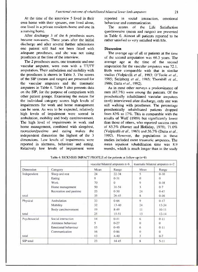
The 2 prostheses users, one traumatic and one vascular amputee, were men with a TT/TT amputation. Their ambulation and mobility with the prostheses is shown in Table 3. The scores of the SIP (means and ranges) are presented for the vascular amputees and the traumatic amputees in Table 4. Table 5 also presents data on the SIP, for the purpose of comparison with other patient groups. Examining the means for the individual category scores high levels of impairments for work and home management can be seen. As was to be expected, relatively high levels of impairment were scored in ambulation, mobility and body care/movement. The high level of impairments in work and home management, combined with sleep/rest, recreation/pastime and eating makes the independent dimension the highest of the 3 dimensions. Low levels of impairments were reported in alertness, behaviour and eating. Relatively low levels of impairment were
reported in social interaction, emotional behaviour and communication.
The scores of the Life Satisfaction questionnaire (mean and ranges) are presented in Table 6. Almost all patients reported to be rather satisfied to very satisfied with life.
Discussion The average age of all or patients at the time
of the second amputation was 66.3 years. The average age at the time of the second amputation for the vascular amputees was 72.1. Both were comparable with that in similar studies (Volpicelli et al., 1983; O'Toole et al., 1985; Steinberg et al., 1985; Thornhill et al., 1986; Datta et al., 1992).
As in most other surveys a predominance of men (67.7%) were among the patients. Of the prosthetically rehabilitated vascular amputees (n=6) interviewed after discharge, only one was still walking with prostheses. The percentage prosthetically rehabilitated patients dropped from 83% to 17%. This is comparable with the results of Wolf (1989) but significantly lower than those of others, who reported success rates of 83.3% (Hunter and Holliday, 1978), 71.0% (Volpicelli et al., 1983) and 56.7% (Datta et al., 1992). However, the populations in these studies included more traumatic amputees. The mean inpatient rehabilitation time was 8.9 months, which is much longer than in the study
Table 4: SICKNESS IMPACT PROFILE of the patients at follow-up (n=8)

22 A. De Frètes, A. M Boonstra and L. D. W. Vos
by Datta et al. (1992). The literature gives an average period of rehabilitation between 12 and 30 weeks (Kerstein et al., 1975; Sakuma et al. 1974; Van de Ven, 1981).
The mean Sickness Impact Profile scores of the bilateral lower limb amputees demonstrates that the consequences of amputation affect
almost all aspects of daily life. The high level of impairments in work was to be expected, because patients had already retired, because of disability or old age. The relatively high level of impairment in home management is comparable with that of stroke patients (Schuling et al., 1993) but much higher than that of patients with
Table 5: Comparison of the SICKNESS IMPACT PROFILE of bilateral amputees with other patient groups

Functional outcome of rehabilitated bilateral lower limb amputees 23
rheumatoid arthritis or chronic low back pain (CLBP) (see Table 5). The individual scores on the physical dimension were high compared to those for rheumatoid arthritis and CLBP patients. In contrast to stroke, rheumatoid arthritis and CLBP patients, bilateral amputees showed fewer impairments in the recreation and pastime categories. The mean score on the psychosocial dimension is comparable to that of the rheumatoid arthritis and stroke patients. The mean score of the total SIP is comparable to that of CLBP and stroke patients.
In the "quality of life" measure patients were found to be rather satisfied to very satisfied with life. The social service system, which for example, enables home adjustments, as well as good family circumstances and the absence of cognitive disabilities were probably reasons which contributed to this result.
A lot of questions remained unanswered. Further studies at different stages of
rehabilitation and subsequent supportive care will be needed to shed more light on the functional outcome of bilateral lower limb amputees.
Acknowledgements The authors thank Miss Heleen Nieborg for
her assistance with data collection.
Table 6: LIFE SATISFACTION of the amputees at follow-up (n=8)
R E F E R E N C E S
BERGNER M , BOBBITT R A , CARTER W B , GILSON B S (1981). The Sickness Impact Profile: development and final revision of a health status measure. Med Care 1 9 , 787-805.
BRODZKA WK, THORNHILL HL, ZARAPKAR SE, MALLY JA, WEISS L (1990). Long-term functions of persons with artherosclerotic bilateral below-knee amputation living in the inner-city. Arch Phys Med Rehabil 1990 7 1 , 895-900.
BROWN P W (1970). Rehabilitation of Bilateral Lower-Extremity Amputees. J Bone Joint Surg 5 2 A , 687-700.
BRUIN DE AF, WITTE DE LP, STEVENS F, DIEDERICKS JPM (1992). Sickness Impact Profile: the state of the art of a generic functional status measure. Soc Sci Med 3 5 , 1003-1014.
COUCH NP, DAVID JK, TILNEY NL, CRANE C (1977). Natural history of the leg amputee. Am J Surg 1 3 3 , 469-473.
DATTA D, NAIR P N , PAYNE J (1992). Outcome of prosthetic management of bilateral lower limb amputees. Disabil Rehabil 1 4 , 98-102.
DEYO RA (1986). Cpmparative validity of the sickness impact profile and shorter scales for functional assessment in low back pain. Spine 1 1 , 951-954.
EVANS WE, JAMES PH, HAYES JP, VERMILLAN BD (1987). Rehabilitation of the bilateral amputee. J Vasc Surg 5 , 589-593.
FOLLICK M J , SMITH TW, AHERN DK ( 1 9 8 5 ) . The sickness impact profile: a global measure of disability in chronic low back pain. Pain 2 1 , 67-76.
GRANGER C V , ALBRECHT G L , HAMILTON, BB (1979). Outcome of comprehensive medical rehabilitation: measurement by PULSES Profile and the Barthel Index. Arch Phys Med Rehabil 6 0 , 145-154.
HUNTER GA, HOLLIDAY P (1978). Review of function in bilateral lower limb amputees. Can J Surg 2 1 , 176-178.
KERSTEIN MD, ZIMMER H , DUGDALE FE, LERNER E (1975). Rehabilitation after bilateral lower-extremity amputation. Arch Phys Med Rehabil 5 6 , 309-311.

24 A De Frètes, A. M. Boonstra and L. D. W. Vos
MCCOLLOUGH N C , JENNINGS JJ, SARMIENTO A ( 1 9 7 2 ) . Bilateral below-the-knee amputation in patients over fifty years of age. J Bone Joint Surg 5 2 A , 1 2 1 7 - 1 2 2 3 .
MELKER DE R A , TOUW-OTTEN F, JACOBS HM, LUTTIK A ( 1 9 9 0 ) . De waarde van de "sickness impact profile " als uitkomstmeting. Ned Tijdschr Geneeskd 1 3 4 ( 1 9 ) , 9 4 6 - 9 4 8 .
MUECKE L , SHEKAR S , DWYER D , ISRAEL E, FLYNN JPG ( 1 9 9 2 ) . Functional screening of lower-limb amputees: a role in predicting rehabilitation outcome. Arch Phys Med Rehabil 73, 8 5 1 - 8 5 8 .
NISSEN SJ, NEWMAN WP ( 1 9 9 2 ) . Factors influencing reintegration to normal living after amputation. Arch Phys Med Rehabil 7 3 , 5 4 8 - 5 5 1 .
O'TOOLE DM, GOLDBERG RT, RYAN B ( 1 9 8 5 ) . Functional changes in vascular amputee patients: evaluation by Barthel index, PULSES Profile and ESCROW Scale. Arch Phys Med Rehabil 6 6 , 5 0 8 - 5 1 1 .
SAKUMA J, HINTERBUCHNER C, GREEN RF, SILBER M ( 1 9 7 4 ) . Rehabilitation of geriatric patients having bilateral lower extremity amputations. Arch Phys Med Rehabil 5 5 , 1 0 1 - 1 1 1 .
SCHULING J, GREIDANUS J, MEYBOOM-DE JONG B ( 1 9 9 3 ) . Measuring functional status of stroke patients with the Sickness Impact Profile. Disabil Rehabil 15, 1 9 - 2 3 .
STEINBERG FU, Sunwoo I, ROETTGER RF ( 1 9 8 5 ) . Prosthetic rehabilitation of geriatric amputee patients: a follow-up study. Arch Phys Med Rehabil 6 6 , 7 4 2 - 7 4 5 .
THORNHILL HL, JONGS J D , BRODZKA W, VANBOCKSTAGLE P ( 1 9 8 6 ) . Bilateral below-knee amputations: experience with 8 0 patients. Arch Phys Med Rehabil 6 7 , 1 5 9 - 1 6 3 .
VAN DE VEN C M C ( 1 9 8 1 ) . An investigation into the management of bilateral leg amputees. Br Med J 2 8 3 , 7 0 7 - 7 1 0 .
VIITANEN M , FUGL-MEYER KS, BERNSPANG B, FUGL-MEYER AR ( 1 9 8 0 ) . Life satisfaction in long-term survivors after stroke. Scand J Rehabil Med 2 0 , 1 7 - 2 4 .
VOLPICELLI LI, CHAMBERS RB, WAGNER FW ( 1 9 8 3 ) . Ambulation levels of bilateral lower extremity amputees. J Bone Joint Surg 6 5 A , 5 9 9 - 6 0 4 .
WITTE DE L ( 1 9 9 1 ) . After the rehabilitation centre: a study into the course of functioning after discharge from rehabilitation. — Amsterdam: Swets & Zeitlinger.
WOLF E , LILLING M , FERBER I, MARCUS J ( 1 9 8 9 ) . Prosthetic rehabilitation of elderly bilateral amputees. Int J Rehabil Res 1 2 , 2 7 1 - 2 7 8 .
Prosthetics and Orthotics International, 1994, 18, 25-33
Upper limb prosthetic use in Slovenia
H. BURGER and Č. MARINČEK
University Rehabilitation Institute Ljubljana, Slovenia
Abstract The article deals with the use of different types of upper limb prostheses in Slovenia.
Four hundred and fourteen upper limb amputees were sent a questionnaire on the type of their prosthesis, its use and reasons for non-use, respectively. The replies were subject to statistical analysis.
Most of the questioned upper limb amputees (70%) wear a prosthesis only for cosmesis. The use of a prosthesis depends on the level of upper limb amputation, loss of the dominant hand, and time from amputation. Prosthetic success appears to be unrelated to age at the time of amputation and the rehabilitation programme.
The most frequent reason for not wearing a prosthesis is heat and consequent sweating of the stump.
More than a third of amputees are dissatisfied with their prostheses.
Introduction Even though upper limb prostheses were
known already in ancient Egypt, they have never succeeded to completely substitute the three functions of the human hand - motor, sensibility and expressive functions. As an extension of the stump, a prosthesis should improve, not impede, its function. It should be even the best prosthesis is only a poor substitute.
used in the way the non-dominant hand is normally used (Hermansson, 1991). However,
As reported by various authors, an upper limb amputee accepts and uses the prosthesis if it is comfortable, functional and has a pleasing appearance. Acceptance and successful use also depend on the quality of the stump, amputation level, level of education and employment status of the user, time from amputation to fitting a first prosthesis, use of a temporary prosthesis, training in use, and patient's motivation (Millstein et al., 1986; Roeschlein and Domholdt 1989). Opinions on the importance of manual dexterity and loss of the dominant hand, respectively, differ considerably. Patients of the same age and with the same amputation level, who after completed rehabilitation and training achieved the same degree of independence, may use the prosthesis throughout the whole day or be reluctant to wear it.
According to different reports, upper limb prosthesis are used with different levels of success (Fletcher, 1970; Herberts et al., 1980; Stein and Walley, 1983; Heger et al., 1985; Vitali et al., 1986; Stürup et al., 1988; Roeschlein and Dumholdt, 1989).
According to Atkins (1989), a high rejection rate of upper limb prostheses can be attributed to development of one-handedness, insufficient training in use, poor comfort, poor construction, and the reactions of other people.
Purpose The objective of this study is to determine the
use and reasons for non-use of different types of upper limb prosthesis in Slovenia. The study also considered how amputees and their environment react to their prostheses.
All correspondence to be addressed to Dr Helena Burger, University Rehabilitation Institute Ljubljana, 61001 Ljubljana, Limbortova 51 , PO Box 381, Slovenia.
25

26 H Burger, C Marincek
Methods of work The dispensary files of all persons who
visited the Orthotic and Prosthetic Outpatient clinic at the University Rehabilitation Institute Ljubljana between 1 January, 1988 and 31 December, 1992 were surveyed. Upper limb amputees with a permanent residence in Slovenia were selected. They were sent a questionnaire on the extent to which they actually used their prostheses.
The responses were statistically analysed by the SPSS program (Statistical Package for Social Sciences). Data were analysed by frequency distribution, Chi-square test, variance analysis test, and Student's t-test.
Respondents In the last five years (1 January, 1988-
31 December, 1992), 414 upper limb amputees were examined in the Prosthetic and Orthotic Outpatient clinic of the University Rehabilitation Institute Ljubljana. Some came for an examination only once, whereas others came several times.
Some 295 responses (71.3%) were received. Of these, 266 (90.2%, representing 64.3% of the questionnaires sent) were filled in, and 29 were left blank (deceased, unknown, departed, etc).
Not everybody answered all questions. All the questions which were answered in these 266 responses were analysed.
At the time of amputation, the persons concerned were fairly young, aged 20.0 years on the average (SD 14.1 years). As many as 238 (of the 266 responses) or 89.5% of the amputations resulted from accidents (127 or 47.7% were war injuries).
More than half (163 or 61.3% of 266) had trans-radial amputations, 54 or 20.3% had trans-humeral amputations and 34 (12.8%) had partial hand amputations; disarticulations in joints were rare (3 or 1.1% had disarticulation at the shoulder, 2 or 0.8% at the elbow, and 10 or 3.7% at the wrist.
Some 245 persons answered the question about handedness before amputation, 132 (53.9%) lost the dominant hand, 85 (34.7%) the non-dominant hand. Twenty-three (9.4%) had a congenital deficiency of the upper limb and in 5 (2%) bilateral amputation had been performed at the same level.
Results and discussion The response rate to the questionnaire was
within normal values. As many as 63.5% (169) of responding upper
Table 1. Use of cosmetic and functional prostheses in daily work and statistically significant differences (p) obtained by Chi-square test.

Upper limb prosthetic use in Slovenia 21
limb amputees in Slovenia had a cosmetic prosthesis and no less than 69.5% (185) possess one single prosthesis. Considering body-powered prostheses, twice as many individuals had a prosthesis with a hand compared to those with a hook. Only 6 patients were provided with a myoelectric prosthesis.
Prosthetic use In Slovenia, upper limb amputees wear a
cosmetic prosthesis for about 10.2 hours and a functional (body powered or myoelectric) prosthesis for about 11.8 hours a day (no significant difference). This is a greater usage than reported by Millstein et al. (1986), according to whom amputees use a prosthesis with a cable operated hook about 8 hours a day, one with a cable operated hand 5 hours a day and a cosmetic prostheses 4 hours a day. However, what that author had in mind was use, whereas in this study the question was of wearing. Upper limb amputees in Slovenia make more extensive use of functional prostheses at housekeeping and farm work (p<0.001), at getting dressed and driving a car (p<0.5), and in personal hygiene (p<0.1) (Table 1). In other activities, prosthetic use is unaffected by the type of prosthesis.
The number of hours per day and the type of occupation in which a person uses a prosthesis depend on the following:
1. time from amputation to the first fitting of the prosthesis (Table 2) 2. amputation level (Tables 3 and 4) 3. age at amputation (Table 5) 4. present age (Table 6) 5. loss of the dominant hand (Table 7).
The time of daily prosthetic use is not related to:
1. the type of prosthesis 2. the evaluation of the rehabilitation programme.
Time from amputation to the first fitting of the prosthesis
The relation between the time after the amputation and the number of hours of prosthetic use per day is completely non-linear (it was established neither by correlation coefficient nor variance analysis test, but only by (Chi-square test). A most relevant point to be considered is whether the patient got his first prosthesis during the first year after the amputation or not (Table 2).
The answers also displayed that at work, all prostheses are more readily used by persons who got them soon after amputation (p<0.1), while in a social setting they are more readily used by those who got them later (p<0.1). Persons, provided with a functional prosthesis soon after the amputation, more often use it in attending to housekeeping (p<0.1), at work (p<0.01), and performing simple repairs at home and at peasant labour (p<0.05).
These findings correspond with the statements by Robinson et al. (1975), Jacobs and Brady (1975), and Bailey (1970) that one of the first reasons underlying unsuccessful prosthetic use is delayed post-surgical fitting in upper limb amputation. After the amputation, amputees quickly learn to compensate for their loss by using the other upper limb. This compensation is often so efficient that the patient finds the prosthesis encumbering and is reluctant to accept it.
Table 2. Influence of time since amputation to the first prosthetic fitting prosthetic use - statistical significance p = 0.5.
Table 3. Amputees with partial hand amputation use their prostheses (all) significantly less than others.

28 H. Burger, C. Marincek
Amputation level Persons with a partial amputation of the hand
wear the prosthesis the lowest number of hours, while there is no difference in daily wearing of prosthesis between trans-radial and trans-humeral amputees (p<0.05) (Table 3).
The level of amputation does not affect prosthetic use at work, in performing simpler home repairs and in peasant labour (Table 4). These occupations seem to be of greatest importance to those persons who are most keen to attend to them. In all other activities, persons with a trans-radial amputation wear their prosthesis more than others. The reason may be that the prostheses hampered others, which many put down as a remark or stated as a reason for their non-use. This corresponds with the conclusions by Millstein et al. (1986) that transradial amputees alone use functional prostheses in daily activities, whereas the others find prostheses useless in attending to daily activities. As reported by Stürup et al. (1988), higher level amputees do not use body powered prostheses, while Roeschlein and Domholdt
(1989) claims that the loss of the elbow is of no consequence to successful prosthetic use. Age at amputation
Functional prostheses are used for slightly longer periods of time by persons who were young at the time of amputation (r = 0.26, p<0.05), and cosmetic prostheses by persons now older (r = 0.30, p=0.001).
Persons using the prosthesis in getting dressed and eating (p<0.05) and in a social setting (p<0.01) were at amputation
Table 4: Influence of amputation level on prosthetic use and statistically significant differences (p) obtained by Chi-square test.
Table 5: Influence of age at amputation on prosthetic use obtained by variance analysis test and F values.

Upper limb prosthetic use in Slovenia 29
characteristically younger than those who do not use their prosthesis in the above-mentioned activities (Table 5). As for attending to other tasks, no significant differences in age at the time of amputation were observed with regard to successful or unsuccessful prosthetic use.
Present age The current age of amputees has even less
influence upon prosthetic use. It is worth noting that persons using a prosthesis at work are now a little younger (p<0.05) than persons who do not, while those using it at peasant work are slightly older (Table 6).
Loss of dominant hand All prostheses (p<0.05), particularly
functional (p<0.01), are worn mostly by amputees who have lost the non-dominant hand and least by bilateral upper limb amputees. The loss of the dominant hand is not significantly related to the time of wearing cosmetic prostheses.
The loss of the dominant hand has the most significant impact upon the use of the prosthesis at eating, work, and recreation, where the prosthesis is most used by bilateral and congenital amputees and more often by non-dominant than dominant hand amputees (Table 7).
The above conclusions are contrary to those drawn by Stürup et al. (1988), according to whom a prosthesis is more widely used by persons who sustained the loss of the dominant hand. Likewise, they do not agree with the statement by Roeschlein and Domholdt (1989) that the loss of the dominant hand exerts no influence upon successful use of a body powered prosthesis.
Most of the respondents failed to indicate at which occupations and recreational activities,
Table 6: Influence of present age on prosthetic use obtained by variance analysis test and F values.
Table 7: Influence of loss of dominant hand on prosthetic use and statistically significant differences (p) obtained by Chi-square test.

30 H. Burger, C Marincek
respectively, they used the prosthesis. Those who answered the question stated that they habitually made extensive use of functional prostheses for peasant work. More than a third of them use functional prostheses for all sorts of peasant labour. In recreational activities, preference is given to the use, or rather, wearing of cosmetic prostheses. From the point of view of different branches of sport (walking, jogging, cycling, skiing, football, gymnastics, bowling), prostheses are mostly irrelevant to function. The respondents most frequently used both types of prostheses at cycling. Sports such as referred to by Millstein et al. (1986) differ from those favoured in other countries where certain
branches of sport enjoy greater popularity than others.
The type of prosthesis The fact that the type of prosthesis is not
related to successful prosthetic use proves that in answering the question the respondents were not really referring to the number of hours per day during which they used the prosthesis but rather to how long they wore it. Thus, there seems no point in dividing persons into active, i.e. successful prosthetic users, and partially active and passive users, as proposed by Kejlaa (1992) and Roeschlein and Domholdt (1989).
Table 8. Reasons for non-use of cosmetic and functional prostheses; statistically significant differences (p), obtained by Chi-square test.
Table 9. Influence of amputation level on reasons for non-use of prostheses and significant differences (p), obtained by Chi-square test.

Upper limb prosthetic use in Slovenia 31
Evaluation of the rehabilitation programme The number of hours of prosthetic use does
not depend on the evaluation of the rehabilitation programme (either for all prostheses together or separately by types of prostheses). No interconnection was established either by the variance analysis test or Chi-square test. This means that there is no interconnection between the rehabilitation programme that the persons were subject to and the number of hours during which they use prostheses.
What stops amputees from using a prosthesis Table 8 refers to reasons that stop amputees
from using a prosthesis, displayed separately for different types of prosthesis. The most frequent reasons are heat and sweating, and only in the third place (17.3%) the loss of sensation, even though McDonnell et al. (1989) state that the prosthesis weakens the amputee's sense of position in space.
The most frequent reasons to stop using a
functional prosthesis are heat and sweating, and the loss of sensation. These two reasons are characteristically affected by the level of amputation (Table 9). Heat and sweating are most disturbing to persons with Trans-humeral amputation (p<0.01), and the loss of sensation to persons with a partial hand amputation (p<0.1).
The amputees, who stop using the prosthesis because of heat and sweating, are now younger (p<0.01) than the others, while other reasons for unsuccessful use are unrelated to age (Table 10).
Age at amputation has a significant influence only upon prosthetic non-use due to the weight of the prosthesis. The persons identifying the weight of the prosthesis as a reason for not using it, were significantly older at the time of amputation (p<0.01) (Table 11).
The persons who resist using the prosthesis due to loss of sensation, were fitted with their first prosthesis later after the amputation
Table 10. Influence of present age on non-use of the prosthesis due to heat and sweating obtained by variance
analysis test.
Table 11. Influence of age at amputation on non-use due to weight of the prosthesis obtained by variance analysis
test.
Fig. 1. Reactions in amputees' social milieu.
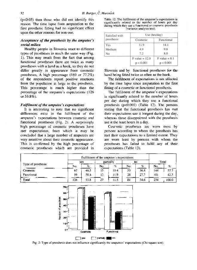
32 H. Burger, C Marincek
(p<0.05) than those who did not identify this reason. The time lapse from amputation to the first prosthetic fitting had no significant effect upon the other reasons for non-use.
Acceptance of the prosthesis by the amputee's social milieu
Healthy people in Slovenia react to different types of prostheses in much the same way (Fig. 1). This may result from the fact that among functional prostheses there are twice as many prostheses with a hand as a hook, so they do not differ greatly in appearance from cosmetic prostheses, A high percentage (169 or 77.2%) of the respondents report positive reactions from the population at large to the prosthesis. This percentage is much higher than the percentage of the amputee's expectations (126 or 53.8%).
Fulfilment of the amputee's expectations It is interesting to note that no significant
differences exist in the fulfilment of the amputee's expectations between cosmetic and functional prostheses (Fig. 2). A surprisingly high percentage of cosmetic prostheses have met expectations, from which it may be concluded that a large number of amputees are very sensitive about their cosmetic appearance. This is confirmed by the high percentage of cosmetic prostheses which are provided in
Slovenia and by functional prostheses for the hand being fitted twice as often as the hook.
The fulfilment of expectations is not affected by the time lapse since amputation to the first fitting of a cosmetic or functional prosthesis.
The fulfilment of the amputee's expectations is significantly related to the number of hours per day during which they use a functional prosthesis (p<0.001) (Table 12). The persons stating that the functional prosthesis has met their expectations use it longest during the day, whereas those disappointed with the prosthesis use it the least hours in a day.
Cosmetic prostheses are worn most by persons according to whom the prosthesis has met their expectations to a limited extent. They are worn least by persons with whom the prostheses has failed to fulfil any of their expectations (Table 13).
Table 12: The fulfilment of the amputee's expectations is significantly related to the number of hours per day during which they use a functional or cosmetic prosthesis
(variance analysis test).
Fig. 2: Type of prosthesis does not influence significantly the amputees' expectations (Chi-square test).

Upper limb prosthetic use in Slovenia 33
The fulfilment of the amputee's expectations is not significantly related to either the age at amputation or the current age.
The majority of persons with a cosmetic prosthesis stated that it had not met their expectations as it was merely cosmetic and unsuitable for work. Secondly the view was expressed that it is aesthetically deficient. The fact that the prosthesis is not applicable at work, being more of an encumbrance than aid, is another reason for non-use.
Surprisingly enough, a third of the respondents were of the opinion that a functional prosthesis, too, is just a cosmetic aid. The reason underlying this belief may be that these amputees were not included in a suitable rehabilitation programme. Another third of the respondents, fitted with a functional prosthesis, stated that the prostheses were poorly made, not strong enough, and unreliable. These are further reasons for unsuccessful prosthetic use.
Conclusion In the last few years, an obvious
improvement in the general attitude to prosthetic use can be observed in Slovenia, though the use of upper limb prostheses is far from satisfactory.
In Slovenia, the use of upper limb prostheses is related to factors which are not under human control (level of amputation, loss of the dominant hand, age at amputation) and also factors that can and must be controlled. Two most outstanding of these are: 1. time from amputation to the first prosthetic fitting 2. type of prosthesis.
Another area open to external control is a suitable rehabilitation programme. Its influence upon the use of functional prostheses however has not been confirmed by this study.
REFERENCES
ATKINS DJ (1989). Adult upper-limb prosthetic training. In: Atkins DJ, Meier RH. Comprehensive management of the upper-limb amputee. - New York: Springer-Verlag, p 39-59.
BAILEY RB (1970). An upper extremity prosthetic training arm. Am J Occup Ther 2 4 , 357-359.
FLETCHER I (1970). Upper limb amputations. Br J Hosp Med 4 , 590-595.
HEGER H , MILLSTEIN S , HUNTER GA (1985). Electrically powered prostheses for the adult with an upper limb amputation. J Bone Joint Surg 6 7 B , 278-281.
HERBERTS P, KoRNER L, CAINE K, WENSBY L (1980). Rehabilitation of unilateral below-elbow amputees with myoelectric prostheses. Scand J Rehabil Med 12, 123-128.
HERMANSSON LM (1991). Structured training of children fitted with myoelectric prostheses. Prosthet Orthot Int 1 5 , 88-92.
JACOBS R R , BRADY WM ( 1 9 7 5 ) . Early post surgical fitting in upper extremity amputations, J Trauma 1 5 , 966-968.
KEJLAA GH (1992). The social and economic outcome after upper limb amputation. Prosthet Orthot Int 1 6 , 25-31.
MCDONNELL PM, SCOTT R, DICKISON J, THERIAULT RA, WOOD B (1989). Do artificial limbs become part of the user? New evidence. J Rehabil Res Dev 2 6 , 17-24.
MILLSTEIN SG, HEGER H , HUNTER GA (1986). Prosthetic use in adult upper limb amputees: a comparison of the body powered and electrically powered prostheses. Prosthet Orthot Int 1 0 , 27-34.
ROBINSON KP, ANDREWS BG, VITALI M (1975). Immediate operative fitting of upper limb prosthesis at the time of amputation. Br J Surg 62, 634-637.
ROESCHLEIN RA, DOMHOLDT E (1989). Factors related to successful upper extremity prosthetic use. Prosthet Orthot Int 1 3 , 14-18.
STEIN RB, WALLEY M (1983). Functional comparison of upper extremity amputees using myoelectric and conventional prostheses. Arch Phys Med Rehabil 64, 243-248
STÜRUP J, THYREGOD HC, JENSEN JS, RETPEN JB, BoBERG G, RaSMUSSEN E, JENSEN S (1988). Traumatic amputation of the upper limb: the use of body-powered prostheses and employment consequences. Prosthet Orthot Int 1 2 , 50-52
VITALI M, ROBINSON KP, ANDREWS BG, HARRIS EE, REDHEAD RG (1986). Amputations and prostheses. l2nd edition - London: Bailliere Tindall. p 1-16, 100-118

Prosthetics and Orthotics International, 1994,18, 34-39
Treatment of congenital subluxation and dislocation of the hip by knee splint harness
M. F U K U S H I M A
Fukushima Orthopaedic Clinic, Hiroshima City, Japan
Abstract The results are reported from a study of 103 cases fitted with the knee splint harness (KSH) which is an orthotic device used for the treatment of congenital subluxation and dislocation of the hip. The knee splint harness consists of a harness attached to posterior plastic shells at the knee which prevent flexion beyond 90 degrees while permitting full and free knee extension. No case was encountered in a 10 year period which failed to reduce. In cases of hip subluxation, reduction was obtained within an average of 5.8 days. In cases of dislocation, reduction was obtained in 6.7 days on average. Follow-up roentgenograms, established that there were no cases presenting with any sign of ischaemia of the epiphysis.
Introduction Congenital dislocation of the hip has been
discussed among Japanese orthopaedic surgeons for many years, being one of the most commonly encountered conditions in Japan. The Lorenz method was introduced into Japan in 1920 and since then has proved a most reliable treatment method. It was considered that aseptic necrosis of the femoral head resulted from the rigid fixation of the hip in frog type plaster casts. The splint described by Von Rosen (1956) which was applied to infants below 2 months old was difficult for the parents to handle. The Pavlik (1957) harness has been
commonly used in Japan since introduced in 1957. In Japan, this harness design has been modified by different manufacturers. Some of these are however considered to be inadequate, especially those where the longitudinal straps are attached at the incorrect location at the knee which may cause knee contracture. The author has used the Pavlik harness since 1966 and occasionally reduction has been ' unsuccessful and contracture of the knee encountered. In these cases, the harness has been removed for a period of time and then reapplied with a successful result achieved in most cases. Reduction was most easily achieved in those cases where in addition to the harness a posterior knee splint (KS) was fitted to the affected side (series 1 - 2 0 cases). In this article we also report upon our experience in the use of the knee splint harness (KSH) (Fig. 1) and the treatment results obtained (series 2 - 8 3 cases).
Materials and Methods One hundred and three cases were examined
and treated at the department of Orthopaedic Surgery, Kure National Hospital and Fukushima Orthopaedic Clinic over the past 10 years.
Series 1 : Pavlik harness was applied coupled with a posterior knee splint (KS) to prevent more than 90 degrees of flexion of the knee (Fig. 2). The splint was applied at the same time as the Pavlik harness or subsequently when reduction was not achieved after a period of 3 weeks.
Series 2: Knee splint harness (KSH) was applied to 30 cases of subluxation and 53 cases of dislocation.
All correspondence to be addressed to M. Fukushima, Fukushima Orthopaedic Clinic Eisen Rakurakuen Bid. 3-22, 4-choume Rakurakuen, Saeki-ku, Hiroshima, Japan 731-51
34

Knee splinl harness 35
Methods At the initial patient visit, the hips were
examined by the following method. The left hip was held by the thumb and index fingers of the left hand and the knee joint held in 90 degrees of flexion by the right hand of the examiner. With the hip joint in a position of horizontal adduction, the femoral head was palpated posteriorly and with the hip joint in a position of horizontal abduction, the head was then palpated anteriorly. Compressive force was applied to the femoral head posteriorly to determine mobility of the head and check joint stability. The right hip was examined in a similar manner.
The classification of acetabular dysplasia and
dislocation according to Dunn (1976) is as follows: Grade 1: Acetabular dysplasia is minimal or zero, the femoral head is slightly subluxed by application of compressive force (subluxation of the hip). Grade 2: Dysplasia is moderate, the femoral head is displaced slightly or moderately as observed roentgenographically. Grade 3: Dysplasia is severe, the superior border of the femoral head is located above the level of the acetabulum as observed roentgenographically.
The KS is a plastic splint comprising posterior thigh and calf sections angled at 90 degrees at the knee joint (Fig. 2). The KSH is a combination of harness and splint. The harness is designed to permit knee flexion from 0 to 90 degrees and hip flexion from 90 degrees to the maximum.
Results Series 1
Nine cases (7 female, 2 male) were treated by Pavlik harness coupled with KS. Reduction was obtained in all cases in 2 to 14 days (averaging 6 days).
Eleven cases were treated initially with the Pavlik harness above, but in 6 of these cases reduction was not obtained after 3 to 12 weeks of treatment. Following the addition of the KS to the harness, reduction was achieved in all 6 cases within 3 weeks (averaging 8.8 days).
Fig 1. Knee splint harness (KSH) (left). The knee splint is composed of posterior plastic shells with circumferential retaining cuffs (right)
Fig 2. Pavlik harness with knee splint (KS) are applied to the left knee joint. The right knee is flexed at more than 90 degrees and the longitudinal strap is displaced posterior to the axis of the knee. The left knee is flexed at less than 90 degrees and the longitudinal strap is located
over the axis of the knee.
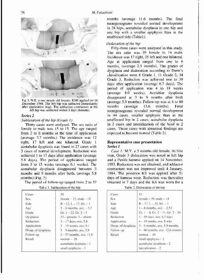
36 M Fukushima
Series 2 Subluxation of the hip (Grade 1 )
Thirty cases were analysed. The sex ratio of female to male was 15 to 15. TTie age ranged from 2 to 6 months at the time of application (average 3.7 months.) The incidence was 12 right, 17 left and one bilateral. Grade 1 acetabular dysplasia was found in 27 cases with 3 cases of normal development. Reduction was achieved 1 to 17 days after application (average 5.8 days). The period of application ranged from 3 to 15 weeks (average 6.1 weeks). The acetabular dysplasia disappeared between 3 months and 9 months after birth, (average 5.8 months) (Fig. 3).
The period of follow-up ranged from 2 to 57
months (average 11.6 months). The final roentgenograms revealed normal development in 28 hips, acetabular dysplasia in one hip and one hip with a smaller epiphysis than in the unaffected side (Table 1).
Dislocation of the hip Fifty-three cases were analysed in this study.
The sex ratio was 39 female to 14 male. Incidence was 17 right, 35 left and one bilateral. Age at application ranged from one to 6 months, (average 2.5 months). The grades of dysplasia and dislocation according to Dunn's classification were 8 Grade 1,11 Grade 2, 34 Grade 3. Reduction was achieved one to 39 days after application (average 6.7 days). The period of application was 4 to 18 weeks (average 8.0 weeks). Acetablar dysplasia disappeared at 3 to 9 months after birth (average 5.9 months). Follow-up was at 4 to 60 months (average 13.6 months). Final roentgenograms revealed normal development in 44 cases, smaller epiphysis than in the unaffected hip in 2 cases, acetabular dysplasia in 2 cases and lateralization of the head in 2 cases. Those cases with abnormal findings are expected to become normal (Table 2).
Representative case presentation Series 1
Case 1 : M.Y. a 3 months old female. At first visit, Grade 3 dislocation was noted in left hip and a Pavlik harness applied on 14 November, 1983. Reduction was not obtained, and adductor contracture was not improved until 4 January, 1984. The posterior KS was applied after 51 days of harness wear. Reduction was thereafter obtained in 7 days and the KS was worn for a
Fig 3. N.K. a one. month old female. KSH applied on 16 December 1988. The left hip was adducted immediately after application (top). The adduction contracture at the
left hip was corrected within 3 days (bottom).
Table 1. Subluxation of the hip Table 2. Dislocation of the hip

Knee splint harness 37
total of 14 days. The Pavlik harness was removed on 12 March 1984 and follow-up roentgenogram at 7 years old revealed a normal hip.
Case 2: O . T a 3 months old female, delivered by Caesarean section. Grade 3 dislocation in left hip. No reduction was obtained after 3 weeks application of the harness. Following application of the KS
reduction was obtained in 5 days. A roentgenogram at age 12 months revealed an almost normal hip (Fig. 4).
Series 2 Case 3: I.C. a 1 month and 3 weeks old
female. At fist examination, Ortolani 's click sign was evident. Roentgenogram revealed Grade 3 dyspasia and dislocation. The KSH was applied on 27 April 1990. On 2 May, reduction was obtained by abducting the left hip. On 9 May stable reduction was obtained and on 13 June 1990, the KSH was removed. The roentgenogram revealed a round epiphysis and disappearance of acetabular dysplasia.
Case 4: H.A. a 4 months old female. Grade 3 dysplasia and dislocation was noted in left hip on 13 September 1991. A Pavlik harness had been worn for 3 weeks at another clinic without reduction being achieved. One week later this same treatment was repeated unsuccessfully. Arthrography of the hip revealed inverted limbus. At time of first examination in our clinic a Pavlik harness was again fitted and the hip appeared to be reduced in the abducted position but posterior dislocation was induced by adduction. The KSH was applied on 7 February 1992 and stable reduction was obtained in a few days. The KSH was removed on 9 March. On 27 August 1992, the roentgenogram revealed that the epiphysis was round although smaller than that of the right side, and acetabular dysplasia was also improved without lateralization of the head.
Discussion In 1957, Pavlik reported a harness for the
treatment of congenital dislocation of the hip. This was introduced into Japan in the same year. It has been proved by many Japanese orthopaedic surgeons that the incidence of aseptic necrosis of the femoral head can be reduced in frequency by this method. Pavlik stated that femoral head necrosis was due to compressive forces exerted on the head by the action of the adductor muscles with circulatory disturbance in the head as a secondary problem. The mechanism of reduction applied by the Pavlik harness is to keep the hip in more than 90 degrees of flexion with the knee in full extension so that the hamstrings induce the femoral head to locate fully in the acetabulum
Fig 4. (Top) Series 1 T.T. a 4 month old female diagnosed as Grade 3 dislocation. Pavlik harness was applied on 14 September 1983.- Two weeks later reduction was still not obtained. On 27 September, knee splint was applied to the right knee. Three days later, reduction appeared to have been achieved but dislocation still occurred when the hip was adducted. The roentgenogram was taken on 14 September 1983 prior
to application of the harness. (Middle) Stable reduction was accomplished on the 17th day of knee splint application. Pavlik harness had been worn for 4 months and 1 week, the knee splint harness for 2 weeks. The roentgenogram was taken on
21 October, 1983. (Bottom) The follow-up roentgenogram at the age of 5
years revealed normal hip.

38 M Fukushima
by the action of the hip extensor mechanism. We, however, consider that the process of
reduction requires that the hip and knee are both kept in 90 degrees of flexion and that the abductive force applied to the hip by the weight of the legs relieves contracture of the adductors, and reduction is thereby induced.
According to Pavlik, the age of application ranged from 2 to 12 months. Takahashi (1985) reported age at the start of initial treatment ranging from 3 to 12 months. The spontaneous reduction rates he achieved were 60% in cases of hip dislocation and 97.9% in subluxation. Twenty-six out of 219 hips required manual reduction and 6 surgical reduction. Aseptic necrosis was observed in 11 joints (6.2%) and lateralization was observed in 21 joints (11.9%). It has been proved in the Series 1 group trial that keeping the knee in 90 degrees flexion or less brings the dislocated hip into reduction. The KSH keeps the hamstrings in tension by blocking knee flexion to more than 90 degrees and blocks hip flexion to less than 90 degrees. Therefore when the hip extends, the hamstrings become tightened and the hip is consequently induced into internal rotation by the strong medial hamstrings which rotate the hip medially. When the hip flexes, it rotates externally because the sartorius and biceps become tightened due to the prevention of more than 90 degrees of knee flexion (Fig. 5). The mechanism of reduction utilised in the early stages is mainly the correction of adduction contracture (Fig. 4). Iwasaki (1983) reported that when the hip was abducted by the harness the adductor muscles were then stretched by the weight of the lower limb. The KSH is more effective in correcting adductor contracture than the Pavlik harness because the former can be
applied at an earlier age and also keeps the hip in abduction. The Pavlik harness tends to hold the hip in a position of more than 90 degrees flexion and the knee in more than 90 degrees flexion. The KSH holds the hip and knee in 90 degrees of flexion so that the adductors are stretched more effectively. The KSH is precisely attached to the transverse axes of the knee so that it permits active knee extension thus preventing the development of knee flexion contractures (Fig. 6). Once the hip is reduced, stability of abduction and adduction, and internal and external rotation are provided by the KSH so that reduction is then maintained by tension of the hamstrings. We have applied the KSH to infants from the age of one month. Stockinette sleeves are used on the limbs to prevent skin problems and infants should also be bathed daily and the skin condition checked by the mother. We have not experienced knee
Fig. 5 KSH applied with stockinette sleeves. The left hip is flexed with abduction and external rotation and the right hip is extended with adduction and internal rotation.
Fig 6. Comparison between Pavlik harness (top) and KSH (bottom) immediately after application. Hip abduction is greater in KSH than in Pavlik harness in the same patient with left dislocation. The longitudinal straps are located posteriorly to the knee joint in Pavlik harness whereas on the KSH the longitudinal straps are located
over the axes of the knee.
Knee splint harness 39
flexion contracture problems. Complications with this harness were limited to mild dermatitis during the early phase of this study which was resolved by use of skin cream. Additionally no delayed reduction or aseptic necrosis of the femoral head has been encountered. No other treatment method or surgery has been required in the treatment of all such cases over the past 10 years. Eight cases have now been followed up for a period of more than 3 years, with no evidence of any Perthes-like change found roentgenographically.
Summary 1. The knee splint harness (KSH) has been used
for the treatment of 103 cases of hip disorders in children since 1983.
2. Age at time of application was one month and upward.
3. No instances of failed reduction occurred and no other method of treatment was required.
4. Reduct ion was obtained in all cases in one to 39 days, (average 7 days).
5. The KSH was used for the treatment of unstable hips which were difficult to keep reduced and which were easily dislocated by adduction of the hip.
6. On follow-up, no ischaemic change of the epiphysis was detected roentgenographically.
7. The period of application ranged from 2 to 3 months. When stable reduction was established collectively by dynamic hip examination, symmetrical abduction, loss of adductor contracture and strong extension of the affected hip, KSH should be removed.
8. There have been no complications encountered in the use of KSH except for mild skin rash.
R E F E R E N C E S
DUNN P M (1976). Perinatal observations on the etiology of congenital dislocation of the hip. Clin Orthop 1 1 9 , 1 1 - 2 2 .
IWASAKI K ( 1 9 8 3 ) . Treatment of congenital dislocation of the hip by the Pavlik harness. Mechanism of reduction and usage. J Bone Joint Surg 6 5 A, 7 6 0 - 7 6 7 .
PAVLIK ( 1 9 5 7 ) . The functional method of treatment using a harness with stirrups as the primary method of conservative therapy for infants with congenital displacement of the hip. Z Orthop 8 9 , 3 4 1 (Reprinted in Clin Orthop 1 9 9 2 , 2 8 1 , 4 - 1 0 ) .
ROSEN S VON ( 1 9 5 6 ) . Early diagnosis and treatment of congenital dislocation of the hip joint. Acta Orthop 2 6 , 1 3 6 - 1 5 5 .
TAKAHASHI I ( 1 9 8 5 ) . Functional treatment of congenital dislocation of the hip using Pavlik harness (Riemenbügel). J Jap Orthop Assoc 5 9 , 9 7 3 - 9 8 4 .

Prosthetics and Orthotics International, 1994,18,40-42
Technical note
A trans-femoral brim adapter for CAD CAM measurements
D. N I E L E N , E. D . L E M A I R E and L. G O U D R E A U
Prosthetics and Orthotics Department, The Rehabilitation Centre, Ottawa, Ontario, Canada
Abstract A computer aided design/computer aided manufacture (CAD CAM) brim measurement adapter was designed for use with a Berkeley casting stand. This measurement adapter accommodates all IPOS/CANFIT-PLUS trans-femoral brim sizes and shapes, allows brim positioning, provides adequate stability, and provides a substantial cost saving by using existing, functional hardware as a base.
Introduction The successful application of computer aided
design/computer aided manufacture (CAD CAM) technology to the production of prosthetic sockets is contingent on the acquisition of reliable input data (Engsberg et al., 1993). In the IPOS/CANFIT-PLUS trans-femoral system the input method involves selecting the appropriate brim for a patient, taking a series of circumference measurements along the stump, and entering the data into a computer. These measurements are used mathematically to produce an image of a quadrilateral or ischial containment socket on a computer screen for modification and/or carving. Since the final socket shape is based on brim dimensions and physical measurements from the patient, accurate measurements from the stump and appropriate brim selection are imperative.
Reliable and accurate CAD CAM measurements cannot be achieved without suitable, stable brim positioning. A brim support system must support the total body weight of the patient, conform to a variety of brim sizes and shapes, be adjustable to the patient, be stable and secure, be relatively inexpensive, and be simple to use and install. Failure to provide a stable brim support system will lead to erroneous measurements due to proximal/distal limb movement inside the brim and/or excessive brim distortion.
The success of the Berkeley casting frame, as well as the abundance of Berkeley stands in the prosthetic and orthotic community, makes this an ideal platform for a C A D CAM oriented measurement stand. A Berkeley casting frame is adjustable for height, has a high degree of stability, and is easy to set up and operate.
An adapter has been developed to integrate IPOS CAD C A M quadrilateral and ischial containment brims with the Berkeley casting stand. The adapter takes advantage of features present in the Berkeley base while providing the necessary options for successful brim selection, brim positioning, and measurement of the stump. This note will describe the design criteria, fabrication procedure, and operation of such a measurement jig.
Method Design criteria
The C A D C A M brim measurement adapter was designed based on the following criteria:
All correspondence to be addressed to Edward Lemaire, Prosthetics and Orthotics Department, The Rehabilitation Centre, 505 Smyth Road, Ottawa, Ontario, Canada K1H 8M2.
40
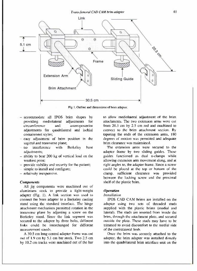
Trans-femoral CAD CAM brim adapter 41
- accommodate all IPOS brim shapes by providing mediolateral adjustments for circumference and anteroposterior adjustments for quadrilateral and ischial containment styles;
- easy adjustment of brim position in the sagittal and transverse plane;
- no interference with Berkeley base adjustments;
- ability to bear 200 kg of vertical load on the weakest point;
- provide stability and security for the patient; - simple to install and configure; - relatively inexpensive.
Components All jig components were machined out of
aluminium stock to provide a light-weight adapter (Fig. 1). A link section was used to connect the brim adapter to a Berkeley casting stand using the standard interface. The hinge attachment mechanism permitted rotation in the transverse plane by adjusting a screw on the Berkeley stand. Since the link segment was secured to the adapter by three bolts, different links could be interchanged for different measurement stands.
A 30.5 cm long central adapter frame was cut out of 1.9 cm by 5.1 cm bar stock. Two 2.5 cm by 10.2 cm tracks were machined out of the bar
to allow mediolateral adjustment of the brim attachments. The two extension arms were cut from 20.3 cm by 2.5 cm rod and machined to connect to the brim attachment section. By tapering the ends of the extension arms, 180 degrees of motion was permitted and adequate brim clearance was maintained.
The extension arms were secured to the adapter frame by two sliding guides. These guides functioned as dual u-clamps while allowing extension arm movement along, and at right angles to, the adapter frame. Since a screw could be placed at the top or bottom of the clamp, sufficient clearance was provided between the locking screw and the proximal shelf of the plastic brim.
Operation Installation
IPOS CAD C A M brims are installed on the adapter using two sets of threaded studs supplied with the plastic brims (medial and lateral). The studs are inserted from inside the brim, through the attachment plate, and secured outside the plate. These studs may have to be trimmed to avoid discomfort to the medial side of the contralateral limb.
Once the brim was securely attached to the adapter, the brim adapter was installed directly into the quadrilateral brim interface unit on the
Fig 1. Outline and dimensions of brim adapter.

42 D Nieten, E.D. Le maire and L. Goudreau
Berkeley stand using standard insertion and locking techniques. Since this unit was integrated into the existing attachment interface, the adapter could be used interchangeably with other Berkeley attachments (i.e. quadrilateral brim, casting rings, e t c ) .
Patient setup Before using this measurement system, the
clinician should pull one ply stockinette across the top of the brim. The patient should be oriented such that he/she faces away from the stand and is securely positioned between the parallel bar supports.
Once the patient is properly positioned, the brim adapter can be raised, using the Berkeley stand, until approximately 90% of the body weight is supported by the brim. Upon loading, brim deformation can be expected in the ischial region. The amount of deformation can be limited by reducing the amount of body-weight that is transferred to the brim.
Using the adapter and the casting stand, the brim can be positioned by adjusting for hip flexion, abduction/adduction, and/or rotation in the transverse plane. Abduction, adduction, and
hip flexion can be adjusted using original Berkeley components while transverse rotation can be performed using the jig outriggers. When the brim is in a satisfactory orientation, the clinician can take distal measurements or a cast from the residual limb as required and outlined by the CAD CAM system.
Maintenance Maintenance of the brim adapter is minimal.
If the adapter is kept clean (i.e. free of plaster build-up that inhibits free movement of the components), no lubrication or other regular maintenance is required.
Conclusion The IPOS brim adapter defined in this paper
provides an easy to use, low maintenance option for using existing casting hardware for CAD CAM orienled measurements. By changing the interface link section this adapter could be used with a variety of measurement stands.
Acknowledgements The authors wish to acknowledge the clinical
feedback of Seth Locke, BSc, CPO(c) and the administrative assistance of Guy Martel, MHSc, CPO(c).
Fig 2. Brim adapter mounted on a Berkeley stand..
Fig 3. Patient position during CAD CAM measurements,
REFERENCES
ENGSBERG JR, CLYNCH GS, LEE AG, ALLAN JS, HARDER JA (1992). A CAD CAM method for custom below-knee sockets. Prosthet Orthot Int 1 6 , 183-188
HANDLING INSTRUCTION FOR AK MEASUREMENT/CASTING STAND (1993). Article No. 05600. - Luneburg. Germany: IPOS Orthopadie Industriell.
IPOS-CANFIT-PLUS AK DESIGN SYSTEM MANUAL (1992). — Vancouver, Canada: Vorum Research Corporation.

Prosthetics and Orthotics International, 1994.18, 43-48
Technical note
How does vacuum forming affect Pelite mechanical properties?
J. E. S A N D E R S * and C. H. D A L Y * *
* Rehabilitation Medicine Department, University of Washington, Seattle, USA **Centre for Bioengineering, University of Washington, Seattle, USA
Abstract Pelite® is a polyethylene closed cell foam commonly used as an interface material in prosthetics. Both normal and vacuum-formed Pelite were tested under compression and under shear loading. For shear testing, the results were not significantly different for normal and vacuum-formed Pelite. For normal Pelite, the slope of the stress-strain curve was 1.17MPa (±0.14) while for vacuum-formed Pelite it was 1.24MPa (±0.22). Compressive results, however, were significantly different. Below 80kPa of applied compression, the slope of the stress-strain curve for normal Pelite was 0.99MPa (±0.11) while for vacuum formed Pelite it was 0.72MPa (±0.12). Between 80kPa and 200kPa of applied compression, the slope of the stress-strain curve for normal Pelite was 0.45MPa (±0.03) while for vacuum formed Pelite it was 0.55MPa (±0.05). Reasons for the differences and their significance to interface mechanics and computer-aided prosthesis design are discussed.
Introduction An amputee using a lower-limb prosthesis
typically wears a cushioning liner between the stump and the prosthetic socket. Pelite, which is an expanded closed cell polyethylene foam made up of units of hydrocarbon chains, is commonly used for this purpose. Typically used in 3mm or 5mm thickness, Pelite is well suited
for use with recently developed computer aided design/computer aided manufacturing (CAD CAM) prosthetic socket fabrication systems (Davies and Russell, 1979). It deforms plastically under heat and pressure to conform to the inside socket shape.
Clinical experience suggests that Pelite is altered by the vacuum forming process. A qualitative description is that it becomes thinner and more rigid. The change is important because the liner has less of a cushioning effect on the stump, i.e. the distribution of the normal and shear forces between the stump and socket is altered. In addition, inconsistencies in mechanical properties in different regions of the liner occur, and this further affects performance. At bony prominences, which require the socket to be curved to relieve interface stress, the Pelite becomes more thin and rigid than the surrounding regions of the liner.
In current clinical practice, the changes in Pelite thickness and stiffness as a result of vacuum forming are usually not planned or desirable. A prosthetist will typically overcome them by altering the liner after vacuum forming, for example placing an additional backing material on the outside of the liner. Alternatively more layers of Pelite could be added before vacuum forming to regions anticipated to become too thin.
In this technical note, changes in Pelite mechanical properties as a result of vacuum-forming are investigated. The purpose is to provide the initial work to determine quantitatively how the changes in Pelite mechanical properties from vacuum forming alter interface stresses. Finite element model
All correspondence to be addressed to J. E. Sanders, Center for Bioengineering, WD-12, University of Washington, Seattle, Washington 98195, USA.
43

•14 J E. Sanders and C. H. Daly
analysis of the stump, liner, and socket will be conducted subsequent to the investigations described here to evaluate interface stress sensitivity to the measured Pelite liner mechanical property changes. If important interface stress changes are found then subsequent work will investigate techniques to overcome the detrimental effects of vacuum-forming and instead alter the liner properties in a favourable way. In other words, we would like to control liner properties during fabrication so that favourable stump prosthetic socket interface stresses as opposed to unfavourable ones are created.
There are a limited number of studies in the rehabilitation literature investigating mechanical properties of interface materials. Most of them have been conducted on shoe insert materials which are similar to the prosthetic materials of interest here, however the stress levels in those studies were higher than those encountered by amputees using prosthetic limbs. Four studies are of relevance to the work in this technical note. Campbell et al. (1982) found that under compression some closed-cell foam interface materials had a three part load-deformation curve: a high initial slope, a moderate intermediate slope, then a high final slope. He called those materials "moderately deformable" and suggested they represented the best class of interface materials because the slope changes were gradual. Materials that have only two distinctly different slopes, low and high, are less optimal, and Campbell called those "very s t i f f " or "highly deformable" materials, depending on their moduli. Loading history effects on Pelite mechanical properties were also investigated. Campbell et al. (1984) found that after release from seven days of sustained loading at 50% strain. 1.6mm thick Pelite suffered 25% elastic strain, i.e. was only 7 5 % of its original thickness. After repetitive loading of 250,000 cycles at 294kPa, approximately 2 5 % elastic strain was found. But in another study where 5mm thick Pelite was loaded at 392kPa for 10,000 cycles at 1Hz, the elastic strain was only 2% (Brodsky et al., 1988). Thus the results suggest elastic strain is dependant on the number of cycles and initial thickness. Kuncir et al. (1990) suggested there are relationships between cell dimensions, uniformity, and reticulation and proposed such relationships are
of tremendous clinical relevance because these properties strongly affect the distribution of stress and the time dependent recovery characteristics of the materials. A more complete description of the theory of closed cell foams where quantitative relationships are described is presented by Gibson and Ashby (1988).
Despite recognition of the importance of liner properties and relationships between cell structure and liner properties, the effects of vacuum forming, a technique commonly used in prosthetics to fabricate a socket liner, on Pelite foam structure or mechanical properties has not been investigated. The purpose of this study was to compare normal and vacuum formed Pelite. Mechanical performance of normal and vacuum formed Pelite under compressive loading and under shear loading was evaluated. Loading magnitudes in the ranges of recent interface stress measurements on trans-tibial amputees (Sanders et al., 1993) were used. Subsequently, beyond the scope of this technical note, effects of the different properties on interface stresses will be investigated using a finite element model.
Methods Specimens
For testing of normal Pelite, sheets of 5 mm thickness of Pelite were obtained from a distributor (Durr-Fillauer Orthopaedic, Incorporated; Chattanooga, TN). For vacuum formed testing, flat specimens were constructed by fabricating parallel pipe plaster/cornstarch casts using computer aided software (Shapemaker®, Prosthetics Research Study, Seattle, Washington). Socket vacuum forming was conducted using a number 2 Pelite truncated cone with end pads. An oven temperature of 220°C and a vacuum pressure of 98kPa were used. Vacuum formed sheet thickness was 2.79mm (±0.27). Normal specimen thickness was 4.95mm (±0.21).
For compression testing, 2.54cm diameter disks were cut using a punch press. Specimens were taken from the central 80% of the width and length of a sheet to avoid thickness changes at the corners. For shear testing, 9.0mm x 9.0mm specimens were cut using a custom designed device that held two scalpel blades parallel during cutting. Specimens were square to within ±0.07mm tolerance. Specimens were
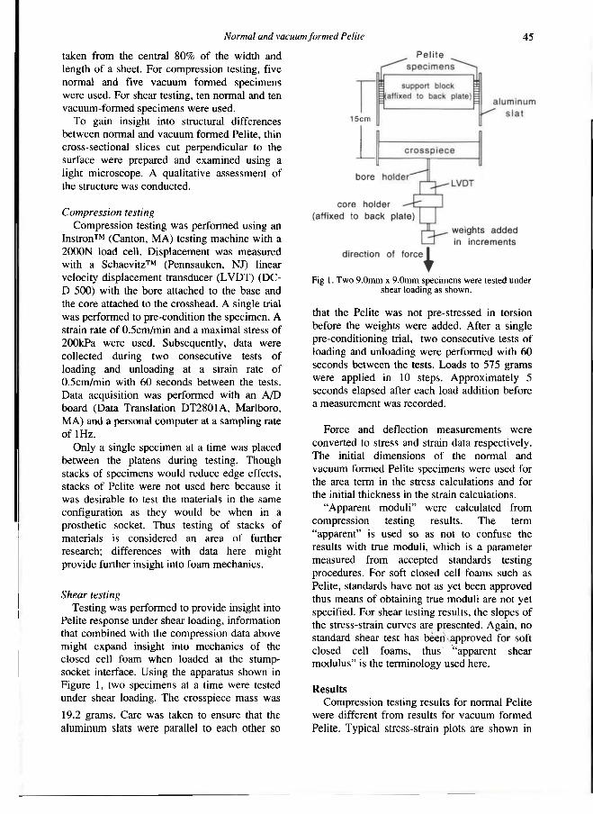
Normal and vacuum formed Pelite 45
taken from the central 80% of the width and length of a sheet. For compression testing, five normal and five vacuum formed specimens were used. For shear testing, ten normal and ten vacuum-formed specimens were used.
To gain insight into structural differences between normal and vacuum formed Pelite, thin cross-sectional slices cut perpendicular to the surface were prepared and examined using a light microscope. A qualitative assessment of the structure was conducted.
Compression testing Compression testing was performed using an
Instron™ (Canton, MA) testing machine with a 2000N load cell. Displacement was measured with a Schaevitz™ (Pennsauken, NJ) linear velocity displacement transducer (LVDT) (DC-D 500) with the bore attached to the base and the core attached to the crosshead. A single trial was performed to pre-condition the specimen. A strain rate of 0.5cm/min and a maximal stress of 200kPa were used. Subsequently, data were collected during two consecutive tests of loading and unloading at a strain rate of 0.5cm/min with 60 seconds between the tests. Data acquisition was performed with an A/D board (Data Translation DT2801A, Marlboro, MA) and a personal computer at a sampling rate of 1Hz.
Only a single specimen at a time was placed between the platens during testing. Though stacks of specimens would reduce edge effects, stacks of Pelite were not used here because it was desirable to test the materials in the same configuration as they would be when in a prosthetic socket. Thus testing of stacks of materials is considered an area of further research; differences with data here might provide further insight into foam mechanics.
Shear testing Testing was performed to provide insight into
Pelite response under shear loading, information that combined with the compression data above might expand insight into mechanics of the closed cell foam when loaded at the stump-socket interface. Using the apparatus shown in Figure 1, two specimens at a time were tested under shear loading. The crosspiece mass was
19.2 grams. Care was taken to ensure that the aluminum slats were parallel to each other so
that the Pelite was not pre-stressed in torsion before the weights were added. After a single pre-conditioning trial, two consecutive tests of loading and unloading were performed with 60 seconds between the tests. Loads to 575 grams were applied in 10 steps. Approximately 5 seconds elapsed after each load addition before a measurement was recorded.
Force and deflection measurements were converted to stress and strain data respectively. The initial dimensions of the normal and vacuum formed Pelite specimens were used for the area term in the stress calculations and for the initial thickness in the strain calculations.
"Apparent moduli" were calculated from compression testing results. The term "apparent" is used so as not to confuse the results with true moduli, which is a parameter measured from accepted standards testing procedures. For soft closed cell foams such as Pelite, standards have not as yet been approved thus means of obtaining true moduli are not yet specified. For shear testing results, the slopes of the stress-strain curves are presented. Again, no standard shear test has been approved for soft closed cell foams, thus "apparent shear modulus" is the terminology used here.
Results Compression testing results for normal Pelite
were different from results for vacuum formed Pelite. Typical stress-strain plots are shown in
Fig 1. Two 9.0mm x 9.0mm specimens were tested under shear loading as shown.

46 J E. Sanders and C. H. Daly
Figure 2. Two apparent moduli, which were linear least square fits to the data, were calculated for each trial, one for the range 0kPa to 80kPa and one for the range 80kPa to 200kPa. The basis for selection of 80kPa as the division point was this was the stress of the greatest slope change in the curves. Only the loading phase of each trial was considered in this analysis. Normal Pelite apparent modulus averaged 0.99 (±0.11) MPa at stresses between 0Kpa and 80kPa while it was 0.45 (±0.03) MPa at stresses between 80kPa and 200kPa. Vacuum formed Pelite apparent modulus was 0.72 (±0.12) MPa ait stresses less than 80kPa while it was 0.55 (±0.05) MPa at stresses between 80kPa and 200kPa (Table 1). The differences between normal and vacuum formed were significant (p<0.01) for both the 0kPa to 80kPa range and the: 80kPa to 200kPa range. Elastic strains after the second and third trials were minimal.
Shear test results were not significantly different for normal and vacuum formed specimens (Fig. 3). Apparent shear moduli were 1.17 (±0.14) MPa and 1.24 (±0.22) MPa respectively. Linear least square fits to the data were used to calculate the slopes. Elastic strains after the second and third trials were minimal.
Examination with a microscope showed that normal and vacuum formed Pelite had different structures. Cells on the surfaces of vacuum formed specimens were collapsed while those in the mid-region of the cross-section were circular or elliptical with their major axes parallel with the surface. Thus there was a "skin" of compressed cells on the top and bottom surfaces. Normal Pelite, however, showed rounded cells throughout with minimal or no "skins" on the surfaces.
Discussion In previous research, maximal stance phase
interfaces stresses on tibial flare, lateral, and posterior regions on trans-tibial amputees ranged from 46kPa to 205kPa for normal stress and 9kPa to 54kPa for shear stress (Sanders et al., 1993). On trans-femoral amputees normal stresses ranged from -6kPa to 345kPa (Appoldt et al., 1968). Thus results presented in this technical note show that within those ranges normal and vacuum formed Pelite demonstrated different mechanical properties.
The differences can be explained by alterations in the cellular structure. Vacuum formed Pelite as evidenced by its elliptical cell structure in the central region lost the integrity
Fig 2. Typical compression testing results for normal and vacuum formed Pelite.
Fig 3. Typical shear testing results for normal and vacuum formed Pelite.
Table 1 : Thicknesses and mechanical properties for normal and vacuum formed Pelite.

Normal and vacuum formed Pelite 47
of vertically oriented structural elements within the foam, resulting in reduced resistance at low loads. Thus the lower part (0kPa to 80kPa) of the compression testing stress-strain curve for vacuum formed Pelite had a lower apparent modulus than that for normal Pelite. At higher loads, however, the cells were collapsed thus loads were transferred across the cell walls. Thus at high loads (80kPa to 200kPa) the apparent modulus was higher than for normal Pelite. Shear testing results provide further insight into cell response to stress. The lack of a significant difference in apparent shear moduli for normal and vacuum formed Pelite but the presence of a difference in structure suggests that vacuum formed cells responded differently than normal cells. Vacuum formed Pelite had "skins" of collapsed cells on the top and bottom surfaces, and presumably those regions underwent minimal lateral deformation. The lack of deformation in the skins, however, was compensated for by excessive deformation in the elliptical cells, thus under shear loading the elliptical cells deformed more easily than the normal cells. This interpretation is consistent with the interpretation of compressive testing results described above. The vacuum formed Pelite had a weaker cell structure. To evaluate the above suggested hypotheses, cellular structural modelling, similar to that suggested by Kuncir et al. (1990) for body support interface materials and more formally described by Gibson and Ashby (1988) for foams in general, should be conducted.
The findings presented here are consistent with results presented in the literature. Campell et al. (1982) suggested that the slope differences for different regions of the stress-strain curves are of clinical relevance. Thus changes in the slopes of normal vs. vacuum formed Pelite might explain the clinically observed performance differences for normal vs. vacuum formed Pelite. In addition, literature results suggest that for a short number of load cycles (less than 10,000 cycles), elastic deformation is minimal, a finding supported by results presented here. Reports from the literature do suggest, however, that long-term loading does affect Pelite mechanical properties, thus further research should consider long-term loading effects on the results.
If subsequent investigation using finite element analysis demonstrates that stump-
socket interface stresses are highly sensitive to Pelite mechanical property changes encountered during vacuum forming, then an exciting concept is introduced to prosthetics. During liner fabrication, control of Pelite local properties might possibly be used to control interface stress distributions. This could be accomplished in a practical manner by altering the cell dimensions in the closed cell Pelite foam, possibly using local thermal control. Thus liners would be custom designed to the needs of the individual patient. The concept of variable mechanical property liners is currently practised clinically, but the methods are time consuming and impractical. A prosthetist typically cuts out a region of the liner and replaces it with a new material that is of more appropriate mechanical properties for that skin region. For example, nylon reinforced silicone is commonly placed at very sensitive soft tissue sites. However, clinical results are not necessarily always beneficial, in part because the edges at the interface of the two linear materials is a source of subsequent skin irritation. In addition, the fabrication processes are extremely time consuming. A liner that was continuous but with local changes in mechanical properties, for example a liner with locally variable micro-cell sizes, would be more optimal.
Acknowledgements Funding for this research was provided by the
Rehabilitation Research and Development Service of the Department of Veterans ' Affairs (John Goldschmidt MD, Director). Assistance from Prosthetics Research Study prosthetic staff in design and manufacture of the liners is gratefully acknowledged. Help from research engineer Richard Harrington of the Orthopaedics Biomechanics Laboratory at the University of Washington in performing compression tests is appreciated.
REFERENCES
APPOLDT FA, BENNETT L, CONTINI R ( 1 9 6 8 ) . Stump-socket pressure in lower extremity prostheses. J Biomech 1, 2 4 7 - 2 5 7 .
BRODSKY J W , KOUROSH S, STILLS M , MOONEY V ( 1 9 8 8 ) . Objective evaluation of insert material for diabetic and athletic footwear. Foot Ankle 9, 1 1 1 - 1 1 6 .

48 J. E. Sanders and C H. Daly
CAMPBELL GJ, MCLURE M, NEWELL EN (1984). Compressive behaviour after simulated service conditions of some foamed materials intended as orthotic shoe insoles. J Rehabil Res Dev 21,57-65.
CAMPBELL GJ, NEWELL EN, MCLURE M (1982). Compression testing of foamed plastics and rubbers for use at orthotic shoe insoles. Prosthet Orthot Int 6, 48-52.
DAVIES RM, RUSSELL D (1979). Vacuum formed thermoplastic sockets for prostheses. In: Disability./ edited by Kenedi RM, Paul JP, Hughes J. - London: MacMillan Press Ltd. p385-390.
GIBSON LJ, ASHBY MF ( 1 9 8 8 ) . Cellular solids: structure and properties. - Oxford: Pergamon Press.
KUNCIR EJ, WIRTA R W , Golbranson FL ( 1 9 9 0 ) . Load-bearing characteristics of polyethylene foam: an examination of structural and compression properties. J Rehabil Res Dev 27, 2 2 9 - 2 3 8 .
SANDERS JE, DALY CH, BURGESS EM ( 1 9 9 3 ) . Clinical measurement of normal and shear stresses on a trans-tibial stump: characteristics of waveform shapes during walking. Prosthet Orthot Int 17, 3 8 - 4 8 .

Prosthetics and Orthotics International, 1994, 18, 49-51
Technical note
New walking aid for primary mobilization of an infant with deficiency of all four limbs
S. B A L O G H , T. G Ö N C Z Y , R. B U J D O S Ó and L. K U L L M A N N
National Institute for Medical Rehabilitation, Budapest, Hungary
Abstract The authors report on a new device for the primary mobilization of a boy with severe deficiencies of all four limbs. This device allows independent mobility.
Introduction The supply with technical aids in the case of
loss of all four limbs is always difficult, especially in respect of primary mobilization. Studying the available literature, the authors found references published mainly in the thalidomide era. On first provision sockets were usually fixed to platforms on casters as by Aitken (1972) and Tippy et al. (1979) or to three wheeled carts, (Motloch and Elliot, 1966). Trunk swing, especially if the upper limbs are not deficient, may provide the means for a slow advancement. The children, as seen from photographs in the publication of the results, did not appear happy with the movement achieved since the motion needed great effort. The other form of provision was a socket fixed on platforms and bars (Marquardt and Martini, 1979; Radford and Steensma, 1957) to achieve an upright position but without mobility. Swivel walkers requiring rocking motions (Motloch and Elliot, 1966) on the other hand are usually not the first aids provided for mobilization. The aim of the authors was the supply to the child of
a device which would permit active communication.
Case history Szabolcs K., male, was born with serious
deficiencies of his four limbs. His upper limbs end between the upper and middle third of the humerus, the right side being somewhat longer. He has free shoulder joint movements. The left
Fig. 1. Szabolcs K with deficiency of four limbs.
All correspondence to be addressed to L. Kullmann, National Institute for Medical Rehabilitation, H-1528 Budapest, 123, P . O . B o x 1, Hungary
[<•)
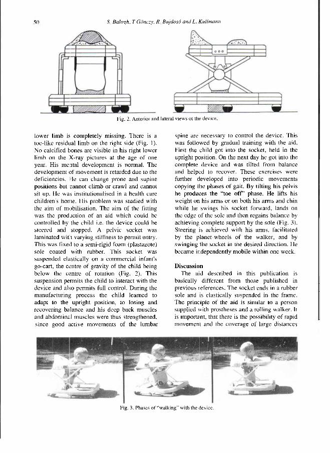
50 S. Bαlogh, T Gönczy, R Bujdosá and L, Kuttmann
lower limb is completely missing. There is a toe-like residual limb on the right side (Fig. 1). No calcified bones are visible in his right lower limb on the X-ray pictures at the age of one year. His mental development is normal. The development of movement is retarded due to the deficiencies. He can change prone and supine positions but cannot cl imb or crawl and cannot sit up. He was institutionalised in a health care children's home. His problem was studied with the aim of mobilisation. The aim of the fitting was the production of an aid which could be controlled by the child i.e. the device could be steered and stopped. A pelvic socket was laminated with varying stiffness to permit entry. This was fixed to a semi-rigid foam (plastazote) sole coated with rubber. This socket was suspended elastically on a commercial infant's go-cart, the centre of gravity of the child being below the centre of rotation (Fig. 2). This suspension permits the child to interact with the device and also permits full control. During the manufacturing process the child learned to adapt to the upright position, to losing and recovering balance and his deep back muscles and abdominal muscles were thus strengthened, since good active movements of the lumbar
spine are necessary to control the device. This was followed by gradual training with the aid. First the child got into the socket, held in the upright position. On the next day he got into the complete device and was tilted from balance and helped to recover. These exercises were further developed into periodic movements copying the phases of gait. By tilting his pelvis he produces the "toe o f f " phase. He lifts his weight on his arms or on both his arms and chin while he swings his socket forward, lands on the edge of the sole and then regains balance by achieving complete support by the sdle (Fig. 3). Steering is achieved with his arms, facilitated by the planet-wheels of the walker, and by swinging the socket in the desired direction. He became independently mobile within one week.
Discussion The aid described in this publication is
basically different from those published in previous references. The socket ends in a rubber sole and is elastically suspended in the frame. The principle of the aid is similar to a person supplied with prostheses and a rolling walker. It is important, that there is the possibility of rapid movement and the coverage of large distances
Fig. 2. Anterior and lateral views of the device.
Fig. 3. Phases of "walking" with the device.

New walker for deficiency of four limbs 5 1
with relatively little muscle work. The danger of accidents is very small. Finally it was possible to teach the use of the aid very quickly. Szabolcs was able to move around indoors safely, without hitting furniture, and became mobile outdoors too. This increased mobility had a remarkable effect on his mental development.
R E F E R E N C E S
AITKEN GT ( 1 9 7 2 ) . The child amputee: an overview. Orthop Clin North Am 3, 4 4 7 - 4 7 2 .
MARQUARDT E , MARTINI AK ( 1 9 7 9 ) . Orthesen und funktionsverbessemde operationen für kinder mit gliedmassenfehlbildungen der unteren extremitäten. Therapiewoche 29, 3 1 3 2 - 3 1 4 6 .
MOTLOCH WM, ELLIOT J ( 1 9 6 6 ) . Fitting and training children with swivel walkers. Artificial Limbs 10(2), 2 7 - 3 X .
RADFORD J, STEENSMA J ( 1 9 5 7 ) . The lower extremity toddler amputee: training procedures. Phys Ther Rev 37, 1-6.
TIPPY LV, KELLER KM, SCHOENECKER PL ( 1 9 7 9 ) . Management of a child with multiple congenital anomalies. ICIB 17(6), 1 1 - 1 6 .

Prosthetics and Orthotics International, 1994, 18, 52-53
Book Review
The manufacture and use of the functional foot orthosis. R. J. Anthony Karger, 1991. ISBN 3-8055-5298-X. 220pp. USD 158.50 GBP 86.10
In his preface the author states that the biomechanical discussions in the volume are "somewhat beyond the basic level" and this is a view that I would strongly endorse. The Introduction comprises an interesting review of the evolution of the "Root" functional orthosis which provides a valuable historical perspective on the development of this relatively new area of orthotic practice.
Chapter 1 reviews the indications for the use of functional foot orthoses and the biomechanical theory underlying their function. Some of this material is quite readily acceptable, however some is less so, indeed the author states that it is "not yet backed up by experimental and clinical research". In addition, as a bioengineer, I had some difficulty with the use of the term "compensation" here and in later chapters, to r≥fer to an abnormal position of the foot joints which is an involuntary consequence of an impairment of the structure or neuromuscular control of the foot, rather than as a result of any voluntary adjustment in alignment.
The following two' chapters deal with negative casting techniques. Five casting techniques are described and their respective advantages and disadvantages discussed. Finally the method of assessing the quality of the cast is described. This material is very detailed, however I found it relatively easy to follow due to the excellent quality of the many illustrations.
Orthosis fabrication is dealt with in Chapter 4 which commences with a rather clear explanation of the differences between the Root functional orthosis and the so-called "standard biomechanical orthosis". This is followed by a detailed description of the fabrication process
used for both designs, including such factors as negative and positive cast preparation, the manufacture of the orthosis shell, the application of posts and finishing procedures. Surprisingly, the author states that "the choice of fabrication process is up to the individual clinician and is based on an understanding of the prescription variations that exist and on the individual's comprehension of the concepts of clinical biomechanics and pathomechanics".
Chapter 5 deals with the subject of prescription writing. A valuable feature of this chapter is the description of the acquisition of the biomechanical data required to formulate an orthosis prescription.
The treatment of the "basic pathological foot types" is described in the following chapter. Foot conditions covered comprise forefoot invertus, forefoot evertus, rearfoot varus and rearfoot valgus and their various causes. This material also is extremely detailed and in contrast to previous chapters perhaps suffers from a lack of appropriate illustrations or figures.
Finally, Chapters 7, 8 and 9 describe the critique of the orthosis, fitting the orthosis and some common problems with orthotic therapy and their management.
This book is packed with information. Probably its strongest aspect is the detailed descriptions of the orthosis fabrication processes and, at least from my view point, the weakest its justification for the prescription criteria. I had considerable difficulty linking the Indications for the Use of Functional Orthosis in Chapter 1 with Prescription Writing in Chapter 5 and Treatment of the Basic Pathological Foot Types in Chapter 6.
This is very definitely a book for foot specialists and regrettably it highlights the problem of the terminological differences which exist between the podiatry profession and most other health care professions. I have no doubt that the functional foot orthosis is an extremely valuable treatment tool when used correctly.
52

Book Review 53
Unfortunately this book does little to help the non-expert understand better their place in the overall management of foot disorders. Finally, it emphasizes the need for objective evaluation of the many theories propounded in it.
David N. Condie Rehabilitation Engineer Dundee Limb Fitting Centre Dundee, Scotland
Proposed Amendment to the Constitution
The following amendment to ISPO's Constitution has been formulated by the Executive Board and will be discussed and voted on by the International Committee at its meeting which will be held in association with the World Congress in Melbourne, 2-7 April, 1995.
The proposed addition to the Constitution, Clause 4.5.10, formally recognises the role of International Consultants to the Executive Board.
Before the International Committee discusses this proposal, the Constitution requires it be published to the International Committee and Members and Fellows for comment. Any such comments should be received by the Honorary Secretary before 1 November, 1994.
4.5.10 The President, with the approval of the Executive Board, may appoint International Consultants with regard to identified tasks in relation to specific countries or geographical regions.

T
WORLD CONGRESS
MELBOURNE, A U S T R A L I A APRIL 2-7. 1995
INTERNATIONAL SOCIETY FOR PROSTHETICS AND
ORTHOTICS
MEET YOU IN MELBOURNE!
APRIL 2-7, 1995
8TH WORLD ISPO CONGRESS
Scientific Programme including: - Instructional Courses - Symposia - Expert Viewpoints - Free Papers - Posters and Videos
Commercial Exhibition
Social programme for registrants and accompanying persons that will introduce you and your family to the Australian way of life.
For information contact:
ICMS, 84 Queensbridge Street, South Melbourne, Victoria 3205, Australia
Telephone: +613 682 0244 Fax: +613 682 0288
54

ISPO Update Course on t ^ f e ^ Lower Limb Amputations and Related Prosthetics %jsiEEm
and Compound Fractures ^ş^E%j? 26th-30th September 1994
Ljubljana, Slovenia
Prosthetics and Orthotics -International, 1994, 18,55-56
Introduction The format of this fourth update course
following the Consensus Conference on Amputation Surgery, in Glasgow, October 1990 will be review lectures based on the presentations at the consensus conference and the points from the discussions leading to contemporary recommendations. Consideration will also be given to the treatment of compound fractures.
Ample time will be allocated for elucidation and discussion with the participants. The course is aimed at orthopaedic surgeons, general surgeons, rehabilitation specialists, prosthetists, orthopaedic technologists and other members of the amputation team, primarily from Central and Eastern Europe.
Venue The Institute for Rehabilitation of the
Republic of Slovenia, 61000 Ljubljana, Linhartova 51, Slovenia.
Course Fee The course fee is DEM 175 which covers
lectures, Consensus Conference Report, coffee and tea. Lunches can be provided at a subsidised cost
Name Male/Female (Delete as appropriate) Mailing address
Institution address
Tel: Fax: Telex: _ Profession
Accommodation Accommodation can be arranged at the
Holiday Inn at a cost of DEM 197 per night for single occupancy and DEM 265 per night for double occupancy; at the Hotel Grand Union or Hotel Slon at a cost of DEM 85 for single occupancy and DEM 122 for double occupancy; or at Hotel Lev at a cost of DEM 73 for single occupancy or DEM 98 for double occupancy. Prices include breakfast. Alternatively accommodation can be arranged in a hostel at a cost of DEM 50 for single occupancy and DEM 70 for double occupancy without breakfast.
Transport By plane to Ljubljana International Airport.
Bus transportation between the airport and Ljubljana is DEM 5 per person and a taxi is DEM 50
Sponsorship Prospective participants are encouraged to
seek sponsorship from local offices of such agencies as Ministry of Health, Ministry of Social Services, British Council, World Health Organisation and any other local agencies.
(continued overleaf)
I require Holiday Inn accommodation in: Hotel Grand Union Single Hotel Slon Double • Hotel Lev
Hostel Expected date of arrival Time Flight No. Expected date of departure Time Flight No. Registration Fee for Update Course DEM 175 Payment enclosed as bank draft Payment to be made on arrival 1 Date Signature
55

56 ISPO Update Course
Collaborators The course is organised in collaboration with
the World Health Organisation, Institute for Rehabilitation of the Republic of Slovenia, the Physical Medicine and Rehabilitation Association of Slovenia, World Orthopaedic Concern and the International Committee of the Red Cross.
Registration Form Mail or fax to Dr. J Steen Jensen, ISPO,
Borgervaenget 5, 2100 Copenhagen 0 , DENMARK. Fax No: (45) 31 18 16 69. Bank drafts should be made payable to ISPO.
Preliminary Programme Monday War injuries Compound Fractures
Tuesday Education, Training and Appropriate Technology NL WHO Viewpoint Amputation Surgery - General Considerations
Wednesday Basic Biomechanics Materials Clinic Team Approach Patient Care including Physiotherapy
Thursday Trans-femoral (Above-knee) Amputations and Prosthetics
Friday Knee Disarticulation Amputations and Prosthetics Trans-tibial (Below-knee) Amputations and Prosthetics
Further Information Should you require further information contact: Dr. J Steen Jensen, ISPO, Borgervaenget 5, 2100 Copenhagen 0 , DENMARK. Fax No: (45) 31 18 16 69.
Provisional Faculty Croatia: B. Lagerkvist (Doctor) Denmark: H. Hermanova (Doctor)
J. Steen Jensen (Surgeon) Germany: G. Neff (Surgeon) •Hungary: L. Kullman (Surgeon) Ireland: A. de Bont (Pros/Orthotist) Slovenia:
V y
C Marincek (Doctor) Sweden: R. Hammer (Surgeon)
B. Persson (Surgeon) Switzerland J. Gehrels (Pros/Orthotist) UK: R. Ham (Physiotherapist)
N. A. Jacobs (Bioengineer) A. Jain (Surgeon)
Local Organising Committee V V
Chairman: Crt Marincek Members: Franc Hočevar
Martin Stefančič
Please send application form to:
Dr. J. Steen Jensen ISPO Borgervaenget 5, 2100 Copenhagen 0 Denmark
Prosthetics and Orthotics International, 1994,18, 57-58
ISPO Update Course on Lower Limb Amputations and Related Prosthetics
and Compound Fractures 14th-18th November 1994
Panama City, Panama
t Introduction
The format of this fifth update course following the Consensus Conference on Amputation Surgery, in Glasgow, October 1990 will be review lectures based on the presentations at the consensus conference and the points from the discussions leading to contemporary recommendations. Consideration will also be given to the treatment of compound fractures.
Ample time will be allocated for elucidation and discussion with the participants. The course is aimed at orthopaedic surgeons, general surgeons, rehabilitation specialists, prosthetists, orthopaedic technologists and other members of the amputation team, primarily from Central and South America and the Carribean.
Venue The El Panama Hotel, Panama City, Panama.
Course Fee The course fee covers lectures, Consensus
Conference Report, coffee and tea and is as follows:
Members Non-members Specialist Doctors US$125 US$150 Residents US$100 US$125 Prosthetists/Therapists US$75 US$100 Medical Students US$65 US$75
Languages Simultaneous translation will be provided
between English and Spanish.
Accommodation Accommodation can be arranged at the El
Panama Hotel at a cost of US$95 per night.
Transport By plane to Panama City International
Airport. Local transportation is available between the airport and Panama City.
Sponsorship Prospective participants are encouraged to
seek sponsorship from local offices of such agencies as Ministry of Health, Ministry of Social Services. British Council, World Health Organisation and any other local agencies.
Collaborators The course is organised in collaboration with
the World Health Organisation, ACOPPRA, the Panamanian Society of Orthopaedic Surgeons, World Orthopaedic Concern and the International Committee of the Red Cross.
(continued overleaf)
Name Male/Female (Delete as appropriate) Mailing address
Institution address.
Tel: Telex:
Fax:
Profession _ ISPO Membership No. Signature
I require accommodation in El Panama
Expected date of arrival Time Flight No. Expected date of departure Time Flight No. Registration Fee for Update Course: Payment enclosed as bank draft Payment to be made on arrival Date
• •
57
58 ISPO Update Course
Registration Form Mail or fax to Dr. J Steen Jensen, ISPO,
Borgervaenget 5, 2100 Copenhagen 0 , DENMARK. Fax No: (45) 31 18 16 69. Bank drafts should be made payable to ISPO.
Preliminary Programme Monday War Environment in Developing Countries Compound Fractures
Tuesday Amputation Surgery — General Considerations Basic Biomechanics
Wednesday Trans-femoral (Above-knee) Amputations, Prosthetics and Physiotherapy Knee Disarticulation Amputations, Prosthetics and Physiotherapy
Thursday Trans-tihial (Below-knee) Amputations, Prosthetics, and Physiotherapy Ankle and Foot Amputations and Prosthetics
Friday Prosthetics supply in Central and South America and the Carribean ACCOPRA Annual Meeting.
Local Organising Committee G. Tejada (Public Relations) F. Cigarruista (Transport) A. Brown (Finance) A. Saldana (General Coordinator) R. Saez (International Coordinator) C. Nieto (Course Arrangements)
Provisional Faculty Denmark: J. Steen Jensen (Surgeon)
Panama: G. Pinilla (Surgeon) R. Saez (Pros/Orthotist) A. Saldana (Surgeon) G. Tejada (Surgeon)
UK: J. Hughes (Bioengineer) N. A.Jacobs (Bioengineer)
USA: J. Craig (Pros/Orthotist) R. Gailey (Physiotherapist) F. Gottschalk (Surgeon) J. Harris (Surgeon) M. Pinzur (Surgeon) M.Schuch (Pros/Orthotist) M. Stills (Orthotist) M. Quigley (Pros/Orthotist)
Further Information Should you require further information contact: Dr. J. Steen Jensen, ISPO, Borgervaenget 5, 2100 Copenhagen, DENMARK. Fax No: (45) 31 18 16 69.
Please send application form to:
Dr. J. Steen Jensen ISPO Borgervaenget 5, 2100 Copenhagen 0 Denmark

Prosthetics and Orthotics International, 1994. 18. 59-61
Calendar of Events
1-5 May, 1994 20th Canadian Medical and Biological Engineering Society Conference, Vancouver, Canada. Information: CMBEC Secretariat, c/o National Research Council, Room 393, Bldg. M-55, Ottawa, Ontario, K1A OR8, Canada.
31 May-2 June, 1994 Annual Meeting of International Medical Society of Paraplegia, Japan. Information: IMMSOP '94 Annual Meeting, Japan Organising Committee, Orthopaedic Dept. of Tokushima University, Kuramotocho, Tokushima-shi, 770, Japan.
31 May-3 June, 1994 8th World Congress of Orthopädie + Reha Technik International, Essen, Germany. Information: Verlag Orthopädie Technik, 4600 Dortmund 1, Reinoldestrasse 7-9, Postfach 10 06 51, Germany.
1-3 June, 1994 'Beyond Normalization Towards One Society For All', Reykjavik, Iceland. Information: 'Beyond Normalization', Iceland Tourist Bureau, Congress Dept., Skogarhlid 18, 101 Reykjavik, Iceland.
4-8 June, 1994 Annual Conference of the American Physical Therapy Association, Toronto, Canada. Information: APTA, 111 N. Fairfax Street, Alexandria, VA 22314, USA.
17-22 June, 1994 Annual Meeting of the Rehabilitation Engineering Society of North America, Nashville, USA. Information: RESNA '94, Suite 700, 1101 Connecticut Ave. NW, Washington DC 20036, USA.
21-24 June, 1994 10th Congress of the International Society of Electrophysiology and Kinesiology, Charleston, USA. Information: Dr. R. Shiavi, Biomedical Engineering, Box 6117, Station B, Vanderbilt University, Nashville, TN 47235, USA.
July, 1994 1 st International Rehabttitation Medicine Conference, Kuala Lumpur, Malaysia. Information: The Secretariat, Rehabilitation Unit, University Hospital, Lembah Pantai, 59100 Kuala Lumpur, Malaysia.
2-6 July, 1994 12th International Symposium of Biomechanics in Sport, Budapest, Hungary. Information: ISBS '94 Symposium Secretariat, Dept. of Biomechanics, Hungarian University of Physical Education, H-l 123 Budapest, Alkotas u.44, Hungary.
5-8 July, 1994 Dundee '94 - International Conference on Clinical Gait Analysis, Dundee, Scotland. Information: Dundee '94 Secretariat, Dundee Limb Fitting Centre, 133 Queen St., Broughty Ferry, Dundee DD5 1AG, Scotland.
59

60 Calendar of Events
10-12 July, 1994 Society for Research in Rehabilitation Summer Meeting, Nottingham, England. Information: Dr. N. Lincoln, Stroke Research Unit, General Hospital, Park Row, Nottingham, NG1 6HA, England.
10-15 July, 1994 2nd World Congress of Biomechanics, Amsterdam, The Netherlands. Information: Biomechanics Section, Institute of Orthopaedics, University of Nijmegen, PO Box 9101, 6500 HB Nijmegen, Netherlands.
20-26 August, 1994 17th International Conference on Medical and Biomedical Engineering, Rio de Janeiro, Brazil. Information: Dr. C. G. Orton, International Organization for Medical Physics, Gershenson Radiation Oncology Center, Harper-Grace Hospitals, 3990 John R., Detroit, MI 48201, USA.
26-27 August, 1994 1st International Symposium on the Lumbar Spine, Brussels, Belgium. Information: International Society for the Study of the Lumbar Spine, c/o Sunnybrook Medical Centre, Room A309, 2075 Batview, CDN Toronto, Ontario M4N 3M5, Canada.
4-9 September, 1994 6th European Regional Conference of Rehabilitation International, Budapest, Hungary. Information: Rehabilitation Secretariat, ISM Ltd., The Old Vicarage, Haley Hill, Halifax HX3 6DR, England.
21-23 September, 1994 Biological Engineering Society AGM and Annual Scientific Meeting, Keele, England. Information: Mrs. B. Freeman, BES, RCS, 35 Lincoln's Inn Fields, London, England.
28 September! October, 1994 International Meeting on Knee Prostheses, Naples, Italy. Information: Prof. V. Montelone, II Divisione Ortopedia e Trautnatologia, Ospedale Cardarelli, Via A. Cardarelli 9, 80131 Napoli, Italy.
9-13 October, 1994 6th Biennial Conference of the International Society for Augrnentive and Alternative Communications, Maastricht, The Netherlands.
Information: Van Namenand Westerlaken, PO Box 1558, 6501 BN Nijmegen, The Netherlands.
14-16 October, 1994 Biomedical Engineering Society Annual Fall Meeting, Tempe, Arizona, USA. Information: B VIES, PO Box 2399, Culver City, CA 90231, USA. 11-15 October, 1994 American Orthotic & Prosthetic Association: Annual National Assembly, Washington, USA. Information: AOPA, 1650 King St., Suite 500, Alexandria, VA 22314, USA.
7-10 December, 1994 8th International Conference on Biomedical Engineering, Singapore. Information: The Secretary, 8th ICBME 1994, Dept. of Orthopaedic Surgery, National University Hospital, Lower Kent Ridge Rd., Singapore 0511.

Calendar of Events 61
1995
17-18 February, 1995 ISPO (UK) Annual Scientific Meeting, Hull, England. Information: Mr. D. Simpson, ISPO Hull '95, NCTEPO, University of Strathclyde, 131 St. James' Rd., Glasgow G4 0LS, Scotland.
16-18 February, 1995 11th International Seating Symposium, Pittsburgh, USA. Information: Elaine Trefler, University of Pittsburgh Medical Centre, Dept. of Conference Management, Nese-Barkan Building, Suite 511, Pittsburgh, PA 15213, USA.
18-19 March, 1995 11th Annual Conference of the Association of Prosthetists and Orthotists, Telford, England. Information: Mr. W. Dykes, APO Conference Co-ordinator, NCTEPO, University of Strathclyde, Curran Building, 131 St. James' Rd., Glasgow G4 0LS, Scotland.
27-31 March, 1995 12th World Congress of the International Federation of Physical Medicine and Rehabilitation, Sydney, Australia. Information: IFPMR Congress Secretariat, DC Conferences, PO Box 629, Willoughby NSW 2068, Australia.
2-7 April, 1995 8th World Congress of the International Society for Prosthetics and Orthotics, Melbourne, Australia. Information: Congress Secretariat, 8th World Congress of the International Society for Prosthetics and Orthotics, 84 Queenbridge St., South Melbourne, Victoria, Australia 3205.
Summer, 1995 Medicon '95: 7th Mediterranean Meeting on Medical and Biological Engineering, Israel. Information: Prof. S. Siderman, c/o Prof. W. Welkowitz, Dept. of Electrical Engineering, PO Box 909, Piscataway, NJ 08854, USA.
16-19 July, 1995 7th International Conference on Mobility and Transport for Elderly and Disabled People, Reading, England. Information: 7th Int. Conf. Secretariat, Disability Unit, Dept. of Transport, Room S10/21, 2 Marsham Street, London SW1P 3EB, England.

Never before has there been a prosthetic knee quite like this one...
Total Knee. The TOTAL KNEE Geometric Locking System, by Century XXII Innovations, is based on a simple, yet totally new polycentric design concept that imitates true knee motion. This unique geometric design creates a locking moment, making it virtually impossible to collapse while in full extension during heel load.
An adjustable stance-flex feature acts as a shock absort>er, simulating the flexing action that occurs in the normal knee, while walking or running.
Assisted by a revolutionary hydraulic system that is a completely new approach to the entire subject of hydraulics, the TOTAL KNEE responds naturally to the patient's gait demands. Your patients will move effortlessly, and expend less energy, wearing the TOTAL KNEE!
C E N T U R Y X X I I INNOVATIONS INC.
517/782-1975 •• FAX 517/780-0404
P 0 BOX 868. JACKSON. Ml 49204. U.S.A.