protective effect of an extract from ascaris suum in ... filewe investigated the effect of an...
TRANSCRIPT

INFECTION AND IMMUNITY, June 2008, p. 2736–2745 Vol. 76, No. 60019-9567/08/$08.00�0 doi:10.1128/IAI.01085-07Copyright © 2008, American Society for Microbiology. All Rights Reserved.
Protective Effect of an Extract from Ascaris suum in ExperimentalArthritis Models�
Francisco Airton Castro Rocha,1* Ana Karine Rocha Melo Leite,1 Margarida Maria Lima Pompeu,2Thiago Mattar Cunha,3 Waldiceu A. Verri, Jr.,3 Fernanda Macedo Soares,4
Rondinelle Ribeiro Castro,1 and Fernando Queiroz Cunha3
Departments of Internal Medicine1 and Pathology,2 Faculty of Medicine, Federal University of Ceara, Fortaleza, Brazil;Department of Pharmacology, Faculty of Medicine, University of Sao Paulo, Ribeirao Preto, Sao Paulo, Brazil3; and
Department of Immunology, Instituto Butantan, Sao Paulo, Brazil4
Received 6 August 2007/Returned for modification 27 September 2007/Accepted 25 March 2008
We investigated the effect of an extract from a helminth (Ascaris suum) in zymosan-induced arthritis (ZYA)or collagen-induced arthritis (CIA). Rats and mice, respectively, received 1 mg and 0.1 mg zymosan intra-articularly (i.a.). Test groups received an A. suum extract either per os (p.o.) or intraperitoneally (i.p.) 30 minprior to i.a. zymosan. Controls received saline. Hypernociception was measured using the articular incapac-itation test. Cell influx, nitrite, and cytokine levels were assessed in joint exudates. The synovia and distalfemoral extremities were used for histopathology. Cartilage damage was assessed through determining gly-cosaminoglycan (GAG) content. DBA/1J mice were subjected to CIA. The test group received A. suum extracti.p. 1 day after CIA became clinically detectable. Clinical severity and hypernociception were assessed daily.Neutrophil influx was determined using myeloperoxidase activity. The A. suum extract, either i.p. or p.o.,significantly and dose-dependently inhibited cell influx and hypernociception in ZYA in addition to reducingGAG loss and ameliorating synovitis. The A. suum extract reduced i.a. levels of NO, interleukin-1� (IL-1�), andIL-10 but not tumor necrosis factor alpha (TNF-�) in rats subjected to ZYA while reducing i.a. IL-10, but notIL-1� or TNF-�, levels in mice. Clinically, mice subjected to CIA treated with the A. suum extract had lesssevere arthritis. Hypernociception, myeloperoxidase activity, and synovitis severity were significantly reduced.These data show that a helminth extract given p.o. protects from arthritis severity in two classical arthritismodels. This A. suum effect is species independent and functions orally and parenterally. The results showclinical and structural benefits when A. suum extract is given either prophylactically or therapeutically.
Genetic influences have been linked to disease susceptibilityand/or severity in autoimmunity (18). However, the relativelylow concordance rate in the incidence of these diseases amongtwins suggests that environmental stimuli play a role in thesemechanisms (18). In this context, microorganisms have beenlinked to the development of arthritis. One possible mecha-nism involves bacterium-derived antigens. After being pro-cessed in the gut and released into the circulation, these anti-gens would be entrapped in joints, where, after the activationof resident cells, an inflammatory response is triggered (33).
An increased incidence of allergic and autoimmune diseasesin highly industrialized countries has been associated with alower prevalence of infections (2). Less exposure to housedust, high ingestion of industrialized food, diminished breastfeeding, and increased vaccination would decrease stimulationto “real-life” germs. Also, the specific protection offered byvaccines would not appropriately prime the immune system,altering the T-cell repertoire (26). Less exposure to helminths,secondary to these lifestyle changes, has been linked to in-creased incidence of immune-mediated diseases (13). It wasproposed that a shift of the balance from a T-helper 1 (Th1)into a Th2 response provoked by fewer infections could explainthe increased prevalence of atopy and autoimmunity in wealthy
populations (2). More recent data showing that helminths ortheir products are able to alter both the function and formationof regulatory T cells added to the complexity of this issue (19).
It has been shown that delay in diagnosis, difficulties inaccess to a specialist, poor compliance with treatment, andfailure or delay in the indication of disease-modifying com-pounds impact the prevalence and severity of autoimmunediseases (24). Therefore, patients from low-income countriesshould display increased morbidity and/or mortality. However,the prevalence of rheumatoid arthritis is higher in developedcountries (1) than that in populations living in tropical climates(3, 17, 27, 38). Regarding severity, a recent report has shownthat systemic lupus erythematosus patients from the northeastpart of Brazil, where intestinal helminth infections are highlyprevalent, had similar organ damage to data reported fromdeveloped countries (38), despite the difficulties in access toappropriate treatment.
A role for helminths in the modulation of inflammation haslong been proposed (12). For example, Schistosoma worms ortheir components ameliorated experimental allergic encepha-lomyelitis, diabetes, and thyroiditis (12). Protozoa may also besimilarly involved, since viable Trypanosoma brucei decreasedinflammation in collagen-induced arthritis (CIA) (20). In ad-dition, ES-62, a glycoprotein derived from a filarial nematode,was shown to suppress disease severity in the CIA model inmice (21).
Considering these epidemiological data and the fact thathelminths may modulate immunoinflammatory reactions, we
* Corresponding author. Mailing address: Department of InternalMedicine, Faculty of Medicine, Federal University of Ceara, Fortaleza60115281, Brazil. Phone and fax: 55 85 32446215. E-mail: [email protected].
� Published ahead of print on 14 April 2008.
2736
on March 30, 2019 by guest
http://iai.asm.org/
Dow
nloaded from
investigated the effect of an Ascaris suum extract in experimen-tal arthritis. This extract was shown to display in vitro anti-inflammatory properties (29). Our present data reveal that theadministration of the A. suum extract provides a remarkableamelioration in clinical and structural parameters in two ar-thritis models, associated with decreased release of the inflam-matory mediators nitric oxide (NO), interleukin-1 (IL-1), andIL-10 into the joints of rats. The extract was equally effective inboth rats and mice when given intraperitoneally (i.p.) or per os(p.o.), both prophylactically and therapeutically. These dataare an experimental proof-of-concept study that oral stimula-tion by helminths influences the pathogenesis of autoimmunearthritis.
MATERIALS AND METHODS
Animals. Male Wistar rats (180 to 200 g) or Swiss mice (25 to 30 g) from ourown animal facilities were used throughout for the zymosan-induced arthritis(ZYA) experiments. CIA was induced in DBA/1J male mice. The experimentalprotocols were approved by the ethical review boards on animal experimentationat the Faculty of Medicine of the Federal University of Ceara and the Faculty ofMedicine of Ribeirao Preto, University of Sao Paulo—Ribeirao Preto, SaoPaulo, Brazil. For all experiments, we used six animals per group.
Induction of ZYA. Rats or mice were subjected to intra-articular (i.a.) injectionof either 1 mg (50 �l total volume) or 0.1 mg (25 �l total volume) of zymosan(Sigma, St. Louis, MO), respectively, dissolved in sterile saline, into the rightknee joints. Control groups received only saline i.a.
Induction and assessment of CIA. Male DBA/1J mice (12 to 14 weeks old)received 200 �g of bovine type II collagen (CII) intradermally in completeFreund’s adjuvant (day 0). A second injection of CII (200 �g in phosphate-buffered saline [PBS]) was given i.p. on day 21. Mice were monitored daily forsigns of arthritis, according to the following severity scores: 0, normal; 1, ery-thema; 2, erythema plus swelling; and 3, extension/loss of function. The total
score was the sum of four limbs. The number of arthritic paws was evaluateddaily, and mechanical hypernociception was assessed using the paw pressure test(9). Disease onset characterized by erythema and/or paw swelling was typicallyseen between days 25 and 35 after CII injection. Paw thickness was measuredwith a plethysmometer (Ugo Basile, Italy) before the CIA induction (Vo) anddaily starting after the beginning of the disease (VT). The difference between VT
and Vo was taken as the edema value, expressed in mm3.Evaluation of joint hypernociception. We used the rat knee joint incapacita-
tion test, as described previously (25). Briefly, after zymosan injection, rats weremade to walk on a steel rotary drum that rotates at 3 rpm. Specially designedmetal gaiters were wrapped around both hind paws. After placement of thegaiters, the animals were allowed to walk freely for habituation. The right pawwas connected to a microcomputer data input/output port. The paw elevationtime (PET) is the time during a 60-s period that the inflamed hind paw is not incontact with the cylinder. This is assumed to reflect joint hypernociception. ThePET was measured at baseline and then hourly, until sacrifice, at 6 h. Results (s/1min) are reported as the maximal PET between 3 and 4 h after injection of thezymosan.
Collection of synovial exudates and analysis of cell influx, cytokines, and NOlevels in the joint cavity. At 6 h after injection of the zymosan, the animals wereanesthetized with chloral hydrate (400 mg/kg i.p.), killed by cervical dislocation,and exsanguinated. The synovial cavity of the knee joints was then washed with0.4 ml (rats) or 0.1 ml (mice) saline containing 10 mM EDTA. The synovialexudates were collected by aspiration, and total cell counts were performed usinga Neubauer chamber. After centrifugation (500 g/10 min), the supernatants werestored at �70°C and used for determination of cytokine release. The concen-trations of tumor necrosis factor alpha (TNF-�), IL-1�, and IL-10 in synovialexudates obtained 6 h after zymosan injection into rats and mice were measuredby enzyme-linked immunosorbent assay (ELISA). Briefly, 96-well microtiterplates (Nunc Immunoplates) were coated overnight at 4°C with immunoaffinity-purified polyclonal antibodies against the respective cytokines. These antibodieswere provided by Steve Poole (National Institute for Biological Standards andControl, United Kingdom). After blocking the plates (1% albumin for 1 h),concentrations of cytokines and samples were loaded in duplicate for 2 h (22°C).A secondary rabbit biotinylated immunoaffinity-purified antibody was added,
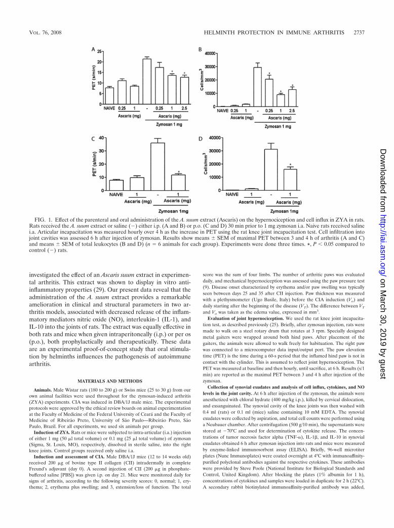
FIG. 1. Effect of the parenteral and oral administration of the A. suum extract (Ascaris) on the hypernociception and cell influx in ZYA in rats.Rats received the A. suum extract or saline (�) either i.p. (A and B) or p.o. (C and D) 30 min prior to 1 mg zymosan i.a. Naive rats received salinei.a. Articular incapacitation was measured hourly over 4 h as the increase in PET using the rat knee joint incapacitation test. Cell infiltration intojoint cavities was assessed 6 h after injection of zymosan. Results show means � SEM of maximal PET between 3 and 4 h of arthritis (A and C)and means � SEM of total leukocytes (B and D) (n � 6 animals for each group). Experiments were done three times. *, P � 0.05 compared tocontrol (�) rats.
VOL. 76, 2008 HELMINTH PROTECTION IN IMMUNE ARTHRITIS 2737
on March 30, 2019 by guest
http://iai.asm.org/
Dow
nloaded from
followed by incubation for 1 h (22°C). Finally, 100 �l of avidin-horseradishperoxidase (1:5,000 dilution; DAKO A/S, Denmark) was added to each well;after 30 min, the plates were washed and the color reagent o-phenylenediamine(40 �g/well) was added. After 15 min, the reaction was stopped with 1 M H2SO4
and the optical density was measured at 490 nm. Cytokine concentration wasexpressed as pg/ml. Total NO2
�/NO3�, as a measure of NO levels, was deter-
mined using a commercially available kit (Cayman Chemical Co.). Briefly, theNO3
� in the synovial exudate supernatants (0.08 ml) is converted to NO2� by
incubation with 0.01 ml nitrate reductase from Aspergillus species (1 U/ml) and0.01 ml NADPH (1 mM) for 30 min at 37°C. NO2
� levels were determinedspectrophotometrically at 540 nm by comparing the absorbance of 0.1 ml sampleafter adding 0.1 ml Griess reagent [1% (wt/vol) sulfanilic acid and 0.1% (wt/vol)N-(1-naphythyl) ethylenediamine in 5% phosphoric acid] to that of an NaNO2
standard.
Determination of neutrophil accumulation in the hind paws. In CIA, myelo-peroxidase (MPO) activity was used as an index of neutrophil accumulation injoint tissues, as previously described (6). Briefly, the mouse paws were collectedin 50 mM K2HPO4 buffer (pH 6.0) containing 0.5% hexadecyl trimethylammo-nium bromide and kept at �80°C until use. Samples were homogenized using aPolytron (PT3100) and centrifuged at 16,100 g for 4 min, and the resultingsupernatant was assayed spectrophotometrically for MPO activity determinationat 450 nm (Spectra max), with three readings in 1 min. The MPO activity ofsamples was compared to a standard curve with the MPO activity obtained froma known number of neutrophils collected from the peripheral blood of mice. Tenmicroliters of sample was mixed with 200 �l of 50 mM phosphate buffer (pH 6.0)containing 0.167 mg/ml O-dianisidine dihydrochloride and 0.0005% hydrogenperoxide. The results were expressed as the MPO activity that reflects thenumber of neutrophils/mg tissue.
Synovial histopathology. After sacrifice, the synovia were excised, fixed in 10%buffered formaldehyde, and processed for routine hematoxylin-eosin (HE) stain-ing. Semiquantitative histopathological grading was performed by an indepen-dent pathologist blinded to group allocation according to the presence of edema,synovial proliferation, cell infiltration, and necrosis, ranging from 0 to 3 (0,absent; 1, mild; 2, moderate; 3, severe), for each parameter. The maximal totalscore was 12. Results are expressed as the median (variation) value for eachgroup of six animals.
Assessment of articular cartilage damage. The glycosaminoglycan (GAG)content of the articular cartilage was determined as follows. The cartilage of thedistal femoral extremities was excised. The samples were weighed after dryingovernight at 80°C. The material was subjected to proteolysis using Prolav 750(Prozyn, SP, Brazil) and further precipitation in absolute ethanol, followed bydilution in distilled water. After this protocol, digestion with specific chondroiti-nases identified chondroitin sulfate as the sole GAG present in the samples. Thismaterial was separated by 0.6% agarose gel electrophoresis. After staining with0.1% toluidine blue, quantitation was made by densitometry (525 nm). Forcomparison, standards of chondroitin 4-sulfate, chondroitin 6-sulfate, and hepa-ran sulfate were subjected to the same protocol. Data were expressed as �gGAG/mg of dried cartilage.
Preparation of the A. suum extract. Adult A. suum worms were obtained fromthe small intestine of pigs from a local abattoir, Frigorıfico FISA, Itapecerica daSerra, Sao Paulo, Brazil, and prepared as described previously (31). Briefly,
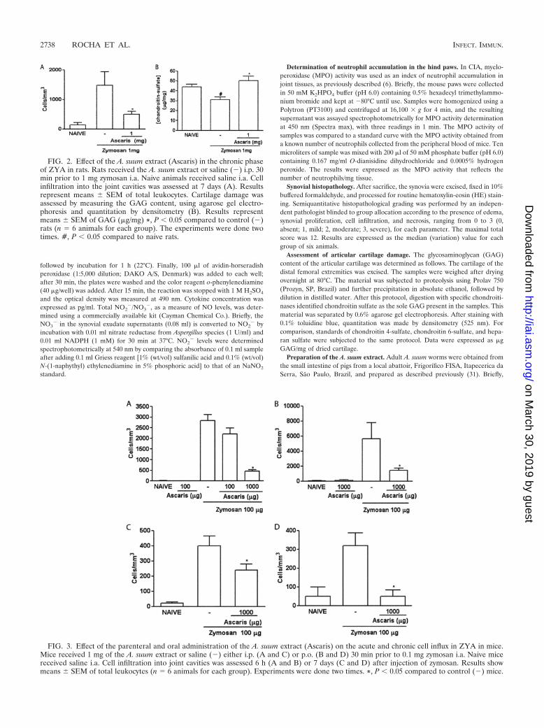
FIG. 2. Effect of the A. suum extract (Ascaris) in the chronic phaseof ZYA in rats. Rats received the A. suum extract or saline (�) i.p. 30min prior to 1 mg zymosan i.a. Naive animals received saline i.a. Cellinfiltration into the joint cavities was assessed at 7 days (A). Resultsrepresent means � SEM of total leukocytes. Cartilage damage wasassessed by measuring the GAG content, using agarose gel electro-phoresis and quantitation by densitometry (B). Results representmeans � SEM of GAG (�g/mg) *, P � 0.05 compared to control (�)rats (n � 6 animals for each group). The experiments were done twotimes. #, P � 0.05 compared to naive rats.
FIG. 3. Effect of the parenteral and oral administration of the A. suum extract (Ascaris) on the acute and chronic cell influx in ZYA in mice.Mice received 1 mg of the A. suum extract or saline (�) either i.p. (A and C) or p.o. (B and D) 30 min prior to 0.1 mg zymosan i.a. Naive micereceived saline i.a. Cell infiltration into joint cavities was assessed 6 h (A and B) or 7 days (C and D) after injection of zymosan. Results showmeans � SEM of total leukocytes (n � 6 animals for each group). Experiments were done two times. *, P � 0.05 compared to control (�) mice.
2738 ROCHA ET AL. INFECT. IMMUN.
on March 30, 2019 by guest
http://iai.asm.org/
Dow
nloaded from
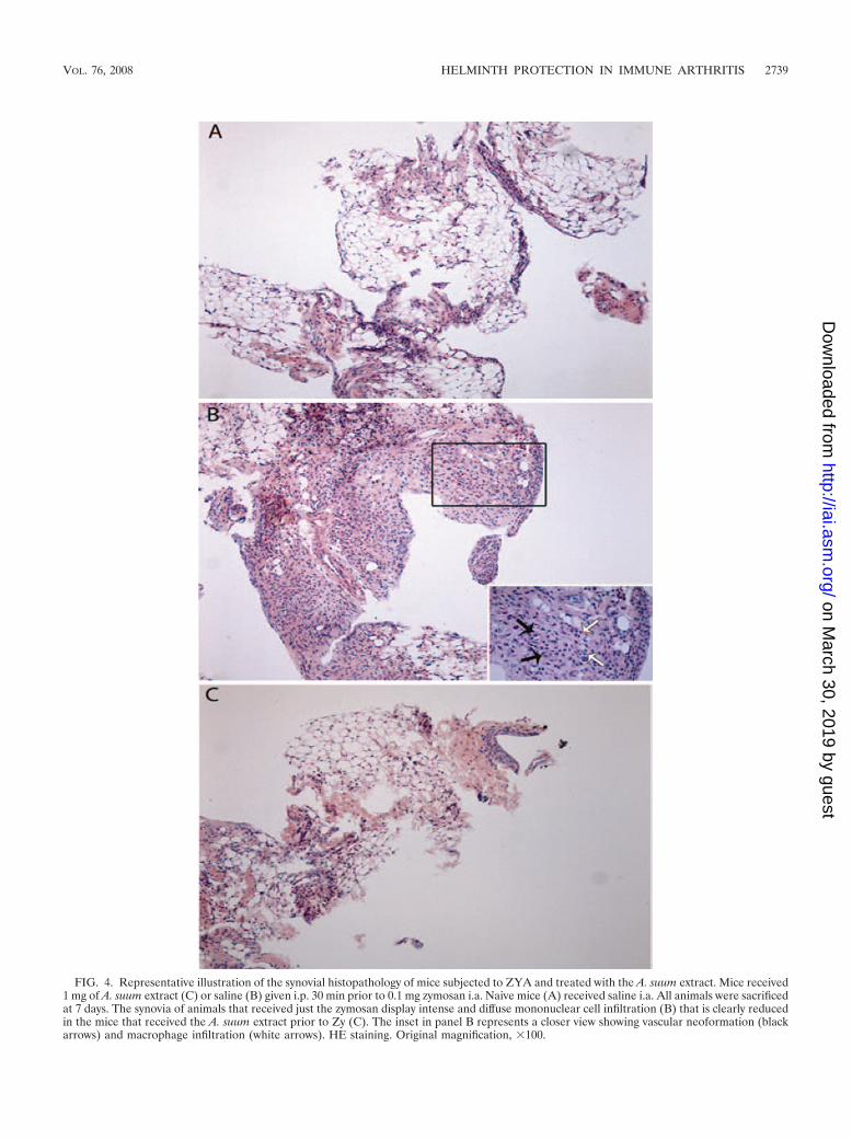
FIG. 4. Representative illustration of the synovial histopathology of mice subjected to ZYA and treated with the A. suum extract. Mice received1 mg of A. suum extract (C) or saline (B) given i.p. 30 min prior to 0.1 mg zymosan i.a. Naive mice (A) received saline i.a. All animals were sacrificedat 7 days. The synovia of animals that received just the zymosan display intense and diffuse mononuclear cell infiltration (B) that is clearly reducedin the mice that received the A. suum extract prior to Zy (C). The inset in panel B represents a closer view showing vascular neoformation (blackarrows) and macrophage infiltration (white arrows). HE staining. Original magnification, 100.
VOL. 76, 2008 HELMINTH PROTECTION IN IMMUNE ARTHRITIS 2739
on March 30, 2019 by guest
http://iai.asm.org/
Dow
nloaded from
worms were washed with saline, minced, and homogenized in PBS (pH 7.2). Thismaterial was then centrifuged (12,000 g for 90 min) at 10°C, and the super-natant was discarded. The precipitate was resuspended again in PBS, underagitation, for 18 h at 4°C. This material was centrifuged (12,000 g for 90 min)at 10°C, and the supernatant was dialyzed for 48 h against distilled water. Thematerial was then aliquoted, lyophilized, and stored at 4°C, until use.
Characterization of components in the A. suum extract. As an initial attemptto identify proteins present in the A. suum extract, dilutions were run on asilver-stained gradient (8 to 25%) polyacrylamide gel electrophoresis. The pres-ence of charged carbohydrates was investigated by separating the A. suum extractby 0.6% (wt/vol) agarose gel electrophoresis in diaminopropane acetate buffer(50 mM [pH 9.0]). This gel was stained with a 0.1% (wt/vol) toluidine bluesolution (5). For comparison, high- and low-molecular-weight standards (Phar-macia Biotech, United Kingdom) as well as standard chondroitin 4-sulfate andchondroitin 6-sulfate (Sigma, St. Louis, MO) were subjected to the same proto-cols, as indicated.
Treatments. Test groups received the A. suum extract, dissolved in sterilesaline, either i.p. or p.o. 30 min prior to the i.a. injection of zymosan. The amountof extract to be administered was based on protein content, using the Bradfordmethod. It has been shown that injection of 1 mg of A. suum extract was able tosuppress immune responses in mice (30). After obtaining a dose-response curvein rats, the concentration of 1 mg of protein was chosen to use for both rats andmice. The experiments done in the ZYA model were considered as prophylacticinterventions, whereas in the CIA, DBA/1J mice were treated with the A. suumextract i.p. or vehicle 1 day after CIA became clinically detectable. The lattergroup was considered as therapeutic. In order to test for the safety of theadministration of the A. suum extract, a group of animals received only theextract. The groups treated with the extract were compared to other groups thatreceived saline either i.p. or p.o. followed by the zymosan i.a. In order to rule outthe effect of an irrelevant protein, a group of animals subjected to the ZYAmodel received 1 mg bovine serum albumin (BSA), either i.p. or p.o.
Statistical analysis. Unless otherwise stated, results are presented as means �standard errors of the mean (SEM) or for histology as medians (range). Differ-ences between groups were analyzed using one-way analysis of variance followedby Tukeys test or Mann-Whitney test, as appropriate. In CIA experiments,two-way analysis of variance was performed. P � 0.05 was considered significant.
RESULTS
A. suum extract reduced cell influx and hypernociception inacute ZYA in rats. Control rats with ZYA displayed hyperno-ciception and intense cell influx into the joint cavity that wasmost prominent 6 h after injection of zymosan (25). Both thei.p. and oral administration of the A. suum extract significantlyand dose-dependently reduced hypernociception (Fig. 1A andC) as well as the acute (Fig. 1B and D) cell influx, compared tothat in vehicle-treated animals. The i.p. injection of 1 mg BSAdid not alter these parameters in rats subjected to ZYA (datanot shown). The effects achieved following the administrationof the A. suum extract were similar whether given p.o. or i.p.Oral administration of the A. suum extract to control (naıve)rats (Fig. 1A to D) did not alter hypernociception or cell influx.Additionally, 1 mg BSA given as an irrelevant protein eitherp.o. or i.p. did not alter hypernociception or cell influx (datanot shown). In order to exclude endotoxin contamination, weshould stress that both the extract and BSA were preparedunder sterile conditions and filtered before administration.Additionally, incubation of the A. suum extract with polymyxinB did not affect its inhibitory effect on ZYA hypernociception(data not shown). Moreover, the oral activity of the extract perse excludes the possibility of endotoxin contamination beingresponsible for the effects observed.
A. suum extract reduced chronic cell influx and preventedcartilage damage in ZYA in rats. Administration of the A.suum extract significantly reduced the cell influx into the jointsof rats, 7 days after injection of the zymosan (Fig. 2A). At thisphase, the numbers of cells are significantly lower, with mono-
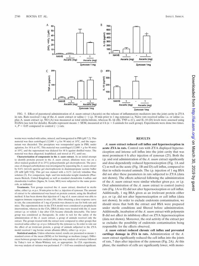
FIG. 5. Effect of parenteral administration of A. suum extract (Ascaris) on the release of inflammatory mediators into the joint cavity in ZYAin rats. Rats received 1 mg of the A. suum extract or saline (�) i.p. 30 min prior to 1 mg zymosan i.a. Naive rats received saline i.a. or saline i.a.plus A. suum extract i.p. NO (A) was measured as total nitrite/nitrate, whereas IL-1� (B), TNF-� (C), and IL-10 (D) levels were assessed usingELISA (see text for details). Results represent means � SEM, measured at 6 h (n � 6 animals for each group). Experiments were done two times.*, P � 0.05 compared to control (�) rats.
2740 ROCHA ET AL. INFECT. IMMUN.
on March 30, 2019 by guest
http://iai.asm.org/
Dow
nloaded from
nuclear cells being predominant (25). Additionally, the A.suum extract prevented the GAG loss that occurs in ZYA (Fig.2B), as compared to vehicle-treated animals.
A. suum extract reduced cell influx in ZYA in mice. Similarto what was observed in rats, either the i.p. or the oral admin-istration of the A. suum extract dose-dependently reduced cellinflux into the joint cavities of mice subjected to ZYA in boththe acute and chronic phases (Fig. 3A to D).
A. suum extract reduced the histopathology changes of thesynovia in ZYA in mice. Pretreatment with the extract (Fig.4C) almost completely abrogated the synovitis in rats subjectedto ZYA, provoking a marked decrease in the number of infil-trating cells, as compared to the group that received zymosanand saline i.p. (Fig. 4B). A naıve synovial sample is shown forcomparison (Fig. 4A). The semiquantitative analysis of thesesamples showed a significant reduction (P � 0.05) of thesynovitis in the group that received the A. suum extract (me-dian, 2; range, 1 to 2), as compared to saline-treated animals(median, 3.5; range, 2 to 5).
A. suum extract alters the release of inflammatory mediatorsin acute ZYA in rats. The A. suum extract significantly reducedNO as well as IL-1� levels released into the joints of ratssubjected to ZYA (Fig. 5A and B, respectively), whereas levelsof TNF-� (Fig. 5C) were not altered. We aimed to evaluate theeffect of the A. suum extract on the in vivo levels of IL-10 in theZYA model. However, levels of IL-10 were similar in the jointsof rats subjected to ZYA to those of naıve joints. Surprisingly,treatment with the A. suum extract reduced IL-10 levels tobelow detection limits both in naıve animals and in those sub-jected to ZYA (Fig. 5D).
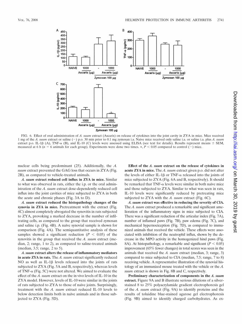
Effect of the A. suum extract on the release of cytokines inacute ZYA in mice. The A. suum extract given p.o. did not alterthe levels of either IL-1� or TNF-� released into the joints ofmice subjected to ZYA (Fig. 6A and B, respectively). It shouldbe remarked that TNF-� levels were similar in both naıve miceand those subjected to ZYA. Similar to what was seen in rats,IL-10 levels were significantly reduced by pretreating micesubjected to ZYA with the A. suum extract (Fig. 6C).
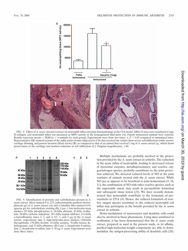
A. suum extract was effective in reducing the severity of CIA.The A. suum extract produced a remarkable and significant ame-lioration of the inflammatory signs in mice subjected to CIA.There was a significant reduction of the articular index (Fig. 7A),number of inflamed paws (Fig. 7B), paw edema (Fig. 7C), andmechanical hypernociception (Fig. 7D), as compared to immu-nized animals that received the vehicle. These effects were asso-ciated with inhibition of the neutrophil influx, shown by the de-crease in the MPO activity in the homogenized hind paws (Fig.8A). At histopathology, a remarkable and significant (P � 0.05)improvement (65% fewer changes) in total scores was seen in theanimals that received the A. suum extract (median, 3; range, 3)compared to mice subjected to CIA (median, 7.5; range, 7 to 8)receiving vehicle. A representative illustration of the synovial his-tology of an immunized mouse treated with the vehicle or the A.suum extract is shown in Fig. 8B and C, respectively.
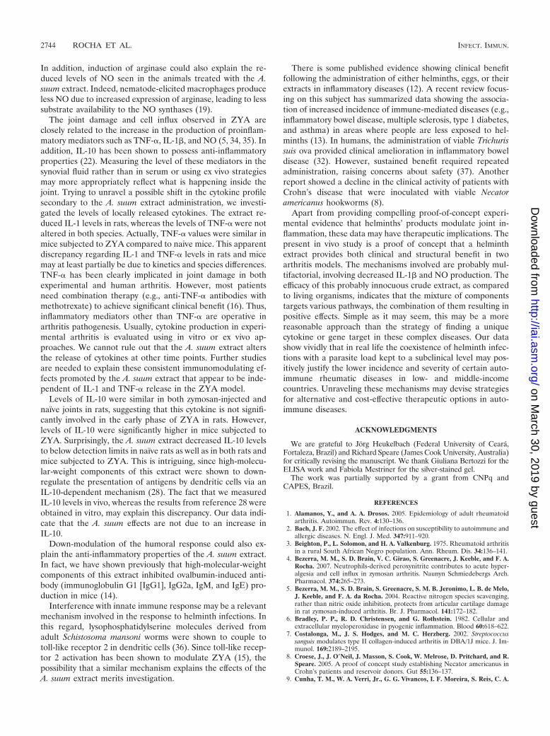
Preliminary characterization of components in the A. suumextract. Figure 9A and B illustrate serious dilutions of a silver-stained 8 to 25% polyacrylamide gradient electrophoresis gelof the A. suum extract (Fig. 9A) to identify proteins and theresults of toluidine blue-stained agarose gel electrophoresis(Fig. 9B) aimed to identify charged carbohydrates. As ex-
FIG. 6. Effect of oral administration of A. suum extract (Ascaris) on release of cytokines into the joint cavity in ZYA in mice. Mice received1 mg of the A. suum extract or saline (�) p.o. 30 min prior to 0.1 mg zymosan i.a. Naive mice received only saline i.a. or saline i.a. plus A. suumextract p.o. IL-1� (A), TNF-� (B), and IL-10 (C) levels were assessed using ELISA (see text for details). Results represent means � SEM,measured at 6 h (n � 6 animals for each group). Experiments were done two times. *, P � 0.05 compared to control (�) mice.
VOL. 76, 2008 HELMINTH PROTECTION IN IMMUNE ARTHRITIS 2741
on March 30, 2019 by guest
http://iai.asm.org/
Dow
nloaded from

pected, there were a wide range of both low- and high-molec-ular-weight proteins shown in the silver-stained gel. Additionalpurification was needed in order to try to identify active com-ponents implicated in the protective effects of the A. suumextract. On the other hand, the absence of charged carbohy-drates, as shown in lane 3 of Fig. 9B, virtually excludes suchcomponents as being responsible for the results achieved.
DISCUSSION
The present study is the first report that a helminth extract,given p.o., has in vivo anti-inflammatory properties in experi-mental arthritis. The reduction of hypernociception, number ofinflamed paws, and visual inflammatory score, assessed in twoarthritis models, indicate clinical benefit. Damage to the artic-ular cartilage using in vivo models can be assessed by directlyquantifying its GAG content, which is reduced in ZYA com-pared to naıve animals (5). Virtual abrogation of the chronicsynovitis, seen at histopathology, and prevention of damage tothe articular cartilage indicate a protective effect of the A.suum extract also at the structural level. Demonstration ofefficacy in different species and models is a crucial step in orderto prove the therapeutic value of a compound. The resultswere dose dependent and not species related, working similarly
in the acute and chronic phases and equally effective regardlessof oral or parenteral (i.p.) administration.
DBA/1J mice subjected to CIA display less-inflammatorychanges when pups are fed shortly postpartum with Strepto-coccus sanguis (7). Infection of rats with the protozoan respon-sible for African sleeping sickness (Trypanosoma brucei) par-tially prevented the development of collagen arthritis (20).More recently, it was shown that a secreted product from thenematode Acanthocheilonema vitae, named ES-62, comprisinga phosphorylcholine-containing glycoprotein, provided clinicaland histological amelioration of collagen arthritis in theDBA/1J mouse, while reducing the ex vivo production of theproinflammatory cytokines TNF-�, IL-6, as well as gammainterferon (21). Our present data add further information tothese previous studies in a number of ways. First, we havedemonstrated that the A. suum extract is active when admin-istered orally. Second, the A. suum extract reduced the hyper-nociception and synovitis both prophylactically and therapeu-tically, indicating intervention could be effective across a wideclinical spectrum. We may also speculate that phosphorylcho-line, present in the A. suum extract, similar to the moleculecontained in ES-62 (21), would be responsible for the anti-inflammatory effects observed in the present study.
FIG. 7. Effect of A. suum extract on clinical score, number of arthritic paws, edema, and hypernociception in the CIA model. DBA/1J mice weresensitized to type II collagen, and the arthritis was evaluated clinically using a clinical score (A), number of arthritic paws (B), hind paw edemameasured by plethysmometry (C), and hypernociception using the paw pressure test (D). Animals received 1 mg of the AS extract i.p. (�), 1 dayafter the arthritis was clinically apparent (arrow). Falsely immunized animals (Œ) were controls. Results represent means � SEM (n � 6 animalsfor each group). Experiments were done two times. *, P � 0.05 compared to immunized mice (f).
2742 ROCHA ET AL. INFECT. IMMUN.
on March 30, 2019 by guest
http://iai.asm.org/
Dow
nloaded from
Multiple mechanisms are probably involved in the protec-tion provided by the A. suum extract in arthritis. The reductionin the acute influx of neutrophils, leading to decreased releaseof lysosomal enzymes, metalloproteinases, and reactive oxy-gen/nitrogen species, probably contributes to the joint protec-tion achieved. We detected reduced levels of NO in the jointexudates of animals treated with the A. suum extract. WhileNO per se appears to be beneficial to joint homeostasis (5, 10,11), the combination of NO with other reactive species, such asthe superoxide anion, may result in peroxynitrite formationand subsequent tissue lesion (23). We have recently demon-strated that neutrophils contribute to the formation of per-oxynitrite in ZYA (4). Hence, the reduced formation of reac-tive oxygen species secondary to the reduced neutrophil cellinflux may participate in the benefit provided by the A. suumextract in arthritis.
Down-modulation of synoviocytes and dendritic cells couldalso be involved in these phenomena. Using mice sensitized toovalbumin, it has been demonstrated that the A. suum crudeextract, prepared as used in the present study, or its affinity-purified high-molecular-weight components are able to down-modulate the antigen-processing ability of dendritic cells (28).
FIG. 9. Identification of proteins and carbohydrates present in A.suum extract. Silver-stained 8 to 25% polyacrylamide gradient electro-phoresis gel of A. suum extract (A) and a toluidine blue-stained 0.6%agarose gel for carbohydrate staining (B). Lane 1, low-molecular-massmarkers (97-kDa phosphorylase b, 66-kDa albumin, 45-kDa ovalbu-min, 30-kDa carbonic anhydrase, 20.1-kDa trypsin inhibitor; 14.4-kDa�-lactoalbumin); lanes 2, 3, and 4, 0.5, 1, and 2 �g of the A. suumextract, respectively; lane 5, high-molecular-mass markers (220-kDathyroglobulin, 170-kDa ferritin, 116-kDa catalase, 76-kDa lactase de-hydrogenase, and 53-kDa albumin). (B) Lane 1, chondroitin-4-sulfate;lane 2, chondroitin-6-sulfate; lane 3, 50 �g A. suum. Experiments weredone three times.
FIG. 8. Effect of A. suum (Ascaris) extract on neutrophil influx and joint histopathology in the CIA model. DBA/1J mice were sensitized to typeII collagen, and neutrophil influx was measured as MPO activity in the homogenized hind paws (A). Falsely immunized animals were controls.Results represent means � SEM (n � 6 animals for each group). Experiments were done two times. *, P � 0.05 compared to immunized mice.Representative HE-stained sections of the ankle joints of mice subjected to CIA that received the vehicle show severe cell infiltration (white arrow),cartilage thinning, and pannus invasion (black arrow) (B), as compared to that of an animal that received 1 mg of A. suum extract i.p., which showspreservation of the cartilage and marked reduction in cell infiltration (C). Original magnification, 40.
VOL. 76, 2008 HELMINTH PROTECTION IN IMMUNE ARTHRITIS 2743
on March 30, 2019 by guest
http://iai.asm.org/
Dow
nloaded from
In addition, induction of arginase could also explain the re-duced levels of NO seen in the animals treated with the A.suum extract. Indeed, nematode-elicited macrophages produceless NO due to increased expression of arginase, leading to lesssubstrate availability to the NO synthases (19).
The joint damage and cell influx observed in ZYA areclosely related to the increase in the production of proinflam-matory mediators such as TNF-�, IL-1�, and NO (5, 34, 35). Inaddition, IL-10 has been shown to possess anti-inflammatoryproperties (22). Measuring the level of these mediators in thesynovial fluid rather than in serum or using ex vivo strategiesmay more appropriately reflect what is happening inside thejoint. Trying to unravel a possible shift in the cytokine profilesecondary to the A. suum extract administration, we investi-gated the levels of locally released cytokines. The extract re-duced IL-1 levels in rats, whereas the levels of TNF-� were notaltered in both species. Actually, TNF-� values were similar inmice subjected to ZYA compared to naive mice. This apparentdiscrepancy regarding IL-1 and TNF-� levels in rats and micemay at least partially be due to kinetics and species differences.TNF-� has been clearly implicated in joint damage in bothexperimental and human arthritis. However, most patientsneed combination therapy (e.g., anti-TNF-� antibodies withmethotrexate) to achieve significant clinical benefit (16). Thus,inflammatory mediators other than TNF-� are operative inarthritis pathogenesis. Usually, cytokine production in experi-mental arthritis is evaluated using in vitro or ex vivo ap-proaches. We cannot rule out that the A. suum extract altersthe release of cytokines at other time points. Further studiesare needed to explain these consistent immunomodulating ef-fects promoted by the A. suum extract that appear to be inde-pendent of IL-1 and TNF-� release in the ZYA model.
Levels of IL-10 were similar in both zymosan-injected andnaıve joints in rats, suggesting that this cytokine is not signifi-cantly involved in the early phase of ZYA in rats. However,levels of IL-10 were significantly higher in mice subjected toZYA. Surprisingly, the A. suum extract decreased IL-10 levelsto below detection limits in naıve rats as well as in both rats andmice subjected to ZYA. This is intriguing, since high-molecu-lar-weight components of this extract were shown to down-regulate the presentation of antigens by dendritic cells via anIL-10-dependent mechanism (28). The fact that we measuredIL-10 levels in vivo, whereas the results from reference 28 wereobtained in vitro, may explain this discrepancy. Our data indi-cate that the A. suum effects are not due to an increase inIL-10.
Down-modulation of the humoral response could also ex-plain the anti-inflammatory properties of the A. suum extract.In fact, we have shown previously that high-molecular-weightcomponents of this extract inhibited ovalbumin-induced anti-body (immunoglobulin G1 [IgG1], IgG2a, IgM, and IgE) pro-duction in mice (14).
Interference with innate immune response may be a relevantmechanism involved in the response to helminth infections. Inthis regard, lysophosphatidylserine molecules derived fromadult Schistosoma mansoni worms were shown to couple totoll-like receptor 2 in dendritic cells (36). Since toll-like recep-tor 2 activation has been shown to modulate ZYA (15), thepossibility that a similar mechanism explains the effects of theA. suum extract merits investigation.
There is some published evidence showing clinical benefitfollowing the administration of either helminths, eggs, or theirextracts in inflammatory diseases (12). A recent review focus-ing on this subject has summarized data showing the associa-tion of increased incidence of immune-mediated diseases (e.g.,inflammatory bowel disease, multiple sclerosis, type 1 diabetes,and asthma) in areas where people are less exposed to hel-minths (13). In humans, the administration of viable Trichurissuis ova provided clinical amelioration in inflammatory boweldisease (32). However, sustained benefit required repeatedadministration, raising concerns about safety (37). Anotherreport showed a decline in the clinical activity of patients withCrohn’s disease that were inoculated with viable Necatoramericanus hookworms (8).
Apart from providing compelling proof-of-concept experi-mental evidence that helminths’ products modulate joint in-flammation, these data may have therapeutic implications. Thepresent in vivo study is a proof of concept that a helminthextract provides both clinical and structural benefit in twoarthritis models. The mechanisms involved are probably mul-tifactorial, involving decreased IL-1� and NO production. Theefficacy of this probably innocuous crude extract, as comparedto living organisms, indicates that the mixture of componentstargets various pathways, the combination of them resulting inpositive effects. Simple as it may seem, this may be a morereasonable approach than the strategy of finding a uniquecytokine or gene target in these complex diseases. Our datashow vividly that in real life the coexistence of helminth infec-tions with a parasite load kept to a subclinical level may pos-itively justify the lower incidence and severity of certain auto-immune rheumatic diseases in low- and middle-incomecountries. Unraveling these mechanisms may devise strategiesfor alternative and cost-effective therapeutic options in auto-immune diseases.
ACKNOWLEDGMENTS
We are grateful to Jorg Heukelbach (Federal University of Ceara,Fortaleza, Brazil) and Richard Speare (James Cook University, Australia)for critically revising the manuscript. We thank Giuliana Bertozzi for theELISA work and Fabiola Mestriner for the silver-stained gel.
The work was partially supported by a grant from CNPq andCAPES, Brazil.
REFERENCES
1. Alamanos, Y., and A. A. Drosos. 2005. Epidemiology of adult rheumatoidarthritis. Autoimmun. Rev. 4:130–136.
2. Bach, J. F. 2002. The effect of infections on susceptibility to autoimmune andallergic diseases. N. Engl. J. Med. 347:911–920.
3. Beighton, P., L. Solomon, and H. A. Valkenburg. 1975. Rheumatoid arthritisin a rural South African Negro population. Ann. Rheum. Dis. 34:136–141.
4. Bezerra, M. M., S. D. Brain, V. C. Girao, S. Greenacre, J. Keeble, and F. A.Rocha. 2007. Neutrophils-derived peroxynitrite contributes to acute hyper-algesia and cell influx in zymosan arthritis. Naunyn Schmiedebergs Arch.Pharmacol. 374:265–273.
5. Bezerra, M. M., S. D. Brain, S. Greenacre, S. M. B. Jeronimo, L. B. de Melo,J. Keeble, and F. A. da Rocha. 2004. Reactive nitrogen species scavenging,rather than nitric oxide inhibition, protects from articular cartilage damagein rat zymosan-induced arthritis. Br. J. Pharmacol. 141:172–182.
6. Bradley, P. P., R. D. Christensen, and G. Rothstein. 1982. Cellular andextracellular myeloperoxidase in pyogenic inflammation. Blood 60:618–622.
7. Costalonga, M., J. S. Hodges, and M. C. Herzberg. 2002. Streptococcussanguis modulates type II collagen-induced arthritis in DBA/1J mice. J. Im-munol. 169:2189–2195.
8. Croese, J., J. O�Neil, J. Masson, S. Cook, W. Melrose, D. Pritchard, and R.Speare. 2005. A proof of concept study establishing Necator americanus inCrohn’s patients and reservoir donors. Gut 55:136–137.
9. Cunha, T. M., W. A. Verri, Jr., G. G. Vivancos, I. F. Moreira, S. Reis, C. A.
2744 ROCHA ET AL. INFECT. IMMUN.
on March 30, 2019 by guest
http://iai.asm.org/
Dow
nloaded from

Parada, F. Q. Cunha, and S. H. Ferreira. 2004. An electronic pressure-meternociception paw test for mice. Braz. J. Med. Biol. Res. 37:401–407.
10. Da Rocha, F. A., and de A. J. Brum-Fernandes. 2002. Evidence that per-oxynitrite affects human osteoblast proliferation and differentiation. J. BoneMiner. Res. 17:434–442.
11. Del Carlo, M., and R. F. Loeser. 2002. Nitric oxide-mediated chondrocytecell death requires the generation of additional reactive oxygen species.Arthritis Rheum. 46:394–403.
12. Dunne, D. W., and A. Cooke. 2005. A worm’s eye view of the immune system:consequences for evolution of human autoimmune diseases. Nat. Rev. Im-munol. 5:420–426.
13. Elliott, D. E., R. W. Summers, and J. V. Weinstock. 2007. Helminths asgovernors of immune-mediated inflammation. Int. J. Parasitol. 37:457–464.
14. Faquim-Mauro, E. L., and M. S. Macedo. 1998. The immunosupressiveactivity of Ascaris suum is due to high molecular weight components. Clin.Exp. Immunol. 114:245–251.
15. Frasnelli, M. E., D. Tarussio, V. Chobaz-Peclat, N. Busso, and A. So. 2005.TLR2 modulates inflammation in zymosan-induced arthritis in mice. Arthri-tis Res. Ther. 75:R370–R379.
16. Furst, D. E., F. C. Breedveld, J. R. Kalden, J. S. Smolen, G. R. Burmester,P. Emery, E. C. Keystone, M. H. Schiff, P. L. Van Riel, M. E. Weinblatt, andM. H. Weisman. 2006. Updated consensus statement on biological agents fortreatment of rheumatic diseases. Ann. Rheum. Dis. 65:iii2–iii15.
17. Greenwood, B. M. 1968. Autoimmune disease and parasitic infections inNigerians. Lancet ii:380–382.
18. Gregersen, P. K., and T. W. Behrens. 2006. Genetics of autoimmune diseases—disorders of immune homeostasis. Nat. Rev. Genet. 7:917–928.
19. Maizels, R. M., and M. Yazdanbakhsh. 2003. Immune regulation by hel-minth parasites: cellular and molecular mechanisms. Nat. Rev. Immunol.3:733–744.
20. Mattsson, L., P. Larsson, H. Erlandsson-Harris, L. Klareskog, and R. A.Harris. 2000. Parasite-mediated down-regulation of collagen-induced arthri-tis (CIA) in DA rats. Clin. Exp. Immunol. 122:477–483.
21. McInnes, I. B., B. P. Leung, M. Harnett, J. A. Gracie, F. Y. Liew, and W.Harnett. 2003. A novel therapeutic approach targeting articular inflamma-tion using the filarial nematode-derived phosphorylcholine-containing gly-coprotein ES-62. J. Immunol. 171:2127–2133.
22. Moore, K. W., R. de Waal Malefyt, R. L. Coffman, and A. O’Garra. 2001.Interleukin-10 and the interleukin-10 receptor. Annu. Rev. Immunol. 19:683–765.
23. Pryor, W. A., K. N. Houk, C. S. Foote, J. M. Fukuto, L. J. Ignarro, G. L.Squadrito, and K. J. Davies. 2006. Free radical biology and medicine: it’s agas, man! Am. J. Physiol. 291:R491–R511.
24. Quinn, M. A., and P. Emery. 2005. Potential for altering rheumatoid arthritisoutcome. Rheum. Dis. Clin. North Am. 31:763–772.
25. Rocha, F. A., A. G. Aragao, Jr., R. C. Oliveira, M. M. Pompeu, M. R. Vale,and R. A. Ribeiro. 1999. Periarthritis promotes gait disturbance in zymosan-induced arthritis in rats. Inflamm. Res. 48:485–490.
26. Rook, G. A., and J. L. Stanford. 1998. Give us this day our daily germs.Immunol. Today 19:113–116.
27. Silman, A. J., W. Ollier, S. Holligan, F. Birrell, A. Adebajo, M. C. Asuzu, W.Thomson, and L. Pepper. 1993. Absence of rheumatoid arthritis in a ruralNigerian population. J. Rheumatol. 20:618–622.
28. Silva, S. R., J. F. Jacysyn, M. S. Macedo, and E. L. Faquim-Mauro. 2006.Immunosuppressive components of Ascaris suum down-regulate expressionof costimulatory molecules and function of antigen-presenting cells via anIL-10-mediated mechanism. Eur. J. Immunol. 36:3227–3237.
29. Soares, M. F., I. Mota, and M. S. Macedo. 1992. Isolation of Ascaris suumcomponents which suppress IgE antibody responses. Int. Arch. Allergy Im-munol. 97:37–43.
30. Souza, V. M. O., E. L. Faquim-Mauro, and M. S. Macedo. 2002. Extracts ofAscaris suum egg and adult worm share similar immunosuppressive proper-ties. Braz. J. Med. Biol. Res. 35:81–89.
31. Strejan, G., and D. H. Campbell. 1967. Hypersensitivity to Ascaris antigens.I. Skin-sensitizing activity of serum fractions from guinea pigs sensitized tocrude extracts. J. Immunol. 98:893–900.
32. Summers, R. W., Jr. J. F. Urban, D. E. Elliott, R. Thompson, and J. V.Weinstock. 2005. Trichuris suis therapy in Crohn’s disease. Gut 54:87–90.
33. Toivanen, P. 2003. Normal intestinal microbiota in the aetiopathogenesis ofrheumatoid arthritis. Ann. Rheum. Dis. 62:807–811.
34. van de Loo, F. A., L. A. B. Joosten, P. L. van Lent, O. J. Arntz, and W. B. vander Berg. 1995. Role of interleukin-1, tumor necrosis factor alpha, anditerleukin-6 in cartilage- and zymosan-induced arthritis. Arthritis Rheum.38:164–172.
35. van de Loo, F. A., O. J. Arntz, F. H. van Enckevort, P. L. van Lent, and W. B.van den Berg. 1998. Reduced cartilage proteoglycan loss during zymosan-induced gonarthritis in NOS2-deficient mice and in anti-interleukin-1-treated wild-type mice with unabated joint inflammation. Arthritis Rheum.41:634–646.
36. van Die, I., S. J. van Vliet, A. K. Nyame, R. D. Cummings, C. M. Bank, B.Appelmelk, T. B. Geijtenbeek, and Y. Van Kooyk. 2003. The dendritic cellspecific C-type lectin DC-SING is a receptor for Schistosoma mansoni eggantigens and recognizes the glycan antigen Lewis-x. Glycobiology 13:471–478.
37. van Kruiningen, H. J., and A. B. West. 2005. Potential danger in the medicaluse of Trichuris suis for the treatment of inflammatory disease. Inflamm.Bowel Dis. 11:515.
38. Vilar, M. J., E. L. Bezerra, and E. L. Sato. 2005. Skin is the most frequentlydamaged system in recent-onset systemic lupus erythematosus in a tropicalregion. Clin. Rheumatol. 24:377–380.
Editor: J. F. Urban, Jr.
VOL. 76, 2008 HELMINTH PROTECTION IN IMMUNE ARTHRITIS 2745
on March 30, 2019 by guest
http://iai.asm.org/
Dow
nloaded from