protein folding by restrained energy minimization and molecular...
TRANSCRIPT

J. Mol. Hiol. (1983) 170, 723-764
Protein Folding by Restrained Energy Minimizationand Molecular Dynamics
MICHAEL LEVITT
Department of Chemical PhysicsWeixmann Institute of kkience .
Rehovot, Israel
(Received 1 November 1982, and in revised form 20 June 1982)
Kative-like folded conformations of bovine pancreatic trvpsin inhibitor protein”are calculated bv searching for conformations with the lowest1 possible potentiala.energy.
Twenty-five random starting structures are subjected to soft-atom restrainedenergy minimization with respect to both the torsion angles and the atomicCartesian co-ordinates. The restraints used to limit the search include the threedisulphide bridges and the 16 main-chain hvdrogen bonds that define the nativesecondary structure. The potential energy functions used are detailed and includeterms that allow bond stretching, bond angle bending, bond twisting, van derWaals’ forces and hydrogen bonds. Novel features of the methods used includesoft-atoms to make restrained energy minimization work, writhing numbers toclassify chain threadings, and molecular dynamics followed by energyminimization to anneal the conformations and reduce their energies further.Conformations are analvsed using writhing numbers, torsion angle distributions,ashvdrogen bonds and accessible surface areas.
“The resulting conformations are very diverse in their chain threadings. energiesand root-mean-square deviations from the X-ray structure. There is a relation-
that theship between the root-mean-square deviation and the energy, 111
lowest energv conformations are also closest’ to the X-rav structure. The bestv Lconformation calculated here has a root-mean-square deviation of onlv 3 A andcshows the same special threading found in the X-ray structure.
The methods introduced here have wide ranging applications: they can be usedtlo build models of protein conformations that have low energv values and obev ac ”wide variet!y of restraints.
1. Introduction
Calctkting the native folded conformation of a protein molecule from the aminoacid sequence is a most challenging intellectual problem. In principle, such acalculation is possible in that it has been shown experimentally that the aminoacid sequence specifies the three-dimensional arrangement of atoms in the nativeconformation (Sela et al., 1957).
The basic method to be used in this calculation of native structure becameobvious after the X-rav analvsis of the myoglobin crystal (Kendrew et al., 1960).al c
7230022-2836/83/3 10723-42 $03.00/O 0 1983 Academic Press h. (London) Ltd.

723 M. LEVITT
The native protein conformation was found to be stabilized by the same types offorces that stabilize small organic molecules. In particular, bond lengths, bondangles and double-bond torsion angles are close to standard values, the atomsinteract to form a close-packed interior, and almost all internal hydrogen bondinggroups are paired to form good hydrogen bonds. The method used to calculate thenative conformation of small organic molecules might, therefore, be applied to aprotein. In this approach, the forces between atoms are represented by simpleempirical functions calibrated on known properties of small molecules, and thenative conformation is found by moving the atoms to give an equilibriumstructure (Hendrickson, 196 1; Lifson & Warshel,’ 1968).
This process is exactly equivalent to moving all the atoms until the potentialenergy function has a minimum value and there is no net force on any atom. Assuch, the method shares the basic deficiencies of all minimization methods,namely, there are usually many different minimum energy conformations. Fromthermodynamic considerations, the predominant conformation at anytemperature will be the one that has the lowest free energy. As a firstapproximation, choosing conformations with the lowest potential energy may beadequate. Thus, the native conformation of a protein could perhaps be calculatedby finding many different equilibrium arrangements of the atoms and choosingthe arrangement with the lowest value of the potential energy.
The method is not guaranteed to work as the native protein conformation maynot have a lower potential energy than all other conformations for two reasons.(I) Some other conformation with a higher potential energy may be entropicallyfavoured and have a lower free energv. (2) The native conformation may be theconformation reached most easily during the folding process; other conformationswith lower free energy would never be encountered due to the huge number ofpossible conformations and the rapidity with which real proteins fold (Levinthal,1968). Problem (1) can be solved by calculating the free energy in the vicinity ofeach equilibrium structure. Problem (2) is much more fundamental and wouldmake the very difficult calculation of the native conformation much moredifficult.
For the present, we follow accepted practice and adopt the working assumptionthat the native structure does have the lowest possible value of the potentialenergy (Levitt, 1982). The problem is then reduced to a search for the set ofatomic co-ordinates that have this lowest energy value. Such a search has twophases: (1) the generation of trial structures, and (2) the refinement of these trialstructures to make them more like the native conformation. The number of trialstructures needed depends on the power of the refinement. If anv trial structurecould be refined to become the conformation with lowest energy, only one trialstructure would be needed.
In the past, all attempts to calculate native conformation have followed thisapproach. Almost all have been tested on the same small protein, bovinepancreatic trypsin inhibitor, whose X-ray structure is known (Huber et al.,
1971). This means the methods can be evaluated on the basis of the root-mean-square deviation of the calculated conformation from the X-rayconformation. One series of calculations tried to avoid the multitude of false
P R O T E I N F O L D I N G BY M I N I M I Z A T I O N 725
minima by using a simplified potential energy function. The atoms of each side-chain were approximated by a single spherical group and r.m.s.Jf deviations of53 A to 6.7 A were obtained (Levitt & Warshel, 1975; Levitt: 1976).Subsequently, the simple energy terms were replaced by sets of constraints on thepositions of C” or CB atoms, and r.m.s. deviations ranging from 34 A to 6.0 A wereobtained (Kuntz et al., 1976J979). In these studies, the energy value was notalways lowest for the conformation with the lowest r.m.s. (Levitt, 1976). Theenergy value could not, therefore, be used to find that conformation. When atomsare treated as spherical groups, the detailed interactions that seem to beresponsible for the stability of the X-ray structure are omitted or greatlyweakened. As a result, the native conformation is no longer especially stable. ’
Energy minimization that includes the details of all interatomic interact)ion ismuch more expensive computationally and has been tested far less extensivelythan for simplified interactions. When such energy minimization was applied to asmall peptide fragment of a protein, there were m a n y different low energyconformations (Gibson & Scheraga, 1969). When the method was used on BPTT,the resulting structures bore little resemblance to the X-ray structure and nor.m.s. deviation was given (Burgess & Scheraga, 1975). Recently, closer agreement,to the X-ray structure (r.m.s. deviation of 4-4 A) has been obtained bv energvminimization that started from a conformation model built manually to “have thksame loop structure as the native protein (Meirovitch & Scheraga, 1981).
Any computational search for the native conformation must satisfy thefollowing criteria. (1) The conformations with the lowest r.m.s. deviation musthave the lowest energy, enabling energy to be used to select the bestconformations. (2) A wide variety of trail structures must be generated so that anative-like conformation is not excluded. (3) The refinement technique must bepowerful enough to bring the trail structures closer to the X-ray structure.
The present studv uses such a scheme to calculate the native conformation ofBPTI. The potential energy is represented in atomic detail so as to be morediscriminating than with the simplified interactions. The vast number of possiblechain foldings is reduced to manageable proportions by using restraints that makeall calculated structures have the same secondarv structure and disulphide bridgepairings as native BPTI. A random set of 25 thal structures are generated by anew method of restrained energy minimization that employs soft-atoms. Thesetrial structures are then refined by a powerful combination of energy minimizationand molecular dynamics that avoids local minima, and changes the conformationby up to 2 A to become more like the native structure. All the conformations areanalysed in terms of energy, torsion angles: hydrogen bonds and atomicaccessibilities. In spite of the common constraints, there are big differencesbetween the calculated conformations which have r.m.s. deviations ranging from3.0 to 6.7 A. The conformations with the lowest energies always have the lowestr.m.s. deviations. The value of the potential energy can therefore be used to selectthe best conformation, whose r.m.s. deviation of 3 A is lower than obtained in allprevious studies. .
t Abbreviations used: r.m.s., root-mean-square; BPTI. bovine pancreatic trypsin inhibitor.

RI. L E V I T T
2. Methods to Generate Conformations
Two classes of methods are needed for the present study: (1) methods used togenerate low energy conformations that satisfy restraints, and (2) methods used toanalvse the generated conformations. These methods are presented in this and thenext: section emphasizing original developments.
(a) Possible approaches
(i) Distance geometry
Previous methods used to generate protein conformations subject to externalrestraints have emphasized the fit to the restraints rather then the energy of theresulting conformation (Kuntz et al., 1976,1979). The method of distancegeometry (Mackay, , 1974; Crippen, 1977) works from individually specified lowerand upper bounds on the distances between all pairs of atoms; energeticconsiderations are represented as distance constraints. For example, bounds areset to hold covalently bonded atoms to the equilibrium bond length separation ort.o keep non-bonded atoms from getting closer than the sum of the van der Waals’radii.
In spite of its elegance and simplicity, the distance geometrv method has twoshortcomings. (1) Computer storage requirements increase as i2 (actually 9n2/2words for n atoms), explaining why the method has so far been used only togenerate simplified protein conformations in which each residue is represented asone “effective” atom (Have1 et al., 1979). (2) Proper representation of a generalinteratomic potential by distance constraints is impossible due to the presence ofnon-quadratic rw6 and r- l2 non-bonded interactions.
(ii) Soft-atom restrained energy minimization
Here a different more direct approach is taken. Methods- used previously tominimize the total potential energv of a macromolecule are modified to includeterms that restrain selected quantities to target values (interatomic distances,angles, distances from the molecular centroid etc.). At first sight this approachseems straightforward and offers the combined advantages that the energy can berepresented properly and that anv number of \Terv general restraints can be used(not’ just) distlanced). However, in preliminary attempts at such calculations,energy minimization made little progress when either there were many restraintsor the restrained variables were initiallv far from their target values. This failureoccurred with a variable metric minimization method, VA09D from the HarwellSubroutine Library (Fletcher, 1970) known for its robustness and rapidconvergence.
“Soft-atoms” were introduced in an attempt to improve the performance ofrestrained energy minimization. For such atoms, the van der Waals’ interaction ismodified so that) the infinitely high energy that results from atoms overlapping isreplaced bv a high but finite value (see Fig. 1). This modification of the van derWaals’ in&action removes all the infinities from the potential and even allowsat)oms t/o pass through each other. With this improvement. anv number ofe
PROTEIN FOLDING
+2-.0 2 4 6
S e p a r a t i o n (8)
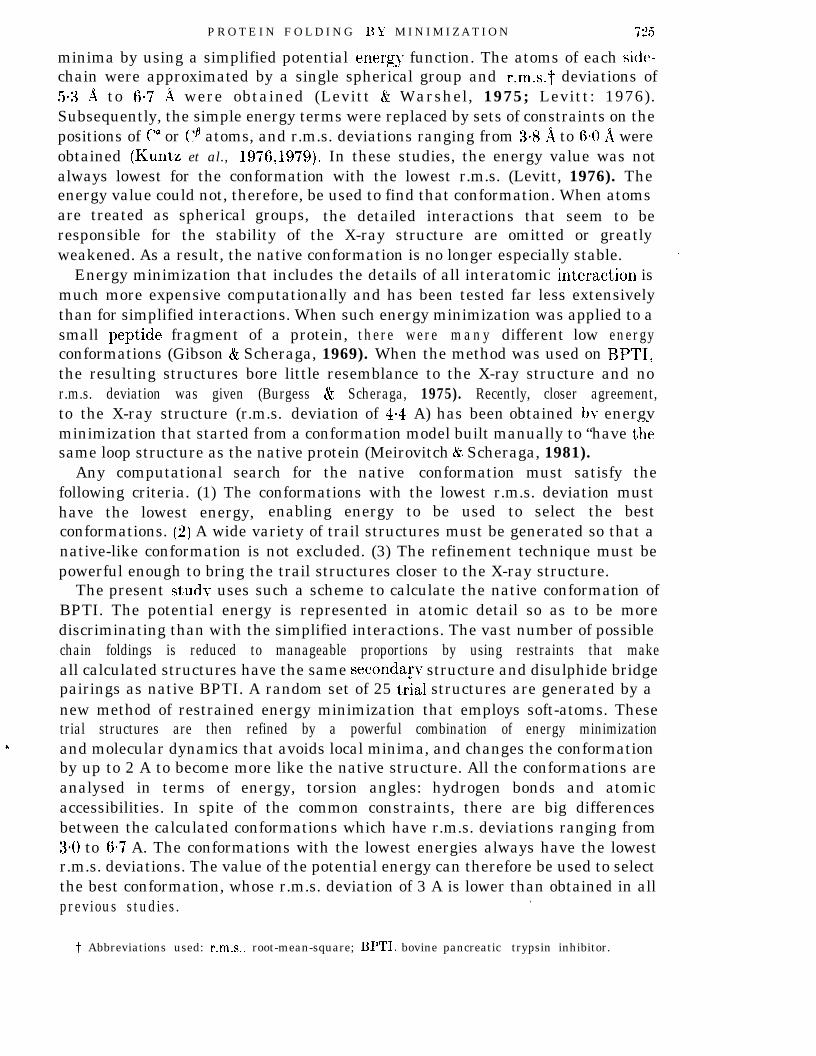
FIG. 1. Comparison of the soft-atom van der Waals’ interaction energy of a pair of nitrogen atoms(broken line) with the corresponding normal-atom van der Waals’ interaction energv (continuous line).The normal potential has the form UVd (r) = A/r12 -B/r6 for the atom pair at a distance r apart. Thesoft-atom pot’ential has the form:
t rsoft =vd ( A l~~~-Bly~}/{(Air’~)(l +O+‘)/h+ l}.
When r is greater than @A/B)As r gets smaller, L’tif’
l/6 the separation at which the energy crvd is minimum. t7tsft. is very like,t’,,d. varies like h( 1 - 04r2), and L~~~f’ = h at r = 0 when 2 atoms are completelvoverlapped. In the present studv h is taken as 10 kcal/mol; tests with h = 3 and 30 kcal/mol gavkLsimilar results.
restraints can be added to the potential energy function (over 1200 were t,ried)without affecting the rapid convergence of energy minimization.
(b) Energy minimization in torsion angle space
(i) The molecule studied
The method outlined above was tested on BPTI, a molecule whoseconformation has been determined by X-ray diffraction studies (Huber et al.,197 1; Deisenhofer & Steigemann, 1975). This same protein has been used in mostprevious folding calculations (Burgess & Scheraga, 1975; Levitt & Warshel? 1975;Kuntz et al., 1976,1979; Levitt, 1976; Goel & Y6as, 1979; Robson & Osguthorpe,1979; Meirovitch & Scheraga, 1981), facilitating comparative evaluation of theresults. BPTI protein has 58 amino acid residues, 892 atoms, 454 non-hvdrogenatoms and 208 4, #;, and x single-bond torsion angles. Omitting all hvdrogen&atoms makes it difficult tchvdrogen at,oms on peptideatoms. (The eight hydrogenare not included to avoid tksingle bond torsion angles.)
(ii) Starting conformations
represent hydrogen bonds realistically, so the 61and amide groups are included giving a total of 515atoms on hydroxyl groups, which are free to rotate,le difficulty of initial positioning and the additional
Different low energv constrained conformations are generated bv starting theminimization from a variety of random chain tionformations. These’sets of atomicco-ordinates of BPTI are built with standard bond lengths, bond angles and
728 M. LEVITT
double-bond torsion angles. Every x1 torsion angle is set to - 60”, the mostcommon value found in real globular protein conformations (Janin et al., 1978).whereas all other x angles are set to HO”, giving a standard tram conform&ion.The initial (4, $) va ues1 of each residue depend on whether the residue is assignedto a-helix or P-sheet secondary structure (see Table 1). For a-helix residues, 4 ischosen randomly to be in the range - 65” and - 55”, and $ is - 45’ to - 35”. Forthe P-sheet residues, C/I is - 130” to - llO” and Ic/ is 140° to 160”. For the otherresidues, the initial $ and @ values are each within 60” of (4, II/) = ( - 90”, go”),giving extended chain structure.
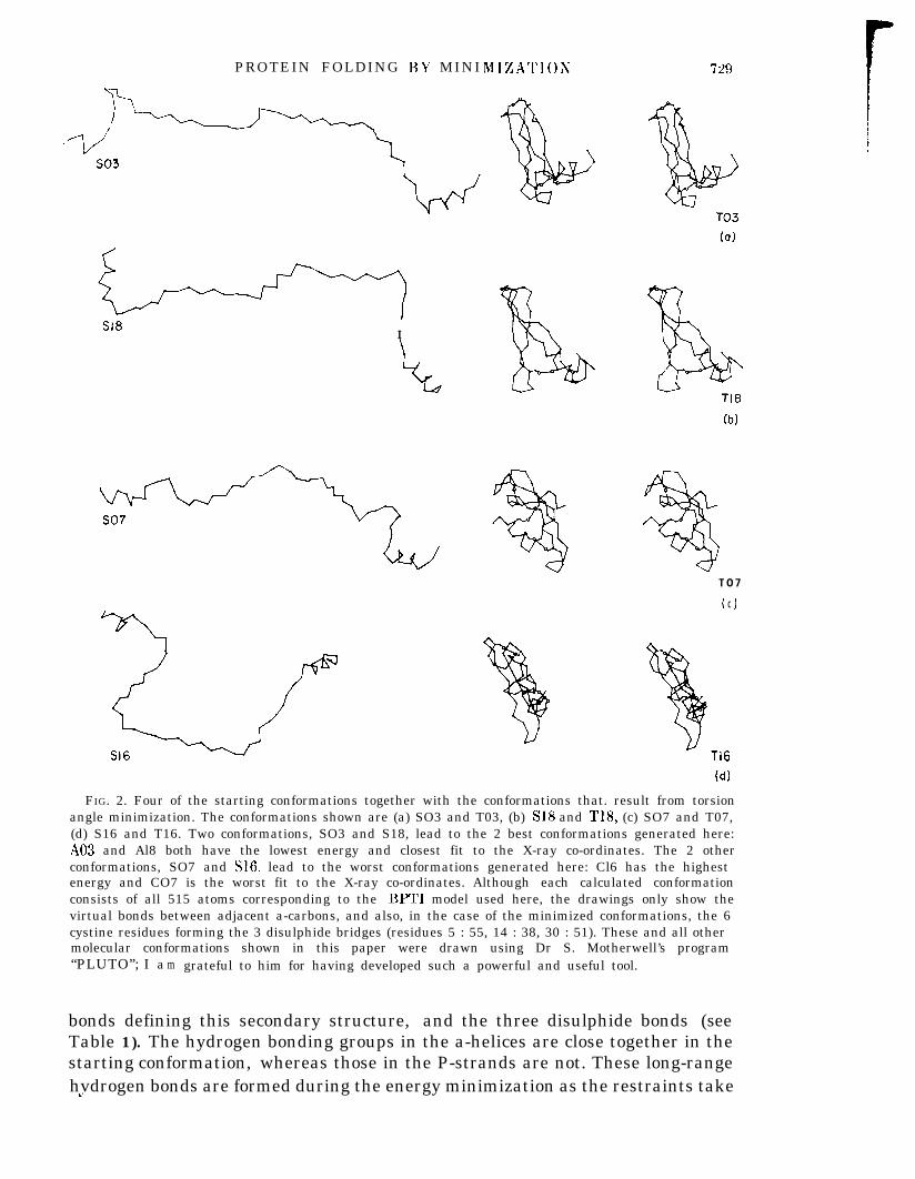
The particular starting conformation depends on the randolm numbergenerator, which is, in turn, controlled by specifving a starting number known as,the “seed”. For simplicity, seed values of 1 to 25 are used to generate 25 different1random starting conformations. These conformations (see Fig. 2) are not at allcompact and contain rod-like segments (the a-helices and P-sheet strands)separated by less regular regions.
(iii) RestraintsThe restraints used to guide the energy minimization include the torsion angles
for the regions assigned to a-helices and P-strands, the 16 main-chain hydrogen
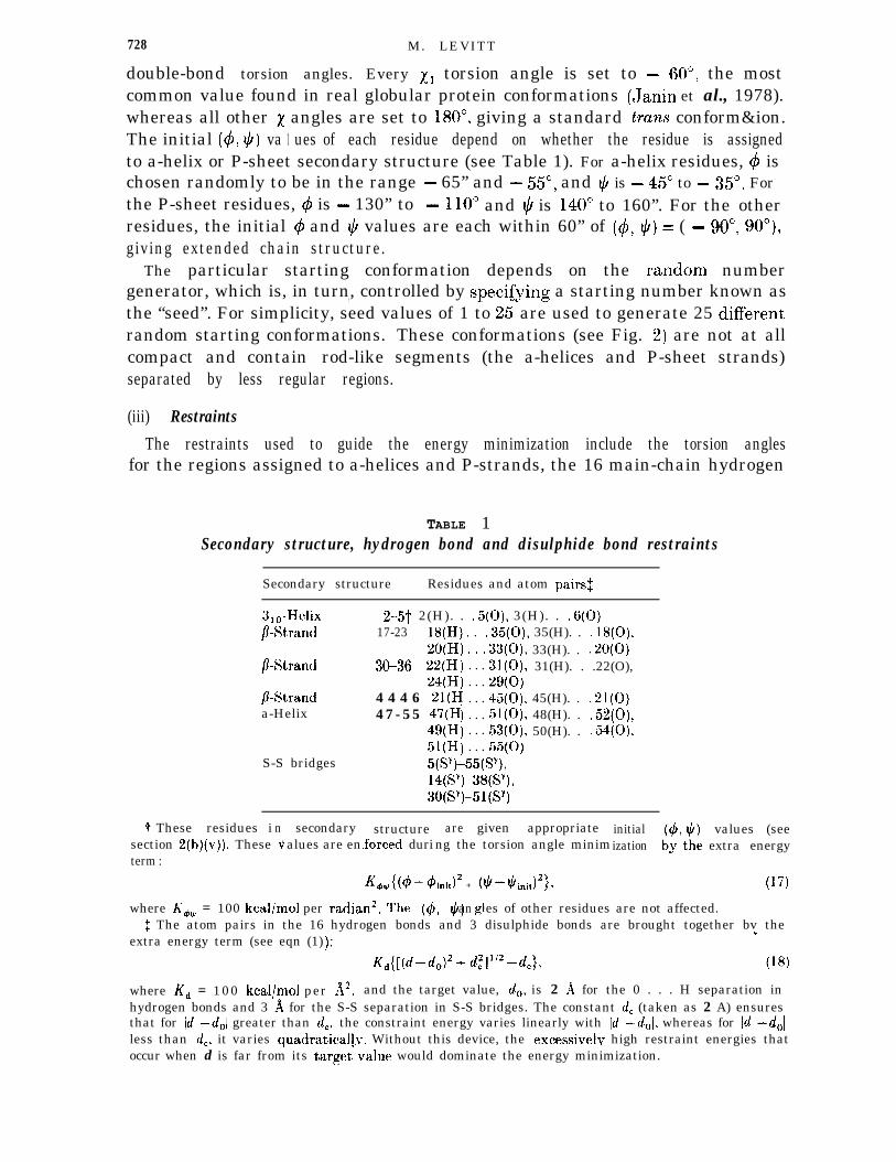
TABLE 1Secondary structure, hydrogen bond and disulphide bond restraints
t These residues isection 2(b#)(v)). Theseterm :
Secondary structure Residues and atom pairsx
3,0-Helix/?-Strand
P-Strand
/?-Stranda-Helix
2-5-t 2(H). . .5(O), 3(H). . .6(O)17-23 18(H). . .35(O), 35(H). . .18(O),
20(H30-36 22(H
24(H4 4 4 6 21(H4 7 - 5 5 47(H
49(H51(H
. . . 33(O), 33(H). . .20(O)
. . . 31(O), 31(H). . .22(O),
. . . 29(o)
. . . 45(O), 45(H). . .21(O)
. . . 51(O), 48(H). . .52(O),
. . . 53(O), 50(H). . .54(O),
. . . 55(O)S-S bridges
n3
secondary structurealues are en.forced duri
where K,, = 100 kcal/mol per$ The atom pairs in the 16
extra energy term (see eqn (1)
radian2. The (4% !h glan es of other residues are not affected.hydrogen bonds and 3 disulphide bonds are brought together bv theu/
areng the
given appropriatetorsion angle minim
K+4g{(4-4init)2 + (ti-tiinit)2}7
initialization
values (seeextra energy
(17)
where K, = 100 kcal/mol per A2, and the target value, d,, is 2 A for the 0 . . . H separation inhydrogen bonds and 3 A for the S-S separation in S-S bridges. The constant d, (taken as 2 A) ensuresthat for Id -d,l greater than d,, the constraint energy varies linearly with Id -&I, whereas for Id -d,(less than d,, it varies quadraticallv. Without this device, the excessivelv high restraint energies thatoccur when d is far from its targetvalue would dominate the energy minimization.
PROTEIN FOLDING BY MINI
98I
IZATION
T O 7
( 1C
l-16
(d)
FIG. 2. Four of the starting conformations together with the conformations that. result from torsionangle minimization. The conformations shown are (a) SO3 and T03, (b) SlS and TN, (c) SO7 and T07,(d) S16 and T16. Two conformations, SO3 and S18, lead to the 2 best conformations generated here:A03 and Al8 both have the lowest energy and closest fit to the X-ray co-ordinates. The 2 otherconformations, SO7 and S16, lead to the worst conformations generated here: Cl6 has the highestenergy and CO7 is the worst fit to the X-ray co-ordinates. Although each calculated conformationconsists of all 515 atoms corresponding to the BPTI model used here, the drawings only show thevirtual bonds between adjacent a-carbons, and also, in the case of the minimized conformations, the 6cystine residues forming the 3 disulphide bridges (residues 5 : 55, 14 : 38, 30 : 51). These and all othermolecular conformations shown in this paper were drawn using Dr S. Motherwell’s program“PLUTO”; I a m grateful to him for having developed such a powerful and useful tool.
bonds defining this secondary structure, and the three disulphide bonds (seeTable 1). The hydrogen bonding groups in the a-helices are close together in thestarting conformation, whereas those in the P-strands are not. These long-rangehvdrogen bonds are formed during the energy minimization as the restraints takeri

730 M. LEVITT
effect. These restraints are equivalent to having prior knowledge of the secondarystructure, including the specific pairing of residues in P-sheets, and the disulphidebridges.
(iv) Choice of variables
The potential energy of this molecule is minimized with respect to the 208single-bond torsion angles ($, $ and x) (53 4 angles, excluding those of Argl ,Pro& Pro& Pro9, and Pro13; 58 $ angles; 97 x angles). Although minimization,with respect to the 1545 atomic Cartesian co-ordinates is used at a later stage ofthis work, torsion angles are preferred for minimization from the open startingconformations. The smaller number of torsion angle variables allows use of thevery efficient variable metric minimization method. Because bond lengths, bondangles, and many torsion angles are rigid in torsion angle minimization, theinitially good standard geometry cannot be distorted by the large forces that mav”arise from close contacts or restraints.
(v) The energy function and first derivatives
The complete express ion for the potent ia l energy ( in kcal/mol;pect to single-bond1 kcal = 4.184 kJ) for restrained energy minimization with res
torsion angles is:
u TOT = c K,[l +cos CnXi)l+ . C K&&~i-#bside-chains secondary structure
+ c Kd([(di-d~)2+d,2]1’2-dc}19 restraints
J2 + (tii-ti,,“)
+ 1 {[A/r,‘,z-B/r: -C]/[(A/r&2)(1 +0+$)/h + l]}s(rJ. (1)atom pairs
The first term represents the preference that certain side-chain ‘torsion angles havefor the staggered conformation; values for the energy parameters K, and n aregiven in Table 2. The second term restrains those (4, $) angles assigned tosecondary structure to their initial values (see Table 1). The third term restrainsdistances di to their target values do and is also described more fully in Table 1.
The final term represents non-bonded interactions, namely the van der Waals’and hydrogen bond interactions between pairs of atoms that are not closeneighbours along the chemical structure (separated bv at least1 4 bonds). Theenergy parameters A and B given in Table 2 for each type of atom were derivedby fitting the crystal dimensions and sublimation energies in a series ofhydrocarbon, amide and amino acid crystals (Levitt & Lifson, unpublished work).The parameter C( = A/612 - B/S6) ensures that the non-bonded energy is zero atithe cutoff separation of 6 A. For r greater than 6 A, non-bonded interactions areneglected. The function S(r) is a polynomial step function (Levit’t, 1976) thatmakes the energy smoothly differentiable in spite of the truncation at’ 6 A. Thesecond bracketed term makes the atoms soft and the parameter h is taken as10 kcal/mol (see Fig. 1 for details).
No hydrogen bond function is used,. but the attraction of hvdrogen bondinggroups is represented by special van der Waals’ parameters.‘ The interaction
P R O T E I N F O L D I N G BY MINIMIZATIOX
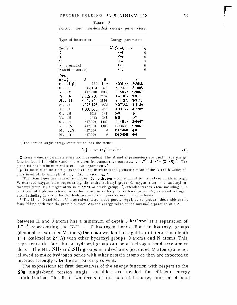
T ABLE 2Torsion and non-bonded energy parameters
731
Type of interaction Energy parameters
Tors ion f4*xx2 (aromatic)x (acid or amide)
Non-bOnd$
H . . . H§ll0 0V:::VY Yii::;Mc . . . cA . . . A0 . . . HV.. .H0 . . .vv . . .NM . . .OTM . . V
A B2 9 0 l-08
145,834 328417,000 1383
3,952,850 25563,952,850 25563,075,695 9 5 31,200,965 425
2913 2412913 241
417,000 1383417,000 1383417,000 0417,000 0
K, (kcal/mol) no-0 0o*o 01.4 30.1 6O*l 6
0*00”100O- 184791=146300.413150.41315OeO73820.037635.05.0b14630l-146300.024860.02486
0
2i5253*10052-90673-81713.81714.31504.2202l-71.72-90672.90674-o4.0
t The torsion angle energy contribution has the form:
K,[l + cos (no)] kcal/mol. (19)
$ These 4 energy parameters are not independent. The A and B parameters are used in the energyfunction (eqn ( 1))) while 6 and r” are given for comparative purposes: E = B2/4A, r” = (2A/B)‘? Thepotential has a minimum value of - E at separation r”.
4 The interaction for atom pairs that are not listed uses the geometric mean of the A and B values ofpairs involved, for example, A,, . y = (A,, . . ()Ay. . &12.. . . *
11 The atom types are defined as follows: H,‘hydrogen atom attached to peptide or amide nitrogen;V, extended oxygen atom representing the entire hydroxyl group; 0, oxygen atom in a carbonyl orcarboxyl group; N, nitrogen atom in peptide or amide group; C, extended carbon atom including 1, 2or 3 bonded hydrogen atoms; A, carbon atom in carbonyl or carboxyl group; M, extended nitrogenatom including 1, 2 or 3 bonded hydrogen atoms in lysine or arginine side-chains.
7 The M . . . 0 and M . . . V interactions were made purely repulsive to prevent these side-chainsfrom folding back onto the protein surface; E is the energy value at the nominal separation of 4 A.
between H and 0 atoms has a minimum of depth 5 kcal/mol at a separation of1.7 A representing the N-H. . . 0 hydrogen bonds. For the hydroxyl groups(denoted as extended V atoms) there,is a weaker but significant interaction (depth1*14 kcal/mol at 2.9 A) with other hydroxyl groups, 0 atoms and N atoms. Thisrepresents the fact that a hydroxyl group can be a hvdrogen bond acceptor ordonor. The NH, NH2 and NH3 groups in side-chains (extended M atoms) are notallowed to make hvdrogen bonds with other protein atoms as they are expected tointeract strongly Ath the surrounding solvent.
The expressions for first derivatives of the energy function with respect to the208. single-bond torsion angle variables are needed for efficient energyminimization. The first two terms of the potential energy function depend

732 M. LEVITT
explicitly on the torsion angle variables and can be differentiated directly. Thethird and fourth terms depend explicitly on the distances between pairs of atoms(di and rij) and must be differentiated in stages:
uTOT ZE 1 h cd%) +z b @ijhi i,j
where +i denotes a 4, Ic/ or x torsion angle and rij denotes either a non-bonded orrestrained interatomic distance. Differentiation with respect to & gives:
a uTOT/a6k = aUI C4kJla4k + C [dU2 Crij)iarijl ’ Larijia4kl* (3)i,j
Because there are many (i, j) pairs and because arij/a& is non-zero for any torsionangle k~ along the chain joining atoms i and j, the transformation in the secondterm is very inefficient. The correct method is to use the Cartesian co-ordinates rn.The second term then becomes:
C 1 La u2 (rij)larijl~arijlarnl~arn~a~kli,j n
= 1 (c La& (rij)larijl ?? Larijlarnl} . [arn/a4d C4)n i,j
Because rij = Iri - I$ ar,,/ar, = 0 unless n = i or j, making the summation over(i,j) very sparse. When the efficient formulation is used (eqn (4)) the derivativecalculation takes less time than the energy calculation alone. When theinefficient formulation is used in an otherwise highly optimized energyminimization program specially written for a super-fast array processor (Pottle etal., 19SO), the derivative calculation takes 170 times longer than the calculation ofthe energy alone.
Because of its high efficiency, the above scheme is described in more detail asfollows. (1) Generate the Cartesian co-ordinates of starting conformations usingstandard geometry and randomly chosen torsion angle values. (2) Calculate thenon-bonded energy for each distance rij and at the same time calculate thecontribution of this term to the Cartesian first derivative XJ,/ar,. (3) Transformthe completed Cartesian first derivative vector to torsion angle space using:
aliJa4k = 1 [au2/arnl ?? [arnlahJ
zz i [au,larnl ’ Lnt#dt x Okrdl~n
(5)
where n4k is the unit vector along the bond about which & operates and r, is theposition of the atom at the end of this bond. Both n4k and r, are calculated in thecurrent Cartesian co-ordinate system. (4) Calculate the torsion angle energy termsand their contributions to the total energy and first derivatives in torsion angleco-ordinates. (5) Use the total energy and first derivatives to get changes in thetorsion angle values A+i. (6) Rotate by A4i about the relevant bonds of thecurrent co-ordinates to generate a new set of Cartesian co-ordinates and repeatthe process from step (2). (For reasons of accuracy, all rotations are actuallyreferred back to the initial Cartesian co-ordinates.) When changing the torsion
PROTEIN FOLDING BY MINIMIZATION 733
angles, it is assumed that the a-carbon of the middle residue (residue 29 in BPTI)is fixed; this is more efficient than fixing an atom at one end of the chain.
(vi > Energy minimization
The robust, very well tested minimization routine VA09D (Fletcher, 1970) isused to minimize the energy by simultaneously changing all 208 torsion anglevariables. On each iteration, the method uses the energy and first derivativevalues from previous steps to approximate the inverse of the second derivativematrix. This matrix, known as the metric of the energy surface, describes the localcurvature and is used to calculate the change in conformation that gets to a localenergy minimum. By using “soft-atoms”, a smoothly truncated energy function(eqn (1 )), and double precision arithmetic (16 significant decimal places), theminimization procedure reaches a precisely defined minimum energyconformation.
More specifically, generation of a minimum energy conformation for BPTIrequires between 849 and 1430 energy and derivative evaluations. The angles ofthese final conformations are accurate to O*OOOOl radians and no component ofthe energy first derivative vector (the torsional forces or couples) exceedsO*OOOOl kcal/mol per radian. Fewer energy and derivative evaluations would havegiven essentially the same final conformation but the small value of the finalforces are a reassurance against errors. A major factor contributing to theefficiency of Fletcher’s (1970) VA09D minimization routine is the fact1 that mostiterations (over 95%) require only one evaluation of the energy and its firstderivatives. Otther variable metric minimizers (Davidon, 1959; Fletcher & Powell,1963) require at least two evaluations per iteration.
(c) Energy minimization and molecular dynamics in Cartesian space
(i) Advantages of Cartesian co-ordinates
The soft-atom energy minimization in torsion angle space described abovegenerates a stereochemically acceptable conformation, which also satisfies theimposed restraints. Additional energy minimization with respect to all the atomicCartesian co-ordinates is necessary for the following reasons. (1) Bond lengths,bond angles and double-bond torsion angles are not able to deviate from standardvalues in torsion angle minimization. (2) Molecular dynamics, needed to annealthe conformations by moving groups over small energy barriers, requiresCartesian co-ordinates as the equations of motion in torsion angle space aretoo complicated. (3) The energy function in torsion angle space (eqn (1)) usesphysically unrealistic soft-atoms introduced onlv to allow restrainedminimization, omits a directional hydrogen bonding term, and lacks a special(4, $) energy term needed to give realistic (4, @) angle distributions.
All these deficiencies are overcome by using Cartesian co-ordinates with thesame energy function used in studies of proline isomerization (Levitt, 1981a) andhydrogen bond dynamics (Levitt, 1981b). This potential behaved well in thesestudies, was better than other potentials in a series of comparative energy
73-t M. LEVITT
minimizations from the X-rav structure (Levitt, 1980) and has been described intidetail (Levitt, 19833).
(ii) The energy function
The energy function is given here to allow comparison with the function used intorsion angle space:
L?T O T = c Kb(bi-b,)2+ 1 h’,(&-&)2
bonds bond angles
+ c K,[l +cos CnXi+6)1+ C FO#C~ $5)torsion angles (49 WI pairs
+ c Alr12- r6.i j Bl 1.lnon-bonded pairs
+. C (A/rb2-B/r$ e-e2’~2+(A’/r12-B’/r6)(1 -e-*2’02). . . H pairs
+ c Kd(di-d,)2.16 restraints
The description and values of the parameters are given elsewhere (Levitt, 19833).Differences from the torsion space energy function include: (1) bond lengths andbond angles can deviate from standard values. (2) All torsion angles canchange and a special potential is used for the (4, $) angle pairs. (3) Van derWaals’ interactions are not softened. No smoothing potential is needed as atompairs in range are listed and used for 100 iterations. (4) The hvdrogen bondinginteraction depends on the 0 . . . N-H angle, 0. (5) The 16* hvdrogen bondrestraints are still imposed but the restraint energy is a simple quadratic function.The disulphide bonds are treated like any other bonds and no restraint is used onthe (4, Ic/) gla n es in regions of secondary structure.
(iii) Energy minimization
Analvtical first derivatives of the energy function are used in energyminimization with respect to all 1545 Cartesian co-ordinates of the 515 atoms inthe molecule. The conjugate gradient minimization routine (Hestenes & Steifel,1952; Fletcher & Reeves, 1964) is used as the variable matrix method wouldrequire memory space for 1,194,285 numbers (1545 x 1546/2), which is impracticalat present. Conjugate 1 gradients minimization requires space for only 1545 x 3numbers Here the minimization is continued for 3000 energy evaluations, at theend of which the r.m.s. force (the first derivative of the energy) is less thanWO5 kcal/mol per At and the energy charge over the last 100 evaluations is lessthan 1 kcal/mol.
(iv) Molecular dynamics1Iolecular dvnamics methods use the values of the energy and its analvtical first
derivative to simulate the motion of the atoms in the presence of thermal energy.Besides vielding a wealth of information about the amplitudes and rates of atomic
motion on the picosecond time-scale (McCammon et aZ., 1977), molecular dynamicshas been shown to “anneal” conformations (Levitt, 1983a). This annealing process
PROTEIN FOLDING l3Y MINIMIZATION 735
removes unfavourable interactions tha*t remain after energy minimization, usingthe thermal energy to hop over small energetic barriers. Subsequent energyminimization is then able to reach a lower energy value. Dvnamic annealing isused here on selected conformations in an at]tempt to get to the lowest possibleminimum energy values.
The iterative solution of the equations of motion that is the basis of moleculardynamics is done as described (Levitt, 19836); the annealing dynamics iscontinued for 30 picoseconds (15,000 energy and derivative evaluations with atime-step of 2 X lo- l5 s). The energy function used for dvnamics is almost thesame as that used for minimization (eqn (6)). In particular,“the restraint on the 16hvdrogen bonds is included. The A and B energy parameters used for dvnamics” ”are slightly different from thothermal expansion. In fact,modified parameters are used(Levitt, 1980,19816,1983a,b).
se used for minimization in an attempt to correct forsuch correction is probably unnecessary but theto be consistent with previous dynamics simulations
3. Methods to Analyse Conformations
Analysing one protein conformation is not straightforward; here about 30conformations have to be analysed and compared. For this task, existing methodshave been extended and new methods introduced.
(a) Comparing atomic co-ordinates
The differences between two sets of co-ordinates i and j is quantified using ther.m.s. deviation of the inter-C’ distances as follows:
AdU -a- $ 1 1 (d;,-d{l)2 “z;kl k>l 1 1 (7)
where dk, is the distance between m-carbon atoms !c and I in conformation i, djkl isthe corresponding distance in conformation j, and nl’k[ is the number of terms inthe summation (58 x 57/2 = 1653 for BPTI). Ad! does not require anysuperposition of the co-ordinates and has been used in most previous comparisonsof protein conformations. Here this r.m.s. deviation is used most often to compareconformation i with the X-ray conformation X and the deviation Adi’ is thenreferred to simply as the r.m.s. deviation.
The distance deviation given above is the same for a conformation and itsmirror-image (Cohen & Sternberg, 1980) and two other deviations are also usedhere:
(8)
where rt is the position of the kth atom in conformation i, ri is the position of the
73ti M. LEVITT
corresponding atom in conformation j, NA is the number of atoms, and N, is thenumber of a-carbon atoms. This deviation does depend on the relative orientationof the two sets of co-ordinates which must be superimposed using matrix algebra(Kabsch, 1976). All the 454 non-hydrogen atoms are included when calculatingthe best superposition even if only the a-carbon positions are used in subsequentcalculation of A$ Projections of the conformation space containing severalconformations are obtained as before (Levitt, 1983c) using A@.
(b) Torsion angles, hydrogen bonds and solvent accessibility
(i) Torsion angles
The (4, $) torsion angles of different conformations were compared as describedabove for Cartesian co-ordinates but this measure was not found useful. Instead,the common scheme of plotting the 4 angle of residue i against the corresponding# angle value is used.
(ii) Hydrogen bonds
Hvdrogen bonds are a very convenient index for describing and comparingconformations. They are few in number, show the spatial proximitv of groups thatmay be distant along the chain, and play an important role in stabilizing thenative conformation. A computer program is used to find hvdrogen bondsautomatically according to the following criteria: (1) the H . . . acclptor separationmust be less than 2=4 a; (2) the donor-H . . . acceptor angle must be within 35” oflinearity. When the hydrogen atom is not explicitly included (hvdroxyl groupsand NH, NH, and NH, groups in arginine and lysine side-chains), It is added withstandard geometry so as to make as good a hydrogen bond as possible. Hydrogenbonds in different conformations are compared by collating the listsautomatically.
(iii) Solvent accessibilityThe area of individual atoms accessible to the solvent is calculated using my
own implementation of the Lee & Richards (1971) algorithm. The radius of thesolvent atom is taken as b4 A, while the solvent exclusion radii of the differenttypes of atoms are taken as: 0 A (H), l-4 A (0 or V), 1.65 A (N), 1.87 A (C), 1.76 A(A), l-85 A (S) and 1.65 ifk (M). Th’is same program and set of radii have been usedextensively in previous solvent accessibility calculations (Chothia, 1976). Theaccessible areas of classes of atoms, individual residues and entire conformationsare obtained by summing up the accessible areas of individual atoms.
(c) Writhing numbers and loop threading
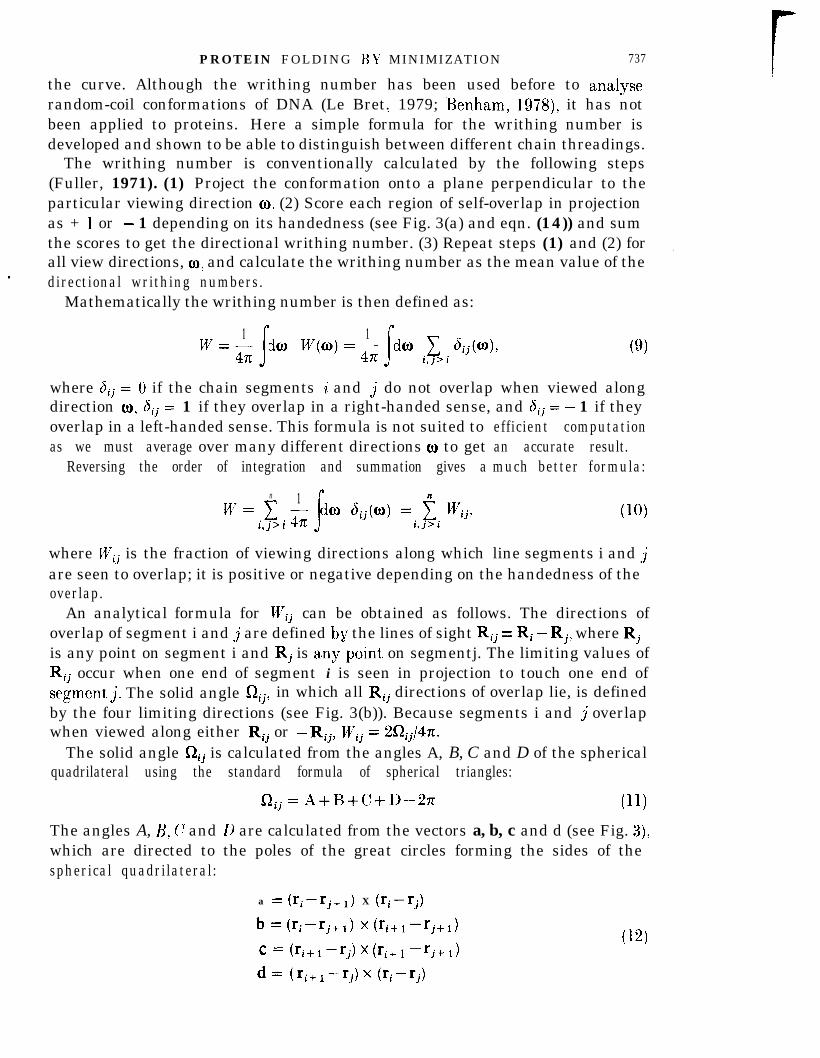
The line joining adjacent a-carbon atoms along the protein backbone traces outa curve in three-dimensional space. The variation of twist and curvature along thecurve has been analysed by formulae of differential geometry (Rackovsky &Scheraga, 1980). Another property of a space curve is the writhing number(Fuller, 1971), which is not a local measure but depends on the overall shape of
PROTEIN FOLDING BY MINIMIZATION 737
the curve. Although the writhing number has been used before to analyserandom-coil conformations of DNA (Le Bret. 1979; Benham, 1978) it has notIbeen applied to proteins. Here a simple formula for the writhing number isdeveloped and shown to be able to distinguish between different chain threadings.
The writhing number is conventionally calculated by the following steps(Fuller, 1971). (1) Project the conformation onto a plane perpendicular to theparticular viewing direction OX (2) Score each region of self-overlap in projectionas + 1 or - 1 depending on its handedness (see Fig. 3(a) and eqn. (14)) and sumthe scores to get the directional writhing number. (3) Repeat steps (1) and (2) for .all view directions, cu, and calculate the writhing number as the mean value of thedirectional writhing numbers.
Mathematically the writhing number is then defined as:
W- 1- z da W(O)=G da 1 dij(m),s
1
s(9)
i,j>i
where 6ij = 0 if the chain segments i and j do not overlap when viewed alongdirection CI), 6ij = 1 if they overlap in a right-handed sense, and 6ij = - 1 if theyoverlap in a left-handed sense. This formula is not suited toas we must average over many different directions 03 to get
Reversing the order of integration and summation gives a
n 1
w=c si,j>iG da 6ij(O) = t WijT
i,j>i
where Wij is the fraction of viewing directions along which line segments i and j
efficient computationan accurate result.much better formula:
are seen to overlap; it is positive or negative depending on the handedness of theoverlap.
An analytical formula for Wij can be obtained as follows. The directions ofoverlap of segment i and j are defined bv the lines of sight Rij = Ri -Rj, where Rjis any point on segment i and Rj is any”point on segmentj. The limiting values ofRij occur when one end of segment i is seen in projection to touch one end ofsegment& The solid angle aij, in which all Rij directions of overlap lie, is definedby the four limiting directions (see Fig. 3(b)). Because segments i and j overlapwhen viewed along either Rij or -RijT Wij = 2Qij/4Za
The solid angle f2ij is calculated from the angles A, B, C and D of the sphericalquadrilateral using the standard formula of spherical triangles:
I2 ij=A+B+C+D-271 (11)
The angles A, B, c and D are calculated from the vectors a, b, c and d (see Fig. 3),which are directed to the poles of the great circles forming the sides of thespherical quadrilateral:
a = (rimrj+l) X (ri-rj)
b zc Crimrj+l) x (Ti+lmrj+l)
c = Cri+l -rj) x Cri+ 1 -‘j+ I>
d= &+I-rj)X (ri-rj)
(12)

M. LEVITT
(a) Start
i j+l
(b)
i,j+l
i+I,j
i j+l
FIG. 3. (a) The total writhing number W can be calculated as a sum of the probabilities, U’iil thatsegments (i, i + 1) and (i, j + 1) overlap when the structure is viewed from a random direction. (b) Wij iscalculated from the positions of the segments. The 2 segments are seen to overlap if the system isviewed from a direction that passes through the spherical quadilateral shown. Consult the text for theequations that give the area of the quadrilateral in terms of angles, A, B, (I! and D and pole unitvectors a, b, c and d.
andA = COS-1 (a ?? d/laildl)R = COS-' (be a/lbllal)? ? ?? ????? ? ? ? ? ? ? b/lcllbl)D = cos-’ (d ?? c/(dlIcl),
where ri is the vector of atomic Cartesian co-ordinates of the ith a-carbon at the

PROTEIX FOLDIXc: BY MINIMIZATIOX 730
beginning of line segment i. The sign of I/c’,, is the same as that of the vector tripleproduct:
[( r.z+1 -rJ x (rj+l -q)] ?? (q-q). (14)
The quantity Wij is the contribution of line segments i and j to the totalwrithing number. Mapping Wij for all (i,j) pairs shows which parts of the chainwrithe most. Plotting 1 Wij against i shows the total writhing contributions of.line segment i. j
An approximate formula for Wij is:
u7’. i j ?? ??? ???X ? ? ? ?? ????????????????
where Si = (ri+l -ri) is the vector along line segment i, and:
ri j = +&+I +ri)-+(rj+l +rj) (16)
is the vector between the midpoints of segments i andj. The approximate formulahas a simple interpretation. The numerator is the area of the region delimited bytlhe limiting viewing directions projected into the plane perpendicular to the linejoining the segment centres; the denominator is simply the surface area of asphere of radius Irijl. This formula is accurate to 10% SO long as IrijJ > ISil. Forproteins ISil = 3+JI A and lrijl is large enough for the error to be small. Neverthelessthe accurate formula is used here.
(d > Computing requirements
The central processing unit times required by one step of torsion angleminimization, Cartesian minimization or Cartesian dynamics are given in Table 3.
TABLE 3Computer requirementst for a single step on BPTI protein
CPTlJ time PercentageProgram section ( )S of total Dependence
Torsion angle minimizationEnergy value l-5 68 nFirst derivatives 0*4 1 8 nVariable metric step 0.3 1 4 n2Total 2.2 100
Owtesian minismization or dynamicsIEnergv value 0.5 62 nFirst derivatives O-2 25 nConjugate gradient or
dvnamics step o-1 1 3 nTotal O-8 100
f All calculations were done on an IBM 370/165 computer with the fastmultiplv optSion. Theprograms were written in a MORTRAX, a rationalized extension of FORTRAN used double precision(64 bit) floating point variables. and were compiled with the FORTRAN & optimizing compilerOPT = 2. The IBM 370/165 is rated at about Z-5 x lo6 instructions/s (mips); a typical multiplication9,as coded in FORTRAN takes 1.4 ps. CPU central processing unit.

740 M. L E V I T T
Most time is spent in calculating the energy. Although very similar potentials areused for torsion and Cartesian co-ordinates, the energy calculation in torsion spacetakes longer. This is because of the different schemes used to find which pairs ofatoms (i,j) are close enough in space to be included in the calculation. In torsionspace., where substantial conformational changes can occur in one it)eration of theminimizer, all (i, j) pairs are scanned on every step (this is done in an efficient wayby not checking distances between atoms in residues whose centroids are very farapart). In Cartesian space, where the conformation is expected to change moreslowlv, a list of (i,j) pairs close enough to interact is made once and usedabout 100 iterations (the precise number depends on the initial value ofenergy and the progress of the minimization).
Calculation of derivatives with respect to the 208 torsion angles increasescomputer time per step by only 30%. In a torsion angle minimization of B I
forthe
the>.TI
employing 186 variables (Pottle et al., 1980), the derivative calculation increasesthe computer time per step by 1 7,OOOo/o (170-fold). The much greater efficiency ofthe present calculation results from proper factorization of the derivativecalculation (see section 2(b)(v), above).
The additional time required to calculate a change in conformation bv either ofthe two minimization methods or the dynamics method is less than 15%. Incomparing these times it is important to remember that there are 208 variables intorsion space and 1545 in Cartesian space. Because the variable matrix algorithminvolves multiplication of vectors by matrices, the time will increase as n2 (for nvariables). In Cartesian space the variable metric step would take ( 1545/208)2times longer than in torsion space (the minimization step would then take 17 s or9 times more than the energy and derivative calculation).
The time required by all the other steps increases linearly with the number ofvariables n, and the methods could be applied to much larger systems. Thememory requirement of the different program sections (see Table 3) has the samedependence on n as the central processing unit time. For BPTI, the programsnow run in 400,000 bytes of memory. Because modern computers have verv largememories (up to 20,000,OOO bytes), memory requirements will not limit the sizeof molecule that can be studied.
4. Results
(a) Generation of conformations
(i) M inimixationAlmost1 100 different conformations of BPTI protein are generated in this
studv; the convention used to name conformations simply and uniquely is givenin Tible 4.
Each of the 25 different starting conformations (SO1 to S25) obtained with aninitial random number of 1 to 25 (the “seed”) is minimized to convergence withrespect to the 208 single-bond torsion angles, using soft-atoms and restraints onthe torsion angles in secondary structure, and the lengths of 16 hvdrogen andthree disulphide bonds (Table 1). Figure 2 shows four of thkse starting

P R O T E I N F O L D I N G BY MIXIMIZATION 741
TA B L E 4Convention used to name conformations
Name Meaning
1 st letters StartingT Torsion space energy minimized
Yc Cartesian space eneigv minimizedA Annealed by Cartesian space molecular dynamics and energv minimization” ”2nd letter .x X-rav co-ordinates (Deisenhofer & Steigemann, 1975)i The 25 starting conformations are labelled SO1 through S25 and were generated bv using i asr/
random number seed in the generation of initial torsion angles
conformations and the corresponding structures after torsion angle minimization.At the start of the minimization, the energy is very high as the pairs ofatoms that are forced to come together to form the hydrogen and thedisulphide bonds are initially very far apart. The use of soft-atoms allows theprogram to deal with these high strain energies and generate a roughly foldedstructure that satisfies the restraints after only 30 steps. Many more cycles ofminimization (between 500 and 1500) are required, however, to improve thepacking of the side-chains and converge to a true energy minimum.
Although the starting conformations are similar in character, the compactfolded structures show a surprising range of energy and r.m.s. deviation values.The energies range from - 109 kcal/mol to 1008 kcal/mol and the r.m.s. deviationsrange from 3.5 A to 6*0 A. A plot of the energy against the deviation (Fig. 4(a))shows no obvious trend: the five conformations with lowest energies (T20, TM,T22, T17 and T03) have r.m.s. deviations of 4.9 A, 4a5 A, 3*7 A, 4.5 A and 3.7 A.Such a diversity of conformations was unexpected as each conformation hasbeen minimized in the same way and fits the same set of constraints.
Next, each of the torsion angle minimized conformations (TO1 to T25) issubjected to further energy minimization with respect to the 1545 atomicCartesian co-ordinates using the more complete energy function (eqn (6)). Thetotal energies and r.m.s. deviations of the resulting minimum energyconformations (CO1 to C25) are shown in Figure 4(b) together with thecorresponding properties of the X-ray (X) and minimized X-ray conformation(CX). Although there is still a large spread in energy and r.m.s. deviations, thereis a clearer trend than after torsion angle minimization (Fig. 4(a)). In particular,the four conformations of lowest energy (C03, C17, Cl8 and C22) are also thosethat have low r.m.s. deviations from the X-ray co-ordinates. None of the 25 Ciconformations has an energy that is as low as the energy of the X-ray minimum(CX); the lowest in energy (CM) has an energy value that is still 51 kcal/molabove that of CX.
There are nine conformations whose energies are less than 100 kcal/mol abovethe. energy of the CX conformation. The energy contributions and r.m.s.deviations of these conformations are listed in Table 5. The bond length, bond

742 M. LEVITT
1 2 0 0rTO6
TO7Tlll6
TO5 C25
T 2 3
T 1 4T15
TO9
T22 TY20
LVV
0 I 2 3 4 5 6 7
Ad, (8)
(a) (b)
0
-100
-IOC
-0
E\02- -200G&
15
-3oc
lr
? ?
cxAx
Cl6
c o 2
Cl0
cm5 co7
c o 5 vt83
cQ&‘9CO6
co1 c21
I. 1. 1. 1. 1. I.
I 2 3 4 5 6 7
A& (8)
Fro. 4. Minimum energv values and r.m.s. a-carbon distance deviations of conformations generatedhere. (a) The 25 T conformations obtained bv energy minimization with respect to the 208 single-bondt,orsion angles, and (b) the 26 C conformatiks obtained by energy minimization with respect, to the1545 atomic Cartesian co-ordinates (including CX), together with the 5 conformations produced bydvnamic annealing (AX., A03, A17, Al8 and A22)..
angle and van der Waals’ energy contributions show a much smaller variationthan the torsion angle and hvdrogen bond contributions. The van der Waals’ andhvdrogen bond energy contributions of conformations CO& C21 and C22 are almostuas favourable as those in the X-ray minimum CX. Much of the difference betweenthe energy of the X-ray minimum and the other minima comes from the torsionangle energy. This suggests that many torsion angles have been forced tounfavourable values to satisfy both the requirements of a close-packed interiorand the 16 hydrogen bond restraints.
(ii) Dynamic annealing
The four lowest energy conformations (C03, C17, Cl8 and C22) are subjected to30 picoseconds of molecular dynamics that are followed bv a, second pass ofCartesian space energy minimization to give annealed conformations A03, Al 7,

PROTEIN FOLDING BY MINIMIZATION
TABLE 5Energy contributions of the nine lowest energy C conformations
743
Conformation Total
Energy contribution (kcal/mol) r.m.s.van der
Bond Angle Torsion Waals’ H bond deviation (A)
CX‘ c o 3
Cl2Cl3Cl7Cl8c20c 2 1c 2 2C24
-342 3- 280 4-262 4- 2 7 4 4- 280 3-291 3-267 4-251 5-293 5- 2 5 9 4
3 3 - 2 7 - 2 2 8 - 1 2 3 o-53 9 6 -221 - 1 0 9 3.43 5 2 - 205 - 9 9 4.84 0 7 -218’ - 108 4.33 4 - 1 -218 - 9 8 3-92 9 - 3 - 2 1 6 - 1 0 5 3.93 5 7 -202 -112 4-45 1 2 2 -222 - 107 5-o4 3 1 0 - 229 -122 3.84 2 1 6 -211 - 1 1 0 3.5
Al8 and A22. The CX conformation is also annealed in this way to giveconformation AX. Table 6 gives the relative energy contributions and r.m.s.deviations of these ten conformations. In every case, annealing leads to aconformation of lower total energy, due mainly to more favourable hydrogenbonding and less torsion angle strain. The energy values of the foldedconformation (Ai) are, however, always greater than those of both the CX andAX conformation derived from X-rav co-ordinates. Annealing changes these four”
TABLE 6Relative energy contributions? before and after annealing
Energy contribution (kcal/mol)a,van der r.m.s. (A)
Conformation Total Bond Angle Torsion Waals’ H bond Adtrev$ Ad,” Ar; R&
After annealingAX (absolute) -359A 0 3 2 6Al7 5 1A 1 8 3 6A 2 2 4 3
Before annealingc x 1 7c o 3 7 9Cl7 7 9Cl8 6 8c 2 2 6 6
3 3 - 3 4 - 2 3 0 -1329 1 9 - 1 - 38 3 3 8 14 1 6 4 1 17 2 4 6 5
0 6 2 9 0.56 4 0 9 2 3 o-91 3 3 1 2 3 4 l-6
- 4 3 1 1 4 2 7 1.51 0 4 4 1 1 0 1.2
o-91*5l-5lb6l-9
1-o3-o3.83*43-9
0*5 O-6394 5.63-9 8963.9 8.33.8 5.9
O-8 lo*95.2 lb08.4 11.47.5 11.46*7 114
lb0119412.012*111.7
7 The energy contributions of all conformations except AX are relative to those of the AXconformation.
$ For the A series, prev refers to the corresponding C conformation e.g. for A03, Ad:“” is measuredto C03. For the C series prev refers to the corresponding T conformation.
5 R, is the radius of gyration calculated as R, = [l/N C (ri -r)2]1’2, where ri is the ith Cartesianposition vector, f is the position of the molecular centroid and the summation extends over all non-hvdrogen atoms. For the X-rav conformation, R, = lo*96 A.. .,25

74-l M. LEVITT
conformations by l-5 A to 1*9 A r.m.s. The change caused by the initial Cartesianco-ordinate minimization is comparable at 0.9 L% to 143 a. Application of the samerefinement techniques to the X-ray co-ordinates causes smaller changes of 0.9 Aand 0.5 A, respectively.
For conformations A03 and AH, the decrease in total energy caused bydynamic annealing is also accompanied by a significant decrease in the r.m.s.deviation from the X-ray co-ordinates (3.4 A to 3.0 A and 3.9 A to 3.4 A). Theenergies of the annealed conformations increase monotonicallv with r.m.s.deviation (see Fig. 4(b)). T h i s same trend is observed for those Cconformationsthat have the lowest energy value for a particular value of the r.m.s. deviation(CZ2, CM, C13, Cl2, C21 and C07). To a first approximaGon the lowest energythat one can get for any A or C conformation varies quadratically with:
E min = - 360 + 3.9 (r.m.s.)2. (17)This indicates that the only way to get to a very low energy value is to becomemore similar to the X-ray co-ordinates. The existence of conformations at eachr.m.s. value with higher energies can be explained bv trapping in high energy localc:minima. The fact that the equilibrium bond length and bond angle values aretaken from the X-ray structure (Levitt, 19833) does not cause a lower total energyat conformation X; all minimized conformations have very low bond and bondangle energies (see Table 6).
The five conformations that have the lowest energies after torsion spaceminimization (T03, T17, TM, T20 and T22) include four of the five conformationsthat, have the lowest energy after Cartesian space minimization (C03, C13, C17,(118 and C22: see Fig. 4). Applying Cartesian co-ordinate minimization only tothose T conformations with low energies would save computer time and allowmany more starting conformations to be generated. It was hoped that the energyvalue after a small number of steps of torsion angle minimization would alsoprovide a selection criteria. Unfortunately, only one of the five conformationswith lowest energy a)fter 300 steps of torsion angle minimization is among the fivebest conformations after Cartesian co-ordinate minimization.
(b) The diversity of conformations
(i) Comparing co-ordinates
All the Cartesian space minimized conformations (26 C and 5 A) are obtainedusing the same potential energy function. All these conformations are forced to,have the 16 hydrogen bonds found in the native secondary structure. ThediversitIy of these conformations is still considerable as evidenced by the range ofr.m.s. deviations from the X-ray structure. This diversity is investigated furtherbv calculating the r.m.s. deviation between all pairs of conformations. The closestpair of conformations (Cl7 and ClS) are 2.3 A apart, while the furthest pair (CO2and C07) are 8*5 A apart
,
A two-dimensional representation of the conformational space is shown inFigure 5 for the 32 X, C and A conformations. In the two-dimensionalrepresentation, the distance between any pair of conformations is a measure of the

P R O T E I N F O L D I N G B Y M I N I M I Z A T I O N 745
6
Cl0C 2 3
Cl5C O ? c21 Cl9
4- C O 8
co9Cl2
Cl6Cl3
C l 1
A 2 2 c20
Y22
Cl4
c o 1C25
A’7-C17
Cl8L/
co3 Al8 co4co2
A 0 3
co5
CXXAX C 2 4
0 2
p (a6
FIG. 5. A 2-dimensional representation of the 15Kdmensional conformational space containingX and the 30 Cr and A minimum energy conformations. In the projection, the distance on paperbetween anv pair of conformations approximates the actual r.m.s. deviation (Ad:) between them(Levitt, 163~). Notice how conformations C02, C06, CO7 and CO8 are least, similar to each other andmap the conformational extremes obtained here.
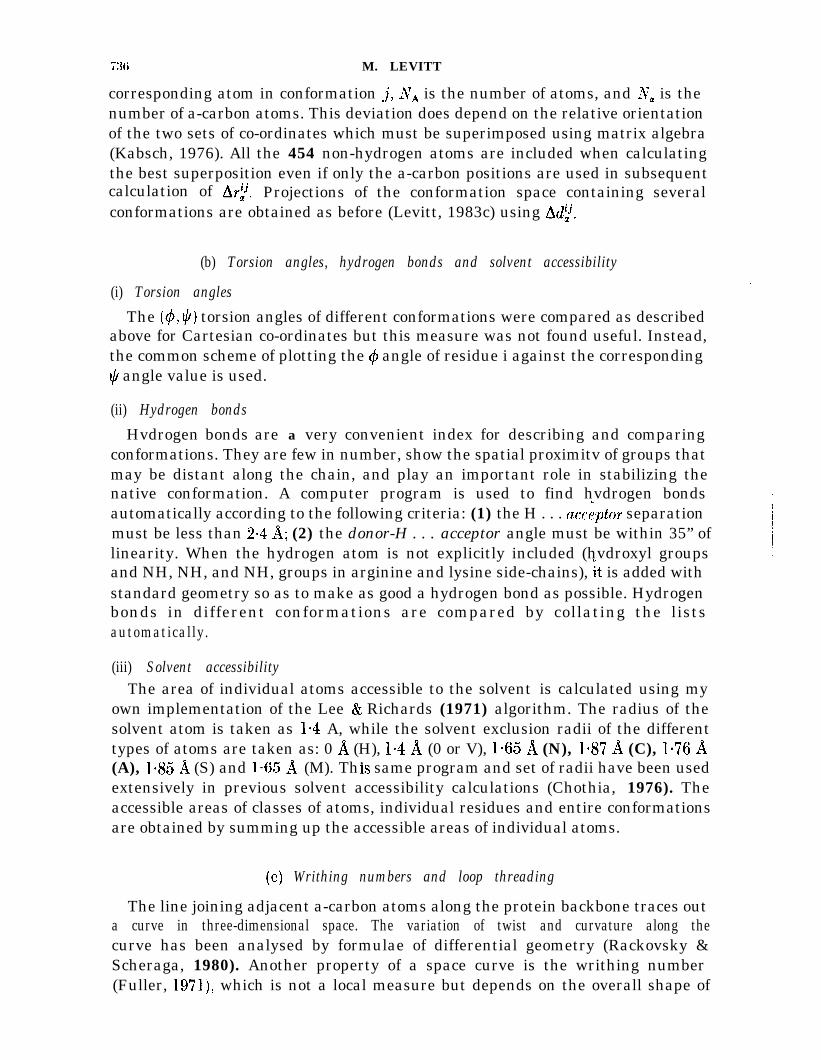
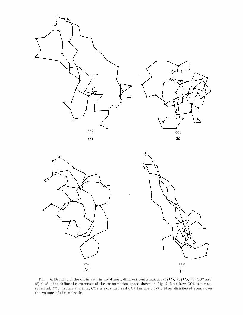
actual r.m.s. deviation between that pair. All but three conformations cluster in apatch that is about 6 a (r.m.s.) across. The native conformations X, CX, and AXare to one side of the patch and are close to it (3 A from A03 to X). The meanr.m.s. deviation between any pair of C conformations is 4.3 A. Conformations CO2,CO6, CO7 and CO8 are most different from one another and define the extremes ofconformation generated by the constrained energy minimization (see Fig. 6).
It is of interest to estimate how many different Ci conformations could begenerated. Let us assume that any two structures with r.m.s. deviation less than1 A are identical (this is the deviation between X and AX). The distribution ofr.m.s. deviation for all pairs of C conformations is approximately a Gaussiandistribution with a mean of 4~3 A and a standard deviation of lb2 A. This gives aprobability of 0.01 that the r.m.s. deviation is less than 1 A. If a space containingn conformations is sampled by choosing m conformations completely at randomthen the probability that they are all different is:
Pdiff = @v- 1)(72-Z) . . . (n-m)/n” 25 ((n-m/Z)ln)“= 1 -m2/2n. (18)
The probability that at least two choices are identical is then simply Pident =1 - Pdiff- In the space of C conformations, Pident is estimated above to be 0.01 andm is 25, giving:
n = m2/2 Pident = 31,250. (19)
Thus, we estimate there are about 30,000 different minimum energy conformations
co2 CO6
(a) (b)
co7 CO8
(d) (cl
FIG. 6. Drawing of the chain path in the 4 most, different conformations (a) CO& (b) (~06, (c) CO7 and(d) CO8 that define the extremes of the conformation space shown in Fig. 5. Note how CO6 is almostspherical, CO8 is long and thin, CO2 is expanded and CO7 has the 3 S-S bridges distributed evenly overthe volume of the molecule.

PROTEIN FOLDING l.W MINIMIZATION 747
that can be generated by the present method of constrained energy minimization.While this is a large number, it is verv much smaller than the astronomicallv largeu ”number of possible conformations of the BPTI polypeptide chain.
(ii) Comparing chain threading
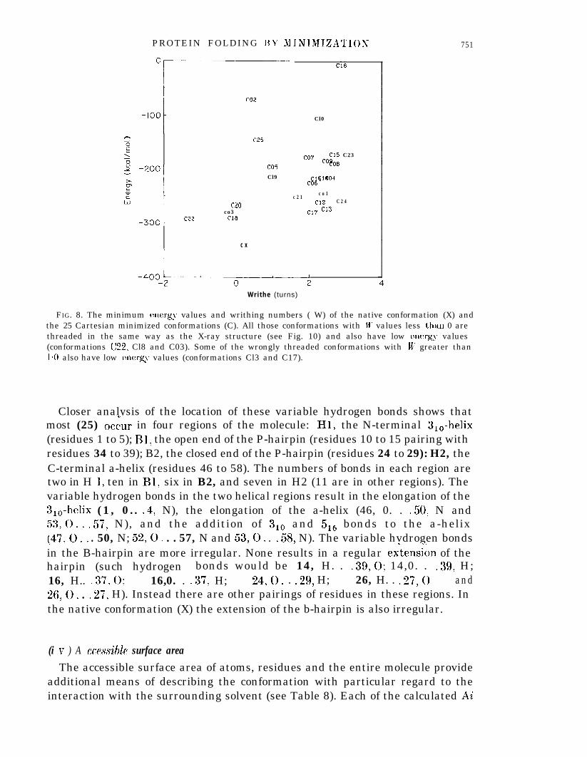
In native BPTI there is an unusual threading with the chain segment consistingof residues 1 to 30 actually passing through the loop formed by the didphide:bond between cystine 30 and 51. None of the BPTI conformations generated byprevious studies shows this native threading in published stereo drawings (Levitt & .Warshel, 1975; Levitt, 1976; Kuntz et al., 1976,1979; Hagler & Honig, 1978: Gael,& YEas, 1979: Robson & Osguthorpe, 1979). The r.m.s. deviation used above toIdistinguish folded conformations is not very sensitive to the chain topology orthreading. Two structures that seem very similar as measured by the r.m.s.deviation may actually have different chain threadings. Threading can be seen bylooking at a stereo drawing of the chain fold (Fig. 7); it can also be detected byusing the writhing number of the chain. The plot of energy against writhingnumber for the C conformations (see Fig. 8) shows that IV varies from - 1.3 turnsto 3-l turns. Most structures have u/’ = 2 turns, which is significantly differentfrom the value of the X-ray conformation (lV = 0.2). Four conformations (C03,Cl& C20 and C22) have IV < 0 and all these also have low energy values.Inspection of the stereo drawings of these conformations shows that theyall have the correct threading, of residues 1 to 25 through the 30-51 loop. Thissame threading is also found in CO2 and C25, which have W values of 0.4 and0*6 turns, respectively, but all 19 other conformations, which have W greater than1 turn, are not threaded. Clearly, the writhing number is a reliable measure of thechain threading and can be used to classify chain folds. Conformations Cl7 andC18, which are the closest pair of C conformations (r.m.s. deviation of 2e3 A, seeFig. 5) have W = 2.1 and W = -0.2 turns, respectively: in order to get fromconformation Cl7 to Cl8 along the shortest path, the chain would have to passthrough itself near the 14 : 38 disulphide bond (see Fig. 7(c) and (d)).
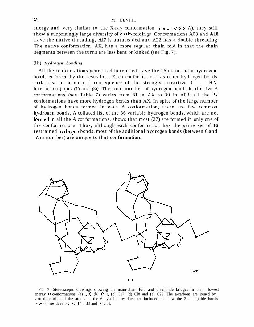
(c) Analysis of the lowest energy conformations
In this section we focus attention on the four C conformations with lowestenergies (C03, Cl 7, Cl8 and C22), the corresponding annealed conformations(A03, A17, Al8 and A22) and the X-ray conformations CX and AX.
(i) Energetics
The four annealed conformations are extremely well-stabilized. Their totalenergies are between 26 and 51 kcal/mol above that of AX (Table 6) with most ofthis difference in the torsion angle energy term. The van der Waals’ and hvdrogenbond energies of A03 are actually more favourable than in AX, the annealedX-ray conformation.
In view of the relatively high torsion angle strain, the distribution of the ($, @)angles was examined and compared with those of the X-ray conformation (Fig. 9).All the distributions have similar clusters of points about the cc-helical and /?-sheet

M. LEVITT
co3
(b)
FIG. 7.
regions. In the folded conformations A03, A17, Al8 and A22 there are many morenon-glycine residues that fall outside these regions. Those conformations withmore of the abnormal conformations (Al7 and A22) have higher torsion angleenergies (see Table 6).

P R O T E I N F O L D I N G B Y M I N I M I Z A T I O N 749
(d)
FIG. 7.
Cl8
(ii) Chain folding
The chain fold in the A conformations is shown in the stereo drawings ofFigure 7 and the schematic drawings of Figure 10. Although the folded Aconformations have the native secondary structure hydrogen bonds. are of low” ad
750 M. LEVITT
energy and very similar to the X-rav conformation (r.m.s. < 3.8 A), they stillshow a surprisingly large diversity of ihain foldings. Conformations A03 and A18have the native threading, Al7 is unthreaded and A22 has a double threading.The native conformation, AX, has a more regular chain fold in that the chainsegments between the turns are less bent or kinked (see Fig. 7).
(iii) Hydrogen bonding
All the conformations generated here must have the 16 main-chain hydrogenbonds enforced by the restraints. Each conformation has other hydrogen bondsthat, arise as a natural consequence of the strongly attractive 0 . . . HNinteraction (eqns (1) and (6)). The total number of hydrogen bonds in the five Aconformations (see Table 7) varies from 31 in AX to 39 in A03; all the Aiconformations have more hvdrogen bonds than AX. In spite of the large numberr/of hydrogen bonds formed in each A conformation, there are few commonhvdrogen bonds. A collated list of the 36 variable hydrogen bonds, which are notfirmed in all the A conformations, shows that most (27) are formed in only one ofthe conformations. Thus, although each conformation has the same set of 16restrained hydrogen bonds, most of the additional hydrogen bonds (between 6 and15 in number) are unique to that conformation.
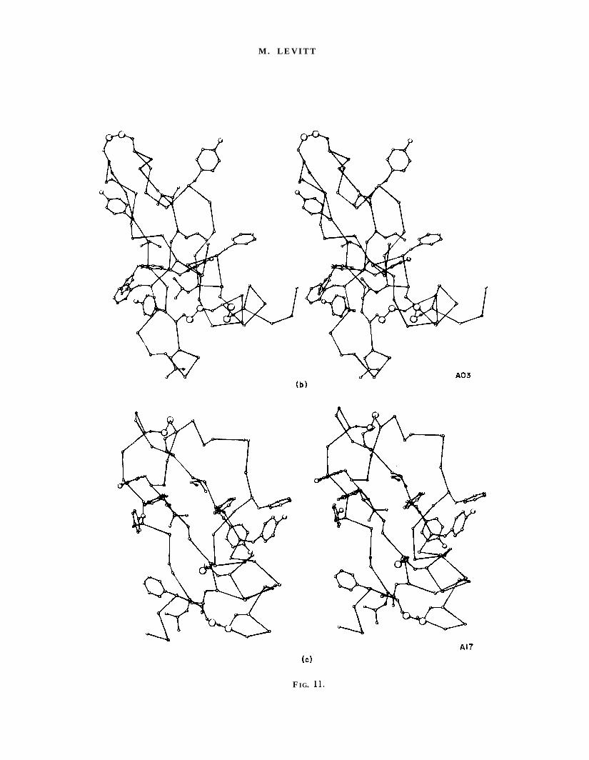
FIG. 7. Stereoscopic drawings showing the main-chain fold and disulphide bridges in the 5 lowestenergy C conformations: (a) CX, (b) CO& (c) C17, (d) Cl8 and (e) C22. The a-carbons are joined byvirtual bonds and the atoms of the 6 cysteine residues are included to show the 3 disulphide bondsbet)ween residues 5 : 55, 14 : 38 and 30 : 51.
PROTEIN FOLDING BY MINIMIZATIOS 751
-
C22
c20co3
Cl8
c x
C25
Cl0
Cl5 C23co7 co$iofj
Cl9C@
g1eo4
c 2 1c o 1
Cl2 C 2 4
Cl7 c13
-2 0 2
W r i t h e (turns)
FIG. 8. The minimum energy values and writhing numbers ( W) of the native conformation (X) andthe 25 Cartesian minimized conformations (C). All those conformations with W values less t.han 0 arethreaded in the same way as the X-ray structure (see Fig. 10) and also have low energy values(conformations C22, Cl8 and C03). Some of the wrongly threaded conformations with W’ greater than1.0 also have low energv values (conformations Cl3 and C17).”
Closer analysis of the location of these variable hydrogen bonds shows thatmost (25) occur in four regions of the molecule: HI, the N-terminal 310-helix(residues 1 to 5); Bl, the open end of the P-hairpin (residues 10 to 15 pairing withresidues 34 to 39); B2, the closed end of the P-hairpin (residues 24 to 29): H2, theC-terminal a-helix (residues 46 to 58). The numbers of bonds in each region aretwo in H 1, ten in Bl, six in B2, and seven in H2 (11 are in other regions). Thevariable hydrogen bonds in the two helical regions result in the elongation of the3,0-helix (1, 0. . .4, N), the elongation of the a-helix (46, 0. . .50, N and53,o.. .57, N), and the addition of 3,, and 516 bonds to the a-helix(47,O.. . 50, N; 52,O. . . 57, N and 53,O. . .58, N). The variable hvdrogen bondsin the B-hairpin are more irregular. None results in a regular exiension of thehairpin (such hydrogen bonds would be 14, H. . .39,0; 14,0. . .39, H;16, H.. .37,0; 16,0. . .37, H; 24,O. . .29, H; 26, H. . .27,0 and26,O. . .27, H). Instead there are other pairings of residues in these regions. Inthe native conformation (X) the extension of the b-hairpin is also irregular.
(i v ) A ccessible surface areaThe accessible surface area of atoms, residues and the entire molecule provide
additional means of describing the conformation with particular regard to theinteraction with the surrounding solvent (see Table 8). Each of the calculated Ai

(a) b)0
P a0 0Oo” O8 X
0d+ 0 0
0%
180 180
O^ 03
“” OOYIB
000 00,
0 0 0
00
1 QX a
x X
0 0
o-̂ 030s 03
0
000 o0 0
0 v
0
n . I -- ’180- 1 8 0 0 180 - 1 8 0 0 180 - 1 8 0 0
0+( )
(e)
0+( )
1 8 0180
do
a0- 03
’ 0X O%
7
0 x0a
0
0
O^ 03
0 x00, 1 .
- 1 8 0 0 180 - 1 8 0 0 180
180
o-̂ 03
X
00 0
O 0 Or
00
*Oo “8
O*
cp0
X X
3 00
, 0 1 .
- 1 8 0 0 180
+(“I0
- 1 8 0 4( )FIG. 9. The distribution of the (4, y?) angl es of the X-ray and 4 annealed conformations (a) A03, (b) A17, (c) X, (d) Al8 and (e) A22. The (4, $) energy
contours at 1 kcal/mol intervals are shown in (f) for alanine dipeptide (CCONHC(C)CONHC:), calculated with the energy function used for the Cartesianminimization.

PROTEIN FOLDING BY MIXIMIZATIOS 753
(a) (bl
N
N C
(e)(d)
FIG. 10. Schematic drawing of the chain path in the 5 annealed A conformations: (a) AX, (b) A03,(c) Al& (d) A22 and (e) ,417. The total energies of these conformations are -359, -333, -3323, -316and -308 kcallmol, respectively, and the r.m.s. deviations from X are l-O? 3.0, 34, 39 and 3.8 A,respectively. The writhing numbers are 0.2, 0, 0, - 1.1 and 2.3 turns, respectively. The loop betweenresidues 30 and 51 is threaded once in (a), (b) and (c), twice in (d) and not at all in (e), explaining thet*rend seen in the writhing numbers.
( 1a
FIG. 11.
M. LEVITT
(b)
FIG.

P R O T E I N F O L D I N G B Y M I N I M I Z A T I O N
(d)
d- :A22
FIG. 11. Stereoscopic drawings showing the main-chain, the disulphide bridges and the largehydrophobic side-chains (Val, Leu, Ile, Met, Phe and Tyr) in the 5 annealed conformations (a) AX,(b) A03, (c) A17, (d) A18 and (e) A22. Interior packing of hvdrophobic groups is clearlv much better in‘* .AX than in the other A conformations.

M. LEVITT
TABLE 7Variable peptide hydrogen bonds
PropertyConformation
AX A03 Al7 Al8 A22
Number ofMain/mainMain/sideSide/side
Main/main variabletThrll, 0.. .Gly36, NGly36, 0. . . Ala16. NAsn24, 0. . . Ala27 ?u‘Asn24, 0 . . . (~1~2% NArg42, 0. . . Asn44. NMet52 0. . . Gly56, NAsp3. 0. . . Glu7, 1JMet52, 0. . . Gly57, xArgl, 0.. .Phe4, NLeu6, 0. . . Lys46? NThrll, 0. . . Asn44, NThrll, 0.. .Arg42, NPro13, 0. ./. Arg39. SLysl5, 0. . . Gly37, NLeu29, 0. . . Ala25, NAla25, 0. . . Leu29, NAla25, 0. . . Gly28, NCys30, 0. . . Ala48, NSer47, 0. . . Asp50. 2u’Arg53, 0. . . Gly56, NArg53, 0. . . Ala58, NPhe4, 0. . . Cvs30, 21’Va134, 0. . . TyrlO. NTyrlO, 0. . . Gly36, NGlv36, 0. . . Gly12, NL&46, 0. . . Asp50, NLvs41, 0. . . Asn44, NPro9, 0. . . Ile19, NArg17, 0.. .Thrll, NAsn43, ‘0. . . Tyr23, NArg53, 0. . . Glv57, NThrll, 0.. .L$41, NCvs38, 0 . . . Glv12: NC&14, 0. . . G&37, NCk14, 0. . . Cv-s38. NAla25, 0. . . Ala27, x
(W(Bl)W)W2)
(HacmVWVW
ww(W&c
cw4
VWWVVW
w(W(WVW
ww
(WVW(W(W
22 3 1 26 23 267 6 1 0 9 1 22 2 1 2 0
XXXXXX
XXXXXXXXXXXXXXX
XX
XX
X
XTxXXX
X
X
XXXXX
x
x
x
X
xXxxxX
t The variable hydrogen bonds listed include all those peptide hydrogen bonds that are formed inany of the 5 conformations. The conserved hydrogen bonds are formed in all 5 conformations andconsist of the 16 that are included as restraints (see Table 1). The variable hydrogen bonds areclassified into four classes given in parenthesis after the bond name (see section 4(c)(iii)).

P R O T E I N F O L D I N G BY M I N I M I Z A T I O N
TABLE 8Accessible surface areasf- of the X and annealed conformations
757
Residue orproperty X
~~
ConformationA03 Al7 A18 A22
Argl 150.41Pro2 43%Asp3 112.2Phe4 37*9cys5 O-0Leu6 99*6Glu7 38.3Pro8 82.7Pro9 52.3TyrlO 75.6Thrll 690 1Gly12 21.5Pro13 86.4Cvsl4 51.2L&l5 174.9Ala16 50.3Arg17 202.4Ile18 59-9Ile19 113*1Arg20 29a6Tyr2 1 60.1Phe22 20.0Tyr23 12.3Ak24 31.8Ala25 50.8Lvs26 180*4Aia27 54.0Gly28 38.8Lei29 78.6cvs30 23.2Gh3 i 67.9Thr32 89.9Phe33 21.8Va134 8198Tvr35 2.9Giy36 0.5Gly37 39.7Cys38 49-5Aig39 174.0Ala40 72.1Lvs4 1 57.1A;g42 131.8Asn43 O-8Asn44 15.2Phe45 39-oLys46 145.5Ser47 23-6Ala48 29*7Glu49 123.0Asp50 55-8cys5 1 o-0Niet52 62.0Arg53 137*9
74- 4 8
6743
- 5 6
- 6 22285
- 5 7
- 5 3
- 6 13 1
6 16549
5 1
29
62433247
39- 9 3
55
- 1 1 4
- 2 1
30
5362
- 2 836
- 3 1
24
5022
27
26- 1 5 4
- 7 054
- 5 2106
94
- 2 2
- 8 569
- 8 174
- 4 9
62136
2284
- 3 3
- 4 7- 4 0
4728
39
- 5 1119
33
29- 4 7
109
24
- 3 9- 2 2- 8 3
- 5 430
36
72
33
742844
62- 4 6
22
- 2 5
8367
74
40
- 2 9
- 6 6- 2 1- 2 1
25- 5 1
376534
22
- 4 1
72
- 2 3- 3 0
- 3 775
85
- 6 6
24- 1 0 0
- 5 438
- 9 2

758
Thr54 48.5cys55 0.3Gly56 56.6Glv57 63*4Al&58 890 1
Total area 3779 4098 4171 4206 3862Non-polar5 975 1528 1423 1252 1368Polar 1829 1618 1910 1904 1701Energy11 O-0 18.2 8.7 4.8 12.4
ConforrntltionA03 Al7 Al8 A22
~-----~~--.________
- 2 9 - 2 5 - 2 969 43 37 68
- 3 8 - 2 5 - 2 143
7 The accessible area is in A2 and is calculated by the Lee-Richards’ method (Lee & Richards, 197 1)using atomic radii given in section 3( b)(iii).
$ The accessible area of each residue is listed for the X conformation only; for the otherconformations the change in area is given if it exceeds 20 A2 in magnitude.
5 The non-polar residues are Val, Leu, Ile, Cys, Met, Phe, Tyr and Trp: the polar residues are Asn,Asp, Gln, Glu, Lys and Arg.
11 The energy is the sum of the change in surface area multiplied by O-024 kcal/mol per A2 for non-polar residues and - 0.024 kcal/mol per A2 for polar residues (Chothia. 1976).
conformations has more solvent accessible surface than does the X-rayconformation (X). Although this increase is small (between 2% and 11%) there isa much bigger increase in the exposed areas of the non-polar side-chains (between28% and 570/b). Inspection of the changes in surface areas of individual residuesshows that some side-chains are more buried or exposed relative to X in more thanone of the Ai conformations. Residues that are consistentlv more exposed includeArgl, Pro2, Phe4, Ala16, Phe22, Tyr23, Phe33, Lys41, Asn43 and Cys55. This listincludes four of the eight aromatic residues in BPTI. Residues that are moreburied include Asp3, Arg17, Lys46, Asp50 and Gly57. This list includes fourcharged residues. Most of the residues that are more buried than in X occur in theresidue ranges 17 to 21 and 46 to 50, whereas those residues that are more exposedoccur mostly in the ranges 1 to 4, 22 to 23, 33 to 35 and 41 to 45. The muchbetter interior packing of hydrophobic groups in the X-ray structure is shownstereoscopically in Figure 11.
5. Discussion
(a) Performance of the methods
(i) Generation of conformations
Generation of folded conformations by torsion angle energy minimization withsoft-atoms and restraints has worked well. This new method produces a set ofatomic Cartesian co-ordinates that has good stereochemistry, few close contactsand obeys the restraints. The energy of these conformations (the Ticonformations) can be used as a selection criterion in that low energyconformations are more likely to be similar to the X-ray conformation. The
PROTEIN FOLDING BY MINIMIZATION 759
method can be used for any restraint that can be expressed as a function of theatomic co-ordinates.
The conformations generated from different randomized sets of initial torsionangles show great diversity although all satisfy the same restraints on S-S bonds,main-chain hydrogen bonds and secondary structure. With such diversity there isa good chance of getting conformations that are like the X-ray structure. Thissame diversity was found when the method of distance geometry was used tocompute a-carbon co-ordinates for BPTI (Have1 et al., 1979). When they used S-Sbonds and secondary structure restraints, the r.m.s. deviation (Ad:) from the *X-ray conformation was 56 +0*4 A. Here the corresponding deviation is4.5 +0*7 A, indicating that the present set of conformations may be more diverse-than would have been obtained with the distance geometry method. Otherimportant differences between the two methods include: (1) distance geometrydoes not provide co-ordinates for all the atoms unless very large matrices areused; (2) distance geometry does not provide an energy value that can be used asa selection criterion; (3) distance geometrv can only deal with restraints that are”expressed as distances.
(ii) Refinement of conformations
The conformations generated by soft-atom restrained energy minimization intorsion angle space can be regarded as starting structures for a more completerefinement aimed at getting conformations of very low potential energy. Here thisis done by a combination of energy minimization and molecular dynamics inwhich all atomic Cartesian co-ordinates are taken as degrees of freedom. Thiscombination provides a powerful tool for annealing conformations, significantlyreducing the potential energy and causing changes of conformation of 24 A to2.8 A (r.m.s. deviation from the corresponding T conformation). Because ther.m.s. differences between the different T conformations are larger than thischange (4-l + 1.2 A) the refinement does not actually change the overall fold of thestarting conformation significantly.
It would have been possible to eliminate torsion angle minimization and startt h e Cartesia’n co-ordinate energy minimization from the open randomconformations (see Fig. 2). The major advantage of torsion angles is that withonly 208 variables, the rapidly convergent variable metric minimizer VA09D canbe used. In a previous study it was concluded that the conformational freedom ofa protein is greatly restricted with torsion angle co-ordinates (van Gunsteren &Karplus, 1980). We have found that the r.m.s. deviation caused by minimizationis comparable with Cartesian and torsion angle co-ordinates providedminimization is continued to convergence (5000 steps of conjugate gradients or500 steps of variable metric minimization); use of 200 steps of conjugate gradients(van Gunsteren & Karplus, 1980) is simply not sufficient.
(iii) Energy as a selection criterion
A most important result obtained here is that the conformations with thelowest energv are also closest to the X-rav conformation (X). Refinement thatCI ”
lowers the energy also brings the conformation closer to X. The energy of theannealed X-ray conformation, AX, is still lower than that of anv of the other*annealed conformations, .
A2 7 indicating that it valid to search for theconformation with the lowest possible potential energy value. For any particularvalue of the r.m.s. deviation there seems to be a lowest value of the minimumpotential energy: it is impossible to get a lower value without becoming moresimilar to the X-ray conformation.
The r.m.s. deviation from X of the lowest energv conformation, A03, is 3 A,which is significantly better than the best values obtained in other studies (3.8 Ato 6 A). Getting such a low deviation depends critically on the number andposition of the restraints used here; without the restraints it would have beenmuch more time-consuming to have found a conformation as close to the X-raystructure. The method can, therefore, only be used on a protein of unknownconformation when either a comparable set of restraint distances are available(perhaps from high-resolution nuclear magnetic resonance; see Wiithrich et al.,1982) or computers become much cheaper and faster. The restraints could inprinciple be furnished by methods that, for example, predict t’he regions ofsecondary structure and pairing of P-strands. Several alternative predictionscould be tested as the conformation with the lowest energy value would always beexpected to be closest to the X-ray structure. Thus, the method provides the basisfor a very general scheme for predicting folded conformations of proteinmolecules.
A careful analysis of the A03 conformation (including building a Labquipmodel) showed that it is still easily distinguishable from a real nativeconformation. The most obvious defects include less regular backbone torsionangles and insufficiently buried aromatic side-chains. Improving the potentialenergy function to eliminate these defects is relatively straightforward and isexpected to make the energy value an even better selector of native conformation.
(iv) Chain threading and the writhing number
BPTT has an unusual chain fold in that part of the chain is threaded through aloop formed by another part. Conformations generated by previous studies ofBPTI (Levitt & Warshel, 1975; Levitt, 1976; Kuntz et al., 1976,1979; Hagler &Honig, 1978; Goel & Yc’as, 1979; Robson & Osguthorpe, 1979) do not show thisthreading, leading to criticism of the methods (Hagler & Honig, 1978). Six of the25 Ci conformations generated here do have the correct threading, and three ofthese (C03, Cl8 and C22) also have very low energies. The use of soft-atoms duringthe torsion angle minimization allows the chain to pass through itself and may beresponsible for the occurrence of a variety of chain threadings.
In the present calculation, the three S-S bridges act as strong restraints thatforce the chain into a compact conformation; threading occurs as the soft-atomsallow the main-chain to pass through itself. This is definitely not the way thechain really folds, as in nature the S-S bridge is a short-range bonding interactionand the chain cannot pass through itself. For the real refolding of BPTI the threenative S-S bridges are formed in a definite sequence (Creighton, 1978). Becauseother non-native S-S bridges also seem to be obligatorv during the BPTIa/

PROTEIN FOLDING BY MINIMIZATION 761
folding pathway, it is not clear whether the results of the calculation could beimproved by activating the S-S bridge restraints at different stages of refolding.
The present concern with chain threading lead to the use of the writhingnumber (W) as a quantitative measure of threading. The novel method used hereto calculate W from the a-carbon co-ordinates is simple and shows the writhing ofa chain to be an additive property of pairs of line segments. The writhing numberdistinguishes the different chain threadings directly and provides an automaticway to classify different chain foldings. It is expected that the formula for Wpresented here will be used to analyse known protein conformation as has alreadybeen done with less quantitative methods (Connolly et al., 1980).
(b) Limitations of the methods
(i) Solvent interactions
The many thousands of water molecules that surround proteins in solution arenot treated explicitly, and the effect of solvent interactions is not included in thepotential energy function used here. This is done here as a deliberate check on theadequacy of the simpler in vacua potential. Although the simple potential workssurprisingly well and the energy value provides a useful indicator of’ nativeconformation, the omission of solvent interactions has a noticeable effect on thecalculated conformations. More specifically, in the Ai conformations mostaromatic residues are insufficiently buried while many charged residues areinsufficiently exposed. Both these trends would be opposed by solvent ef?ects inwhich hydrophobic side-chains are repelled bv the solvent, whereas charged side-r/chains are attracted to it.
Now that this defect of the potential has been identified, it can be remedied byusing the Ai conformations themselves. Inclusion of a realistic solvent effectshould raise the energy of the Ai conformations relative to that of AX. A simplesolvent effect can be obtained by increasing the attractive van der Waals’interaction between the atoms in the large non-polar side-chains (Val, Leu, Ile,Phe, Tyr and Trp) and decreasing this attraction between atoms in polar side-chains (Asp, Asn, Glu, Gln, Lys and Arg) and all other atoms. A more realisticsolvent effect may be derived from the potential of mean force calculated from amolecular dynamics simulation of BPTI surrounded by 1850 water molecules(Levitt & Sharon, unpublished work).
(ii) Computational requirements
The method used here requires large amounts of computer time. Generation of asingle low energy annealed conformation involves soft-atom minimization intorsion angle space (40 min of IBM 370/165 central processing unit time),Cartesian space energy minimization (40 min), molecular dynamics (180 min) andfinally more Cartesian energy minimization (40 min). In practice manyconformations will have to be generated before the best can be selected on thebasis of having the lowest energy value. For BPTI, the present results show thatas many as 30,000 starting conformations would be needed to get within 1 A (r.m.s.deviation) of the native conformation (even when constraints are used).

762 M. LEVITT
Certain characteristics of the present scheme can be used to reduce the requiredcomputer time. (1) Preselection, in that not all starting conformations need to berefined by Cartesian co-ordinate energy minimization and molecular dynamics. Inthe present study, it would have sufficed to refine only the five Ti conformationswith the lowest energy. Candidates for further refinement could also be selectedusing the writhing number to classify the starting conformations. (2) Lessannealing, in that molecular dynamics need not be continued for 15,000 steps atroom temperature. A shorter run at a higher temperature may work equally wellat getting to a lower energy minimum. (3) Parallel computation, in that acollection of n independent microprocessors could generate n folded conformationsat the same time. This level of parallelism is trivial to implement and only dependson the availability of sufficiently powerful cheap components. It should be notedthat the five-hour calculation on the rather old-fashioned IBM 370/165 would takeabout ten minutes on a Cray 1 or Cyber 205 supercomputer.
(iii) Is energy minimization a valid approach?
The present study rests on the working assumption that the nativeconformation of a globular protein will have a lower value of the potential energythan all other conformations. Proving the validity of the assumption bycomputation would be very difficult as all energy minima would have to besearched. The present results are encouraging, however, as none of the calculatedBPTT conformations has an energy that is lower than that of the annealed X-rayconformation. At the present time we see no workable alternative to theassumption; if globular proteins have metastable conformations that aredetermined by kinetic factors (Levinthal, 1968), the protein folding problem willbe much more difficult. Fortunately, at present, experimental evidence supportsthe idea that the native conformation is thermodynamically most stable (Baldwin& Creighton, 1980).
(c) Future applications
Soft-atom restrained energy minimization introduced here can be regarded as atool that builds macromolecular conformations having very low energies andobeying arbitrary restraints. The method is extremely robust and has someobvious applications as follows.
(1) Building an unknown protein conformation from a known one. Methods arepresently being developed to use the known conformation to detect sequencehomology, align the two amino acid sequences and provide starting co-ordinatesfor atoms in common between the structures. The method is being tested on theserine proteases and on the antibody variable domains. One advantage of the useof the present method compared to other studies (Greer, 1980; Padlan et al., 1976)is that the calculation of a model conformation is completely automatic and canbe repeated to give a family of possible conformations.
t
(2) Extreme perturbation of the native conformation. Small perturbations ofthe X-ray conformation have helped explain how aromatic side-chains flip over(Gelin & Karplus, 1975), prolines influence rates of refolding (Levitt, 1981a) anddomains move relative to one another (McCammon et al., 1976). The present
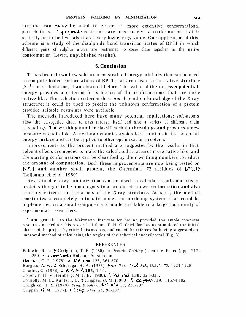
PROTEIN FOLDING BY MINIMIZATION 7 63
method can easilv be used to generate more extensive conformationalperturbations. Appiopriate restraints are used to give a conformation that issuitably perturbed yet also has a very low energy value. One application of thisscheme is a study of the disulphide bond transition states of BPTI in whichdifferent pairs of sulphur atoms are restrained to come close together in the nativeconformation (Levitt, unpublished results).
6. Conclusion
Tt has been shown how soft-atom constrained energy minimization can be usedto compute folded conformations of BPTI that are closer to the native structure(3 a r.m.s. deviation) than obtained before. The value of the in vacua potentialenergy provides a criterion for selection of the conformations that are morenative-like. This selection criterion does not depend on knowledge of the X-raystructure; it could be used to predict the unknown conformation of a proteinprovided suitable restraints were available.
The methods introduced here have many potential applications: soft-atomsallow the polvpeptide chain to pass through itself and give a variety of different, chainthreadings. *he writhing number classifies chain threadings and provides a newmeasure of chain fold. Annealing dynamics avoids local minima in the potentialenergy surface and can be applied to other optimization problems.
Improvements to the present method are suggested by the results in thatsolvent effects are needed to make the calculated structures more native-like, andthe starting conformations can be classified by their writhing numbers to reducethe amount of computation. Both these improvements are now being tested onBPTT and another small protein, the C-terminal 72 residues of L7/L12(Leijonmarck et al., 1980).
Restrained energy minimization can be used to calculate conformations ofproteins thought to be homologous to a protein of known conformation and alsoto study extreme perturbations of the X-ray structure. As such, the methodconstitutes a completely automatic molecular modeling system- that could beimplemented on a small computer and made available to a large community ofexperimental researchers.
I am grateful to the Weizmann Institute for having provided the ample computerresources needed for this research. I thank F. H. C. Crick for having stimulated the initialphases of the project by critical discussions, and one of the referees for having suggested animproved method of calculating the angles of the spherical quadrilateral (Fig. 3).
REFERENCESBaldwin, R. L. & Creighton, T. E. (1980). In Protein Folding (Jaenicke. R.. ed.), pp. 217-
259, Elsevier/Eorth Holland, Amsterdam.Benham, C. J. (1978). J. i&ZoZ. Biol. 123, 361-370.Burgess, A. W. & Scheraga, H. A. (1975). Proc. Nat. Acad. Sci., U.S.A. 72. 1221-1225.Chothia, C. (1976). J. Mol. BioZ. 105, 1-14.Cohen, F. H. & Sternberg, M. J. E. (1980). J. Mol. BioZ. 138, 32 l-333.Connolly, M. L., Kuntz, I. D. & Crippen, G. M. (1980). Biopolymers, 19, 1167-l 182.Creighton. T. E. (1978). Prog. Biophys. Mol. BioZ. 33, 231-297.Crippen, G. M. (1977). J. Camp. Phys. 24, 96-107.
76-1 M. LEVITT
Davidon, W. C. (1959). A .E.C. Research and Development report, AM,-5990, @&ridgeNational Laboratory.
Deisenhofer, J. & Steigemann, W. W. (1975). Acta Crystallogr. sect. B, 31, 238-250.Fletcher, R. (1970). Computer J. 13, 317-322.Fletcher, R. & Powell, M. J. D. (1963). Computer J. 6, 163-168.Fletcher, R. & Reeves, C. M. (1964). Computer J. 7, 149-154.Fuller, F. B. (1971). hoc. Nat. Acad. Sci., U.S.A. 68, 815-819.Gelin, B. R., & Karplus, M. (1975). Proc. Nat. Acad. Sci., U.S. A. 72, 2002-2006.Gibson, K. D. & Scheraga, H. A. (1969). Proc. Nat. Acad. Sci., U.S.A. 63, 9-15.Goel, N. S. & YEas, M. (1979). J. Theoret. Biol. 77, 253-305.Greer, J. (1980). Proc. Nat. Acad. Sci., U.S.A. 77, 3393-3397.Hagler, A. T. & Honig, B. (1978). Proc. Nat. Acad. Sci., U.S. A. 75, 554-558.Have], T. F., Crippen, G. M. & Kuntz, I. D. (1979). Biopolymers, 18, 73-81.Hendrickson, J. B. (1961). J. k4mer. Chem. Sot. 83, 4537-4547.Hestenes, M. R. & Stiefel, E. (1952). J. Res. Nat. Bur. Stand. 49, 409-436.Huber, R., Kukla, I)., Ruhlmann, A. & Steigemann, W. (1971). Cold Spring Harbor Symp.
Quant. BioZ. 36, 141-150.Janin, J.. Wodak, S., Levitt, M. & Maigret. B. (1978). J. Mol. BioZ. 125, 357-386.Kabsch, W. (1976). Acta Crystallogr. sect. A, 32, 922-923.Kendrew. J. C.? Dickerson, R. E., Strandberg. B. E.! Hart! R. G.. Davies. D. R., Phillips,
D. C. & Shore, V. C. (1960). Nature (London), 185. 422427.Kuntz, I. D., Crippen, G. M., Kollman, P. A. & Kimelman, D. (1976). J. Mol. BioZ. 106,
983-994.Kuntz, I. D., Crippen, G. M. & Kollman, P. A. (1979). Biopolymers, 18, 939-957.Le Bret, M. (1979). Biopolymers 18, 1’709-1725.Lee, B. & Richards, F. H. (1971). J. Mol. BioZ. 55, 379400.Leijonmarck, M., Ericksson, S. & Liljas, A. (1980). Nature (London), 286, 824-826.Levinthal? C. (1968). J. Chim. Phys. 65, 44-45.Levitt, M. (1976). J. Mol. BioZ. 104, 59-107.Levitt, M. (1980). In Protein Folding (Jaenicke, R., ed.), pp. 17-39, Elsevier/North
Holland, Amsterdam.Levitt. M. (1981a). J. MoZ. Biol. 145, 251-263.Levitt, M. (1981 b). Nature (London), 294, 379-380.Levitt, M. (1982). Annu. Rev. Biophys. Bioeng. 11, 251-271.Levitt. M. (1983a). CoZd Spring Harbor Symp. &uant. BioZ. 47. 251-262.Levitt, M. (19833). J. Mol. Biol. 168, 595-620.Levitt, M. (1983c). J. Mol. BioZ. 168, 621-657.Levitt, M. & Warshel, A. (1975). Nature (London), 253: 694-698.Lifson, S. & Warshel. A. (1968). J. Chem. Phys. 49, 5116-5129.MacKay, A. L. (1974). Acta Crystallogr. sect. A, 30, 440447.McCammon, J. A., Gelin, B. R.. Karplus, M. & Wolynes, P. G. (1976). Nature (London),
262, 325-327.McCammon, J. A., Gelin, B. R. & Karplus, M. (1977). Nature (London), 267, 585-589.Meirovitch, H. & Scheraga, H. A. (1981). Proc. Nat. Acad. Sci., U.S.A. 78, 6584-6587.Padlan, E. A., Davies, D. R., Pecht, I., Givol, D. & Wright, C. E. (1976). Cold Spring
Harbor Symp. Quant. Biol. 41. 627-637.Pottle, C., Pottle, M. S., Tuttle, R. W., Kinch, R. J. & Scheraga, H. A. (1980). J. Comp.
Chem. 1, 46-58.Rackovsky, S. & Scheraga? H. *!I. (1980). MacromolecuLes, 13, 1440-1453.Robson, B. & Osguthorpe. .D. J. (1979). J. Mol. BioZ. 132, 19-51.Sela, M.. White, F. H. & Anfinsen, C. B. (1957). Science, 125, 691-692.van Gunsteren, W. F. & Karplus, M. (1980). J. Comp. Chem. 1. 266-274.Wiithrich, K., Wider, G., Wagner, G. & Braun, W. (1982). J. Mol. BioZ. 155. 311-319.
Edited by S. Brenner