protein structure. each protein has a characteristic shape, size and function : 1. structural...
TRANSCRIPT

PROTEIN STRUCTURE
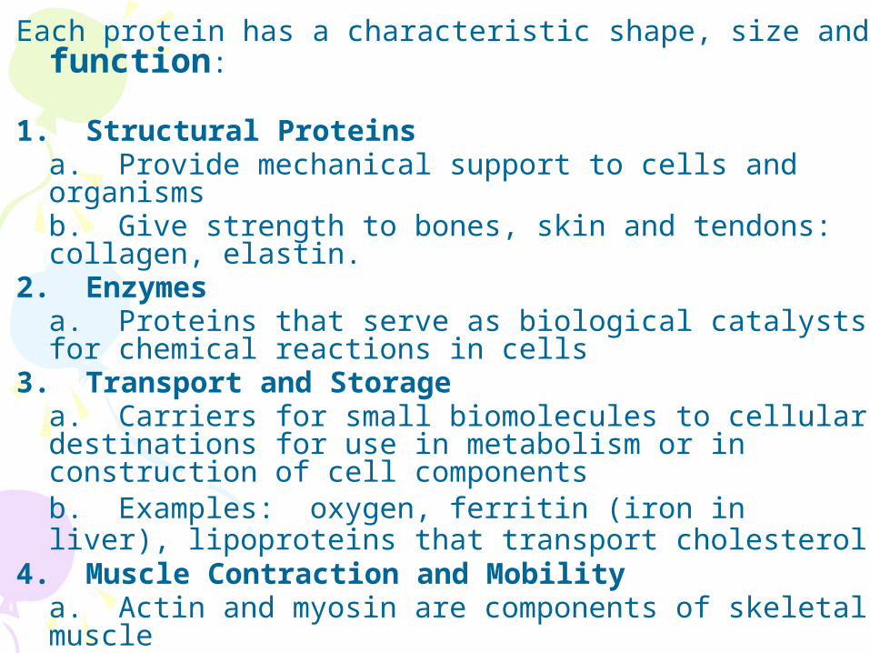
Each protein has a characteristic shape, size and function:
1. Structural Proteins
a. Provide mechanical support to cells and organisms b. Give strength to bones, skin and tendons: collagen, elastin.
2. Enzymes a. Proteins that serve as biological catalysts for chemical reactions in cells
3. Transport and Storage a. Carriers for small biomolecules to cellular destinations for use in metabolism or in construction of cell components b. Examples: oxygen, ferritin (iron in liver), lipoproteins that transport cholesterol
4. Muscle Contraction and Mobility a. Actin and myosin are components of skeletal muscle
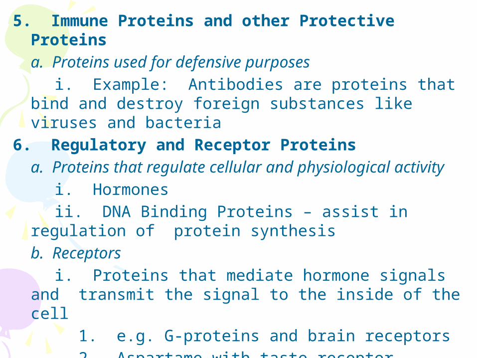
5. Immune Proteins and other Protective Proteins a. Proteins used for defensive purposes
i. Example: Antibodies are proteins that bind and destroy foreign substances like viruses and bacteria
6. Regulatory and Receptor Proteins a. Proteins that regulate cellular and physiological activity
i. Hormones ii. DNA Binding Proteins – assist in regulation of protein synthesis
b. Receptors i. Proteins that mediate hormone signals and
transmit the signal to the inside of the cell 1. e.g. G-proteins and brain receptors 2. Aspartame with taste receptor


COMMON THEME: RECOGNITION!!
**Proteins have three-dimensional shapes!!
- Proteins interact selectively with other proteins or molecules through NON-covalent interactions in oder to function.
- Reactant with enzyme - Transported molecule with transporter - Protein-protein interactions - Protein-DNA interactions - Non-covalent interactions: • Electrostatic interactions • Sterics • Van der Waals interactions • Hydrogen bonds


PROTEIN STRUCTURE On the basis of shape and solubility, proteins :
1) Can be globular o Spherical or near-spherical o Soluble in water – lots of charged groups o Dynamic and flexible
2) Can be fibrous o Elongated and threadlike o Not soluble in water – lots of hydrophobic,
non-polar R groups o Tough o Examples: hair, nails, skin
3) Can be membrane proteins - Can be monomers – single polypeptide chain - Can be oligomers – multiple polypeptide chains
o Held together by non-covalent interactions o Each polypeptide chain = subunit if part of
a complex

- Size expressed in terms of mass: units of Daltons or kilodaltons
- One Dalton = one atomic mass unit o Estimate size = # amino acids x
average molecular weight aa § 110 g/mole per amino acid

(a) Proteins having structural roles in cells are typically fibrous and often water insoluble. Collagen is composed of three polypeptide chains that intertwine.
(b) Soluble proteins serving metabolic functions can be characterized as compactly folded globular molecules, such as myoglobin.
(c) Membrane proteins fold so that hydrophobic aa side chains are exposed in their membrane-associated regions. Bacteriorhodopsin is a typical membrane protein; it binds the light-absorbing pigment, cis-retinal


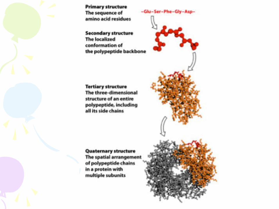
FOUR LEVELS OF PROTEIN STRUCTURE- Primary (1°) o Linear sequence of
amino acids in a protein
- Secondary (2°) o Local 3-dimensional
structure of the PEPTIDE BACKBONE
- Tertiary (3°) o Global arrangement
of secondary structure, side chains (R groups), and other prosthetic groups (e.g. metals)
- Quaternary (4°) o Arrangement of
multiple proteins into complexes
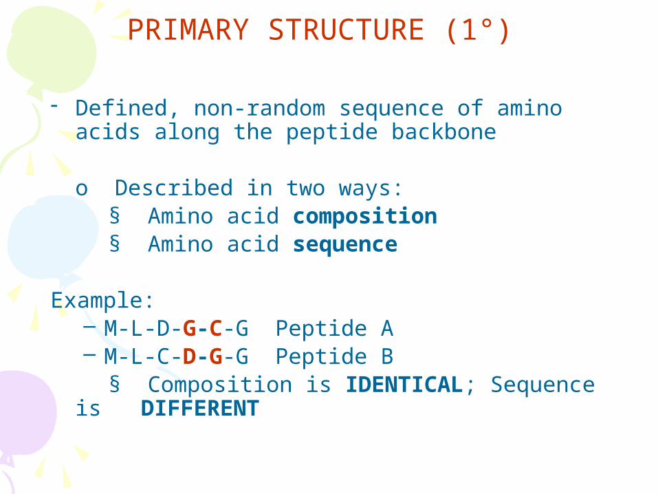
PRIMARY STRUCTURE (1°)
- Defined, non-random sequence of amino acids along the peptide backbone
o Described in two ways: § Amino acid composition § Amino acid sequence
Example: – M-L-D-G-C-G Peptide A – M-L-C-D-G-G Peptide B
§ Composition is IDENTICAL; Sequence is DIFFERENT
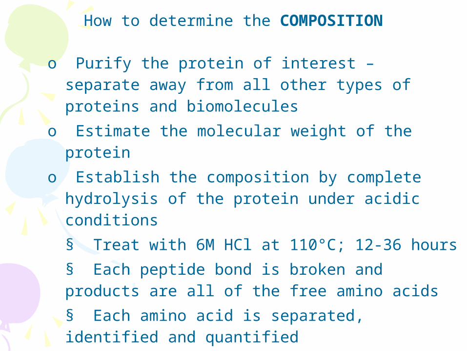
How to determine the COMPOSITION
o Purify the protein of interest – separate away from all other types of proteins and biomolecules
o Estimate the molecular weight of the protein
o Establish the composition by complete hydrolysis of the protein under acidic conditions
§ Treat with 6M HCl at 110°C; 12-36 hours
§ Each peptide bond is broken and products are all of the free amino acids
§ Each amino acid is separated, identified and quantified
§ Final result: Know HOW MANY of each amino acid present in the original


How to determine the ORDER
o Determine the C-terminal amino acid
§ Use carboxypeptidase – enzyme that removes the last (C-terminal) amino acid in a free form by breaking the peptide bond
• Hydrolyzes the peptide bond nearest the C-terminus o Identify the N-terminal amino acids in order
§ Process called SEQUENCING § Often difficult to characterize an intact protein § Instead, employ a “divide and conquer” approach to
analyze peptide fragments of the intact protein§ Cut large proteins into smaller parts § Use enzymes called PROTEASES
• Cleave peptide bond in a specific way
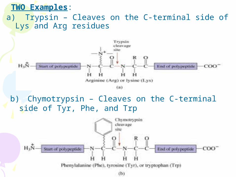
TWO Examples: a) Trypsin – Cleaves on the C-terminal side of Lys and
Arg residues
b) Chymotrypsin – Cleaves on the C-terminal side of Tyr, Phe, and Trp
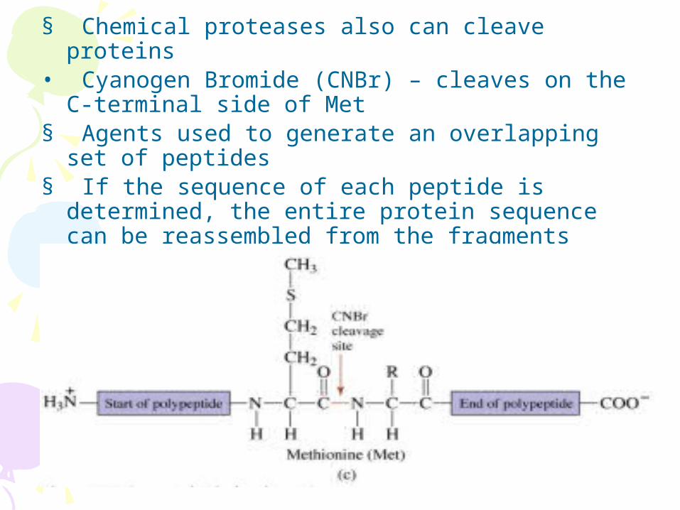
§ Chemical proteases also can cleave proteins • Cyanogen Bromide (CNBr) – cleaves on the C-
terminal side of Met § Agents used to generate an overlapping set of
peptides § If the sequence of each peptide is determined, the
entire protein sequence can be reassembled from the fragments


SECONDARY STRUCTURE (2°): HYDROGEN BONDING IS KEY!
- Three-dimensional structure of the peptide backbone
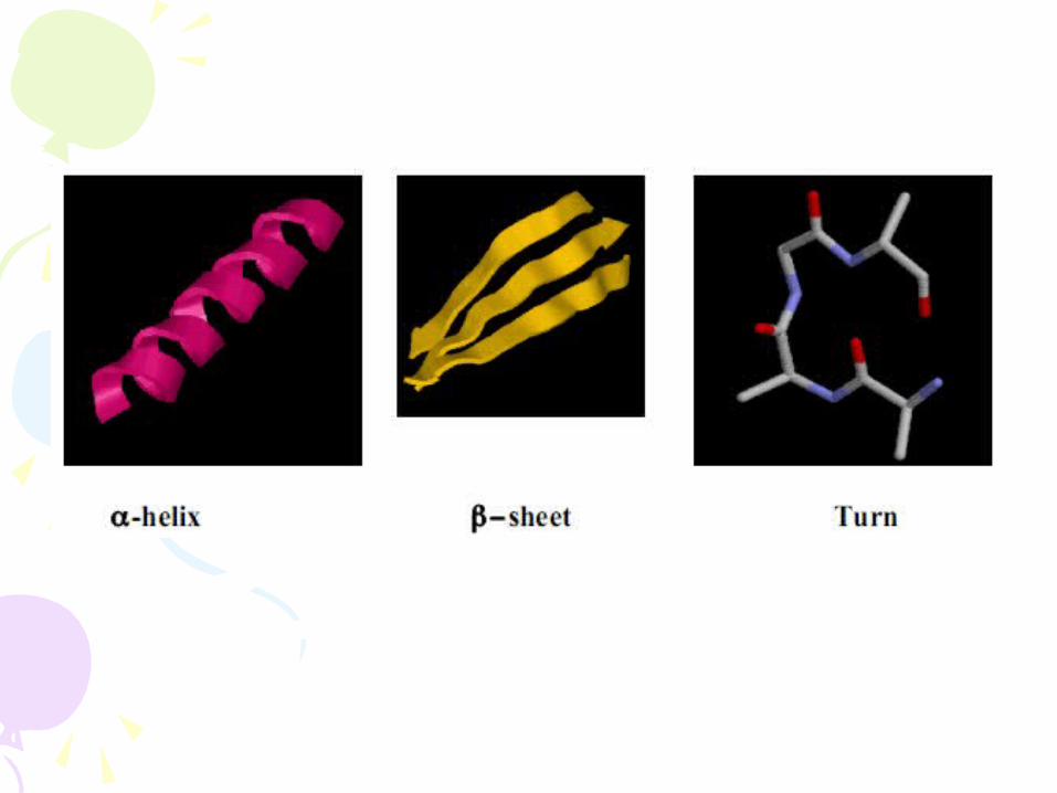
- 3 major classes of secondary structure are dictated by the RIGIDITY a PLANARITY of the peptide bond and the nature of the side chains o α-helix o β-sheet o turns and random coil

1) a-helix - Rod-like structure (phone cord) - Involves only one polypeptide chain - Main chain atoms on the INSIDE - R-group side chains on the outside – stick out - Stabilized by HYDROGEN BONDS - Carbonyl (C = O) of each amino acid is H-bonded to
the amide (N-H) of the aa that is 4 aas further toward the C-terminus n+4 (e.g. aa 1 is H-bonded to aa 5 – see model)
- a-helices have sidedness: n+4 on the same side of the helix
- 3.6 aa’s per turn (about 4aa) - Pitch of the helix (i.e. one turn = 5.4 Å) - Overall dipole moment – positively charged N-
terminus -> negatively charged C-terminus - Helices can be right or left handed - § PROTEINS ARE RIGHT HANDED

- Factors that affect stability of an a-helix
Although the helix is defined by the H-bonding of the peptide backbone, the nature of the side chains can affect overall stability
§ Adjacent bulky amino acids unfavorable (steric hindrance)
§ Proline unfavorable – creates bends; helix-breaker
§ Glycine unfavorable – too mobile (no side chain but H)
§ Too many + or – charged groups near each other in space are unfavorable – electrostatic repulsion
•
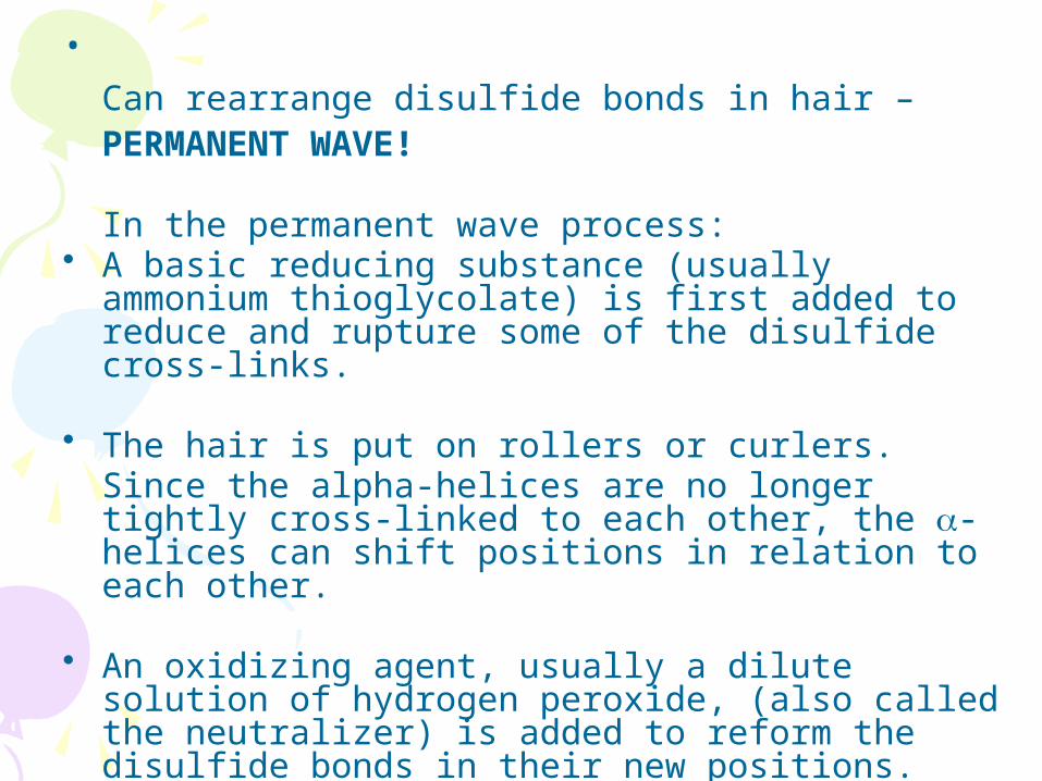
Can rearrange disulfide bonds in hair – PERMANENT WAVE!
In the permanent wave process:• A basic reducing substance (usually ammonium
thioglycolate) is first added to reduce and rupture some of the disulfide cross-links.
• The hair is put on rollers or curlers. Since the alpha-helices are no longer tightly cross-linked to each other, the a-helices can shift positions in relation to each other.
• An oxidizing agent, usually a dilute solution of hydrogen peroxide, (also called the neutralizer) is added to reform the disulfide bonds in their new positions.

2) β−sheet Proteins with major β-pleated sheet secondary structure are generally fibrous, such as silk, but pleated sheet is observed as a significant part of secondary stucture in other proteins.
- Generally have rod-like shapes and are not so soluble in water.
- β sheets consist of β strands connected laterally by
at least two or three backbone hydrogen bonds, forming a generally twisted, pleated sheet.
- Unlike the α-helix, β-sheets can involve one or more
polypeptide chains – interchain or intrachain interactions
- A β-strand is a stretch of polypeptide chain typically 3
to 10 amino acids long with the peptide backbone almost completely extended

- R-groups stick UP and DOWN from β-sheets – alternating on either side of the strand
o Usually small compact side chains like Gly, Ser, Ala
o In an amphipathic β-sheet the amino acids alternate with polar/charged and non-polar (hydrophobic) aas.
Thus, the hydrophobic residues are on one side and the polar/charged are on the other.
o Stabilized by Hydrogen bonds (near perpendicular to direction of peptide backbone)
o Carbonyl of each aa is H-bonded to the NH of another aa.

Beta Sheet

Parallel and Anti-Parallel β-sheets
- Adjacent chains can be PARALLEL or ANTI-PARALLEL
- Parallel β−sheet: H-bonds between 2 β−strands running in the same direction
- Anti-parallel β−sheet: H-bonds between 2 β−strands running in opposite directions

ANTI-PARALLEL BETA SHEET

PARALLEL BETA SHEET

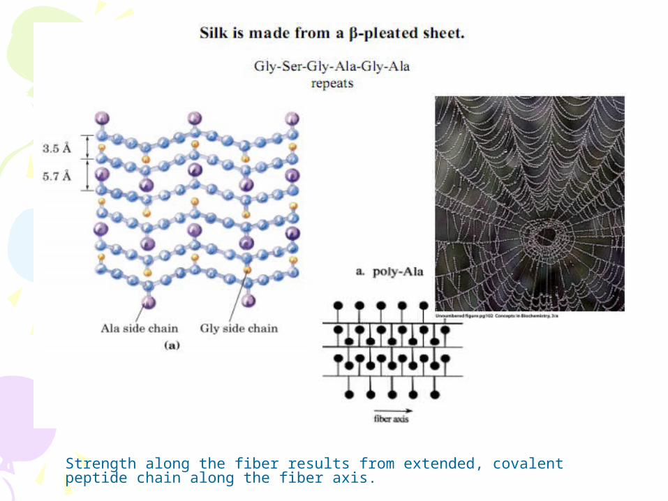
Strength along the fiber results from extended, covalent peptide chain along the fiber axis.

2) Loops or Turns
- Small regions of peptide backbone that can form small loops
- Often contain Gly (small and mobile) and Pro (causes kinks)
- Reverse direction of the main polypeptide chain - Connects regions of more regular secondary
structure - Not periodic; irregular - Extended bend = loop and contains 6 -16 aas; ~10 Å
long
- β-bend or hairpin turn – connects anti-parallel β-sheets. Example below. Forms a loop to change direction in the polypeptide chain.

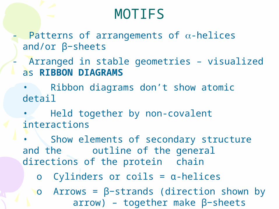
MOTIFS- Patterns of arrangements of a-helices and/or
β−sheets
- Arranged in stable geometries – visualized as RIBBON DIAGRAMS
• Ribbon diagrams don’t show atomic detail
• Held together by non-covalent interactions
• Show elements of secondary structure and the outline of the general directions of the protein chain
o Cylinders or coils = α-helices
o Arrows = β−strands (direction shown by arrow) – together make β−sheets
o Ribbons = bends, loops and random

β−sheets (each arrow is a β−strand)

EXAMPLES
a) aa motif (helix-loop-helix motif)\ Helix–loop–helix
b) βαβ motif parallel (Note that the beta strands are parallel (direction of the arrows)

Fibrous Proteins - Usually perform a structural role - Most prominent structural protein = collagen (major protein in skin, bone & tendons) - Collagen contains repeating units Pro – Gly – X OR Hyp (hydroxyproline) – Gly – X - Rich in Pro, therefore unable to fold into a-helices or
β-sheets - Form a triple helix – three extended helical chains wrapped together - Rope-like structure - Helices held together by hydrogen bonding and covalent cross-links - High tensile strength


TERTIARY STRUCTURE (3°) Global 3-dimensional arrangement of ALL atoms in a protein
o Includes: § 2° structural elements (alpha helices and beta sheets) § Amino acid side chains § Prosthetic groups
Small organic molecule or metal ion associated with a protein
o Regions of SECONDARY structure INTERACT to give a
protein it TERTIARY structure - Major forces stabilizing tertiary structure are
hydrophobic interactions among nonpolar side chains in the compact core of the proteins.


QUATERNARY STRUCTURE (4°)
- Arrangement of multiple protein molecules into COMPLEXES
- The three dimensional structure of a protein made of >1 polypeptide
- Complexes of 2, 3, 4 etc… protein molecules are called dimers, trimers, tetramers…oligomers
- Oligomers may be: o Formed with identical protein monomers = HOMOOLIGOMER o Formed with different protein monomers = HETEROOLIGOMERS o Example: Hemoglobin:
§ 2 alpha subunits § 2 beta subunits
- Protein subunits of oligomers interact through NON-COVALENT interactions

Liver Alcohol Dehydrogenase (2 subunits) Can be either a homo- or heterodimer
Hemoglobin (4 subunits) Heterotetramer

What are the structural and functional advantages driving
quaternary association?
• Stability: reduction of surface to volume ratio
• Genetic economy and efficiency
• Bringing catalytic sites together
• Cooperativity

PROTEIN FOLDING Goal: To achieve the LOWEST energy state
Formation of hydrophobic domains often the primary driving force for tertiary structure
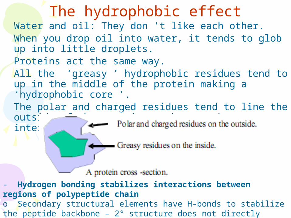
The hydrophobic effectWater and oil: They don ’t like each other. When you drop oil into water, it tends to glob up into little droplets.Proteins act the same way. All the ‘greasy ’ hydrophobic residues tend to up in the middle of the protein making a ‘hydrophobic core ’.The polar and charged residues tend to line the outside of the protein as they are happy interacting with water.
- Hydrogen bonding stabilizes interactions between regions of polypeptide chain o Secondary structural elements have H-bonds to stabilize the peptide backbone – 2° structure does not directly involve side chains

What forces determine the structure?
• Primary structure - determined by covalent bonds
• Secondary, Tertiary, Quaternary structures - all determined by weak forces
• Weak forces: – H-bonds,– ionic interactions,– van der Waals interactions, – hydrophobic interactions

Hydrogen bonds
- Hydrogen bonds occur when a proton (hydrogen) is shared between a donor group and the unpaired electrons of an acceptor oxygen.
-Proteins fold such that all hydrogen bonding groups participate in a hydrogen bond.

Other forces involving side chains that influence how proteins fold: o Metal ion coordination to negatively charged amino acid side chains o Hydrophobic interactions between NON-POLAR side chains
§ Favored on interior – not exposed to water o Ionic/Electrostatic Interactions between charged side chains
§ Favored on the outside § Sometimes on inside if near opposite charge
o Hydrogen bonding among side chains of polar amino acids
o Disulfide bridges between Cys aas stabilize tertiary structure COVALENTLY (only covalent interaction – rest are non-covalent) Note: Once folded, proteins are not rigid; highly dynamic

Weak Forces are Responsible for Protein Folding
What are they? What are the relevant numbers?
• van der Waals: 0.4 - 4 kJ/mol
• hydrogen bonds: 12-30 kJ/mol
• ionic bonds: 20 kJ/mol
• hydrophobic interactions: <40 kJ/mol

FORCES THAT STABILIZE STRUCTURE OF PROTEINS

How do we determine the 3-D structure? 1. X-ray crystallography – use crystal of pure protein 2. NMR (2-D NMR) – measures magnetic characteristics
of each atom - Both methods are extremely difficult and require lots
of computer power to make sense of data

CAN WE UNFOLD PROTEINS ONCE THEY ARE FOLDED? YES!
- Proteins can be unfolded = DENATURED
o Lose most levels of structure
o Protein adopts a random coil conformation
o Primary amino acid sequence is maintained
o Loss of protein function – enzymatic etc…
- Go from NATIVE (correctly folded, biologically
active state) to DENATURED and UNFOLDED
UNFOLDED (loss of organized structure and
function)

Use denaturing agents: Interfere with the forces that stabilize protein folding

Is this process REVERSIBLE? – i.e. can we restore a protein, once denatured to its original configuration and restore function?
• Yes – Denaturation CAN BE reversible
o Heat treatment usually is not reversible

- The renaturation of the protein RIBONUCLEASE A (an enzyme that cleaves DNA) won Christian Anfinsen the Nobel Prize in 1972.
- Experiment:
1. Denatured pure Ribonuclease A by treatment with UREA and β-mercaptoethanol to give a completely unfolded, denatured protein
o β-mercaptoethanol used to reduce disulfide bonds
o Urea breaks H-bonds and hydrophobic interactions
2. Then he removed the denaturants and exposed the protein to air
3. The protein had folded back into its original 3-D shape and activity was restored!!

Anfinsen’s Ribonuclease A Denaturation and Renaturation Experiment
ANFINSEN: AMINO ACID SEQUENCE DETERMINES PROTEIN SHAPE

*This experiment suggested that the unfolded polypeptide refolded by itself in the test tube
* Further experiments determined that it DID refold back to its original state
CONCLUSION: All the necessary information as to how a protein folds is encoded into the primary sequence!
1° SEQUENCE DICTATES 2° AND 3° STRUCTURE!

Primary Sequence = Structure
Proteins can self-assemble! All the information needed to make a working 3-D machine is encoded in the amino acid sequence!
Unfortunately, we haven’t figured out the code yet. We can’t effectively predict 3-D structure of a protein from looking at the primary aa sequence.

Molecular Chaperones
• Why are chaperones needed if the information for folding is inherent in the sequence?
– to protect nascent proteins from the concentrated protein matrix in the cell
– perhaps to accelerate slow steps
• Chaperone proteins were first identified as "heat-shock proteins" (hsp60 and hsp70)

Other Chemical Groups in Proteins
Proteins may be "conjugated" with other chemical groups
• If the non-amino acid part of the protein is important to its function, it is called a prosthetic group.
• Be familiar with the terms: – glycoprotein, – lipoprotein, – nucleoprotein, – phosphoprotein, – metalloprotein, – hemoprotein, – flavoprotein.

Some Proteins Have Chemical Groups Other Than Amino Acids
– Simple proteins: consist of only aa and contain no other chemical groups.
(Examples: the enzyme ribonuclease and the contractile protein actin).
– Conjugated proteins: contain various chemical constituents as an integral part of their structure.

Table 5.4
Representative Conjugated Proteins
Class Prosthetic Group
Percent by
Weight (approx.)
Glycoproteins contain carbohydrate Fibronectin -Globulin Proteoglycan
Lipoproteins contain lipid Blood plasma lipoproteins: High density lipoprotein (HDL) (-lipoprotein) Low density lipoprotein (LDL) (-lipoprotein)
Triacylglycerols, phospholipids, cholesterol Triacylglycerols, phospholipids, cholesterol
75 67
Nucleoprotein complexes contain nucleic acid Ribosomes Tobacco mosaic virus Adenovirus HIV-1 (AIDS virus)
RNA RNA DNA RNA
60 5
Phosphoproteins contain phosphate Casein Glycogen phosphorylase
Phosphate groups Phosphate groups
Metalloproteins contain metal atoms Ferritin Alcohol dehydrogenase Cytochrome oxidase Nitrogenase Pyruvate carboxylase
Iron Zinc Copper and iron Molybdenum and iron Manganese
35
Hemoproteins contain heme Hemoglobin Cytochrome c Catalase Nitrate reductase Ammonium oxidase

Prosthetic group: the nonprotein part is crucial to the protein’s function
• If the nonprotein moiety is not covalently linked to the protein, it can usually be removed by denaturing the protein structure.
• If the conjugate is covalently joined to the protein, it may be necessary to carry out acid hydrolysis of the protein into its component aa in order to release it.
• Conjugated proteins are typically classified according to the chemical nature of their nonamino acid component

• Glycoproteins
– are proteins that contain carbohydrate
– are characteristically destined for an extracellular location
For example:• fibronectin and proteoglycans are important
components of the extracellular matrix that surrounds the cells of most tissues in animals.
• Immunoglobulin G molecules are the principal antibody species found circulating free in the blood plasma.
• Many membrane proteins are glycosylated on their extracellular segments.

• Lipoproteins:
• the class of proteins conjugated with lipid
Examples: Blood plasma lipoproteins:
– function primarily in the transport of lipids to sites of active membrane synthesis.
– Serum levels of low density lipoproteins (LDLs) are often used as a clinical index of susceptibility to vascular disease.

• Nucleoproteins:
-have many roles in the storage and transmission of genetic information.
– Ribosomes are the sites of protein synthesis.
– Virus particles and even chromosomes are protein-nucleic acid complexes.
• Phosphoproteins
– have phosphate groups esterified to the hydroxyls of Ser,Thr, or Tyr residues.
– Casein, the major protein of milk, contains many phosphates and serves to bring essential phosphorus to the growing infant.
– Many key steps in metabolism are regulated between states of activity or inactivity, depending on the presence or absence of phosphate groups on proteins.
Glycogen phosphorylase a is one well-studied example.
• Metalloproteins
are either metal storage forms (as in the case of ferritin) or enzymes in which the metal atom participates in a catalytically important manner.
There are many examples of the vital metabolic functions served by metalloenzymes.

• Hemoproteins
• are actually a subclass of metalloproteins because their prosthetic group is heme, the name given to iron protoporphyrin IX (Fig. 5.15).
• Because heme-containing proteins enjoy so many prominent biological functions, they are considered a class by themselves.
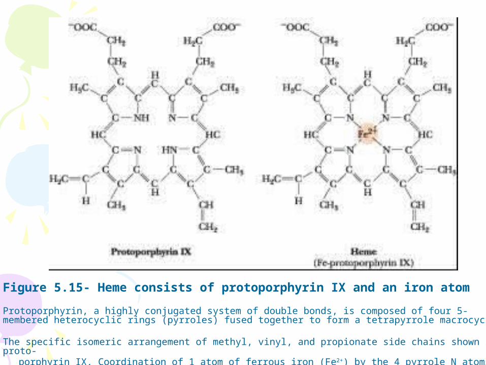
Figure 5.15- Heme consists of protoporphyrin IX and an iron atom
Protoporphyrin, a highly conjugated system of double bonds, is composed of four 5-membered heterocyclic rings (pyrroles) fused together to form a tetrapyrrole macrocycle.
The specific isomeric arrangement of methyl, vinyl, and propionate side chains shown is proto- porphyrin IX. Coordination of 1 atom of ferrous iron (Fe2+) by the 4 pyrrole N atoms yields heme
• Flavoproteins
Flavin is an essential substance for the activity of a number of important oxidoreductases.
(We discuss the chemistry of flavin and its derivatives, FMN and FAD, in the chapter on electron transport and oxidative phosphorylation)

Sickled and Normal Red Blood Cells

STRUCTURE OF HEMOGLOBIN
Tetramer (4 subunits)
o 2 alpha (α) subunits
o 2 beta (β) subunits
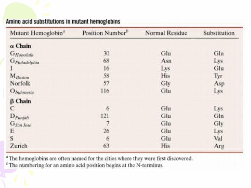
o Mutation occurs in the beta subunit (Glu -> Val; position 6)
o Sickle cell has 2 abnormal β-chains and 2 normal α-chains

Mutations:
- Most common
o Single a.a change from Glu -> Val at position 6
o Places hydrophobic side chain on surface of the protein
o When deoxygenated, having this hydrophobic group on the surface causes a decrease in protein solubility and rod-like structure production
- Heterozygotes
o Carriers without symptoms
o Selective advantage
§ Survive malarial outbreaks
- Homozygotes – have the disease

A sickled red cell filled with sickle hemoglobin fibers
