proteomic analyses of cells isolated by laser microdissection
TRANSCRIPT

3
Proteomic Analyses of Cells Isolated by Laser Microdissection
Valentina Fiorilli1, Vincent P. Klink2 and Raffaella Balestrini1* 1Istituto per la Protezione delle Piante del CNR
and Dipartimento Biologia Vegetale dell’Università di Torino, Torino
2Department of Biological Sciences, Harned Hall, Mississippi State University - Mississippi State
1Italy 2USA
1. Introduction
Living organisms conduct biological processes by transducing biotic and abiotic stimuli through gene regulation into well-orchestrated growth and development. Analyzing RNA from a transcribed genome (the transcriptome) is fairly easy due to the availability of various nucleotide sequencing technologies. The translation of a transcriptome provides a blueprint of tens of thousands of different proteins that is known as the proteome (Wasinger et al., 1995). The analysis of proteins from whole tissues, organs or organisms has been made easier thanks to various technologies, including the Edman degradation technique, which is used in sequencing polypeptides (Edman, 1950), and 2 dimensional gel electrophoresis (2-DE), which could resolve up to 10,000 polypeptides (Barrett & Gould, 1973; O'Farrell, 1975; O'Farrell et al., 1977). Thus, the problems of assaying a transcriptome and a proteome from samples isolated from tissues, organs or whole organisms have largely been overcome. However, although it is fairly easy to assay a transcriptome and a proteome at these higher levels of biological organization, assaying a proteome at a cellular resolution level involves a set of problems that in primarily centered on the ability to collect sufficient cells for meaningful studies. The central focus of this chapter is a discussion on the technologies that have allowed the proteomic analyses of cells, isolated from complex samples thanks to a procedure that was first called laser microdissection (Isenberg et al., 1976).
2. The diversity of genome activity
Thanks to the recent advances in high-throughput technologies, the past decade has witnessed an explosion of global transcriptome profiling studies, which have produced novel insights into many developmental, physiological and medicinal aspects. Although a great deal of information can be obtained from transcriptome profiling, it is however insufficient for a comprehensive delineation of biological systems. A single approach cannot fully
* Corresponding Author
www.intechopen.com

Integrative Proteomics
48
unravel the complexity of living organisms (Persidis, 1998). In addition, enzymatic reactions and signaling pathways depend on the activity of proteins, and protein quantities are regulated by protein synthesis and degradation. These processes may be independent of transcriptional control or have only a weak correlation (Lu et al., 2007; Nie et al., 2007). By generating information on the proteome at cellular resolution, a greater understanding of biological complexity is gained, including post-translational modification, isoforms, and splice variants, which may lead to the identification of important cell-specific protein entities (Schulze & Usadel, 2010). The proteomics approach can shed light on a number of protein species that can be translated from a single gene as a result of alternative splicing (AS) or PTMs. Proteomics analyses can also provide the biological meaning of each variant (Kim et al., 2007; Witze et al., 2007). For example, the Drosophila Dscam1 gene, which encodes a membrane receptor protein, has 115 exons. The various combinations permit the possibility of 38,016 different proteins to be produced and many have been identified (Schmucker et al., 2000; Chen et al., 2006; Meijers et al., 2007). On the basis of large-scale EST-cDNA alignments and bioinformatics analyses on the genomes of Arabidopsis thaliana (thale cress) and Oryza sativa (rice), it has been estimated that approximately 30–35% of the their genes are alternatively spliced (Cambpell et al., 2006; Xiao et al., 2005), while in humans up to 95% of multi-exon genes undergo alternative splicing (Pan et al., 2008). The number of alternatively spliced genes in plants is still likely to be underestimated because of the relatively low EST coverage and depth of sequencing of many plant transcripts (Simpson et al., 2008; Xiao et al., 2005). Extensive AS variation has been shown in some Arabidopsis-specific gene families, for example in genes encoding serine/arginine-rich proteins, and this results in a five-fold increase in transcriptome complexity (Palusa et al., 2007; Tanabe et al., 2007). In addition, stress conditions seem to dramatically alter the splicing pattern of many plant genes (Ali & Reddy, 2008). For these reasons, there is growing interest in complementing transcriptomic studies with proteomics, which should be considered as part of a multidisciplinary integrative analysis that extend from the gene to the phenotype through proteins.
3. Developmental plasticity of protein complexes
Many processes and structures are composed of protein complex aggregates. Protein complexes can vary in size and composition, and range from mega-Dalton assemblies of dozens of proteins (such as the ribosome and the spliceosome) to smaller clusters of just a few proteins. The composition and stability of protein complexes is highly regulated in both a context dependent manner, such as cell-type-specific differences, and a time-dependent manner (Michnick et al., 2004). This biological variability of proteins and their range of physicochemical properties reflect the difficulty of characterizing the structure and the function of protein complexes (Cravatt et al., 2007). In addition, in proteomics, the sample amount is often a limiting factor since, unlike transcript profiling, proteomic approaches cannot benefit from amplification protocols. It should be evident that sensitivity, resolution and speed in data capture are all significant problems with proteomics techniques. In order to circumvent these problems, methods have been developed to extract, separate, detect and identify a wide range of proteins from small sample amounts (Gutstein & Morris, 2007). Technical advances in mass spectometry have facilitated major progress in both the qualitative and quantitative analysis of proteins (Kaspar et al., 2010). Most of these improvements have occurred over the last decade and proteomics has developed a broad range of new protocols, platforms and workflows.
www.intechopen.com

Proteomic Analyses of Cells Isolated by Laser Microdissection
49
4. Proteomic workflow
The workflow of a standard proteomics experiment is crucial for the success of any experiment and it usually includes a good experimental design, an appropriate extraction/fractionation/purification protocol that considers the needs of different samples (tissue/cells or organelle), a suitable separation protocol, protein identification, statistical analysis and validation. The use of proteomics in plant biology research has increased significantly over the last few years with an improvement in both quality and quantitative analysis, inaugurating a new phase of “Second Generation Plant Proteomics” (Jorrín et al., 2009). This growing interest in plant proteomics has continually produced a large number of developmental studies on plant cell division, elongation, differentiation, and formation of various organs using various proteomics approaches (Hochholdinger et al., 2006; Takàč et al., 2011, Miernyk et al., 2011). Most of the studies published in the plant field concern the proteome of Arabidopsis and rice. The work has focused on profiling organs, tissues, cells, and/or subcellular proteomes (Rossignol et al., 2006; Komatsu et al., 2007; Jorrin et al., 2007; Jamet et al., 2008; Baerenfaller et al., 2008; Jorrin et al., 2009; Agrawal & Rakwal, 2011) and studying developmental processes and responses to biotic (Mehta et al., 2008) and abiotic stresses (Nesatyy & Suter 2008) using differential expression strategies. However, proteomics research results have recently appeared on several non-model herbaceous non-crop species, woody plants, fruit and forest trees (Table 1). Furthermore, over the past year, proteome analysis has increasingly been applied to the study of cereal grains with the aim of
model species
Lycosersicon esculentum tomato Sheoran 2007
Hordeum vulgare wheat Song et al., 2007
Glycine max soybean Djordjevic et al., 2007
Zea mays maize Dembinsky et al., 2007
Medicago truncatula alfalfa de Jong et al., 2007
non-model species
Elymus elongatum wheatgrass Gazanchian et al., 2007
Nicotiana alata jasmine tobacco Brownfield et al., 2007
Boea hygrometrica Jiang et al., 2007
Xerophyta viscose Ingle et al., 2007
Solanum chacoense chaco potato Vyetrogon et al., 2007
Citrullus lanatus wild watermelon Yoshimura et al., 2008
Citrus sinensis Lliso et al., 2007
Pinus nigra Australian pine Wang et al., 2006
Pinus radiate Californian pine Fiorani Caledon et al., 2007
Eucalyptus grandis rose gum eucalyptus Lippert et al., 2007
Picea sitchenisis sitka spruce Valledor et al., 2008
Pyrus communis conference pears Pedreschi et al., 2007
Table 1. Proteomics analyses perfomed on model and non-model plants
www.intechopen.com

Integrative Proteomics
50
providing knowledge that will facilitate the improvement of crop quality, either in terms of resistance to biotic and abiotic stress, or in terms of nutritional processing quality (Salekdeh & Komatsu, 2007; Finnie et al., 2011).
5. Proteomic approaches
Comparative plant proteomic approaches are still largely based on traditional two dimentional polyacrilamide gel electrophorsis (2D PAGE) with isoelectric focusing in the first dimension and SDS-PAGE in the second dimension. This technique was initially considered the most suitable method to visualize the differences between protein samples derived from samples grown under different conditions and/or from different tissues. Complex protein mixtures can be resolved efficiently, and the detection of differences in bands or spot intensities is intuitive. Currently, it is possible to visualize over 10,000 protein spots, corresponding to over 1,000 proteins, on single 2D gels (Görg et al., 2009). In many cases, however, individual spots may consist of more than one protein. The differences in spot composition can only be identified by means of mass-spectrometry. The quantitative mass-spectrometry-based proteomics field is constantly evolving, with continuous improvement in protocols, machines and software. Most of the early developments quantitative mass-spectrometry-based proteomic applications were driven by research on yeast and mammalian cell lines. However, in plant physiology analyses, mass spectrometry-based proteomics is no longer only used as a descriptive tool. Instead, well-designed quantitative proteomics has been applied to various aspects of organelle biology, growth regulation and signaling (Schulze & Usadel, 2010). These efforts have greatly improved our knowledge of protein diversity during complex processes. Encouraging pioneer studies on specific subproteomes in plants have revealed candidate proteins that are phosphorylated under specific stress conditions (Oda et al., 1999; Benshop et al., 2007; Niittylä et al., 2007) or during the light independent cycle of photosynthesis (Reiland et al., 2009). Protein abundance changes have been monitored in response to heat shock (Palmblad et al., 2008), during leaf senescence (Hebeler et al., 2008) and during the protein turnover of photosynthetic proteins, monitored using pulse-chase labeling in combination with mass-spectrometry (Nowaczyk et al., 2006). The combination of subcellular fractionation techniques and mass-spectrometry has led to the extensive characterization of the plant subcellular proteome which in turn has led to the discovery of new metabolic pathways (Dunkley et al., 2006). Organelle proteomes were also characterized, such as chloroplasts (Kleffmann et al., 2007; Mejaran et al., 2005; Peltier et al., 2000; Pevzner et al., 2001; Reiland et al., 2009) and plasma membranes and their microdomains (Kierszniowska et al., 2009; Nelson et al., 2006a).
6. Problems with proteomic analyses
Although quantitative methods and their results are desirable, the proteomics data that is usually produced is very complex and often variable in quality. The main problem is incomplete data, since the most advanced mass spectrometers cannot sample and fragment all the peptide ions that are present in complex samples. In fact, only a subset of the peptides and proteins present in a sample can be identified. The first step in primary data extraction is the manual validation of the identity of a peptide and quantification through the revision of the spectra assigned to each sequence. The identification of proteins through
www.intechopen.com

Proteomic Analyses of Cells Isolated by Laser Microdissection
51
the use of algorithms has long been practiced and has been well documented (Eng et al., 1994; Pevzner et al., 2001; Craig & Beavis, 2004; Geer et al., 2004; Tanner et al., 2005). The development of robust algorithms to extract quantitative information from multidimensional proteomic experiments, based on mass spectrometry, is instead a more recent development (Schulze & Usadel, 2010 and references therein). Parallel investigations that provide complete genome sequences for several important agricultural crops will make proteomics-based analyses more useful and increase confidence in proteomic identification and characterization. Unfortunately, genome sequencing is still a relatively new approach and is still fairly expensive therefore most plant species of interest have not yet been sequenced, with consequent gaps in the databases. In such cases, it is possible to exploit the homology-driven proteomics for the characterization of proteomes (Junqueira et al., 2008). The availability of fairly large databases of genomic data from model systems has made it feasible to explore the proteomics of single cell types isolated from complex tissues through a procedure known as laser microdissection. The remainder of this chapter is focused on the use of laser microdissection-assisted proteomic analyses on plant tissues.
7. Laser microdissection in plant biology
Plants are considered to have about 40 different cell types (Martin et al., 2001). Therefore, the gene expression profiles, protein levels and chemical composition of these cell types are destined to be different, even when they are directly adjacent to each other. For this reason, it is important that the sampling and analysis of data are generated in an ever more spatio-temporal cognizant manner, to allow for a far greater resolution in gene expression (Moco et al., 2009). For many years, in situ hybridization and experiments with transgenic plants expressing promoter-gene reporter fusion constructs have been used to identify the expression of individual genes in specific cell types (Jefferson et al. 1987; reviewed in Balestrini & Bonfante, 2008). While these techniques cannot be developed with a high-throughput capability, there is a clear need to analyze a transcriptome and proteome at the specific cell-type level (Klink et al., 2007, 2009, 2010a, 2010b, 2011a, 2011b). It is well known that cell-type specific differences occur in gene expression. Identifying these differences in gene expression is complicated by the complexity of the cells that compose the tissues and organs. Thus, the primary reason for obtaining gene expression information from specific cell types is to minimize the dilution effect caused by the cellular complexity found in tissues and organs. This limitation has been overcome by the laser microdissection (LM) technique which was first described by Isenberg et al. (1976) and then developed at the NIH (National Institute of Health, U.S.) for the dissection of cells from histological tissue sections (Emmert-Buck et al., 1996). Laser microdissection permits the rapid procurement of selected cell populations from a section of heterogeneous tissue in a manner conducive to the extraction of DNA, RNA or proteins. Since it was re-designed for histological sections, LM technology has been used routinely in mammalian (Kamme et al., 2003; Kim et al., 2003; Mouledous et al., 2003) and, in more recent years, in plant systems (Asano et al., 2002; Nakazano et al., 2003; Kerk et al., 2003; Day et al., 2005; Klink et al., 2005). The LM apparatus is generally attached to a light microscope and the dissection of the region of interest is computer-controlled. Several instruments are commercially available to isolate individual cells or groups of cells from intact tissues and they are based on two major methods: laser capture microdissection (LCM) and laser cutting (Day et al., 2005; Nelson et al., 2006b). In LCM, the target cells are attached to a thermoplastic film, which covers an optically clear
www.intechopen.com

Integrative Proteomics
52
tube cap, using a pulsed infrared laser. The laser is manipulated so that it melts and fuses the film onto the desired cells. When the cap is removed, the target region is selectively pulled away from the surrounding tissues (Emmert-Buck et al., 1996). An alternative approach uses a UV laser to excise target regions from tissue sections. In the first system, the excised fragment is catapulted upwards into a tube cap (laser microdissection pressure catapulting, LMPC), whereas in the second, the sample falls into the collection tube without any extra forces (LMD). These two instruments allow the collection of a single cell and/or a group of cells or tissue regions. A new generation of LCM systems includes both an infrared laser and a UV laser that allow both laser excised microdissection and capture. Some recent reviews have highlighted the increasing interest of the scientific community in the application of this approach in plant biology (Day et al., 2005, 2006; Nelson et al., 2006b; Ramsay et al., 2006; Balestrini et al., 2009). The preparation of plant samples has been described extensively (Asano et al. 2002; Nakazono et al. 2003; Kerk et al., 2003; Inada & Wildermuth 2005; Klink et al. 2005; Tang et al., 2006; Yu et al., 2007; Balestrini et al., 2007; Klink et al. 2007) with additional details being provided in several reviews (Day et al., 2005, 2006; Nelson et al., 2006b).
8. Tissue processing for LM
The tissues for LM are first fixed and sectioned and then the target cells are isolated from the non-target cells under the LM microscope. Sample preparation for LM requires a balance between two contrasting aims: to preserve enough visual detail to identify specific cells during the harvest, and to allow the maximum subsequent recovery of the nucleic acids/proteins from the harvested cells (Figure 1). Two methods have been adopted to prepare sample sections for LM: cryosectioning and paraffin sectioning. Cryosectioning is commonly used in animal research, due to its speed, and it is better at preserving intact molecules, including RNAs and proteins. Although cryosectioning has been described in plant studies (Nakazono et al., 2003), its applicability should be judged on a case-by-case basis (V.K., unpublished observations). Freezing procedures can cause the formation of ice crystals inside vacuoles and air spaces between cells in mature plant tissues: both these features compromise tissue cytology, and eventually lead to the disassembly of cell structures. Cryosectioning of more mature or vacuolated plant material generally requires fixation as well as a cryoprotectant treatment using for example 10–15% sucrose, in order to alleviate the tissue damage caused by freezing. As an alternative, samples are embedded in paraffin after fixation when a more satisfactory preservation of tissue histology is required for target identification. Although this protocol provides excellent cytology, the RNA and protein yield is reduced compared with that from frozen samples. Therefore, it is clear that tissue fixation and paraffin embedding could result in a considerable loss in quality and quantity of the extracted material during RNA studies (Ramsay et al., 2006). Nevertheless, satisfactory amounts of RNA have been obtained from paraffin-embedded material (Kerk et al., 2003; Klink et al., 2005; Tang et al., 2006; Klink et al., 2007, 2009; Hacquard et al., 2010) and an improved morphology is sometimes essential to identify the appropriate cell types for collection purposes. The embedding of Medicago truncatula roots in Steedman’s wax has recently been used as an alternative to paraffin, and sections of satisfactory morphology and improved RNA quality have been obtained (Gomez & Harrison, 2009). A method for preparing serial sections that reduces RNA degradation has been recently described by using a microwave method (Takahashi et al., 2010). As far as the analysis of nucleic acids is
www.intechopen.com

Proteomic Analyses of Cells Isolated by Laser Microdissection
53
Fig. 1. Experimental proteomics workflow. The classical proteomics workflow has been adapted for a targeted analysis of microdissected samples.
concerned, the possibility of amplifying the RNA extracted from laser microdissected cells allows a transcriptome to be explored by means of microarrays (Nakazono et al., 2003, Casson et al., 2005; Jiang et al., 2006; Klink et al., 2007, 2009; Hacquard et al., 2010) or mRNA-seq techniques based on pyrosequencing platforms, such as 454 Roche and Illumina/Solexa (Graveley, 2008; Simon et al., 2009). In recent years, LM technology has been applied to gene expression analysis on specific plant cell-types (Day et al., 2005; Nelson et al., 2006b; Ohtsu et al., 2007; Balestrini & Bonfante, 2008; Day et al., 2006; Nelson et al., 2008). The gene expression profile of a number of plant vegetative tissues or cell types, including root cortical cells, vascular bundles, parenchyma, meristem, incipient leaves, syncytia developed from nematode parasitism and abscission zones have been analyzed using the LM technique in several plants (Klink et al., 2005; 2007, 2009, 2010a, 2010b, 2011a, 2011b; Ramsay et al., 2006; Cai & Lashbrook, 2008; Augusti et al., 2009; Nelson et al., 2008 and reference therein). Recently, LM has also been used to provide new insight into fruit development and physiology through the collection of epidermal and subepidermal cells from green, expanding Citrus clementina fruit (Matas et al., 2010). A few studies have also focused on the application of LM to gene expression in plant-microbe interactions (Tang et al., 2006; Balestrini et al., 2007; Gomez et al., 2009; Guether et al., 2009a, 2009b; Fiorilli et al., 2009; Chandran et al., 2010; Hacquard et al., 2010).
www.intechopen.com

Integrative Proteomics
54
9. Proteomics/metabolomics and LM
The proteome varies in different cells and various cells respond differently to physiological perturbations. Obtaining a better understanding of tissue complexity could be accomplished by isolating specific cells and analyzing them through proteomic analyses, that could compliment mRNA studies. Over the last few years, the combined use of LM and proteomic analysis has been widely adopted in animal biology and significant progress has been made in adapting the technology to the study of plant cellular processes (Gutstein & Morris, 2007). A list of papers on the application of LM in proteomic and metabolomic studies in plant biology is showed in Table 2. However, difficulties in upstream tissue processing, for example achieving cellular morphological integrity and extracting specific types of protein from cells have limited the efficiency of this approach. The most critical step involves extracting as many proteins as possible from the sample of interest. The wide range of chemical properties of proteins implies that the extraction of all the different types of proteins cannot occur with the same efficiency. Despite these difficulties, recent studies have shown that it is possible to obtain useful information from samples as small as those of single cells (Rubakhin et al., 2003; Hummon et al., 2006). Two general classes of fixatives are usually used in LM analysis: cross-linking and precipitating. Cross-linking fixatives generally have little effect on genomic DNA recovery, but have profound effects on RNA (Goldsworthy et al., 1999) and proteins (Rekhter et al., 2001). Therefore, precipitating fixatives such as ethanol and Methacarn are preferred for protein work (Shibutani et al., 2000; Ahram et al., 2003). It has been demonstrated that brief ethanol post fixation and LM using the IR-laser method does not adversely affect proteomic profiling by 2DE (Banks et al., 1999). In plant biology UV laser seems the most used for proteomic studies (Table 2). This could be probably related to the fact that in more recent years the UV-laser systems are the more widespread and also instruments with IR laser cell capture are combined with UV-laser tissue cutting (Balestrini et al., 2009; Nelson et al., 2006b). It has also been showed that paraffin embedding can have only a slight effect on proteomic profiling whether the tissue is processed properly (Ahram et al., 2003; Hood et al., 2006). This is an interesting observation because it opens the way towards the proteomics analyses of LM-collected cells, above all for plant tissues that are particularly prone to cell morphology damage during cryosectioning. Several studies on animal systems have suggested the staining of the tissue section with such dyes as hematoxylin and eosin to guide the dissection process. However, it has been demonstrated that conventional histological staining methods such as cresyl, hematoxylin/eosin and tolouidine blue, as well as some non-conventional methods such as chlorazol black E and Sudan black B, are incompatible with the 2DE-based proteomic analysis of samples isolated by LM (Banks et al., 1999; Craven & Banks, 2001; Moulédous et al., 2002; Craven et al., 2002; Sitek et al., 2005). As previously mentioned, many efforts have been made to ensure that sample collection methods involving LM do not interfere with the subsequent proteomic analysis. Extractions can be performed both physically and chemically, or as a combination of mechanical disruption and chemical treatments. A wide range of methods has been described to physically disrupt cells for protein analysis: homogenization, ultrasonication, freeze-thawing, pressure cycling, and bead mills (Butt & Coorssen, 2006; Rabilloud et al., 1996). Cellular homogenization and ultrasonication methods are generally more applicable for a wide variety of biological samples. Chemical extraction and protein solubilization have improved substantially over the past few years. The used approaches include denaturation,
www.intechopen.com

Proteomic Analyses of Cells Isolated by Laser Microdissection
55
Subject Tissue preparation
LM system
Technique Reference
Optimization of several tissue fixing and embedding procedures, and of protein extraction methods from Arabidopsis thaliana stem microdissected vascular bundle
Fixation in - 70% ethanol - ethanol/acetic acid (75:25 v/v) Paraffin embedding
(30 m) Cryosectioning
(30 m)
LMPC (UV)
2-DE LC-MS/MS
Schad et al., 2005a
Comparison of gene expression and protein accumulation in pericycle cells of maize root
Fixation in ethanol/acetic acid 3:1 Cryosectioning
(10 m)
PixCell II LCM
2-DE ESI-MS/MS
Dembinsky et al., 2007
Analysis of tissue-specific differences in proteome profiles during barley grain development
Cryosectioning
(20 m)
LMPC (UV)
nanoUPLC combined with ESI-Q-TOF MS
Kaspar et al., 2010
Micromethod for the analysis of amino acid concentrations in NP and ETC cell-type populations from developed barley grain
Cryosectioning
(15 m)
LMPC (UV)
UPLC Thiel et al., 2009
Metabolite measurement in microdissected vascular bundle samples from A. thaliana stem
Cryosectioning
(30 m)
LMPC (UV)
GC-TOF MS Schad et al., 2005b
Analysis of cell wall carbohydrates from lignified and unlignified parenchyma cells, and xylem fibres of Urtica dioica
Fixation in 0.2% glutaraldehyde and 2% formaldehyde Paraffin embedding
(4 m)
LCM (UV)
GC-MS Angeles et al., 2006
Identification of secondary plant metabolities in specific cells from Norway spruce
Cryosectioning
(30 m)
LMD (UV)
NMR MS
Li et al., 2007
www.intechopen.com

Integrative Proteomics
56
Subject Tissue preparation
LM system
Technique Reference
Analysis of metabolite profiling in leaf and flower secretory cavities from fresh and dried sample of Dilatris plants
Cryosectioning
(60 m)
LMD (UV) LMPC (UV)
NMR HPLC
Schneider & Hölscher, 2007
Combined analysis of RNA transcripts abundance, enzyme activity and metabolite profiles in individual specialized tissues from white spruce stems
Cryosectioning
(25 m)
LMD (UV)
GC-MS Abbot et al., 2010
Table 2. Application of LM in proteomic and metabolomic studies in plant biology
osmotic shock, the use of membrane solvents and enzymatic lysis (Asenjo & Andrews, 1990; Hopkins et al., 1991). When using chemical methods, it is important to reduce the interactions between the proteins, as well as the interactions between the proteins and other substances, including nucleic acids and lipids. It is also important to remove contaminants and interfering substances, and prevent protein precipitation during the separation process (Rabilloud et al., 1996, Gutstein & Morris, 2007). Once the proteins have been extracted, the resultant complex mixture needs to be separated for the subsequent detection, abundance and differential expression analyses.
10. Separation technologies used for proteins isolated from LM cells
One of most common methods used to perform protein quantification, which can be coupled with LM technology, is 2D gel electrophoresis (Table 2). At the same time, advances in high-efficiency liquid chromatography (LC), in conjunction with tandem mass spectrometry (MS/MS) have also been reported (Table 2). Although the application of LM to plant biology has been focused above all on cell-specific gene expression profiling, its application to protein analysis has rarely been reported for plant tissues (Nelson et al., 2008; Balestrini & Bonfante, 2008; Hölscher & Schneider, 2008). This is probably because of the difficulties encountered due to the relatively large amount of proteins that are needed to achieve successful protein profiling (Schad et al., 2005a). As previously mentioned, unlike transcript profiling, which can be performed from very small sample amounts due to efficient amplification strategies, no in vitro amplification procedure is yet available for proteins. However, the applicability of 2-DE and high-efficiency liquid chromatography (LC), in conjunction with tandem mass spectrometry (MS/MS), to plant LM material has recently been demonstrated (Schad et al., 2005a). Schad and colleagues (2005a) have compared and optimized several tissue fixation and embedding procedures to obtain the cross sections of Arabidopsis thaliana stem tissue, which enabled the microdissetion of
www.intechopen.com

Proteomic Analyses of Cells Isolated by Laser Microdissection
57
vascular bundles, as well as an efficient extraction of proteins. They demonstrated that cryosectioning retains a reasonable morphology and, at the same time, allows an efficient protein extraction. The analysis of proteins from 5000 vascular bundles (~ 250,000 cells yielding about 25 µg total protein) by means of analytical 2-DE has indicated that this tissue processing procedure does not lead to protein degradation/modification. Furthermore, they also optimized the LC-MS/MS approach, starting from a lower amount of material (400 vascular bundles, ~ 20,000 cells, about 2 µg total protein). This resulted in the identification of 131 proteins from 20 stem sections without vascular bundles and 33 specific proteins from 400 vascular bundles. The advantages of the LC-MS/MS approach include the possibility to use a lower amount of material, the capacity for high throughput, no bias against protein classes and high detection ability. The work of Schad et al. (2005a) has certainly increased interest in the application of this procedure, demonstrating that it is a very promising alternative for tissue-specific protein profiling. The number of studies that have employed LM techniques for protein identification and profiling in plant cells has increased significantly over the last years. For example, Dembinsky and colleagues (2007) have analyzed the transcriptome and proteome of pericycle cells in the primary root of maize (Z. mays) versus non-pericycle cells. For the proteomics experiments, about 1,000 rings of pericycle cells (200,000 cells) have been isolated from root cross sections, extracting
approximately 30 g of proteins, which were separated by 2-DE. The 56 most abundant protein spots were picked from a representative 2-D gel, digested with trypsin and the eluted peptides were subjected to liquid chromatography-tandem mass spectrometry (LC-MS/MS). The pericycle reference map was made in triplicate from indipendent protein preparations and all the identified proteins were detected in all the replicates. Twenty of the 56 proteins were identified by matching known plant proteins, thus defining a reference dataset of the maize pericycle proteome. In another study, Kaspar et al. (2010) focused their attention on tissue-specific differences in the proteome during barley grain development. In order to address this issue, nucellar projection (NP) and endosperm transfer cells (ETC) of barley grain were collected by LM. Proteins were subsequently extracted, digested with trypsin and analyzed through nanoLC separation combined with ESI-Q-TOF mass spectrometry. This procedure requires material from between 40 and 75 sections per sample. Three independent extractions showed highly reproducible chromatograms. Quantitative and qualitative protein profiling led to the identification of a number of proteins with tissue specific expression. For example, 137 proteins were identified from ETC and 44 from the NP. Among the identified proteins, 31 were identified in both tissues. The major differences between ETC and NP protein profiles concerned cell wall and protein synthesis (in the ETC but not in the NP) and the disease response (with a greater representation in NP), which is in agreement with previously published transcript analyses (Thiel et al., 2008). These experiments have shown that nanoLC-based separation in combination with MS detection can be considered a suitable platform for identifying proteins present in laser-microdissected samples, which contains only small quantities of proteins (Kaspar et al., 2010).
11. Metabolomic studies in cells isolated by LM
The last decade has seen an increase in metabolomic-based studies, which are crucial to understand cellular processes because they can connect metabolite profiles and metabolic changes to protein activity, and thus leading to a detailed and more comprehensive understanding of the phenotype of the organism of interest. So far, studies in this field have
www.intechopen.com

Integrative Proteomics
58
mainly been performed on whole plants, organs, such as fruits (Moco et al., 2006; Fraser et al., 2007), leaves (Kant et al., 2004; Glauser et al., 2008), tubers (Roessner et al., 2001; Sturm et al., 2007), flowers (Kazuma et al., 2003; Wang et al., 2004), and roots (Opitz et al., 2002; Hagel et al., 2008;). However, some studies have also been performed on specific tissues (Moco et al., 2007; Fait et al., 2008) and even on specific cells (Li et al., 2007; Schneider & Hölscher, 2007). Metabolite analysis at a microscale level from sectioned tissues or cells is a major challenge since metabolities (usually < 1500 Da) show an enormous chemical diversity and for this reason general multiple approaches are required for extraction, fractionation and analysis. Moreover, there is a higher turnover of metabolites than large biomolecules and there is a dynamic range of metabolite concentrations. Micromethods have been adapted from animal biology in order to determine the spatial distribution of small molecules in plant tissue (Schneider & Hölscher 2007; Fait et al., 2008; Hagel et al., 2008). Among the two different methods of LM, laser capture microdissection (LCM) and laser cutting, this last seems to be the most useful method for harvesting samples for metabolite analysis because, in contrast to LCM, it is contact-free and avoids potential contamination from the melting foil (Moco et al., 2009). In addition, most of the analyses have exploited the cryosection method, thus avoiding any further chemical treatment of the material (Table 2). Using standard tissue fixation and embedding protocols, metabolites can in fact either be extracted by means of dehydrating solvents, or washed out by embedding agents (Schad et al., 2005b). Paraffin embedding has been used for the carbohydrate analysis of the polysaccharides from the walls of lignified and unlignified parenchyma cells, and of xylem fibres of Urtica dioica (Angeles et al., 2006). The carbohydrate composition of different cell wall types was obtained by the combination of laser microdissection and GC-MS analysis. For metabolite analyses based on LM, GC-TOF-MS, LC-MS, GC-MS and NMR-related
strategies have been used (Schad et al., 2005b; Lisec et al., 2006; Moco et al., 2006). MS-based
analytical methods probably ensure a higher identification power for small molecules than
NMR measurements. In the first study in which LM was applied successfully to analyze the
spatial distribution of metabolites in plant tissues, Schad and colleagues (2005b) used the
GC-TOF MS technique to investigate vascular bundles obtained from Arabidopsis thaliana
cross sections. Cryo-sectioned stem material of 30 m section thickness was subjected to
LMPC. Vascular bundles were dissected and catapulted into the collection device, which
was filled with ethanol to inactivate the metabolic enzymes and protect the cell contents
from undesired enzymatic modification. An ethanol extract of approximately 100 collected
vascular bundles (~5,000 cells) was derivatized with N-methyl-N-(trimethylsilyl)
trifluoroacetamide (MSTFA) and subjected to GC-time-of-flight (TOF) MS analysis to
simultaneously detect compounds of different classes. Sixty-eight metabolites were detected
in the vascular bundles; sixty-five metabolites were instead identified in control samples,
which are sections without vascular bundles.
As an alternative, Thiel et al. (2009) used a combination of LMPC-based microdissection and
liquid chromatography (UPLC) to analyze the amino acid concentrations in nucellar
projections (NP) and endosperm transfer cells (ETC) from developing barley grains. In order
to guarantee a sufficient amount of material to produce consistent values and detect the
differences in the amino acid concentrations between the two tissues, the authors prepared
10-20 cryosections for one sample and analyzed 4-5 biological replicates/sample. UPLC
technology was used to measure free amino acid concentrations from microdissected tissues
and the sum of all the measured amino acids was 98 and 112 amol m-3 for NP and ETC,
www.intechopen.com

Proteomic Analyses of Cells Isolated by Laser Microdissection
59
respectively. This metabolite approach based on LM was combined with a transcriptome
analysis. On the basis of these studies, it has been concluded that combining metabolite data
with a transcriptome approach leads to a better understanding of the metabolism,
interconversion and transfer of amino acids at the maternal–filial boundary of growing
barley seeds.
Methods have been also developed to analyze laser-microdissected samples by means NMR
spectroscopy (http://www.ice.mpg.de/ext/769.html). For instance, high-resolution 1H NMR
spectroscopy has been used, in combination with LM, as a tool to analyze the contents of the
secretory cavities from fresh leaves and herbarium specimen of Dilatris plants
(Haemodoraceae) (Schneider & Hölscher, 2007). The secretory cavity sections show a typical
storage cell surrounded by a thin layer of glandular epithelial cells. Their low water content
makes them well accessible to LM (Moco et al., 2009). The dissected cavities were localized
under a stereomicroscope. They were then picked up using an extremely sharp dissecting
needle and transferred directly to a microcentrifuge tube containing the extraction solvent
(acetone/water 20:1). In some experiments, the dissected material was transferred directly to
the NMR tube without centrifugation, and extracted using the NMR solvent (deuterated
acetone) in an ultrasonic bath. The extracts were subjected to cryogenic 1H NMR spectroscopy
and reversed-phase high-performance liquid chromatography (HPLC). The results obtained
from 180-year-old herbarium specimens of Dilatris corymbosa and Dilatris viscosa showed that
phenylphenalenones, which are typical secondary metabolites of Hemodoraceae, were
identified in secretory cells of leaves and flower petals (Schneider & Hölscher, 2007).
LM has not been widely applied to woody plant tissue. Cell-specific metabolic profilings have been conducted on special cells harvested from the bark of Norvegian spruce (Picea
abies) (Li et al., 2007) by means a combination of LM, NMR, and MS. Sclereids (stone cells) were detected in cryosections of the bark taking advantage of their characteristic fluorescence and this was followed by laser microdissection. Non-fluorescing phloem tissue was microdissected from the same cryosections and used as a control sample. The collected samples were then transferred to NMR tubes to which deuterated methanol was added for extraction. 1H and 2D NMR spectra were measured using a cryogenically cooled probehead. The results indicate that both sclereids and the adjacent parenchymatic tissue show similar phenolic components. Comparison with the spectra of reference compounds, together with MS analysis, revealed that astringin (major component) and dihydroxyquercetin 3′-O-β-D-glucopyranoside (minor component) are present in both the sclereids and the control cells. The control cells (sclereid-surrounding cells) showed higher levels of the two components. Abbott and colleagues (2010) have recently reported, in a methodology article, the successful use of LMD technology to isolate individual specialized tissues from the stems of the woody perennial Picea glauca (white spruce), suitable for subsequent combined analysis of RNA transcripts abundance, enzyme activity and metabolite profiles. In agreement with previous papers, the authors underlined that sample preparation protocols for LM can vary substantially on the basis on the type of tissue and down-stream analysis. A tangential cryosectioning approach was essential to obtain large quantities of cortical resin ducts (CRD) and cambial zone (CZ) tissues using LM. Gene expression results showed a differential expression of genes involved in terpenoid metabolism between the CRD and CZ tissues, and in response to methyl jasmonate (MeJA). In addition, terpene synthase enzyme activity has been identified in CZ protein extracts and terpenoid metabolites were detected, by means of GC-MS, in both the CRD and CZ tissues. These analyses supported by LM seem to be very
www.intechopen.com
Integrative Proteomics
60
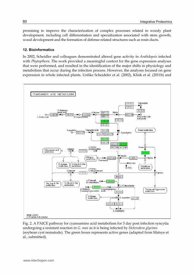
promising to improve the characterization of complex processes related to woody plant development, including cell differentiation and specialization associated with stem growth, wood development and the formation of defense-related structures such as resin ducts.
12. Bioinformatics
In 2002, Scheidler and colleagues demonstrated altered gene activity in Arabidopsis infected
with Phytopthora. The work provided a meaningful context for the gene expression analyses
that were performed, and resulted in the identification of the major shifts in physiology and
metabolism that occur during the infection process. However, the analyses focused on gene
expression in whole infected plants. Unlike Scheideler et al. (2002), Klink et al. (2011b) and
Fig. 2. A PAICE pathway for cyanoamino acid metabolism for 3 day post infection syncytia undergoing a resistant reaction in G. max as it is being infected by Heterodera glycines (soybean cyst nematode). The green boxes represents active genes (adapted from Matsye et al., submitted).
www.intechopen.com

Proteomic Analyses of Cells Isolated by Laser Microdissection
61
Matsye et al. (submitted) have used the same principle, adapting the publically available
Kyoto Encyclopedia of Genes and Genomes (KEGG) (http://www.genome.jp/kegg/
catalog/org_list.html) and modifying it so that gene expression can be visualized using a
KEGG application called Pathway Analysis and Integrated Coloring of Experiments
(PAICE). PAICE was developed in the laboratories of Dr. Benjamin Matthews (USDA;
Beltsville, MD) and Dr. Nadim Alkharouf (Towson University, Baltimore, MD) (Hosseini et
al., unpublished) and is freely available (http://sourceforge.net/projects/paice/). PAICE
has been used on LM cells infected with parasitic nematodes, and it provides a deeper
understanding of the biochemical and metabolic activities during multiple defense reactions
in multiple G. max genotypes compared to both pericycle control cell populations and the
susceptible reaction (Klink et al., 2011; Matsye et al., submitted). However, the analyses
were based on RNA isolated from the specific cell types and not on proteins or metabolites
(Figure 2). It is believed that PAICE could be expanded to provide a comprehensive
understanding of any cell isolated by LM and analyzed for its proteomic and metabolic
content.
13. Conclusion
To have a better understanding of tissue and organ-defined processes and functions, it is
necessary to study the biochemical activity at a cellular resolution level by analyzing the
proteome. This has become increasingly important, since it has been demonstrated, in
several comparative studies, that protein expression and abundance often poorly correlate
with the mRNA levels in the same cell types (Schad et al., 2009a). Many proteins are the
primary determinant molecules of physiological processes and are often restricted to
specific tissues and cell types. Thus, the monitoring of protein expression at a very high
spatial resolution could help enhance our understanding of the biological processes that
control plant growth and development. At the same time, the use of different strategies and
protocols for the characterization of a wide number of metabolites from a single cell or
tissue have increased significantly over the last decade making the broad applicability of
these analyses tractable. In order to address these issues, sampling methods, for example
LM in plants, have been adopted to extract highly specific tissue regions and homogeneous
cell-type populations with limited damage, and have led to the discovery of functions of
genes/proteins/metabolites that contribute to cell specialization (Galbraith & Birnbaum
2006). Despite these considerable efforts, the current strategies used for protein/metabolite
characterization still face significant obstacles. These challenges are mainly caused by the
cellular complexity and spatial and temporal distribution of localized gene activity within
living tissues, including metabolic processes. Other challenges concern the identification of
the high degree of chemical diversity of the different cell types that can be affected by the
analysis procedures. Technical improvements are still required to achieve reliable protein
and metabolite profilings in small samples. The introduction of statistical analysis, applied
to the handling and manipulation of data from proteomics and metabolomics, will lead to
the development of promising strategies that can be used to extract precious information
from large data sets and to identify new proteins and metabolites. Although most of these
restrictions have already been solved in the field of genomics and transcriptomics, the
problem still remains of adapting these computational strategies for proteomic and
metabolic analyses.
www.intechopen.com

Integrative Proteomics
62
14. Acknowledgements
Contributions to this chapter have been partly funded by CNR (Premio DAA 2009) to RB. VF was supported by a grant from BIOBIT-CIPE (Piedmont Region project). VPK would like to thank the Mississippi Soybean Promotion Board for the funding and critical reading of the manuscript by Prachi D. Matsye.
15. References
Abbott, E., Hall, D., Hamberger, B. & Bohlmann, J. (2010). Laser microdissection of conifer stem tissues: isolation and analysis of high quality RNA, terpene synthase enzyme activity and terpenoid metabolites from resin ducts and cambial zone tissue of white spruce (Picea glauca), BMC Plant Biology 10:106.
Ahram, M., Flaig, M.J., Gillespie, J.W., Duray, P.H., Linehan, W.M., Ornstein, D.K., Niu, S., Zhao, Y., Petricoin, E.F. 3rd & Emmert-Buck, M.R. (2003). Evaluation of ethanol-fixed, paraffin-embedded tissues for proteomic applications, Proteomics 3: 413–421.
Ali, G.S. & Reddy, A.S.N. (2008) Regulation of Alternative Splicing of Pre-mRNAs by Stresses, Current Topics in Microbiology and Immunology 326: 257-275.
Agrawal, G.K. & Rakwal, R. (2011). Rice proteomics: A move toward expanded proteome coverage to comparative and functional proteomics uncovers the mysteries of rice and plant biology, Proteomics 11: 1630–1649.
Angeles, G., Berrio-Sierra, J., Joseleau, J.P., Lorimier, P., Lefebvre, A. & Ruel, K. (2006). Preparative laser capture microdissection and single- pot cell wall material preparation: a novel method for tissue-specific analysis, Planta 224: 228–232.
Asano, T., Masumura, T., Kusano, H., Kikuchi, S., Kurita, A., Shimada, H. & Kadowaki, K. (2002). Construction of a specialized cDNA library from plant cells isolated by laser capture microdissection: toward comprehensive analysis of the genes expressed in the rice phloem, The Plant Journal 32: 401-408.
Asenjo, J.A. & Andrews, B.A. (1990) Enzymatic Cell Lysis for Product Release, in J.A. Asenjo, Marcel Dekker (eds), Separation Processes in Biotechnology, New York, pp. 143-175.
Augusti, J., Merelo, P., Cercós, M., Tadeo, F.R. & Talón, M. (2009) Comparative transcriptional survey between laser-microdissected cells from laminar abscission zone and petiolar cortical tissue during ethylene-promoted abscission in citrus leaves, BMC Plant Biology 9: 127.
Baerenfaller, K., Grossmann, J., Grobei, M.A., Hull, R., Hirsch-Hoffmann, M., Yalovsky, S., et al. (2008). Genome-scale proteomics reveals Arabidopsis thaliana gene models and proteome dynamics, Science 320: 938–41.
Balestrini, R., Gòmez-Ariza, J., Lanfranco, L. & Bonfante, P. (2007). Laser Microdissection reveals that transcripts for five plant and one fungal phosphate transporter genes are contemporaneously present in arbusculated cells, Molecular Plant–Microbe Interactions 20: 1055–1062.
Balestrini, R. & Bonfante, P. (2008). Laser Microdissection (LM): Applications to plant materials, Plant Biosystems 142: 331-336.
Balestrini, R., Gòmez-Ariza, J., Klink, V.P. & Bonfante P. (2009). Application of laser microdissection to plant pathogenic and symbiotic interactions, Journal of Plant Interactions 4: 81-92.
www.intechopen.com

Proteomic Analyses of Cells Isolated by Laser Microdissection
63
Banks, R.E., Dunn, M.J., Forbes, M.A., Stanley, A., Pappin, D., Naven, T., Gough, M., Harnden, P. & Selby, P.J. (1999). The potential use of laser capture microdissection to selectively obtain distinct populations of cells for proteomic analysis - Preliminary findings, Electrophoresis 20: 689–700.
Barrett, T.H. & Gould H. (1973) Tissue and species specificity of non-histone chromatin proteins, Biochimica et Biophysica Acta 294: 165-170.
Benschop, J.J., Mohammed, S., O’Flaherty, M., Heck, A.J., Slijper, M. & Menke, F.L. (2007). Quantitative phospho-proteomics of early elicitor signalling in Arabidopsis, Molecular and Cellular Proteomics 6: 1705–13.
Bligny, R. & Douce, R. (2001) NMR and plant metabolism. Current Opinion in Plant Biology 4: 191-196.
Brownfield, L., Ford, K., Doblin, M.S., Newbigin, E., Read, S. & Bacic, A. (2007). Proteomic and biochemical evidence links the callose synthase in Nicotiana alata pollen tubes to the product of the NaGSL1 gene, The Plant Journal 52: 147–56.
Butt, R.H. & Coorssen, J.R. (2006). Pre-extraction Sample Handling by Automated Frozen Disruption Significantly Improves Subsequent Proteomic Analyses, Journal of Proteome Research 5: 437-448.
Cai, S. & Lashbrook, C.C. (2006). Laser capture microdissection of plant cells from tape-transferred paraffin sections promotes recovery of structurally intact RNA for global gene profiling, The Plant Journal 48: 628–637.
Campbell, M.A., Haas, B.J., Hamilton, J.P., Mount, S.M. & Buell, C.R. (2006). Comprehensive analysis of alternative splicing in rice and comparative analyses with Arabidopsis, BMC Genomics 7:327.
Casson, S., Spencer, M., Walker, K. & Lindsey, K. (2005). Laser capture microdissection for the analysis of gene expression during embryogenesis of Arabidopsis, The Plant Journal 42: 111-123.
Celedon, P.A.F., Andrade, A., Meireles, K.G.X., Carvalho, M.C.C.G., Caldas, D.G.G., Moon, D.H., Carneiro, R.T., Franceschini, L.M., Oda, S. & Labate, C.A. (2007). Proteomic analysis of the cambial region in juvenile Eucalyptus grandis at three ages, Proteomics 7: 2258–2274.
Chandran, D., Inada, N., Hather, G., Klindt, C.K. & Wildermuth M.C. (2010). Laser microdissection of Arabidopsis cells at the powdery mildew infection site reveals site-specific process and regulators, Proceedings of the National Academy of Science USA 107: 460-465.
Chen, B.E., Kondo, M., Garnier, A., Watson, F.L., Püettmann-Holgado, R., Lamar, D.R. & Schmucker, D. (2006). The molecular diversity of Dscam is functionally required for neuronal wiring specificity in Drosophila, Cell 125: 607-620.
Ciobanu, L. & Pennington, C.H. (2004). 3D micron-scale MRI of single biological cells, Solid State Nuclear Magnetic Resonance 25: 138-141.
Craig, R. & Beavis, R.C. (2004). TANDEM: matching proteins with mass spectra, Bioinformatics 20: 1466–67.
Cravatt, B.F., Simon, G.M. & Yates, III J.R. (2007). The biological impact of mass-spectrometry-based proteomics, Nature 450: 991-1000.
Craven, R. & Banks, R. (2001). Laser capture microdissection and proteomics: Possibilities and limitation, Proteomics 1: 1200-1204.
Craven, R.A., Totty, N., Harnden, P., Selby, P.J. & Banks, R.E. (2002). Laser capture microdissection and two-dimensional polyacrylamide gel electrophoresis:
www.intechopen.com

Integrative Proteomics
64
evaluation of tissue preparation and sample limitations, American Journal of Pathology 160: 815–822.
Day, R.C., Grossniklaus, U. & Macknight, R.C. (2005). Be more specific! Laser-assisted microdissection of plant cells, Trends in Plant Science 10: 397–405.
Day, R.C., McNoe, L.A. & Macknight, R.C. (2006). Transcript analysis of laser microdissected plant cells, Technical Focus, Physiologia Plantarum 129: 267–282.
de Jong, F., Mathesius, U., Imin, N., Rolfe, B.G. (2007). A proteome study of the proliferation of cultured Medicago truncatula protoplasts, Proteomics 7: 722-36.
Dembinsky, D., Woll, K., Saleem, M., Liu, Y., Fu, Y., Borsuk, L.A., Lamkemeyer, T., Fladerer, C., Madlung, J., Barbazuk, B., Nordheim, A., Nettleton, D., Schnable, P.S. & Hochholdinger F. (2007). Transcriptomic and proteomic analyses of pericycle cells of the maize primary root, Plant Physiology 145: 575-88.
Djordjevic, M.A., Oakes, M., Li, D.X., Hwang, C.H., Hocart, C.H. & Gresshoff, P.M. (2007). The glycine max xylem sap and apoplast proteome, Journal of Proteome Research 6: 3771-9.
Dunkley, T.P., Hester, S., Shadforth, I.P., Runions, J., Weimar, T., Hanton, S.L., Griffin, J.L., Bessant, C., Brandizzi, F., Hawes, C., Watson, R.B., Dupree, P. & Lilley, K.S. (2006). Mapping the Arabidopsis organelle proteome, Proceedings of National Academy Science USA 103: 6518–23.
Edman, P. (1950). Method for determination of the amino acid sequence in peptides, Acta Chemica Scandinavica 4: 283-293.
Emmert-Buck, M.R., Bonner, R.F., Smith, P.D., Chuaqui, R.F., Zhuang, Z., Goldstein, S.R., Weiss, R.A. & Liotta, L.A. (1996). Laser capture microdissection, Science 274: 998–1001.
Eng, J., McCormack, A.L. & Yates, J.R.I. (1994). An approach to correlate tandem mass spectral data of peptides with amino acid sequences in a protein database, Journal of the American Society for Mass Spectrometry 5: 976–89.
Fait, A., Hanhineva, K., Beleggia, R., Dai, N., Rogachev, I., Nikiforova, V.J., Fernie, A.R. & Aharoni, A. (2008). Reconfiguration of the chene and receptacle metabolic networks during strawberry fruit development, Plant Physiology 148: 730–750.
Finnie, C., Sultan, A. & Grasser, K.D. (2011). From protein catalogues towards targeted proteomics approaches in cereal grains, Phytochemistry 72: 1145-1153.
Fiorilli, V., Catoni, M., Miozzi, L., Novero, M,. Accotto, G.P. & Lanfranco, L. (2009). Global and cell-type gene expression profiles in tomato plants colonized by an arbuscular mycorrhizal fungus, New Phytologist 184: 975-987.
Fraser, P.D., Enfissi, E.M.A., Goodfellow, M., Eguchi, T. & Bramley, P.M. (2007). Metabolite profiling of plant carotenoids using the matrix-assisted laser desorption ionization time-of-flight mass spectrometry, The Plant Journal 49: 552–564.
Galbraith, D.W. & Birnbaum, K. (2006). Global studies of cell type-specific gene expression in plants, Annual Review of Plant Biology 57:451-75.
Gazanchian, A., Hajheidari, M., Sima, N.K. & Salekdeh, G.H. (2007). Proteome response of Elymus elongatum to severe water stress and recovery, Journal of Experimental Botany 58: 291–300.
Geer, L.Y., Markey, S.P., Kowalak, J.A., Wagner, L., Xu M, Maynard, D.M., Yang, X., Shi, W. & Bryant, S.H.2004. Open mass spectrometry search algorithm, Journal of Proteome Research 3: 958–64.
Glauser, G., Guillarme, D., Grata, E., Boccard, J., Thiocone, A., Carrupt, P.A., Veuthey, J.L., Rudaz, S., & Wolfender, J.L. (2008). Optimized liquid chromatography-mass
www.intechopen.com
Proteomic Analyses of Cells Isolated by Laser Microdissection
65
spectrometry approach for the isolation of minor stress biomarkers in plant extracts and their identification by capillary nuclear magnetic resonance, Journal of Chromatography 1180: 90–98.
Goldsworthy, S.M., Stockton, P.S., Trempus, C.S., Foley, J.F. & Maronpot, R.R. (1999) Effects of fixation on RNA extraction and amplification from laser capture microdissected tissue, Molecular Carcinogenesis 25: 86-91.
Gomez, S.K. & Harrison, M.J. (2009). Laser microdissection and its application to analyze gene expression in the arbuscular mycorrhizal symbiosis. Pest Management Sciences 65: 504-511
Gomez, K.S., Javot, H., Deewatthanawong, P., Torres-Jerez, I., Tang, Y., Blancaflor, B.E., Udvardi, M.K. & Harrison, J. M. (2009). Medicago Truncatula and Glomus Intraradices gene expression in cortical cells harboring arbuscules in the arbuscular mycorrhizal symbiosis, BMC Plant Biology 9:10.
Görg, A., Drews, O., Lück, C., Weiland, F. & Weiss, W. 2-DE with IPGs, Electrophoresis 30: S122-32.
Graveley, B.R. (2008) Molecular biology: power sequencing, Nature 453: 1197-8. Guether, M., Balestrini, R., Hannah, M., He, J., Udvardi, M.K. & Bonfante, P. (2009a).
Genome-wide reprogramming of regulatory networks, transport, cell wall and membrane biogenesis during arbuscular mycorrhizal symbiosis in Lotus japonicus, New Phytologist 182: 200–212.
Guether, M., Neuhauser, B., Balestrini, R., Dynowski, M., Ludewig, U. & Bonfante P. (2009b). A mycorrhizal-specific ammonium transporter from Lotus Japonicus acquires nitrogen released by arbuscular mycorrhizal fungi, Plant Physiology 105: 73-83.
Gutstein, H.B. & Morris, J.S. (2007). Laser capture sampling and analytical issue in proteomics, Expert Review of Proteomics 4: 627-37.
Hacquard, S., Delaruelle, C., Legué, V., Tisserant, E., Kohler, A., Frey, P., Martin, F. & Duplessis S. (2010) Laser capture microdissection of uredinia formed by Melampsora larici-populina revealed a transcriptional switch between biotrophy and sporulation, Molecular Plant-Microbe Interactions 23: 1275-1286.
Hagel, J.M., Weljie, A.M., Vogel, H.J. & Facchini, P.J. (2008). Quantitative H-1 nuclear magnetic resonance metabolite profiling as a functional genomics platform to investigate alkaloid biosynthesis in opium poppy, Plant Physiology 147: 1805-1821.
Hebeler, R., Oeljeklaus, S., Reidegeld, K.A., Eisenacher, M., Stephan, C., Sitek, B., Stühler, K., Meyer, H.E., Sturre, M.J., Dijkwel, P.P. & Warscheid, B. (2008). Study of early leaf senescence in Arabidopsis thaliana by quantitative proteomics using reciprocal 14 N/15 N labeling and difference gel electrophoresis, Molecular and Cellular Proteomics 7:108–20.
Hinse, C., Sheludko, Y.V., Provenzani, A., Stöckigt, J.H.H. (2001). In vivo NMR at 800 MHz to monitor alkaloid metabolism in plant cell cultures without tracer labeling, Journal of America Chemical Society 123: 5118-5119.
Hochholdinger, F., Sauer, M., Dembinsky, D., Hoecker, N., Muthreich, N., Saleem, M. & Liu, Y. (2006). Proteomic dissection of plant development, Proteomics 6: 4076–4083.
Hölscher, D., Schneider, B. (2008). Application of Laser-Assisted Microdissection for Tissue and Cell-Specific Analysis of RNA, Proteins, and Metabolites, Progress in Botany 69: 141-167.
Hood, B.L., Conrads, T.P. & Veenstra, T.D. (2006). Unravelling the proteome of formalin-fixed paraffin-embedded tissue, Briefings in Functional Genomics and Proteomics 5: 169–175.
www.intechopen.com

Integrative Proteomics
66
Hopkins, T.R. (1991). Physical and chemical cell disruption for the recovery of intracellular proteins, Bioprocess Technology 12: 57–83.
Hummon, A.B., Amare, A. & Sweedler, J.V. (2006). Discovering new invertebrate neuropeptides using mass spectrometry, Mass Spectrometry Reviews 25: 77–98.
Inada, N. & Wildemuth, M.C. (2005). Novel tissue preparation method and cell-specific marker for laser microdissection of Arabidopsis mature leaf, Planta 221: 9–16.
Ingle, R.A., Schmidt, U.G., Farrant, J.M., Thomson, J.A. & Mundree, S.G. (2007). Proteomic analysis of leaf proteins during dehydration of the resurrection plant Xerophyta viscosa, Plant Cell and Environment 30: 435-46.
Isenberg, G., Bielser, W., Meier-Ruge, W. & Remy, E. (1976) Cell surgery by laser micro-dissection: a preparative method, Journal of Microscopy 107: 19–24.
Jamet, E., Boudart, G., Borderies, G., Charmont, S., Lafitte, C., Rossignol, M., Canut, H. & Pont-Lezica R.F. Isolation of plant cell wall proteins, Methods Molecular Biology 425: 187–201.
Jefferson, R.A., Kavanagh, T.A. & Bevan, M.W. (1987). GUS fusions: -glucuronidase as a sensitive and versatile gene fusion marker in higher plants, EMBO Journal 6: 3901-3907.
Jiang, G., Wang, Z., Shang, H., Yang, W., Hu, Z., Phillips, J. Deng, X. (2007). Proteome analysis of leaves from the resurrection plant Boea hygrometrica in response to dehydration and rehydration, Planta 225: 1405-20.
Jiang, K., Zhang, S., Lee, S., Tsai, G., Kim, K., Huang, H., Chilcott, C., Zhu, T. & Feldman, L.J. (2006). Transcription profile analyses identify genes and pathways central to root cap functions in maize, Plant Molecular Biology 60: 343–363.
Jorrin, J.V., Maldonado, A.M. & Castillejo, M.A. (2007). Plant proteome analysis: a 2006 update, Proteomics 7: 2947–62.
Jorrín-Novo, J.V., Maldonado, A.M., Echevarría-Zomeño, S., Valledor, L., Castillejo, M.A., Curto, M., Valero, J., Sghaier, B., Donoso, G. & Redondo, I. (2009). Plant proteomics update (2007–2008): Second-generation proteomic techniques, an appropriate experimental design, and data analysis to fulfill MIAPE standards, increase plant proteome coverage and expand biological knowledge, Journal of Proteomics 72: 285-314.
Junqueira, M., Spirin, V., Santana Balbuenaa, T., Thomasa, H., Adzhubeib, I., Sunyaevb, S. & Shevchenko, A. (2008). Protein identification pipeline for the homology-driven proteomics, Journal of Proteomics 71: 346-356.
Kamme, F., Salunga, R., Yu, J., Tran, D.T., Zhu, J., Luo, L., Bittner, A., Guo, H.Q., Miller, N., Wan, J. & Erlander, M. (2003). Single-cell microarray analysis in hippocampus CA1: demonstration and validation of cellular heterogeneity, Journal of Neuroscience 23: 3607-3615.
Kant, M.R., Ament, K., Sabelis, M.W., Haring, M.A. & Schuurink, R.C. (2004). Differential timing of spider mite-induced direct and indirect defenses in tomato plants, Plant Physiology 135: 483- 495.
Kaspar, S., Weier, D., Weschke, W., Mock, H.P. & Matros, A. (2010). Protein analysis of laser capture micro-dissected tissues revealed cell-type specific biological functions in developing barley grains, Analytical and Bioanalytical Chemistry 398: 2883-93.
Kazuma, K., Noda, N. & Suzuki, M. (2003). Flavonoid composition related to petal color in different lines of Clitoria ternatea, Phytochemistry 64: 1133-1139.
www.intechopen.com

Proteomic Analyses of Cells Isolated by Laser Microdissection
67
Kerk, N.M., Ceserani, T., Tausta, S.L., Sussex, I.M. & Nelson, T.M. (2003). Laser capture microdissection of cells from plant tissues, Plant Physiology 132: 27–35.
Kierszniowska, S., Seiwert, B. & Schulze, W.X. (2009). Definition of Arabidopsis sterol-rich membrane microdomains by differential treatment with methyl-ß-cyclodextrin and quantitative proteomics, Molecular and Cellular Proteomics 8: 612–23.
Kim, E., Magen, A. & Ast, G. (2007). Different levels of alternative splicing among eukaryotes, Nucleic Acids Research 35: 125–31.
Kim, J.O., Kim, H.N., Hwang, M.H., Shin, H.I., Kim, S.Y., Park, R.W., Park, E.Y., Kim, I.S., van Wijnen, A.J., Stein, J.L., Lian, J.B., Stein, G.S. & Choi, J.Y. (2003). Differential gene expression analysis using paraffin-embedded tissues after laser microdissection, Journal Cell Biochemistry 90: 998-1006.
Kleffmann, T., von Zychlinski, A., Russenberger, D., Hirsch-Hoffmann, M., Gehrig, P, Gruissem, W. & Baginsky, S. (2007). Proteome dynamics during plastid differentiation in rice, Plant Physiology 143: 912–23.
Klink, V.P., MacDonald, M., Alkharouf, N. & Matthews, B.F. (2005). Laser capture microdissection (LCM) and expression analyses of Glycine max (soybean) syncytium containing root regions formed by the plant pathogen Heterodera glycines (soybean cyst nematode), Plant Molecular Biology 59: 969-983.
Klink, V.P., Overall, C.C., Alkharouf, N., MacDonald, M.H. & Matthews, B.F. (2007). Laser capture microdissection (LCM) and comparative microarray expression analysis of syncytial cells isolated from incompatible and compatible soybean roots infected by soybean cyst nematode (Heterodera glycines), Planta 226: 1389-1409.
Klink, V.P., Hosseini, P., Matsye, P., Alkharouf, N. & Matthews, B.F. (2009). A gene expression analysis of syncytia laser microdissected from the roots of the Glycine max (soybean) genotype PI 548402 (Peking) undergoing a resistant reaction after infection by Heterodera glycines (soybean cyst nematode), Plant Molecular Biology 71: 525-567.
Klink, V.P., Hosseini, P., Matsye, P., Alkharouf, N. & Matthews, B.F. (2010a). Syncytium gene expression in Glycine max[PI 88788] roots undergoing a resistant reaction to the parasitic nematode Heterodera glycines, Plant Physiology and Biochemistry 48: 176-193.
Klink, V.P., Overall, C.C., Alkharouf, N., MacDonald, M.H. & Matthews, B.F. (2010b). Microarray detection calls as a means to compare transcripts expressed within syncytial cells isolated from incompatible and compatible soybean (Glycine max) roots infected by the soybean cyst nematode (Heterodera glycines), Genome Article ID 491217: 1-30.
Klink, V.P., Matsye, P.D. & Lawrence, G.W. (2011a). Cell-specific studies of soybean resistance to its major pathogen, the soybean cyst nematode as revealed by laser capture microdissection, gene pathway analyses and functional studies in Aleksandra Sudaric (ed.) Soybean - Molecular Aspects of Breeding, Intech Publishers pp. 397-428.
Klink, V.P., Hosseini, P., Matsye, P.D., Alkharouf, N. & Matthews, B.F. (2011b). Differences in gene expression amplitude overlie a conserved transcriptomic program occurring between the rapid and potent localized resistant reaction at the syncytium of the Glycine max genotype Peking (PI 548402) as compared to the prolonged and potent resistant reaction of PI 88788, Plant Molecular Biology 75: 141-165.
Komatsu, S., Konishi, H. & Hashimoto, M. (2007). The Proteomics of plant cell membranes, Journal Experimental Botany 58: 103–12.
www.intechopen.com

Integrative Proteomics
68
Krishnan, P., Kruger, N.J. & Ratcliffe, R.G. (2005). Metabolite fingerprinting and profiling in plants using NMR. Journal of Experimental Botany 56: 255-265.
Li, S.H., Schneider, B. & Gershenzon J. (2007). Microchemical analysis of laser-microdissected stone cells of Norway spruce by cryogenic nuclear magnetic resonance spectroscopy, Planta 225: 771-779.
Lippert, D., Chowrira, S., Ralph, S.G., Zhuang, J., Aeschliman, D., Ritland, C., Ritland, K., Bohlmann, J. (2007). Conifer defense against insects: proteome analysis of Sitka spruce (Picea sitchensis) bark induced by mechanical wounding or feeding by white pine weevils (Pissodes strobi), Proteomics 7: 248-70.
Lisec, J., Schauer, N., Kopka, J., Willmitzer, L. & Fernie, A.R. (2006). Gas chromatography mass spectrometry-based metabolite profiling in plants, Nature Protocols 1: 387–396.
Lliso, I., Tadeo, F.R., Phinney, B.S., Wilkerson, C.G. & Talón, M. (2007). Protein changes in the albedo of citrus fruits on postharvesting storage, Journal of Agriculture Food Chemistry 55: 9047–53.
Lu, P., Vogel, C., Wang, R., Yao, X. & Marcotte, E.M. (2007). Absolute protein expression profiling estimates the relative contributions of transcriptional and translational regulation, Nature Biotechnology 25: 117–24.
Martin, C., Bhatt, K. & Baumann, K. (2001). Shaping in plant cells, Current Opinion in Plant Biology 4: 540-9.
Matsye, P.D., Kumar, R., Hosseini, P., Jones, C.M., Alkharouf, N., Matthews, B.F.& Klink VP. Mapping cell fate decisions that occur during soybean defense responses. (Submitted)
Matas, A.J., Augusti, J., Tadeo, F.R., Talón, M. & Rose, J.K. (2010). Tissue-specific transcriptome profiling of the citrus fruit epidermis and subepidermis using laser capture microdissection, Journal of Experimental Botany 61: 3321-3330.
Mehta, A., Brasileiro, A.C., Souza, D.S., Romano, E., Campos, M.A., Grossi-de-Sá, M.F., Silva, M.S., Franco, O.L., Fragoso, R.R., Bevitori R. & Rocha, T.L. (2008). Plant–pathogen interactions: what is proteomics telling us?, FEBS Journal 275: 3731–3746.
Meijers, R., Puettmann-Holgado, R. Skiniotis, G., Liu, J.H., Walz, T., Wang, J.H. & Schmucker, D. (2007). Structural basis of Dscam isoform specificity, Nature 449: 487-491.
Michnick, S.W. (2004). Proteomics in living cells, Drug Discovery Today 9: 262–267. Miernyk, J.A., Pret'ova, A., Olmedilla, A., Klubicova, K., Obert B., Hajduch M. et al. (2011).
Using proteomics to study sexual reproduction in angioperms, Sex Plant Reproduction 24: 9–22.
Moco, S., Bino, R.J., Vorst, O., Verhoeven, H.A., de Groot, J., van Beek, T.A., Vervoort, J. & De Vos, R.C.H. (2006). A liquid chromatography-mass spectrometry-based metabolome database for tomato, Plant Physiology 141: 1205–1218.
Moco, S., Capanoglu, E., Tikunov, Y., Bino, R.J., Boyacioglu, D., Hall, R.D., Vervoort, J., De Vos, R.C.H. (2007). Tissue specialization at the metabolite level is perceived during the development of tomato fruit, Journal Experimental Botany 58: 4131-4146.
Moco, S., Forshed, J., De Vos, R.C.H., Bino, R.J. & Vervoort, J. (2008). Intra- and inter-metabolite correlation spectroscopy of tomato metabolomics data obtained by liquid chromatography-mass spectrometry and nuclear magnetic resonance, Metabolomics 4: 202–215.
Moco, S., Schneider, B. & Vervoort, J. (2009). Plant micrometabolomics: the analysis of endogenous metabolites present in a plant cell or tissue, Journal of Proteome Research 8: 1694-1703.
www.intechopen.com

Proteomic Analyses of Cells Isolated by Laser Microdissection
69
Moulédous, L., Hunt, S., Harcourt, R., Harry, J., Williams, K.L. & Gutstein, H.B. (2002). Lack of compatibility of histological staining methods with proteomic analysis of laser-capture microdissected brain samples, Journal of Biomolecular Techniques 13: 258-264.
Moulédous, L., Hunt, S., Harcourt, R., Harry, J., Williams, K.L. & Gutstein, H.B. (2003). Navigated laser capture microdissection as an alternative to direct histological staining for proteomic analysis of brain samples, Proteomics 3: 610-615.
Nakazono, M., Qiu, F., Borsuk, L.A. & Schable, P.S. (2003). Laser capture microdissection, a tool for the global analysis of gene expression in specific plant cell types: identification of genes expressed differentially in epidermal cells or vascular tissue of maize, The Plant Cell 15: 583-596.
Nelson, C.J., Hegeman, A.D., Harms, A.C. & Sussman, M.R. (2006a). A quantitative analysis of Arabidopsis plasma membrane using trypsin-catalyzed 18 O labeling. Molecular Cell Proteomics 5: 1382–95.
Nelson, T., Tausta, S.L., Gandotra, N. & Liu, T. (2006b). Laser microdissection of plant tissue: What you see is what you get, Annual Review of Plant Biology 57: 181–201.
Nelson, T., Gandotra, N. & Tausta, S.L. (2008). Plant cell types: reporting and sampling with new technologies, Current Opinion in Plant Biology 11: 567–573.
Nesatyy, V.J. & Suter, M.J. (2008). Analysis of environmental stress response on the proteome level, Mass Spectrometry Reviews 27: 556-74.
Nie, L., Wu, G., Culley, D.E., Scholten, J.C. & Zhang W. (2007). Integrative analysis of transcriptomic and proteomic data: challenges,solutions and applications, Critical Reviews in Biotechnology 27: 63–75.
Niittylä, T., Fuglsang, A.T., Palmgren, M.G., Frommer, W.B. & Schulze, W.X. (2007). Temporal analysis of sucrose-induced phosphorylation changes in plasma membrane proteins of Arabidopsis, Molecular Cell Proteomics 6: 1711–26.
Nowaczyk, M.M., Hebeler, R., Schlodder, E., Meyer, H.E., Warscheid, B. & Rögner, M. (2006). Psb27, a cyanobacterial lipoprotein, is involved in the repair cycle of photosystem II, The Plant Cell 18: 3121–3177.
Oda, Y., Huang, K., Cross, F.R., Cowburn, D. & Chait, B.T. (1999). Accurate quantitation of protein expression and site-specific phosphorylation. Proceedings of the National Academy of Science USA 96: 6591–96.
O'Farrell, P.H. (1975). High resolution two-dimensional electrophoresis of proteins, The Journal of Biological Chemistry 250: 4007-4021.
O'Farrell, P.Z., Goodman, H.M. & O'Farrell P.H. (1977). High resolution two dimensional electrophoresis of basic as well as acidic proteins, Cell 12: 1133-1142.
Opitz, S. & Schneider, B. (2002). Organ-specific analysis of phenylphenalenone-related compounds in Xiphidium caeruleum, Phytochemistry 61: 819–825.
Ohtsu, K., Takahashi, H., Schnable, P.S. & Nakazono, M. (2007). Cell type-specific gene expression profiling in plants by using a combination of laser microdissection and high-throughput technologies, Plant Cell Physiology 48: 3-7.
Palmblad, M., Mills, D.J. & Bindschedler, L.V. (2008). Heat-shock response in Arabidopsis thaliana explored by multiplexed quantitative proteomics using differential metabolic labeling, Journal of Proteome Research 7: 780–85.
Palusa, S.G., Ali. G.S. & Reddy A.S.N. (2007). Alternative splicing of pre-mRNAs of Arabidopsis serine/arginine-rich proteins: regulation by hormones and stresses, The Plant Journal 49: 1091–107.
www.intechopen.com

Integrative Proteomics
70
Pan, H. & Lundgren, L.N. (1995). Phenolic extractives from root bark of Picea abies, Phytochemistry 39: 1423-8.
Pan, Q., Shai, O., Lee, L.J., Frey, B.J. & Blencowe, B.J. (2008). Deep surveying of alternative splicing complexity in the human transcriptome by high-throughput sequencing, Nature Genetic 40: 1413-5.
Pedreschi, R.,Vanstreels, E., Carpentier, S., Hertog, M., Lammertyn, J., Robben J., Noben, J.P., Swennen, R., Vanderleyden, J. & Nicolai, B. (2007). Proteomic analysis of core breakdown disorder in conference pears (Pyrus communis L.), Proteomics 7: 2083-99.
Peltier, J.B., Friso, G., Kalume, D.E., Roepstorff, P., Nilsson, F., Adamskaa, I. & van Wijk K.J. (2000). Proteomics of the chloroplast: systematic identification and targeting analysis of lumenal and peripheral thylakoid proteins, The Pant Cell 12: 319–342.
Persidis, A. (1998). Proteomics, Nature Biotechnology 4: 393-394. Pevzner, P.A., Mulyukov, Z., Dancik, V. & Tang, C.L. (2001). Efficiency of database search
for identification of mutated and modified proteins via mass spectrometry, Genome Research 11: 290–99.
Purea, A., Neuberger, T. & Webb, A.G. (2004). Simultaneous NMR micro-imaging of multiple single-cell samples, Concepts Magn Reson Part B 22B: 7–14.
Rabilloud, T. (1996). Solubilization of proteins for electrophoretic analyses, Electrophoresis 17: 813-829.
Ramsay, K., Jones, M.G.K. & Wang, Z. (2006). Laser capture microdissection: A novel approach to microanalysis of plant–microbe interactions, Molecular Plant Pathology 7: 429–435.
Reiland, S., Messerli, G., Baerenfaller, K., Gerrits, B., Endler, A., Grossmann, J., Gruissem W. & Baginsky, S. (2009). Large-scale Arabidopsis phosphoproteome profiling reveals novel chloroplast kinase substrates and phosphorylation networks, Plant Physiology 150: 889–903.
Rekhter, M.D. & Chen, J. (2001). Molecular analysis of complex tissues is facilitated by laser capture microdissection: Critical role of upstream tissue processing, Cell Biochemistry and Biophysics 35: 103–113.
Roessner, U., Willmitzer, L. & Fernie, A.R. (2001). High-resolution metabolic phenotyping of genetically and environmentally diverse potato tuber systems. Identification of phenocopies, Plant Physiology 127: 749–764.
Rossignol, M., Peltier, J.B., Mock, H.P., Matros, A., Maldonado, A.M. & Jorrin J.V. (2006). Plant proteome analysis: a 2004–2006 update, Proteomics 6: 5529-48.
Rubakhin, S.S., Greenough, W.T. & Sweedler, J.V. (2003). Spatial profiling with MALDI MS: distribution of neuropeptides within single neurons, Analytical Chemistry 75: 5374-5380.
Salekdeh, G.H. & Komatsu, S. (2007). Crop proteomics: aim at sustainable agriculture of tomorrow, Proteomics 7: 2976–96.
Schad, M,. Lipton, M.S., Giavalisco, P., Smith, R.D. & Kehr, J. (2005a). Evaluation of two-dimensional electrophoresis and liquid chromatography–tandem mass spectrometry for tissue-specific protein profiling of laser-microdissected plant samples, Electrophoresis 26: 2729–2738.
Schad, M., Mungur, R., Fiehn, O. & Kehr, J. (2005b). Metabolic profiling of laser microdissected vascular bundles of Arabidopsis thaliana, Plant Methods 1:2.
www.intechopen.com

Proteomic Analyses of Cells Isolated by Laser Microdissection
71
Schmucker, D., Clemens, J.C., Shu, H., Worby, C.A., Xiao, J., Muda, M., Dixon, J.E. & Zipursky, S.L. (2000). Drosophila Dscam is an axon guidance receptor exhibiting extraordinary molecular diversity, Cell 101: 671-684.
Schneider, B. & Hölscher, D. (2007). Laser microdissection and cryogenic nuclear magnetic resonance spectroscopy: an alliance for cell type-specific metabolite profiling, Planta 225: 763–770.
Schulze, W.X. & Usadel, B. (2010). Quantitation in mass-spectrometry-based proteomics, Annual Review in Plant Biology 61: 491–516.
Sheoran, I.S., Ross, A.R., Olson, D.J. & Sawhney, V.K. (2007). Proteomic analysis of tomato (Lycopersicon esculentum) pollen, Journal of Experimental Botany 58: 3525–35.
Shibutani, M., Uneyama, C., Miyazaki, K., Toyoda, K. & Hirose, M. (2000). Methacarn Fixation: A Novel Tool for Analysis of Gene Expressions in Paraffin-Embedded Tissue Specimens. Lab Invest. 80: 199–208.
Simon, S.A., Zhai, J., Nandety, R.S., McCormick, K.P., Zeng, J., Mejia, D. & Meyers, B.C. (2009). Short-Read Sequencing Technologies for Transcriptional Analyses, Annual Review of Plant Biology 60: 305–33.
Simpson, C.G., Lewandowska, D., Fuller, J., Maronova, M., Kalyna, M., Davidson, D., et al. (2008) Alternative splicing in plants, Biochem Soc Trans 36: 508–10.
Sitek, B., Luttges, J., Marcus, K., Kloppel, G., Schmiegel, W., Meyer, H. E., Hahn, S. A. & Stuhler, K. (2005). Application of fluorescence difference gel electrophoresis saturation labelling for the analysis of microdissected precursor lesions of pancreatic ductal adenocarcinoma, Proteomics 5: 2665–2679.
Song, X., Ni, Z., Yao, Y., Xie, C., Li, Z., Wu, H, Zhang, Y. & Sun, Q. (2007). Wheat (Triticum aestivum L.) root proteome and differentially expressed root proteins between hybrid and parents, Proteomics 27: 3538–57.
Sturm, S., Seger, C., Godejohann, M., Spraul, M. & Stuppner, H. (2007). Conventional sample enrichment strategies combined with high- performance liquid chromatography-solid phase extraction-nuclear magnetic resonance analysis allows analyte identification from a single minuscule Corydalis solida plant tuber, Journal of Chromatography 1163: 138–144.
Takáč, T., Pechan, T. & Samaj, J. (2011). Differential proteomics of plant development, Journal of Proteomics 74: 577-88.
Takahashi, H., Kamakura, H., Sato, Y., Shiono, K., Abiko, T., Tsutsumi, N., Nagamura, Y., Nishizawa, N.K., Nakazono, M. (2010). A method for obtaining high quality RNA from paraffin sections of plant tissues by laser microdissection, Journal of Plant Research 123: 807-813.
Tanabe, N., Yoshimura, K., Kimura, A., Yabuta, Y. & Shigeoka, S. (2007). Differential expression of alternatively spliced mRNAs of Arabidopsis SR protein homologs, atSR30 and atSR45a, in response to environmental stress, Plant Cell Physiology 48: 1036–1049.
Tang, W., Coughlan, S., Crane, E., Beatty, M. & Duvick, J. (2006). The application of laser microdissection to in planta gene expression profiling of the maize anthracnose stalk rot fungus Colletotrichum graminicola, Molecular Plant–Microbe Interaction 19: 1240–1250.
Tanner, S., Shu, H., Frank, A., Wang, L.C., Zandi, E., Mumby, M., Pevzner, P.A. & Bafna, V. (2005). InsPecT: identification of posttranslationally modified peptides from tandem mass spectra, Analytical Chemistry 77: 4626–39.
www.intechopen.com

Integrative Proteomics
72
Thiel, J., Weier, D., Sreenivasulu, N., Strickert, M., Weichert, N., Melzer, M., Czauderna, T., Wobus, U., Weber, H., Weschke, W. (2008) Different hormonal regulation of cellular differentiation and function in nucellar projection and endosperm transfer cells—a microdissection-based transcriptome study of young barley grains, Plant Physiology 148: 1436–1452.
Thiel, J., Müller, M., Weschke, W. & Weber, H. (2009). Amino acid metabolism at the maternal–Wlial boundary of young barley seeds: a microdissection-based study, Planta 230: 205–213.
Valledor, L., Castillejo, M.A., Lenz, C., Rodríguez, R., Cañal, M.J. & Jorrín, J. (2008). Proteomic analysis of Pinus radiata needles: 2-DE map and protein identification by LC/MS/MS and substitution-tolerant database searching, Journal of Proteome Research 7: 2616–31.
van der Weerd, L., Claessens, M.M.A.E., Efde, C. & Van As, H. (2002). Nuclear magnetic resonance imaging of membrane permeability changes in plants during osmotic stress, Plant Cell and Environment 25: 1539-1549.
Vyetrogon, K., Tebbji, F., Olson, D.J., Ross, A.R. & Matton, D.P. (2007). A comparative proteome and phosphoproteome analysis of differentially regulated proteins during fertilization in the self-incompatible species Solanum chacoense Bitt., Proteomics 7: 232–47.
Wang, Y.L., Tang, H.R., Nicholson, J.K., Hylands, P.J., Sampson, J., Whitcombe, I., Stewart, C.G., Caiger, S., Oru, I., Holmes, E. (2004). Metabolomic strategy for the classification and quality control of phytomedicine: a case study of chamomile flower (Matricaria recutita L.), Planta Medica 70: 250-5.
Wang, D., Eyles, A., Mandich, D. & Bonello, P. (2006). Systemic aspects of host–pathogen interactions in Austrian pine (Pinus nigra): a Proteomics approach. Physiological and Molecular Plant Pathology 68: 149–57.
Ward, J.L., Harris, C., Lewis, J. & Beale, M.H. (2003). Assessment of H-1 NMR spectroscopy and multivariate analysis as a technique for metabolite fingerprinting of Arabidopsis thaliana, Phytochemistry 62: 949–957.
Wasinger, V.C., Cordwell, S.J., Cerpa-Poljak, A., Yan, J.X., Gooley, A.A., Wilkins, M.R., Duncan, M.W., Harris, R., Williams, K.L. & Humphery-Smith, I. (1995). Progress with gene-product mapping of the Mollicutes: Mycoplasma genitalium, Electrophoresis 16: 1090-1094.
Wenzler, M., Hölscher, D., Oerther, T. & Schneider, B. (2008). Nectar formation and floral nectary anatomy of Anigozanthos flavidus: a combined magnetic resonance imaging and spectroscopy study, Journal of Experimental Botany 59: 3425-3434.
Witze, E.S., Old, W.M., Resing, K.A. & Ahn N.G. (2007). Mapping protein post-translational modifications with mass spectrometry, Nature Methods 4: 798–806.
Xiao, Y.L., Smith, S.R., Ishmael, N., Redman, J.C., Kumar, N., Monaghan, E.L., Ayele, M., Haas, B.J., Wu, H.C. & Town C.D. (2005). Analysis of the cDNAs of hypothetical genes on Arabidopsis chromosome 2 reveals numerous transcript variants, Plant Physiology 139: 1323-37.
Yoshimura, K., Masuda, A., Kuwano, M., Yokota, A. & Akashi, K. (2008). Programmed proteome response for drought avoidance/tolerance in the root of a C3 xerophyte (wild watermelon) under water deficits, Plant and Cell Physiology 49: 226–41.
Yu, Y.Y., Lashbrook, C. & Hannapel, D. (2007). Tissue integrity and RNA quality of laser microdissected phloem of potato, Planta 226: 797–803.
www.intechopen.com

Integrative ProteomicsEdited by Dr. Hon-Chiu Leung
ISBN 978-953-51-0070-6Hard cover, 442 pagesPublisher InTechPublished online 24, February, 2012Published in print edition February, 2012
InTech EuropeUniversity Campus STeP Ri Slavka Krautzeka 83/A 51000 Rijeka, Croatia Phone: +385 (51) 770 447 Fax: +385 (51) 686 166www.intechopen.com
InTech ChinaUnit 405, Office Block, Hotel Equatorial Shanghai No.65, Yan An Road (West), Shanghai, 200040, China
Phone: +86-21-62489820 Fax: +86-21-62489821
Proteomics was thought to be a natural extension after the field of genomics has deposited significant amountof data. However, simply taking a straight verbatim approach to catalog all proteins in all tissues of differentorganisms is not viable. Researchers may need to focus on the perspectives of proteomics that are essentialto the functional outcome of the cells. In Integrative Proteomics, expert researchers contribute both historicalperspectives, new developments in sample preparation, gel-based and non-gel-based protein separation andidentification using mass spectrometry. Substantial chapters are describing studies of the sub-proteomes suchas phosphoproteome or glycoproteomes which are directly related to functional outcomes of the cells.Structural proteomics related to pharmaceutics development is also a perspective of the essence.Bioinformatics tools that can mine proteomics data and lead to pathway analyses become an integral part ofproteomics. Integrative proteomics covers both look-backs and look-outs of proteomics. It is an ideal referencefor students, new researchers, and experienced scientists who want to get an overview or insights into newdevelopment of the proteomics field.
How to referenceIn order to correctly reference this scholarly work, feel free to copy and paste the following:
Valentina Fiorilli, Vincent P. Klink and Raffaella Balestrini (2012). Proteomic Analyses of Cells Isolated by LaserMicrodissection, Integrative Proteomics, Dr. Hon-Chiu Leung (Ed.), ISBN: 978-953-51-0070-6, InTech,Available from: http://www.intechopen.com/books/integrative-proteomics/proteomic-analyses-of-cells-isolated-by-laser-microdissection

© 2012 The Author(s). Licensee IntechOpen. This is an open access articledistributed under the terms of the Creative Commons Attribution 3.0License, which permits unrestricted use, distribution, and reproduction inany medium, provided the original work is properly cited.