protocolo de rehabilitación para la reconstrucción del ... · normal en la rodilla que los...
TRANSCRIPT

Protocolo de Rehabilitación para la Reconstrucción del Ligamento Cruzado Anterior
Servicios ortopédicos de Stephen M. Howell, MD
ALÍNEESE CON EL MEJOR

2 ��������������������������������������������������������������������������������������� DrSteveHowell.com
PROTOCOLO DE REHABILITACIÓN PARA LA RECONSTRUCCIÓN DEL ACL ������������������������������������������ 3
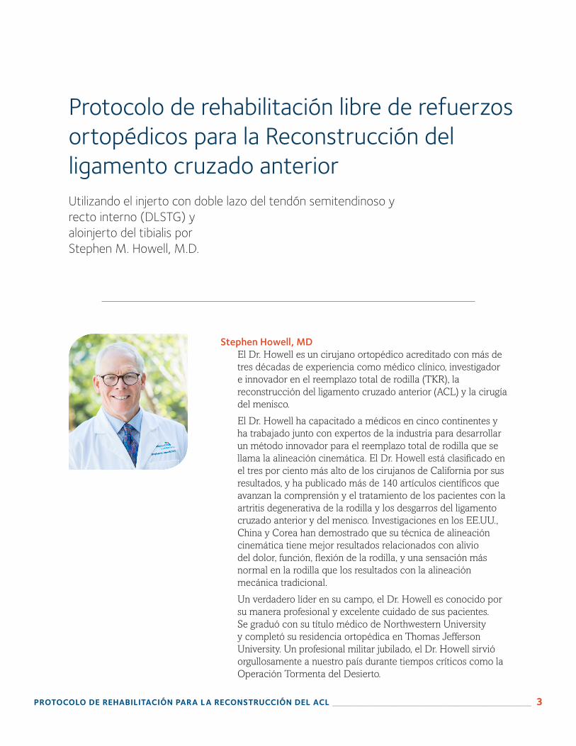
Stephen Howell, MDEl Dr. Howell es un cirujano ortopédico acreditado con más de tres décadas de experiencia como médico clínico, investigador e innovador en el reemplazo total de rodilla (TKR), la reconstrucción del ligamento cruzado anterior (ACL) y la cirugía del menisco.
El Dr. Howell ha capacitado a médicos en cinco continentes y ha trabajado junto con expertos de la industria para desarrollar un método innovador para el reemplazo total de rodilla que se llama la alineación cinemática. El Dr. Howell está clasificado en el tres por ciento más alto de los cirujanos de California por sus resultados, y ha publicado más de 140 artículos científicos que avanzan la comprensión y el tratamiento de los pacientes con la artritis degenerativa de la rodilla y los desgarros del ligamento cruzado anterior y del menisco. Investigaciones en los EE.UU., China y Corea han demostrado que su técnica de alineación cinemática tiene mejor resultados relacionados con alivio del dolor, función, flexión de la rodilla, y una sensación más normal en la rodilla que los resultados con la alineación mecánica tradicional.
Un verdadero líder en su campo, el Dr. Howell es conocido por su manera profesional y excelente cuidado de sus pacientes. Se graduó con su título médico de Northwestern University y completó su residencia ortopédica en Thomas Jefferson University. Un profesional militar jubilado, el Dr. Howell sirvió orgullosamente a nuestro país durante tiempos críticos como la Operación Tormenta del Desierto.
Protocolo de rehabilitación libre de refuerzos ortopédicos para la Reconstrucción del ligamento cruzado anteriorUtilizando el injerto con doble lazo del tendón semitendinoso y recto interno (DLSTG) y aloinjerto del tibialis por Stephen M. Howell, M.D.

4 ��������������������������������������������������������������������������������������� DrSteveHowell.com
MetaRestablecer el rango de movilidad (ROM), la fuerza y la confianza en la rodilla mientras se protege el injerto del ligamento cruzado anterior (ACL) de estiramiento y ruptura.
Información importante · La transformación del injerto en un ligamento fuerte y duradero requiere cuatro meses.
· Un injerto que se estire o se rompa durante este período de tiempo no se puede reparar.
· Se debe seguir este protocolo para prevenir lesiones en el injerto de ACL.
· Cualquier desviación de este régimen puede comprometer innecesariamente su resultado final.
Para recordarCuando usted y yo acordamos reconstruir su ACL desgarrado, usted se comprometió voluntariamente a un programa de rehabilitación autoadministrado de cuatro meses, sin refuerzos ortopédicos. Usted debe entender que el resultado final depende en gran medida de su disciplina, motivación y perseverancia en realizar el programa de ejercicios. Sin su compromiso y energía, la cirugía podría no cumplir con sus expectativas. Con su cooperación y dedicación usted tiene una excelente probabilidad de recuperar la fuerza, estabilidad y confianza en su rodilla que tenía antes de su herida.
Muchos pacientes están demasiado ocupados con las demandas del trabajo y la familia para participar en el programa formal de fisioterapia que requiere su atención con regularidad a horas inconvenientes durante el día. Afortunadamente, en la mayoría de los casos, se puede realizar la rehabilitación en casa, en el gimnasio o si está viajando, en el gimnasio de un hotel usando una bicicleta fija, la piscina y equipo de ejercicios. El siguiente programa de ejercicios debe seguirse diariamente por su propia cuenta para alcanzar las metas esperadas al final de cada intervalo de tiempo. Este detallado protocolo se diseñó como una referencia específicamente para usted y su entrenador o fisioterapeuta.
Programación de tres visitas de seguimientoVamos a monitorear cuidadosamente su progreso en cada una de estas visitas y recomendarle unas metas claras que usted debe esforzarse por cumplir antes de su próxima visita.
SEMANA 2
SEMANA 8
SEMANA 16
PROTOCOLO DE REHABILITACIÓN PARA LA RECONSTRUCCIÓN DEL ACL ������������������������������������������ 5
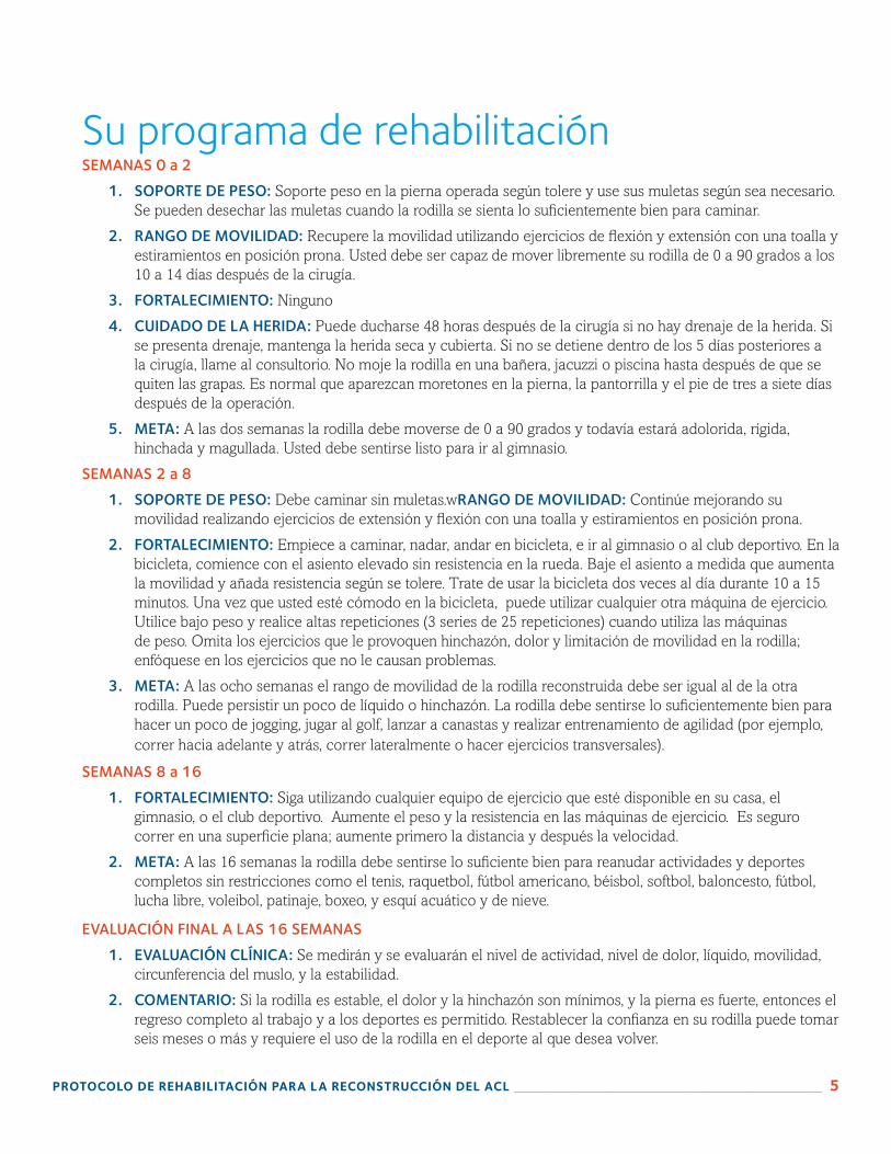
Su programa de rehabilitaciónSEMANAS 0 a 2
1. SOPORTE DE PESO: Soporte peso en la pierna operada según tolere y use sus muletas según sea necesario. Se pueden desechar las muletas cuando la rodilla se sienta lo suficientemente bien para caminar.
2. RANGO DE MOVILIDAD: Recupere la movilidad utilizando ejercicios de flexión y extensión con una toalla y estiramientos en posición prona. Usted debe ser capaz de mover libremente su rodilla de 0 a 90 grados a los 10 a 14 días después de la cirugía.
3. FORTALECIMIENTO: Ninguno
4. CUIDADO DE LA HERIDA: Puede ducharse 48 horas después de la cirugía si no hay drenaje de la herida. Si se presenta drenaje, mantenga la herida seca y cubierta. Si no se detiene dentro de los 5 días posteriores a la cirugía, llame al consultorio. No moje la rodilla en una bañera, jacuzzi o piscina hasta después de que se quiten las grapas. Es normal que aparezcan moretones en la pierna, la pantorrilla y el pie de tres a siete días después de la operación.
5. META: A las dos semanas la rodilla debe moverse de 0 a 90 grados y todavía estará adolorida, rígida, hinchada y magullada. Usted debe sentirse listo para ir al gimnasio.
SEMANAS 2 a 8
1. SOPORTE DE PESO: Debe caminar sin muletas.wRANGO DE MOVILIDAD: Continúe mejorando su movilidad realizando ejercicios de extensión y flexión con una toalla y estiramientos en posición prona.
2. FORTALECIMIENTO: Empiece a caminar, nadar, andar en bicicleta, e ir al gimnasio o al club deportivo. En la bicicleta, comience con el asiento elevado sin resistencia en la rueda. Baje el asiento a medida que aumenta la movilidad y añada resistencia según se tolere. Trate de usar la bicicleta dos veces al día durante 10 a 15 minutos. Una vez que usted esté cómodo en la bicicleta, puede utilizar cualquier otra máquina de ejercicio. Utilice bajo peso y realice altas repeticiones (3 series de 25 repeticiones) cuando utiliza las máquinas de peso. Omita los ejercicios que le provoquen hinchazón, dolor y limitación de movilidad en la rodilla; enfóquese en los ejercicios que no le causan problemas.
3. META: A las ocho semanas el rango de movilidad de la rodilla reconstruida debe ser igual al de la otra rodilla. Puede persistir un poco de líquido o hinchazón. La rodilla debe sentirse lo suficientemente bien para hacer un poco de jogging, jugar al golf, lanzar a canastas y realizar entrenamiento de agilidad (por ejemplo, correr hacia adelante y atrás, correr lateralmente o hacer ejercicios transversales).
SEMANAS 8 a 16
1. FORTALECIMIENTO: Siga utilizando cualquier equipo de ejercicio que esté disponible en su casa, el gimnasio, o el club deportivo. Aumente el peso y la resistencia en las máquinas de ejercicio. Es seguro correr en una superficie plana; aumente primero la distancia y después la velocidad.
2. META: A las 16 semanas la rodilla debe sentirse lo suficiente bien para reanudar actividades y deportes completos sin restricciones como el tenis, raquetbol, fútbol americano, béisbol, softbol, baloncesto, fútbol, lucha libre, voleibol, patinaje, boxeo, y esquí acuático y de nieve.
EVALUACIÓN FINAL A LAS 16 SEMANAS
1. EVALUACIÓN CLÍNICA: Se medirán y se evaluarán el nivel de actividad, nivel de dolor, líquido, movilidad, circunferencia del muslo, y la estabilidad.
2. COMENTARIO: Si la rodilla es estable, el dolor y la hinchazón son mínimos, y la pierna es fuerte, entonces el regreso completo al trabajo y a los deportes es permitido. Restablecer la confianza en su rodilla puede tomar seis meses o más y requiere el uso de la rodilla en el deporte al que desea volver.

6 ��������������������������������������������������������������������������������������� DrSteveHowell.com
Respuestas a preguntas frecuentesP: ¿Cuánto tiempo estaré en el hospital?
R: La cirugía que se realiza es ambulatoria. Los pacientes se van a casa 4 a 5 horas después de la cirugía luego de caminar con muletas. Si tiene sus propias muletas, por favor lléveselas al hospital.
P: ¿Cuánto tiempo tarda la cirugía?
R: La cirugía generalmente requiere de 30 a 40 minutos para realizarse. Reparar o remover un menisco desgarrado requiere unos 15 a 20 minutos adicionales.
P: ¿Cuándo puedo manejar un carro?
R: Puede volver a manejar cuando se sienta seguro y tenga confianza al volante, lo cual puede tomar de una a dos semanas o más. No maneje si está tomando pastillas para el dolor.
P: ¿Cuándo puedo volver al trabajo o a la escuela?
R: Las personas motivadas que quedan sentados o tienen un trabajo de oficina generalmente pueden regresar al trabajo a los 7 a 10 días después de la cirugía. Los trabajadores en la construcción requieren de 2 a 4 meses, dependiendo de las exigencias del trabajo.
P: ¿Cuál es la tasa de éxito de la cirugía?
R: El éxito depende del grado de otros daños en la rodilla en el momento de la reconstrucción del ACL, tales como desgarros del menisco, lesiones del cartílago articular, la artritis y otros desgarros de ligamentos. Aproximadamente el 95% de los pacientes con solo un desgarro del ACL y ninguna otra lesión puede volver a las actividades completas sin restricción sin ningún tipo de refuerzo ortopédico.
P: ¿Es posible volver a lesionar el injerto?
R: Sí, es posible volver a lesionarse. Recuerde que se le rompió su propio ligamento natural, entonces en teoría sería posible romper su injerto. Afortunadamente, es poco común la ruptura del injerto. Existe la misma probabilidad de desgarrar el ACL en la otra rodilla que volver a desgarrar el ligamento reconstruido.
P: Infórmeme sobre el uso de un tendón aloinjerto de un donante en lugar de mi propio tendón.
R: La ventaja de un aloinjerto es que hay menos dolor y no debilita los músculos de la pierna, lo cual sí ocurre al quitarle un tendón a su cuerpo para usar como injerto del ACL. Existe el bajo riesgo de transmisión de enfermedades con el aloinjerto y la tasa de fracaso puede ser mayor cuando se trata el injerto con irradiación o productos químicos. Nosotros utilizamos un aloinjerto no irradiado y no químicamente tratado de la transmisión musculoesquelética que no pierde la función durante 7 años. Vea lo siguiente.

PROTOCOLO DE REHABILITACIÓN PARA LA RECONSTRUCCIÓN DEL ACL ������������������������������������������ 7
Understanding Allografts:What they are and the role they’ll play in your surgery
Mission statement
MTF is a non-profit service organization dedicated
to providing quality tissue through a commitment to
excellence in education, research, recovery and care
for recipients, donors and their families.
125 May Street n Edison, NJ 08837 n 732-661-0202 n mtf.org
©2010 Musculoskeletal Transplant Foundation 4/10 0005 CI
8 ��������������������������������������������������������������������������������������� DrSteveHowell.com
You have a surgery coming up…and your doctor will be using an allograft in your procedure. What is an allograft?
An allograft is a bone, ligament, cartilage, tendon or section of skin that is transplanted from one person to another.
Every year in the United States, doctors use more than a million allografts to help…
• athletes who need knee reconstruction• people suffering from back pain• cancer patients who need tumor surgery
These are just a few examples. Surgeons have used allografts successfully—in all kinds of procedures—for decades. Allografts have improved millions of people’s lives. Your doctor thinks an allograft is a good choice for you, too.
Where do allografts come from?
Allografts come from donors—people who died in accidents or from sudden illnesses. Many times, just one donor’s gift can help a lot of people.
Donating tissue is a wonderful thing for someone to do. But it’s also important for you to know that not every donor is accepted. The bones, cartilage, tendons, skin and ligaments they donate need to be healthy. So, every donor is carefully screened by medical professionals. If the donor’s tissue is suitable, it is cleaned, processed and tested for sterility before it is sent to your doctor.
Can my doctor use my own tissue for my surgery?
Yes, but many doctors prefer to use donated tissue (allografts). When a doctor uses your own tissue (called an autograft), it has to come from another part of your body, which usually means a second surgical site and possibly more pain and recovery time.
Allografts are available, safe, and ready to use. There’s no need for a second surgery, so your recovery and healing will be easier.
Your allograft will come from the Musculoskeletal Transplant Foundation (MTF)—the largest tissue bank in the country. MTF is a non-profit organization and is highly respected in the medical community. Your doctor chose MTF because of our strong reputation and the quality and safety of our allografts.
Are allografts safe?
Yes. Since 1987, MTF has provided almost 4.2 million allografts from nearly 80,000 donors. MTF has an exceptional safety record because we are directed by surgeons from world-renowned hospitals, universities and medical institutions. These surgeons set MTF’s high standards and oversee which donors are accepted.
Want to know more?
If you have more questions about allografts, ask your doctor, visit MTF’s website at mtf.org or call 1.800.433.6576.
PROTOCOLO DE REHABILITACIÓN PARA LA RECONSTRUCCIÓN DEL ACL ������������������������������������������ 9
The Effect of Graft Tissue on Anterior Cruciate LigamentOutcomes: A Multicenter, Prospective, Randomized Controlled
Trial Comparing Autograft Hamstrings With Fresh-FrozenAnterior Tibialis Allograft
Keith W. Lawhorn, M.D., Stephen M. Howell, M.D., Steve M. Traina, M.D.,John E. Gottlieb, M.D., Thomas D. Meade, M.D., and Howard I. Freedberg, M.D.
Purpose: To compare the results and outcome of anterior cruciate ligament (ACL) reconstruction usingautogenous hamstring tendon versus fresh-frozen allograft anterior tibialis tendon. Methods: A prospec-tive randomized study was conducted from September 2002 to October 2006. We randomized 147patients to undergo ACL reconstruction with either autogenous hamstring or fresh-frozen allograft anteriortibialis tendon. Of these patients, 102 (69%) completed a minimum of 2 years’ follow-up. There were 54patients in the hamstring group (73% of those originally enrolled in the group) and 48 patients in theallograft group (66%). All patients underwent standardized subjective and objective evaluation withfunctional outcome assessments (International Knee Documentation Committee [IKDC]), and standard-ized radiographs were also obtained. Results: The mean age was 32.0 years for the autograft group and33.3 years for the allograft group. There was no difference in stability between the 2 groups (P � .05).The mean IKDC subjective score was 91.0 for the autograft group and 90.9 for the allograft group (P �.05). The functional IKDC scores for the autograft group were normal in 46 patients (85%), nearly normalin 7 patients (13%), and severely abnormal in 1 patient. For the allograft group, the functional IKDCscores were normal in 43 patients (90%) and nearly normal in 5 (10%) (P � .05). There were 4reoperations in the allograft group and 3 reoperations in the autograft group. No patient underwent revisionACL surgery or planned to undergo revision surgery because of instability in either group during the studyperiod despite the 1 patient in the autograft group with a pivot shift and a maximum manual KTmeasurement (MEDmetric, San Diego, CA) of 5 mm. Conclusions: The use of fresh-frozen anteriortibialis allograft (non-treated) for ACL reconstruction produced similar subjective and functional out-comes at 24 months’ minimal follow-up compared with patients undergoing ACL reconstruction withautograft hamstring tendon. Level of Evidence: Level II, prospective comparative study.
With the advent of improved fixation devices andmore exacting tunnel techniques, hamstring
tendon and tibialis allografts for anterior cruciate lig-ament (ACL) reconstruction continue to grow in pop-ularity.1,2 Many clinical studies comparing the out-comes of hamstring and bone–patellar tendon–bone(BPTB) grafts for ACL reconstruction show similaroutcomes.2-6 Despite the high success rates with au-tograft tissues, some surgeons continue to favor allo-graft use to avoid all harvest-site morbidity.
The effectiveness of allografts, however, remainscontroversial based on the current body of literature.Retrospective case-control studies and systematic re-views predominate the current body of literature, withthese studies showing few to no control groups re-garding graft preparation, types of allografts, tunnel
From Commonwealth Orthopaedics and Rehabilitation (K.W.L.),Fairfax, Virginia; Private Practice Orthopedics (S.M.H.), Sacra-mento, and Department of Mechanical Engineering (S.M.H.), Uni-versity California at Davis, California; Western Orthopedics(S.M.T.), Denver, Colorado; Private Practice (J.E.G.), Vail, Col-orado; Private Practice (T.D.M.), Allentown, Pennsylvania; andSuburban Orthopaedics (H.F.), Bartlett, Illinois, U.S.A.
The authors report that they have no conflicts of interest in theauthorship and publication of this article.
Received September 25, 2011; accepted May 8, 2012.Address correspondence to Keith W. Lawhorn, M.D., Common-
wealth Orthopaedics and Rehabilitation, 3620 Joseph Siewick Dr,Ste 100, Fairfax, VA 22033, U.S.A. E-mail: [email protected]
© 2012 by the Arthroscopy Association of North America0749-8063/11610/$36.00http://dx.doi.org/10.1016/j.arthro.2012.05.010
1079Arthroscopy: The Journal of Arthroscopic and Related Surgery, Vol 28, No 8 (August), 2012: pp 1079-1086
10 ������������������������������������������������������������������������������������� DrSteveHowell.com
1 3
Knee Surg Sports Traumatol ArthroscDOI 10.1007/s00167-016-4351-3
KNEE
Anterior laxity and patient‑reported outcomes 7 years after ACL reconstruction with a fresh‑frozen tibialis allograft
Emily Meike1 · S. M. Howell2 · M. L. Hull3
Received: 27 April 2016 / Accepted: 28 September 2016 © European Society of Sports Traumatology, Knee Surgery, Arthroscopy (ESSKA) 2016
significant (p = 0.08), and the average increase in anterior laxity of 2.7 ± 2.3 mm between the day of surgery and 7 years was significant (p < 0.001). There were no signifi-cant declines in activity (median Tegner score, 6 at 1 year, 6 at 7 years), function (average Lysholm score, 94 at 1 year, 91 at 7 years), and subjective satisfaction (average Interna-tional Knee Documentation Committee score, 90 at 1 year, 87 at 7 years) between 1 and 7 years after surgery.Conclusion In demonstrating that the ACL graft construct remains functional in the long term, this study supports the use of a fresh-frozen tibialis allograft in patients with an average age of 37 years at the time of surgery when used in conjunction with a surgical technique which avoids roof and PCL impingement, uses slippage-resistant fixation devices, and allows brace-free, self-paced rehabilitation.Level of evidence IV.
Keywords Anterior cruciate ligament · Roentgen stereophotogrammetry · Long-term follow-up · Ligamentization · Graft maturation
Introduction
An increase in anterior laxity following anterior cruciate ligament (ACL) reconstruction is worrisome because it can cause recurrent instability, and a reduction in activity level, function, and patient satisfaction [7]. The causes of an increase in anterior laxity are several and can be broadly categorized as short term, which extends over the period of graft incorporation into the bone tunnels and is limited to 3–4 months [29, 39], and long term which extends over the period of graft maturation beyond 3–4 months.
Focusing on soft tissue allografts, depending on the graft construct, surgical technique, and rehabilitation regimen,
Abstract Purpose After reconstructing a torn ACL with a soft tissue allograft, the long-term healing process of graft maturation following the short-term healing process of graft incorpora-tion into the bone tunnels might lead to recurring instability and concomitant decreases in the activity level, function, and patient satisfaction. Relying on roentgen stereopho-togrammetric analysis (RSA), the primary purpose was to determine whether anterior laxity increased and whether patient-reported outcomes declined between 1 and 7 years for a particular graft construct, surgical technique, and rehabilitation programme.Methods Eighteen of 19 patients, who participated in an earlier RSA study which extended to 1 year after the surgi-cal procedure, were contacted 7 years after the surgical pro-cedure. An examiner, different from the treating surgeon, measured anterior laxity under 150 N of anterior force using RSA in 16 patients and obtained outcome scores in 17 patients. One patient moved abroad and could not be contacted. One patient reinjured his reconstructed ACL and was excluded.Results The average increase in anterior laxity of 1.5 ± 2.1 mm between 1 and 7 years after surgery was not
* M. L. Hull [email protected]
1 Biomedical Engineering Graduate Group, University of California, Davis, CA 95616, USA
2 Department of Biomedical Engineering, Biomedical Engineering Graduate Group, University of California, Davis, CA 95616, USA
3 Department of Mechanical Engineering, Department of Biomedical Engineering, Biomedical Engineering Graduate Group, University of California, Davis, CA 95616, USA
PROTOCOLO DE REHABILITACIÓN PARA LA RECONSTRUCCIÓN DEL ACL ����������������������������������������� 11
Notas:

12 ������������������������������������������������������������������������������������� DrSteveHowell.com
Adventist Health Physicians Network Medical Office - Orthopedics
8120 Timberlake Way, Suite 112
Sacramento, CA 95823
916-689-7370
Adventist Health Lodi Memorial Medical Office – Orthopedics
1235 W. Vine St, Suite 22
Lodi, CA 95240
209-334-8535