purification and characterization of transporter proteins ... site pdfs/25-wang... · detergents,...
TRANSCRIPT
20
Purification and Characterization of TransporterProteins From Human Erythrocyte Membrane
Da-Neng Wang, M. Joanne Lemieux, and Jonathan M. Boulter
1. IntroductionFunctions and biochemical properties of several membrane transporter pro-
teins from human erythrocyte, in particular, the glucose transporter (Glut1) andanion exchanger (AE1, also called Band 3) have been extensively characterized.Glut1 is a member of the mammalian facilitative glucose transporter familyGlut1-13 (1,2). The 50-kDa integral membrane protein is expressed in allhuman cells, and is particularly abundant in human erythrocytes, fibroblasts andendothelial cells. In erythrocytes it is responsible for the uptake of glucose—thecell’s major energy source. Glut1 consists of 492 amino acids and is N-glycosy-lated on residue Asn45 in a highly heterogeneous manner. The protein, whichfunctions as a monomer, is predicted to span the membrane 12 times in the formof �-helices (1).
A member of the anion exchanger family, AE1 is a 95-kDa integral mem-brane protein with multiple functions (3). It is responsible for the electroneutralexchange of HCO3
− and Cl− across the erythrocyte membrane, thereby facilitat-ing CO2 transport by the blood. The AE1 protein consists of two structurallydistinct domains, an amino-terminal cytosolic domain (residues 1–360) and acarboxyl-terminal membrane domain (residues 361–911) (4). The cytosolicdomain is approx 43 kDa, and provides binding sites for hemoglobin andcytoskeletal proteins. On the other hand, the 52-kDa membrane domain(AE1MD), which is glycosylated, is solely responsible for the protein’s iontransport activity, and retains functionality following enzymatic removal of thecytosolic domain.
Easy access to human blood and their abundance in the erythrocyte mem-
239
From: Methods in Molecular Biology, vol. 228:Membrane Protein Protocols: Expression, Purification, and Characterization
Edited by B.S. Selinsky © Humana Press Inc., Totowa, NJ
18649_ch20_wang.239-256.qxd 1/27/03 4:31 PM Page 239
240 Wang, Lemieux, and Boulter
brane have made Glut1 and AE1MD prototypes of integral membrane proteinsfor biochemical and biophysical studies (5,6). In this work, we present twopurification protocols for Glut1 and AE1MD. The protocol for Glut1, devel-oped in our laboratory, introduces a positive chromatography step. As a result,less lipid is copurified whilst the protein still retains ability to bind to itsinhibitor cytochalasin B in detergent solution (7). The protocol for purifyingAE1MD is derived from a procedure by Casey et al. (8), to which an additionalmembrane stripping and chromatography step is introduced. The first Q anionexchange column is followed by deglycosylation of the protein, and the sec-ond Q column removes glycosidase and detached sugars and delipidates theprotein (9). Stability and monodispersity of purified Glut1 and AE1MD aresubsequently analyzed under a wide range of conditions by analytical size-exclusion high-pressure liquid Chromatography (HPLC). AE1MD generatedby our procedure yields three-dimensional crystals, diffracting to 14 Å reso-lution (9).
2. Materials2.1. Equipment
1. Preparative centrifuge: Sorvell RC 5B centrifuge with GS-3 and SA-600 rotors (DuPont, Wilmington, DE).
2. Ultracentrifuge: Beckman XL-90 ultracentrifuge with Ti45 and Ti70.1 rotors(Beckman, Palo Alto, CA).
3. FPLC system: controller LCC-501 PLUS, peristaltic pump P-1, pumps P-500,valves MV-7 and MV-8, detector UV-M II, fraction collector FRAC-100, andrecorder REC-102 (Amersham Pharmacia, Piscataway, NJ). The FPLC system iskept at 4°C in an Isotemp incubator (Fisher Scientific, Pittsburgh, PA).
4. Preparative ion-exchange chromatography columns: HiTrap Q 1 mL and 5 mLcolumns (Amersham Pharmacia).
5. Vertical electrophoresis system: Mini-Protean II xi vertical electrophoresis cell andgel casting system, and PowerPac 1000 or 3000 power supply (Bio-Rad, Hercules,CA).
6. HPLC system: Waters solvent delivery system (controller 600 and pump 626), con-trolled by the Millennium software on a PC, and photodiode array detector Waters996 (Waters, Milford, MA).
7. Spectrophotometer: Agilent UV-Visible spectrophotometer 8453 (Agilent Tech-nologies, Palo Alto, CA).
8. Analytical size-exclusion chromatography columns: Shodex KW803 and KW804columns (Showa Denko, Tokyo, Japan).
9. Transmission electron microscope: CM12 electron microscope with a single-tilt,room-temperature specimen holder (Philips, Eindhoven, The Netherlands).
18649_ch20_wang.239-256.qxd 1/27/03 4:31 PM Page 240

Purification of Erythrocyte Membrane Transporters 241
2.2. Reagents
1. Detergents, solubilization grade or higher (Anatrace, Maumee, OH).2. Phospholipids (Avanti Polar Lipids, Alabaster, AL).3. Peptide: N-glycosidase F (PNGase F) (New England BioLabs, Beverly, MA).4. Trypsin (Worthington Chemicals, Toronto, Canada).5. Micro-BCA Protein Assay Kit (Pierce, Rockford, IL).6. Electron microscopy copper grids (Ted Pella, Redding, CA).7. Filter paper type #41 (Whatman, Clifton, NJ).8. All other reagents are from Sigma (St. Louis, MO) and are of analytical grade or
higher.9. All purification steps are carried out at 4°C unless specified otherwise. Purified pro-
teins samples are stored at 4°C.
2.3. Buffers for Glut1 Purification
1. Phosphate-buffered Saline (PBS) buffer: 5 mM Na2HPO4, pH 7.4, and 150 mM NaCl.2. Haemolysis buffer: 0.1 M phosphate buffer, pH 7.4, 0.5 mM ethylene diamine
tetraaretic acid (EDTA), and 1 mM phenylmethyl sulfonyl fluoride (PMSF).3. Stripping buffer: 15 mM NaOH, pH 12, 2 mM EDTA, and 0.2 mM PMSF.4. Solubilization buffer: 20 mM Bis-Tris, pH 7.0, 50 mM NaCl, 1 mM EDTA, 1 mM
PMSF, 10% glycerol, and 2% decylmaltoside (DM).5. Loading buffer: 10 mM Bis-Tris, 0.5 mM EDTA, pH 6.0, and 0.2% DM.6. HPLC buffer: 20 mM Tris-HCl, pH 8.0, 0.2 M Na2SO4, and 0.15%DM
2.4. Buffers for AE1MD Purification
1. Hemolysis buffer: 0.1 M phosphate buffer, pH 7.4, 0.5 mM EDTA, 1 mM PMSF.2. Stripping buffer: 15 mM NaOH, pH 12, 2 mM EDTA, and 0.2 mM PMSF.3. Solubilization buffer: 20 mM imidazole, pH 7.0, 50 mM NaCl, 1 mM EDTA, 1 mM
PMSF, 10% glycerol, and 1% dodecylmaltoside (DDM) or 1% C12E8 detergent.4. Loading buffer: 20 mM imidazole, pH 7.0, 1 mM EDTA, 0.5 mM PMSF, 1 mM
NaN3, 10% glycerol, and 0.1% DDM or C12E8.5. HPLC buffer: 20 mM Tris-HCl, pH 8.0, 0.2 M Na2SO4, and 0.05% DDM.
3. Methods3.1. Purification of Glut1 Protein
1. Source: Human erythrocyte “ghost” membrane is prepared from packed outdatedred cells (obtained from Bloodbank). Wash cells twice in PBS buffer. Centrifuge at3000g (4000 rpm in GS-3 rotor) for 30 min and collect pellet.
2. Ghost preparation: Lyse cells directly in ice-cold haemolysis buffer. Wash cellscontinuously in hemolysis buffer and centrifuge at 9000g for 15 min at 4°C. Repeatthe washing step 10 times until the membrane appears white.
18649_ch20_wang.239-256.qxd 1/27/03 4:31 PM Page 241

242 Wang, Lemieux, and Boulter
Fig. 1
Fig. 2
3. Cell stripping: Remove cytoskeletal and peripheral membrane proteins fromghost membrane by washing with 10 vol of stripping buffer for 30 min at 37°C.Centrifuge at 16000g for 20 min. Store aliquoted membrane at −20°C. Typi-cally, one unit of packed red blood cells produces 20 mL of stripped ghost mem-brane.
4. Erythrocyte membrane solubilization (see Notes 1 and 2): Solubilize 4 mL ofstripped ghost membrane in 20 mL solubilization buffer. Stir solution for 30 min at4°C. Remove unsolubilized materials and large aggregates by centrifuging at10000g for 15 min (See Fig. 1).
5. Q anion exchange chromatography (see Notes 3 and 4): Load solubilized sampleonto a 1-mL HiTrap Q anion exchange column on FPLC, preequilibrated withloading buffer, at a rate of 0.5 mL/min. Wash with 15-mL loading buffer. Elutesample in the same buffer with a 15-mL linear 0–500 mM NaCl gradient at 0.25mL/min (see Fig. 2). Collect 0.5 mL fractions.
Fig. 1. Selection of detergent for solubilization of human erythrocyte membrane andextraction of Glut1 and AE1MD. The Coomassie Blue-stained SDS-PAGE shows theerythrocyte membrane solubilized with different detergents. PNGase F is included inthe SDS buffer to remove the oligosaccharide and sharpen the protein bands. Of all thedetergents tested here, DM and NG are most effective at extracting Glut1. NG, however,causes aggregation of the glucose transporter protein and DM is, therefore, selected forsolubilization and subsequent purification of the protein. DDM and C12E8 are selectedfor extraction and purification of AE1MD.
18649_ch20_wang.239-256.qxd 1/27/03 4:31 PM Page 242

Purification of Erythrocyte Membrane Transporters 243
Fig. 3
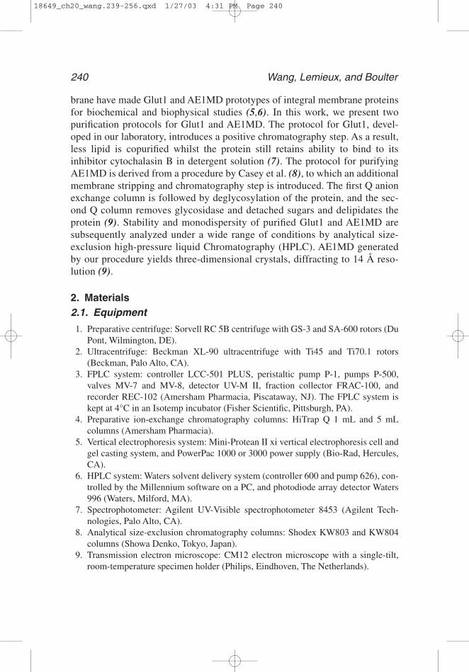
6. Sodium dodecyl Sulfate-polyacxylamide gel electrophoresis (SDS-PAGE) analysis(see Note 5): To better assess protein purity, deglycosylate Glut1 with 5000 U/mgPNGase F in the presence of 1% SDS at 20°C. Analyze immediately by CoomassieBlue-stained SDS-PAGE (see Fig. 3).
7. Protein concentration measurement. Determine protein concentration by the MicroBCA assay using spectrophotometer. Include 10 mM Bis-Tris in the blank to mini-mize interference with the assay.
3.2. Purification of AE1 Membrane Domain
1. Source (see Note 6): The same as Subheading 3.1., Step 1, or freshly donatedhuman blood.
2. DIDS labeling (see Note 7): Prepare diluted cells of 25% hematocrit in PBS con-taining 0.0005% DIDS. Incubate for 60 min at 37°C. Wash cells with PBS and cen-trifuge to remove unbound DIDS. Cross-link DIDS by incubating with 10 vol of0.1 M NaHCO3, pH 9.5, for 60 min 37°C. Remove excess DIDS from supernatantby centrifuging for 20 min in GS-3 rotor at 7500g).
3. Ghost preparation: Lyse DIDS-labeled or unlabeled cells as described in Sub-heading 3.1., Step 2.
Fig. 2. Elution of DM-solubilized erythrocyte membrane proteins from HiTrap Qanion exchange column. Protein is monitored at 280-nm absorbance and Glut1 eluted ataround 100 mM NaCl at a concentration of 0.5–1.0 mg/mL. Column buffer is 10 mMBis-Tris pH 6.0, 0.5 mM EDTA, 0.2% DM. (Adapted from ADDIN ENRfu (7) with per-mission.)
18649_ch20_wang.239-256.qxd 1/27/03 4:31 PM Page 243
244 Wang, Lemieux, and Boulter
4. AE1 membrane domain generation: Dilute ghost membrane two-fold with hemoly-sis buffer lacking PMSF. Add trypsin to a final concentration of 5 �/mL. Stir for 60min at 4°C, and then add PMSF to a final concentration of 2 mM to stop the trypsindigestion. Centrifuge for 15 min 35000g. Wash with 10 vol. of hemolysis buffer.Centrifuge again and collect pellet.
5. Cell stripping (see Note 8): Repeat twice the stripping procedure as described inSubheading 3.1., step 3 but at 4°C. Store membrane at −20°C.
6. Erythrocyte membrane solubilization (see Notes 1 and 2): Solubilize 4 mL ghostmembrane in 20 mL of solubilization buffer. Stir for 30 min at 4°C. Centrifuge toremove unsolubilized materials (35,000 rpm in Ti45 rotor for 30 min).
7. First anion exchange column (see Note 9): Load solubilized membrane onto a5-mL HiTrap Q anion chromatography column on FPLC, preequilibrated withloading buffer, at a rate of 0.5 mL/min. Wash with 5 mL loading buffer. Elute pro-tein with the same buffer, using a 20-mL linear 0.05–1 M NaCl gradient at 0.25mL/min. Collect 0.5 mL fractions (see Fig. 4).
8. Protein deglycosylation: Deglycosylate AE1MD with 5 U/mg PNGase Fovernight at 20°C. Verify completeness of deglycosylation by SDS-PAGE analysis(see Fig. 4).
Fig. 3. Coomassie Blue-stained SDS-PAGE showing Glut1 purification and deglyco-sylation. (A) Lane 1: Protein molecular weight standards. Lane 2: Glut 1 purified in DMdetergent by HiTrap Q anion-exchange column, with a purity of approx 95% purity. Theprotein is highly glycosylated. Lane 3: After PNGase F treatment in the presence ofSDS, polysaccharide is cleaved from Glut1, resulting a sharpened band that shifted to43 kDa. (B). When Glut1 is incubated with PNGase F, followed by separation fromPNGase F using a size-exclusion column, and then loaded onto SDS-PAGE, the broadband of glycosylated Glut1 monomer remains intact. Minimal amounts of Glut1 dimercan form owing to delipidation. 10 �g protein is loaded in each lane. (Reproduced fromADDIN ENRfu (7) with permission.)
Fig. 4
18649_ch20_wang.239-256.qxd 1/27/03 4:32 PM Page 244

Purification of Erythrocyte Membrane Transporters 245
9. Second anion exchange column: Dialyze deglycosylated peak fractions from thefirst Q column against column buffer containing 50 mM NaCl. Load protein onto a1-mL HiTrap Q anion exchange chromatography column on FPLC, in the sameloading buffer that is used for the first column. The detergent used for the second Qcolumn is DDM (0.1%), undecylmaltoside (UDM, 0.1%), DM (0.1%), decylth-iomaltoside (DTM, 0.1%), Cymal-6 (0.25%), Cymal-5 (0.5%), C12E8 (0.1%), C10E6(0.1%), or C8E5 (0.7%). Elute protein from column in five column volumes of load-ing buffer with a 0.05–2-M NaCl linear gradient to ensure high concentration frac-tions. Collect 0.5 mL fractions (see Fig. 4).
10. SDS-PAGE analysis (see Note 10): Analyze protein samples from differentpurification stages by Coomassie Blue- or silver-stained SDS-PAGE for proteinpurity.
11. Protein concentration measurements (see Note 11): Determine protein concentra-tion by the Micro BCA assay using spectrophotometer.
Fig. 4. Coomassie Blue-stained SDS-PAGE showing purification and deglycosyla-tion of human erythrocyte anion exchanger membrane domain. Following purifica-tion with the first Q anion exchange chromatography column, the protein is treatedwith PNGase F to deglycosylated AE1MD and a second Q column is introduced tofurther purify and delipidate the protein. (Reproduced from ADDIN ENRfu (9) withpermission.)
18649_ch20_wang.239-256.qxd 1/27/03 4:32 PM Page 245
246 Wang, Lemieux, and Boulter
3.3. Determination of Stokes Radius of Glut1 and AE1MD by Size-Exclusion HPLC
1. Inject 50 �g of purified Glut1 or AE1MD sample onto a Shodex KW803 orKW804 analytical size-exclusion column on HPLC, preequilibrated with HPLCbuffer. Develop in the same buffer at 0.5 mL/min. Monitor UV absorption spectrumat 280 nm (see Note 12).
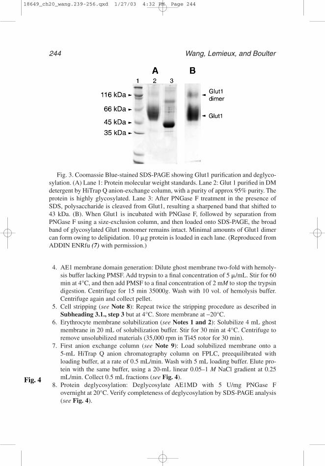
2. Record retention times, t, for Glut1 and AE1MD (see Fig. 5).3. Record retention times of a set of soluble proteins of known Stokes radius (RS) (10):
thyroglobulin (86 Å), apoferritin (63 Å), catalase (52 Å), and aldolase (46 Å).4. Determined Stokes radii of Glut1 and AE1MD by comparison with the retention
times of standard proteins.
3.4. Determination of Glut1 and AE1MD Stability and Monodispersity
Stability and monodispersity of Glut1 and AE1MD proteins in various deter-gent solutions and under a wide range of conditions are measured by a modifiedprotocol from the literature (11,12), using analytical size-exclusion chromatog-raphy on HPLC (7,9). The following parameters can be screened for their effecton protein stability and monodispersity: detergent, pH, additive and tempera-ture. On a Shodex KW803 or 804 column, each such run takes 30 min and onlya 50-�g protein sample is needed. Accordingly, a number of conditions can be
Fig. 5. Size exclusion HPLC chromatograms of AE1MD in C12E8 (thin line) and NTG(thick line) solution. The samples are run on a Shodex KW-804 column, equilibrated with50 mM Tris, pH 8.0, 200 mM N2SO4, 3 mM NaN3, and 0.1% C12E8. Peaks are identifiedby their retention time. AE1MD in complex with detergent had a Stokes radius of 66 Å,corresponding to a protein dimer. Elution times of standard proteins of known Stokesradius are indicated: TG = thyroglobulin (86 Å), AF = apoferritin (63 Å), CA = catalase(52Å), AL = aldolase (46Å). (Reproduced from ADDIN ENRfu (9) with permission.)
Fig. 5
18649_ch20_wang.239-256.qxd 1/27/03 4:32 PM Page 246

Purification of Erythrocyte Membrane Transporters 247
screened within a short period of time. Those detergents and conditions thatkeep the protein stable and monodisperse can be used for its purification andcrystallization (see Note 13).
3.4.1. Sample preparations
1. To study detergent effect: Incubate 50 �L Glut1 or AE1MD of 1 mg/mL, purified in0.2% DM or 0.1% C12E8, respectively, in the presence of various detergents at con-centrations 0.2% above their respective CMC (added from 10% stock solutions) for2 to 16 h at 25°C.
2. To study pH effect: Titrate 50 �L samples of purified Glut1 (in 10 mM Bis-Tris, pH6.0), or AE1MD (in 20 mM imidazole, pH 7.0), to the desired pH by adding 5 �Lof 1 M buffer relevant pH. Incubate for 2 to 16 h at 25°C. Use following buffers: pH4: acetate/acetic acid, pH 5: acetate/acetic acid, pH 6 (control): Bis-Tris-HCl, pH 7:Bis-Tris-HCl, pH 8: Tris-HCl, pH 9: Tris-HCl, pH 10: carbonate-HCl.
3. To study additive effect: Add following additives to purified Glut1 samples: glyc-erol to 20% (v/v), cytochalasin B to 10 mM (from a 2-mM stock in ethanol, storedat −20°C), glucose to 100 mM, adenosine triphosphate (ATP) to 1 mM, and dithio-threitol (DTT) to 5 mM. Incubate for 16 h at 37°C.
4. To study lipid effect: Delipidate Glut1 by size-exclusion HPLC and collect proteinfractions. Add individual phospholipids to protein samples to a final concentrationof 0.1 mg/mL. Incubate for 16 h at 4°C.
5. To study temperature effect: Incubate purified Glut1 and AE1MD samples at 4, 25,and 37°C. Withdraw aliquots at specific time intervals.
3.4.2. Protein Monodispersity analysis by Size-Exclusion HPLC
1. Load 50 �g of sample, prepared as described above, onto a Shodex KW803 orKW804 size-exclusion column equilibrated with HPLC buffer. Develop at 0.5mL/min.
2. Collect absorption spectrum at 280 nm. Monitor shape and height of protein peak.3. Compare with control sample (see Fig. 5). Extract and integrate chromatograms
and calculate proportion of the protein in the peak relative to the total amount (seeTables 1–3) (see Notes 14–17).
3.5. Protein Homogeneity Analysis by Electron Microscopy (EM)
Transmission electron microscopy (EM) of negatively stained specimens is aconvenient way of assessing the homogeneity of a membrane protein prepara-tion (13), provided an EM facility is accessible.
1. Specimen preparation: Apply a 2-�L aliquot of purified protein sample (concentra-tion of about 1 mg/mL) to a carbon-coated electron microscopy grid. Remove sam-ple from grid with filter paper after 10. Stain grid twice with 2% uranyl acetate, for20 s each time. Dry grid thoroughly with filter paper.
Table1
Table2
Table3
18649_ch20_wang.239-256.qxd 1/27/03 4:32 PM Page 247

Table 1Monodispersity of Purified Glut1 Protein Under Various Conditions
% monomer1 Effect on Reagent Conditions remaining monodispersity
(A) Detergents:
DM 25°C 22 controlNM overnight 22 −UDM without 22 +/−DDM glycerol 25 +DTM 26 +Cymal-3 19 −OG 5 −−−NG 4 −−−DG 13 −−C12E8 6 −−−C10E8 4 −−−MEGA−10 7 −−−(B) pH:
4.0 25°C 10 −5.0 overnight 70 ++6.0 95 +++7.0 90 +++8.0 65 ++9.0 15 −(C) Additives:
None 37°C 24 control20% Glycerol overnight 59 ++++10�M Cytochalasin B 42 +++100mM Glucose 36 ++1mM ATP 19 −−5mM DTT 24 ; ms/+
(D) Lipids:
None 4°C 61 controlPhosphatidylcholine overnight 75 +Phosphatidylethanolamine 56 −Sphingomyelin 63 +/−Cholesterol 59 −Phosphatidylinositol 79 ++Phosphatidylserine 93 +++Total Erythrocyte Lipids 81 ++
1 Effect of (a) lipid, (b) detergent, (c) pH, and (d) additives on the stability of Glut1. Concentra-tions of reagents areas follows: 0.1 mg/mL lipid (added to delipidated Glut1), 1% detergent. Theproportion of monomeric Glut1 is 100% before treatment. Each parameter is assayed separately,and combination of the positive factors identified in such a way is used to preserve Glut1 monodis-persity for an extensive period of time. (Reproduced from et (7) with permission from Academic.)
18649_ch20_wang.239-256.qxd 1/27/03 4:32 PM Page 248
Purification of E
rythrocyte Mem
brane Transporters249
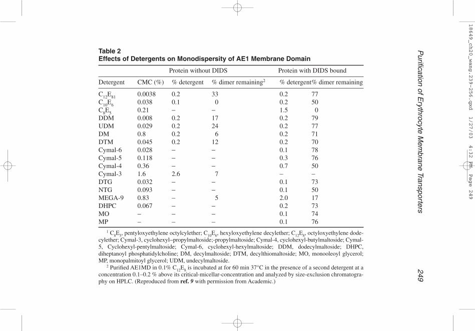
Table 2Effects of Detergents on Monodispersity of AE1 Membrane Domain
Protein without DIDS Protein with DIDS bound
Detergent CMC (%) % detergent % dimer remaining2 % detergent% dimer remaining
C12E81 0.0038 0.2 33 0.2 77C10E6 0.038 0.1 0 0.2 50C8E5 0.21 − − 1.5 0DDM 0.008 0.2 17 0.2 79UDM 0.029 0.2 24 0.2 77DM 0.8 0.2 6 0.2 71DTM 0.045 0.2 12 0.2 70Cymal-6 0.028 − − 0.1 78Cymal-5 0.118 − − 0.3 76Cymal-4 0.36 − − 0.7 50Cymal-3 1.6 2.6 7 − −DTG 0.032 − − 0.1 73NTG 0.093 − − 0.1 50MEGA-9 0.83 − 5 2.0 17DHPC 0.067 − − 0.2 73MO − − − 0.1 74MP − − − 0.1 76
1 C8E5, pentyloxyethylene octylcylether; C10E6, hexyloxyethylene decylether; C12E8, octyloxyethylene dode-cylether; Cymal-3, cyclohexyl−propylmaltoside;-propylmaltoside; Cymal-4, cyclohexyl-butylmaltoside; Cymal-5, Cyclohexyl-pentylmaltoside; Cymal-6, cyclohexyl-hexylmaltoside; DDM, dodecylmaltoside; DHPC,diheptanoyl phosphatidylcholine; DM, decylmaltoside; DTM, decylthiomaltoside; MO, monooleoyl glycerol;MP, monopalmitoyl glycerol; UDM, undecylmaltoside.
2 Purified AE1MD in 0.1% C12E8 is incubated at for 60 min 37°C in the presence of a second detergent at aconcentration 0.1–0.2 % above its critical-micellar-concentration and analyzed by size-exclusion chromatogra-phy on HPLC. (Reproduced from ref. 9 with permission from Academic.)
18649_ch20_wang.239-256.qxd 1/27/03 4:32 PM Page 249

250 Wang, Lemieux, and Boulter
Fig. 6
Table 3Effects of pH on monodispersity of AE1 membrane domain with DIDS bound
pH Buffer1 % dimer remaining2
4.5 Acetate 35.5 Citrate 726.0 Bis-Tris 727.0 Imidazole 807.0 Phosphate 807.5 HEPES 807.5 Tris-HCl 828.5 TAPS 829.0 Glycine 81
10.0 Glycine 811 Buffers are prepared at a concentration of 1.0 M.2 50 �g samples of purified AE1MD (10 mM imidazole, pH
7.0) are titrated to the relevant pH by addition of 5 �L of 1 Mbuffer of the desired pH, and then incubated at 25°C for 30 min,followed by analysis using size-exclusion chromatography onHPLC. (Reproduced from ref. 9 with permission from Academic.)
2. Electron microscopy: Mount grid onto a room-temperature, single-tilt holder. Insertinto electron microscope. Search at low magnification (2000–3000). Observe andrecord electron micrographs at higher magnification (40000–100000) (see Fig. 6).
4. Notes1. Detergent selection: A variety of detergents are tested for extraction of Glut1 from
erythrocyte membrane (see Fig. 1). They include Triton X-100, C12E8, octylgluco-side (OG), nonylglucoside (NG), HECAMEG, DDM, DM, CHAPS, CHAPSO,sodium cholate and SB-12. For this purpose, 50 �L membrane in 50 mM Tris/HClpH 7.4, 0.5 mM EDTA, is solubilized with 1% micellar detergent, i.e., at a detergentconcentration 1% above its critical micellar concentration (CMC). After incubationat 4°C for 30 min with stirring and removal of unsolubilized membrane, 25 �L ofthe supernatant is taken for SDS-PAGE analysis, followed by Coomassie bluestaining. Of all the detergents tested here, DM and NG are most effective at extract-ing Glut1. However, NG causes aggregation of the glucose transporter protein, andDM is, therefore, selected for solubilization of Glut1 and subsequent purification.For comparison, four detergents extract AE1 well: C12E8, NG, DDM, and DM.Among them, the two with long carbon-chain (C12), C12E8 and DDM, keep theprotein stable and are therefore used for AE1MD extraction and purification.
2. Membrane solubilization completeness: It is critical that the ghost membrane iscompletely solubilized for subsequent protein purification—the solution shouldbecome translucent following 30 min stirring at 4°C in the presence of 1–2%
18649_ch20_wang.239-256.qxd 1/27/03 4:32 PM Page 250

Purification of Erythrocyte Membrane Transporters 251
Fig. 6. EM of purified Glut1 sample. Purified Glut in DM detergent is very homoge-neous. The protein sample is negatively stained with 2% uranyl acetate on carbon-coated EM grid. The protein molecules adhere to the carbon support film in variousorientations, resulting in different shapes and sizes. Collapsing of the protein moleculeon the grid and the surrounding detergent micelle make the protein’s dimensions appearto be somewhat larger than in solution, scale bar represents 1000 Å.
micellar detergent. Otherwise add 20% more solubilization buffer and stir the solu-tion for another 15 min.
3. Glut1 purification yield: For Glut1 purification, erythrocyte membrane is com-pletely solubilized by 2% DM. In this detergent, Glut1 binds to the HiTrap Q anionexchange column at pH 6.0, and is eluted as a sharp peak at approximately 100 mMNaCl, with a protein concentration of 0.5–1.0 mg/mL (see Fig. 2). Other erythro-cyte membrane proteins, such as AE1, do not bind significantly to the columnunder these conditions and, therefore, the Glut1 peak dominates the chromatogram,yielding a positive purification procedure. The protein in the peak fractions isapprox 95% pure. The yield of the Glut1 peak is 5 ± 1% (n=5) of total protein in thestripped erythrocyte membrane, although the entire yield of Glut1, including non-peak fractions, is estimated to be 8–10% of total protein.
4. Glut1 binding to Q column: The binding of Glut1 to an anion exchange matrix atpH 6.0 needs explanation, given the high isoelectric point of Glut1 of around 8.4(14). At this pH, Glut1 should be positively charged (cationic) and should not bindto an anion exchange column. We hypothesize that negatively charged lipids copu-rifying, such as phosphatidylserine and phosphatidic acid, could bridge the posi-tively charged protein and the positively charged column matrix, resulting in thebinding of Glut1 to the anion exchange resin. Interestingly, the Q column-purifiedGlut1 will not bind to the same anion exchange column again, but will bind weaklyto a cation exchange column instead, suggesting that the localized negative charges
18649_ch20_wang.239-256.qxd 1/27/03 4:32 PM Page 251

252 Wang, Lemieux, and Boulter
holding the protein to the first Q column have become dispersed upon release fromthe resin.
5. Glut1 deglycosylation: PNGase F is added to purified Glut1 sample at 5000 �/mgprotein and subsequently loaded onto SDS-PAGE. Consequently, the Glut1 bandbecomes sharpened and shifts to about 43 kDa, indicating complete deglycosyla-tion of the protein (see Fig. 3A). However, the amount of glycosidase needed is1000 times more than that needed to deglycosylate AE1MD. When Glut1 is sepa-rated from PNGase F using a size-exclusion column following incubation, thebroad band of glycosylated Glut1 on SDS-PAGE remains intact (see Fig. 3B). We,therefore, conclude that PNGase F is only able to deglycosylate Glut1 when theprotein is unfolded in the presence of SDS and is inactive against folded Glut1.
6. AE1MD purification: The purification protocol presented here differs from the pro-tocol of Casey et al. (8) in several ways. An additional membrane-stripping step isemployed here to further remove contaminating proteins. A strong anion exchangerresin, instead of diethylaminoethyl (DEAE), is used in the first chromatography stepand the pH in the column is changed from 8.0 to 7.0, resulting in less backgroundcontaminant. Furthermore, the newly introduced second Q column removes detachedoligosaccharide and the glycosidase used, and further delipidates the protein.
7. AE1MD DIDS-labeling: The covalently bound DIDS locks AE1 into a fixed con-formation and stabilizes the protein. Mild trypsinization cleaves off the cytosolic42 kDa domain from the membrane domain. Both are necessary for three-dimensional (3-1) crystal formation of the protein (9).
8. Membrane stripping: Repeating the membrane stripping step twice removes a largeproportion of glycophorin A, a single-span membrane protein that otherwise con-taminates the preparation. This is particularly important because its presence is dif-ficult to detect by Coomassie Blue-stained SDS-PAGE.
9. AE1MD purification and deglycosylation: Both C12E8 and DDM completely solu-bilize the stripped red cell membrane and effectively extract AE1MD from themembrane. The first HiTrap Q anion exchange column produced 90% pure AE1membrane domain (see Fig. 4). Deglycosylation of AE1MD with PNGase F at 5U/mg protein overnight at 20°C cleaves off the oligosaccharide from the proteincompletely. Unlike in the case of Glut1, the deglycosylation reaction does occur onthe intact AE1MD in the absence of SDS, yielding a markedly more homogeneouspreparation. AE1MD elutes from the second Q column as a sharp peak at 0.8 MNaCl, and the steep salt gradient results in concentrated (1–2 mg/mL) AE1MDfractions at a relatively low (0.1%) detergent concentration. The final protein purityis close to 95%. In addition, this Q column can also be used to exchange detergentfor subsequent crystallization experiments.
10. Membrane protein SDS-PAGE analysis: In contrast to soluble proteins, membraneprotein samples prepared for SDS-PAGE should not be boiled. Boiling the samplesin SDS causes severe aggregation. Prolonged incubation of the samples with thesample buffer at the room temperature, or even 4°C, also leads to aggregation. Thebest results are obtained with freshly prepared samples on newly cast gels. Ready-made gels from commercial sources often yield blurred bands. In addition,hydrophobic proteins with multiple transmembrane segments like Glut1 and
18649_ch20_wang.239-256.qxd 1/27/03 4:32 PM Page 252
Purification of Erythrocyte Membrane Transporters 253
AE1MD tend to migrate to lower positions on SDS-PAGE, compared with solubleproteins with similar molecular weights. This is probably due to the larger amountsof SDS bound to the polypeptide or partially folded structures of the membraneproteins in SDS.
11. Membrane protein concentration measurement: We have tested various proteinassays for protein concentration determination, including Lowry, Bradford, BCA,and Micro-BCA. Micro-BCA from Pierce has the broadest compatibility withdetergents, and it produced very consistent results, within 15% from those mea-sured by absorption at 280 nm. We therefore choose this assay for protein concen-tration determination.
12. Stokes radius measurement: From a size-exclusion chromatography column onHPLC, both Glut1 in DM and AE1MD in DDM elute as a single major protein peak(see Fig. 5). By comparison with the retention times of soluble protein standards,the Stokes radii of the Glut1 and AE1MD particles are determined to be 50 Å and66 Å, respectively. Such Stokes radii are of the protein-detergent complexes, not ofthe proteins along. Compared with the Stokes radii of other membrane transporterswith similar molecular weights (11,15), the Glut1 protein is likely to be a monomerin DM and AE1MD a dimer in DDM.
13. Protein monodispersity and stability: To maintain the proteins in a monodispersestate for crystallization, various factors are screened for their ability to influence thestability of the protein. They include detergent, pH, additive, lipid, and temperature.Conditions are eventually found to maintain the protein monodisperse for weeks(see Tables 1–3).
14. Detergent effect: Detergents can be assayed for their effect on the stability of Glut1and AE1MD in solution. Maltoside detergents generally retain Glut1 monodisper-sity, with DTM being the best, followed by DM and DDM (see Table 1A). Simi-larly, AE1MD in complex with DIDS is also particularly stable in maltosidedetergents, including: DDM, UDM, DM, DTM, Cymal-6, Cymal-5, and Cymal-4(see Table 2). In addition, it remains monodisperse in long-chain CmEn and gluco-side detergents like C10E6, decylthioglucoside (DTG), and nonylthioglucoside(NTG). Short-chain detergents, including C8E5, octylthioglucoside (OTG) and hep-tathioglucoside (HTG), however, destabilize AE1MD.
15. pH effect: A pH range of 6–7 is optimal for stabilizing the Glut1 protein at 25°C(see Table 1B). In contrast, the DIDS-bound dimeric AE1 membrane domain staysmonodisperse over a wider pH range, from 5.5 to 10.0 (see Table 3),
16. Additive effect: Several reagents can prevent the Glut1 and AE1MD from aggre-gating and thus increased their effective lifetime (see Table 1C and D). Amongthem, glycerol at 20–30% is most effective, increasing the stability of both proteinssignificantly. This is in agreement glycerol’s stabilizing effect for soluble proteins(16). We, therefore, include glycerol in all solubilization and purification steps. Theinhibitor, cytochalasin B, also has a positive effect for Glut1, as does glucose.
17. Temperature effect: Combination of the stabilizing factor identified above resultsin conditions that maintain Glut1 and AE1MD monodispersity over a wide tem-perature range. At 4°C Glut1 undergoes minimal amounts of aggregation overtime. Moreover, in the presence of 20% glycerol, Glut1 could be stored for at
18649_ch20_wang.239-256.qxd 1/27/03 4:32 PM Page 253
254 Wang, Lemieux, and Boulter
least 6 mo at −20°C without any apparent aggregation. Freezing, however, causesirreversible aggregation of AE1MD. Assuming a monoexponential decay, thehalf-life of the monodisperse Glut1 in DM at pH 6 (with 50% monomer retained)is calculated to be 13 h, 8 d, and 5 wk, respectively, at 37, 25, and 4°C.
AcknowledgmentThe authors would like to thank Dr. Xiao-Dan Li and Dr. Reinhart Reith-
meier for helpful discussions, and Heather Griffith for critical reading of themanuscript. The research was financially supported by NIH (DK-53973).
References1. Mueckler, M., Caruso, C., Baldwin, S. A., Panico, M., Blench, I., Morris, H. R., et
al. (1985). Sequence and structure of a human glucose transporter. Science 229,941–945.
2. Joost, H. G. and Thorens, B. (2001) The extended GLUT-family of sugar/polyoltransport facilitators: nomenclature, sequence characteristics, and potential func-tion of its novel members (review). Mol. Membr. Biol. 18, 247–256.
3. Passow, H. (1986) Molecular aspects of band 3 protein-mediated anion transportacross the red blood cell membrane. Rev. Physiol. Biochem. Pharmacol. 103, 61–203.
4. Wang, D. N. (1994). Band 3 protein: structure, flexibility and function. FEBS Lett.346, 26–31.
5. Fairbanks, G., Steck, T. L., and Wallach, D. F. (1971) Electrophoretic analysis ofthe major polypeptides of the human erythrocyte membrane. Biochemistry 10,2606–2617.
6. Baldwin, J. M., Lienhard, G. E., and Baldwin, S. A. (1980). The monosaccharidetransport system of the human erythrocyte. Orientation upon reconstitution.Biochim. Biophys. Acta. 599, 699–714.
7. Boulter, J. M. and Wang, D. N. (2001) Purification and characterization of humanerythrocyte glucose transporter in decylmaltoside detergent solution. Prot. Expr.Purif. 22, 337–348.
8. Casey, J. R., Lieberman, D. M., and Reithmeier, R. A. (1989) Purification and char-acterization of Band 3 protein. Meth. Enzymol. 173, 494–512.
9. Lemieux, M. J., Reithmeier, R., and Wang, D. N. (2002) Importance of detergentand phospholipid in the crystallization of the human erythrocyte anion exchangermembrane domain. J. Struct. Biol. In press.
10. Le Maire, M., Aggerbeck, L. P., Monteilhet, C., Andersen, J. P., and Moller, J. V.(1986) The use of high-performance liquid chromatography for the determinationof size and molecular weight of proteins: a caution and a list of membrane proteinssuitable as standards. Anal. Biochem. 154, 525–535.
11. Casey, J. R. and Reithmeier, R. A. (1993). Detergent interaction with Band 3, amodel polytopic membrane protein. Biochemistry 32, 1172–1179.
12. Harlan, J. E., Picot, D., Loll, P. J., and Garavito, R. M. (1995). Calibration of size-exclusion chromatography: use of a double Gaussian distribution function todescribe pore sizes. Anal. Biochem. 224, 557–563.
18649_ch20_wang.239-256.qxd 1/27/03 4:32 PM Page 254

Purification of Erythrocyte Membrane Transporters 255
13. Stauffer, K. A., Kumar, N. M., Gilula, N. B., and Unwin, N. (1991). Isolation andpurification of gap junction channels. J. Cell. Biol. 115, 141–250.
14. Haneskog, L., Andersson, L., Brekkan, E., Englund, A. K., Kameyama, K., Liljas,L., et al. (1996). Monomeric human red cell glucose transporter (Glut1) in non-ionic detergent solution and a semi-elliptical torus model for detergent binding tomembrane proteins. Biochim. Biophys. Acta 1282, 39–47.
15. Wang, D. N., Kühlbrandt, W., Sarabia, V. E., and Reithmeier, R. A. F. (1993). Two-dimensional structure of the membrane domain of human Band 3, the anion trans-port protein of the erythrocyte membrane. EMBO J. 12, 2233–2239.
16. Deutcher, M. P. (1990). Maintaining protein stability, in Methods in Enzymology(Deutcher, M. P., ed.), vol. 182, Acedamic, San Diego, CA, pp. 83–89.
18649_ch20_wang.239-256.qxd 1/27/03 4:32 PM Page 255

18649_ch20_wang.239-256.qxd 1/27/03 4:32 PM Page 256