purified outer membranes of serpulina hyodysenteriae contain
TRANSCRIPT
JOURNAL OF BACTERIOLOGY,0021-9193/97/$04.0010
Sept. 1997, p. 5414–5421 Vol. 179, No. 17
Copyright © 1997, American Society for Microbiology
Purified Outer Membranes of Serpulina hyodysenteriaeContain Cholesterol
HAMILTON PLAZA,1 THELMA R. WHELCHEL,1 STEVE F. GARCZYNSKI,2
ELIZABETH W. HOWERTH,3 AND FRANK C. GHERARDINI1*
Departments of Microbiology1 and Entomology2 and Department of Pathology, College of Veterinary Medicine,3
University of Georgia, Athens, Georgia 30602
Received 18 November 1996/Accepted 15 June 1997
We have isolated outer and inner membranes of Serpulina hyodysenteriae by using discontinuous sucrosedensity gradients. The outer and inner membrane fractions contained less than 1 and 2%, respectively, of thetotal NADH oxidase activity (soluble marker) in the cell lysate. Various membrane markers including lipo-oligosaccharide (LOS), the 16-kDa outer membrane lipoprotein (SmpA), and the C subunit of the F1F0 ATPaseindicated that the lowest-density membrane fraction contained outer membranes while the high-densitymembrane fraction contained inner membranes and that both are essentially free of contamination by theperiplasmic flagella, a major contaminant of membranes isolated by other techniques. The outer membranefractions (r 5 1.10 g/cm3) contained 0.25 mg of protein/mg (dry weight), while the inner membrane samples(r 5 1.16 g/cm3) contained significantly more protein (0.55 mg of protein/mg [dry weight]). Lipid analysisrevealed that the purified outer membranes contained cholesterol as a major component of the membranelipids. Treatment of intact S. hyodysenteriae with different concentrations of digitonin, a steroid glycoside thatinteracts with cholesterol, indicated that the outer membrane could be selectively removed at concentrationsas low as 0.125%.
Swine dysentery, caused by Serpulina (formerly Treponema)hyodysenteriae (9, 10), is a highly infectious, acute to chronicdisease that can result in a debilitating mucohemorrhagic di-arrhea and death. The first stage of the disease is spirochetalcolonization and proliferation in the enteric epithelium, result-ing in a significant decrease in the absorption of nutrients, ions,and water (12). However, the spirochetes do not invade be-yond the lamina propria, and putative virulence factors, such ashemolysins and lipooligosaccharide (LOS), probably cause thecellular damage (16, 21–24, 35). Because a close association ofthe bacteria with the intestinal lining is critical in the develop-ment of disease, surface components of the spirochetes mustplay a pivotal role in the process.
Previously, different groups have attempted to isolate outermembranes (OMs) from S. hyodysenteriae with less than favor-able results. This was mainly due to the contamination of OMfractions by periplasmic flagella. For example, Joens et al. (11)found seven major proteins, ranging from 42 to 32 kDa in size,in OMs isolated from S. hyodysenteriae B204 by a Sarkosylextraction technique. However, six of the seven bands ap-peared to be derived from the periplasmic flagella. Similarly,Chatfield et al. (5) and Wannemuehler et al. (39) found thatthe majority of the putative OM proteins isolated with deter-gent ranged in size from 45 to 29 kDa and strongly resembledthe protein profile observed for the flagellar proteins. Thesedata suggested that membranes isolated by these techniqueswere heavily contaminated by flagella. Therefore, a more ef-fective technique is needed to better define the OM of S.hyodysenteriae.
We have developed a membrane separation technique thatpermits the isolation of S. hyodysenteriae OMs and inner mem-
branes (IMs) with minimal contamination by flagella. Analysisof the OMs indicated that they are quite interesting. First, theOM of S. hyodysenteriae has a lower density than the IM, whichis very unusual for bacteria. In addition, the OM containssignificant levels of cholesterol. Previously, Mycoplasma spe-cies (6), Borrelia hermsii (15), and S. hyodysenteriae (30, 33) hadbeen described as having sterols in their membranes; however,the distributions of the cholesterol in various membrane frac-tions in S. hyodysenteriae and B. hermsii were not known. Thedata presented in this report suggest that the OM containsvirtually all of the cell-associated cholesterol. By defining thecomponents of the OM of S. hyodysenteriae, we will gain abetter understanding of the role of these surface structures inthe pathogenesis of swine dysentery.
MATERIALS AND METHODS
Bacterial strains and culture conditions. Stock cultures of S. hyodysenteriaeB204 were provided by T. B. Stanton, National Animal Disease Center, Ames,Iowa. S. hyodysenteriae cells were cultured in brain heart infusion (Difco Labo-ratories, Detroit, Mich.) supplemented with 5% heat-inactivated normal rabbitserum (Atlanta Biologicals, Norcross, Ga.) under an N2-CO2-O2 (85:10:5, vol/vol/vol) atmosphere. Cells were enumerated by dark-field microscopy as de-scribed by Miller (18). Frozen stocks were prepared by resuspending cell pelletsin a solution of sterile 30% glycerol in brain heart infusion medium that was thenflushed with N2 for 3 min. Borrelia burgdorferi B31 (passage 5) was grown aspreviously described (3).
Membrane separation technique for S. hyodysenteriae. The technique for theisolation of IMs and OMs was based on the method of Bledsoe et al. (3) but withextensive modifications (Fig. 1). All sucrose solutions were made in 20 mMHEPES buffer containing 50 mM NaCl (pH 7.6) (buffer I), and the concentra-tions are expressed as percentages (wt/vol). All procedures were performed at4°C unless stated otherwise. When a 4-liter culture reached a cell density of 5 3108 cells/ml, bacteria were harvested by centrifugation (10,000 3 g for 10 min).The cell pellet was collected, washed twice with 100 ml of buffer I, and resus-pended in 30 ml of buffer I containing 10% sucrose and 2 mM EDTA. In someexperiments, 500 mg of Pefabloc SC (Boehringer Mannheim, Indianapolis, Ind.)per ml was added to inhibit protease activity. DNase type 1 and RNase type A (40mg each) were added, and the cell suspension was passed through a 22-gaugeneedle to break up cellular aggregates. The cells were lysed by two passagesthrough a cold French pressure cell (15,000 lb/in2), and cell debris was removed
* Corresponding author. Mailing address: 546 Biological SciencesBuilding, Department of Microbiology, University of Georgia, Athens,GA 30602. Phone: (706) 542-4112. Fax: (706) 542-2674. E-mail:[email protected].
5414
on February 12, 2018 by guest
http://jb.asm.org/
Dow
nloaded from

by centrifugation (12,000 3 g for 15 min). A 1-ml portion of the supernatant (celllysate) was removed and stored at 220°C for future analysis.
The remaining cell lysate in 10% sucrose was divided in half, and each portionwas gently layered onto a step gradient consisting of 8 ml of 52% and 14 ml of37% sucrose (Fig. 1). The gradients were subjected to centrifugation with aBeckman SW28 rotor (100,000 3 g for 2 h). The material that moved into thelower, 52% sucrose layer was diluted to 40 ml with buffer I, harvested bycentrifugation (100,000 3 g for 3 h), and resuspended in 500 ml of buffer I (Fig.1, FLA I). This fraction contained mainly flagella.
The middle, 37% sucrose layer (high-density fraction, putative IM enrich-ment) was collected, transferred to a clean tube, and brought to a final volumeof 35 ml with buffer I. A 2-ml volume of 30% sucrose was placed at the bottomof the tube, and each high-density enrichment was concentrated by centrifuga-tion (100,000 3 g for 3 h). The material that moved into the 30% sucrose pad wascollected, diluted with 2 ml of buffer I to a final sucrose concentration of 17.5%,layered directly on a discontinuous gradient (consisting of 3 ml of 60%, 4 ml of55%, 16 ml of 50%, 4 ml of 40%, and 3 ml of 30% sucrose), and centrifuged(100,000 3 g for 14 to 16 h). Two distinct bands, an upper membrane band(putative IMs) and a lower flagellum band (Fig. 1, FLA II), were observed. Theflagellum band was collected, diluted in 35 ml of buffer I, and harvested bycentrifugation (100,000 3 g for 3 h). The membrane enrichment band wascarefully harvested, layered on a second discontinuous gradient consisting of 5 mlof 55%, 3 ml of 50%, 16 ml of 40%, 4 ml of 30%, and 3 ml of 25% sucrose, andcentrifuged (100,000 3 g for 14 to 16 h). Two distinct bands were observed, anupper, intermediate-density band (putative hybrid membrane [HM], an interme-diate-density fraction that contains both IM and OM components), and a lower,high-density band (putative IMs). Fractions (1 ml) were collected from thebottom of each tube with gradient fractionator (Hoefer Scientific, San Francisco,Calif.).
The upper layer (in 10% sucrose) from the original step gradient (low-densityfraction, putative OM enrichment) was collected, transferred to a clean centri-
fuge tube, and brought to a final volume of 35 ml with buffer I. A 2-ml volumeof 25% sucrose was gently placed at the bottom of the tube, and the membraneenrichments were concentrated by centrifugation (100,000 3 g for 3 h). Thematerial that moved into the pad was collected, diluted with 2 ml of buffer I toa final sucrose concentration of 12.5%, layered directly on a discontinuousgradient (consisting of 4 ml of 60%, 3 ml of 50%, 19 ml of 36%, and 8 ml of 30%sucrose), and centrifuged (100,000 3 g for 14 to 16 h). Two distinct bands wereobserved, an upper, low-density band (putative OMs) and a lower, intermediate-density band (putative HMs). Fractions (1 ml) were collected from these gradi-ents as previously described.
The protein concentration (measured by monitoring the absorbance at 280 nm[A280] and density (grams per cubic centimeter) of each fraction were measuredwith a Beckman DU 640 spectrophotometer and a Bausch & Lomb refractom-eter, respectively. The protein peaks were pooled, diluted with buffer I, andharvested by centrifugation (200,000 3 g for 3 h). The membrane pellets wereresuspended in buffer I and harvested a second time. The final membrane pelletswere resuspended in 0.5 ml of 20% glycerol in buffer I and stored at 220°C. Theprotein concentration of the membrane samples was determined as described byMarkwell et al. (17). Dry weights were determined by the method of Kotarskiand Salyers (13).
Gel electrophoresis, immunoblotting, and sera. Membrane proteins were sep-arated by sodium dodecyl sulfate-polyacrylamide gel electrophoresis (SDS-PAGE) as described by Kotarski and Salyers (13). The molecular weight stan-dards were purchased from Bio-Rad Laboratories, Richmond, Calif. (low-molecular standards). The proteins were visualized by staining with silver (19) orwith Coomassie blue.
Immunoblotting was performed by the method of Towbin et al. (37). Proteinswere transferred to nitrocellulose (0.45-mm-pore-size Protran membrane; Schlei-cher & Schuell, Keene, N.H.) with a Bio-Rad Trans Blot cell and visualized withPonceau red (0.1% Ponceau red in 1% acetic acid), and the standards weremarked. After probing, immunoreactive bands were detected with the enhancedchemiluminescence Western blotting detection kit from Amersham Life Science,Arlington Heights, Ill. Reactive proteins were quantitated by scanning the filmwith a scanning densitometer (no. 300A; Molecular Dynamics Corp., Sunnyvale,Calif.).
The anti-FlaB and FlaA antisera were obtained from M. Jacques, Faculte demedecine veterinaire, Universite de Montreal, Quebec, Canada. The monoclo-nal antibody (MAb) against the S. hyodysenteriae serotype 2 LOS was obtainedfrom L. A. Joens, Department of Veterinary Science, University of Arizona,Tucson, Az. (40). The MAb against the 16-kDa surface membrane lipoprotein(SmpA) was obtained from Richard Sellwood, Institute for Animal Health,Compton Newbury, England (29, 36). Rabbit polyclonal antiserum against the Csubunit of the F1F0 ATPase complex of Escherichia coli was obtained from R.Fillingame, Department of Biological Chemistry, University of Wisconsin, Mad-ison, Wis.
Two-dimensional nonequilibrium pH gradient electrophoresis, 2D-NEPHGEwas performed by the method of Bledsoe et al. (3) with the following modifica-tions. Briefly, a 50-ml sample containing 50 mg of membrane protein was pre-cipitated by adding an equal volume of cold acetone. The sample was incubatedfor 30 min at 4°C and harvested by centrifugation (16,000 3 g for 15 min at 4°C).The protein pellet was resuspended in 100 ml of 50% cold acetone, harvested bycentrifugation, and dried at 37°C. The pellet was resuspended in 125 ml of samplesolution (9 M urea, 2% 2-mercaptoethanol, 4% Nonidet P-40, 0.4% ampholytes[60% pH 3 to 10 ampholytes and 40% pH 2 to 11 ampholytes]) and incubated for2 h at 26°C. The solubilized membrane samples were then applied to tube gels(0.2 by 16.0 cm), and electrophoresed for 2,800 V-h. The gels were equilibratedin SDS equilibration buffer (2% SDS, 125 mM Tris-HCl, 10% glycerol, 100 ml of0.4% bromphenol blue [pH 6.8]), and the proteins were separated in the seconddimension by SDS-PAGE (12.5% polyacrylamide). After electrophoresis, theproteins were visualized by silver staining as previously described.
Lipid analysis. IMs, OMs, and HMs (protein content, 100 mg) were collectedby centrifugation (100,000 3 g for 1 h at 4°C) and extracted with chloroform-methanol (2:1) at 26°C. The extracted membrane fractions were centrifuged(13,000 3 g for 5 min at 26°C), the supernatant was saved, and the pellet wasfurther extracted with chloroform-methanol (2:1). The organic extracts werecombined and analyzed by thin-layer chromatography.
Lipids were separated by high-performance thin-layer chromatography onaluminum plates precoated with silica gel 60 (VWR Scientific). The solventsystem used for plate development was chloroform-methanol-H2O (65:35:5) orchloroform-acetone (85:15). Phosphate and lipid detection was done with mo-lybdenum blue spray reagent as specified by the manufacturer (Sigma Chemical,St. Louis, Mo.). Phosphatidylglycerol, phospholipid mixture (phosphatidyleth-anolamine, phosphatidylinositol, and phosphatidylcholine), cholesterol, and cho-lestanol standards were obtained from Sigma. The total cholesterol for mem-brane fractions was determined enzymatically with the Abbott Spectrum high-performance diagnostic system.
Digitonin treatment of S. hyodysenteriae. Cultures (10 ml) of S. hyodysenteriaeB204 and B. burgdorferi B31 were grown to a density of 5 3 108 cells/ml. Aliquots(1 ml) of the cultures were transferred to Eppendorf tubes, and the cells wereharvested by centrifugation (7,000 3 g for 10 min at 26°C). The cell pellets wereresuspended in 1 ml of 50 mM NaCl–10% sucrose–20 mM HEPES (pH 7.6)(buffer II), harvested by centrifugation, and resuspended in 450 ml of buffer II.
FIG. 1. Schematic drawing of the membrane separation technique.
VOL. 179, 1997 OUTER MEMBRANE OF SERPULINA HYODYSENTERIAE 5415
on February 12, 2018 by guest
http://jb.asm.org/
Dow
nloaded from
Various amounts of a 5% suspension of digitonin (Sigma) solution in buffer IIwere added to each tube, and the reaction mixture volume was increased to 500ml with buffer II. The resulting digitonin concentrations ranged from 0 to 0.5%.The tubes were incubated for 1 h at 26°C with gentle agitation, and the cells wereharvested by centrifugation (7,000 3 g for 10 min at 26°C). The treated cells wereresuspended in 250 ml of buffer II, 10-ml aliquots were negatively stained (7), andsamples were examined under a JEOL C3 100 electron microscope at 80 keV.
Enzyme assays. The NADH oxidase activities in subcellular fractions weredetermined by measuring the rate of NADH oxidation (decrease in A340) asdescribed by Stanton and Jensen (34). Briefly, all measurements were done induplicate at 24°C with a Beckman DU 640 spectrophotometer equipped with anautomatic cuvette sampler. The molar extinction coefficient for NADH was6.22 3 103 M21 cm21 at 340 nm.
RESULTS
Membrane separation technique. Initially, we attempted toseparate OMs and IMs from S. hyodysenteriae B204 by themethod described by Bledsoe et al. (3) for the purification ofmembranes from B. burgdorferi. When this method was usedfor strain B204, the membrane fractions were heavily contam-inated with endoflagella as determined by SDS-PAGE (datanot shown). The highest-density material from the sucrosegradients (r 5 1.23 to 1.25 g/cm3) consisted primarily of en-doflagella, while other fractions (r 5 1.10 to 1.16) containedfewer flagellar components and significantly more membranevesicles, as determined by electron microscopy (data notshown). We took advantage of the differences in density be-tween the membranes and endoflagella to develop a techniquethat effectively separated OM and IM fractions essentially freeof endoflagella. The final procedure (Fig. 1) is described indetail in Materials and Methods.
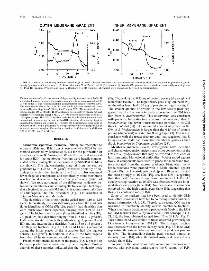
The densities of the protein peaks varied from 1.10 to 1.16g/cm3. Interestingly, the lowest-density peak from the gradients(later identified as OMs) (Fig. 2A, peak I), had densities thatwere much lower than expected, ranging from 1.09 to 1.12g/cm3. The highest-density peak (later identified as IMs) (Fig.2B, peak IV) had densities ranging from 1.15 to 1.17 gm/cm3.HMs were isolated from both gradients (Fig. 2, peaks II andIII) and had intermediate densities from 1.13 to 1.15 gm/cm3.The flagellar fractions (Fig. 1, FLA I and FLA II) recoveredduring the initial stages of the separation had the highestdensity (1.22 g/cm3). In addition, .95% of the total flagellafrom the cell lysate were recovered in these fractions.
Fractions that included each of the peaks (Fig. 1, peaks I toIV) were pooled and concentrated by centrifugation. Proteinanalysis of these samples indicated that the low-density peak
(Fig. 2A, peak I) had 0.25 mg of protein per mg (dry weight) ofmembrane isolated. The high-density peak (Fig. 2B, peak IV),on the other hand, had 0.55 mg of protein per mg (dry weight).The smaller amount of protein in the low-density peak sug-gested that this fraction potentially represented the OM frac-tion from S. hyodysenteriae. This observation was consistentwith previous freeze-fracture analysis that indicated that S.hyodysenteriae had fewer transmembrane particles in its OMthan E. coli did (38). The measured amount of protein in theOM of S. hyodysenteriae is larger than the 0.15 mg of proteinper mg (dry weight) reported for B. burgdorferi (3). This is alsoconsistent with the freeze-fracture data that suggested that S.hyodysenteriae OMs had more transmembrane particles thandid B. burgdorferi or Treponema pallidum (38).
Membrane markers. Several investigators have identifiedand characterized major antigens and other components of theOMs of S. hyodysenteriae that may be involved in virulence orhost immunity. Monoclonal antibodies (MAbs) raised againsttwo OM components were used to probe the membrane frac-tions isolated from the sucrose gradients. First, when mem-brane fractions were probed with a MAb directed againstSmpA (29), the lowest-density peak (r 5 1.10 g/cm3) reactedthe most strongly at 16 kDa (Fig. 3A, lane OM), suggestingthat this peak contained significant amounts of OMs. Anequally strong reaction at 16 kDa was observed with the inter-mediate-density peak (lane HM). No measurable reaction wasobserved with the high-density peak (lane IM), suggesting thatthis peak contained mostly IMs.
Some of the markers that have been used to identify OMsfrom other spirochetes have led to confusing results and erro-neous information (3, 4, 25). Therefore, a second OM markerwas used to tentatively identify various membrane fractions.When membrane fractions were probed with MAb to the LOS(an OM marker) from S. hyodysenteriae B204 serotype 2 (11,21, 31), the band obtained ranged from 16 to 26 kDa (Fig. 3).This diffuse band was similar to those observed previously forLOS from S. hyodysenteriae B204 (35). The strongest reactionwas observed with the lowest-density peak (Fig. 3B, lane OM)supporting the original observation that this peak was primar-ily OM. The intermediate-density peak reacted almost asstrongly (lane HM), while the high-density peak reacted veryweakly (lane IM).
To confirm the localization data, membrane fractions wereprobed with polyclonal antiserum to the C subunit of F1F0
FIG. 2. Analysis of sucrose step gradients. Fractions (1 ml) were collected from inner and outer membrane sucrose gradients and analyzed for protein (A280) anddensity (grams per cubic centimeter). (A) Peak I (fractions 23 to 27) and peak II (fractions 6 to 8) from the OM gradient were pooled and harvested by centrifugation.(B) Peak III (fractions 19 to 23) and peak IV (fractions 7 to 11) from the IM gradient were pooled and harvested by centrifugation.
5416 PLAZA ET AL. J. BACTERIOL.
on February 12, 2018 by guest
http://jb.asm.org/
Dow
nloaded from

ATPase (an IM marker) of E. coli. A 10-kDa protein in thehigh-density peak reacted the most strongly with antiserum(Fig. 3C, lane IM), confirming this fraction as IM, while noband was detected in the low-density fraction, identifying thisfraction as OM. A 9.5-kDa band was detected in the controllane containing total membranes from E. coli (lane TM).These data also strongly suggest that the low-density peak isOM while the high-density peak is IM.
Further analysis of membrane fractions. First, to measurethe contamination of the membrane fractions by soluble pro-tein, membrane fractions were assayed for NADH oxidaseactivity (a soluble marker) (34) (Table 1). The OMs contained2% of the total NADH oxidase activity from the cell lysate,while the IMs contained 1%. These data indicated that thevarious membrane samples showed only minor contaminationby soluble protein.
To measure the levels of cross-contamination among variousmembrane samples, OMs, HMs, and IMs of S. hyodysenteriaeB204 were probed with MAb to SmpA, MAb to serotype 2LOS, and polyclonal antiserum to the F1F0 ATPase C subunit(Fig. 3). Reactive bands were quantitated by densitometry, and
the results are shown in Table 1. Analysis with LOS as an OMmarker indicated 3% cross-contamination of IMs by OMs. Theanti-LOS MAb reacted with a diffuse band ranging from 14 to26 kDa. Joens et al. (11) had noted a similar reactive patternfor LOS from S. hyodysenteriae serotype 2 when membraneswere probed with anti-LOS polyclonal serum. Similar resultswere obtained with the MAb to SmpA, which demonstrated,1% cross-contamination. More than 97% of these markerswere measured in the OM and HM fractions. Less than 1% ofthe IM marker (F1F0 ATPase) was detected in the OM frac-tion, and 96% localized to the IM and HM fractions. Thesedata indicate that there were very low levels of cross-contam-ination between the OM and IM fractions.
Previous attempts by other groups to purify OMs from S.hyodysenteriae have been hindered by the periplasmic flagella.These complex structures are a major contaminant of mem-branes isolated by other techniques (11, 39, 40). To evaluatehow effectively the membrane separation technique eliminatedperiplasmic flagella from isolated samples, membrane frac-tions (as well as the flagellar fractions FLA I and FLA II [Fig.1]) were probed with antiserum to FlaB (Fig. 4). The antiserumreacted with three bands of 39, 35, and 33 kDa, which repre-sent the three different forms of FlaB. The OMs, HMs, andIMs showed little contamination by FlaB (Fig. 4, lanes OM,
FIG. 3. Localization of membrane markers. Immunoblots of membrane frac-tions probed with MAb to SmpA (A), MAb to serotype 2 LOS (B) or polyclonalantiserum raised against the 9.5-kDa C subunit of F1F0 ATPase from E. coli (C).The lanes contained OM, HM, or IM from S. hyodysenteriae B204, or totalmembranes (TM) from E. coli (C). Each lane contained 30 mg of protein.Reactive proteins were visualized by enhanced chemiluminescence (AmershamCorp.). The asterisks in the gel in panel C indicate reactive bands in the hybridand inner membrane fractions. The numbers on the left indicate the relativemobilities of molecular mass standards.
FIG. 4. Localization of flagellar proteins. An immunoblot of subcellular frac-tions probed with antisera raised against FlaB as shown. Lanes: STD, molecularmass standards; CL, cell lysate; OM, outer membranes (Fig. 2A, peak I); HM,hybrid membranes (Fig. 2B, peak III); IM, inner membranes (Fig. 2B, peak IV);FLA I and FLA II, periplasmic flagella (Fig. 1). Reactive proteins were visualizedby enhanced chemiluminescence.
TABLE 1. Physical and biochemical characteristics of membrane fractions
Fraction (density) [g/cm3])
% of:Amt of cholesterol
(mg/ml)Soluble marker(NADH oxidase)a
OM markers IM marker(F1F0 ATPase)b
Fla marker(FlaB)b
16-kDa SMLPb,c LOSb
OM (1.10) 2 52 60 ,1 2 380HM (1.14) 2 47 37 26 3 130IM (1.16) 1 ,1 3 70 5 30Fla (1.22) ,1 ,1 2 ,1 90 NDd
a Determined as a percentage of the total from cell lysate.b Determined by densitometry and expressed as a percentage of the total.c SMLP, 16-kDa surface membrane lipoprotein.d ND, not determined.
VOL. 179, 1997 OUTER MEMBRANE OF SERPULINA HYODYSENTERIAE 5417
on February 12, 2018 by guest
http://jb.asm.org/
Dow
nloaded from

HM, and IM). When the levels of contamination were mea-sured by densitometry, 90% of the FlaB was isolated in theFLA I and FLA II fractions. Less than 2% of the total FlaBwas detected in the OM fraction, while less than 5% localizedto the IM fraction (Table 1). Very similar results were obtainedwhen membrane and FLA fractions were probed with anti-serum to FlaA (data not shown). These data indicate that themembrane purification technique permitted the isolation ofOMs and IMs with minimal contamination by periplasmic fla-gella.
SDS-PAGE analysis of OMs and IMs of S. hyodysenteriaeB204. The OM, HM, and IM fractions were analyzed by SDS-PAGE (12.5% polyacrylamide) (data not shown). The purifiedOM contained 30 to 35 polypeptides, most of which wereunique to these membranes. The most prominent band was 39kDa, and this protein did not appear to be related to any of theflagellar proteins, as determined by immunoblotting. WhenOM proteins were analyzed by SDS-PAGE with or withoutheat treatment, the 39-kDa protein showed altered mobility,suggesting that it was heat modified (data not shown). The IMcontained 50 to 55 polypeptides with no predominant bands.However, most of the polypeptides appeared to be specific forthe IM. The HMs contained polypeptides from both OMs andIMs. This is consistent with the hypothesis that HMs are fu-sions between OMs and IMs.
Analysis of OMs and IMs of S. hyodysenteriae B204 by 2D-NEPHGE. Purified S. hyodysenteriae B204 OMs and IMs werecompared by 2D-NEPHGE, and the results are shown in Fig.5. The separation of proteins from OMs (Fig. 5A) resulted in40 to 45 discrete spots of different intensities. There wereseveral more polypeptides than were observed by SDS-PAGE,and additional proteins were identified which migrated withproteins with similar molecular masses when OMs were ana-lyzed by SDS-PAGE. The IM (Fig. 5B) protein separationresulted in 55 to 60 discrete spots of different intensities.Again, additional spots were observed compared to the num-ber observed by SDS-PAGE. Seven to ten polypeptides werecommon to both IMs and OMs, in particular, polypeptideswith apparent molecular masses of 31, 29, 28, 25, 24, and 22kDa. Polypeptides common to both OMs and IMs were de-scribed for purified B. burgdorferi membranes (e.g., OspA [3]).
Therefore, IM and OM markers better reflect the purity ofisolated fractions. It should also be noted that no protein withan apparent molecular mass consistent with serum albumin (66kDa) was observed in the OMs or IMs when analyzed by2D-NEPHGE (Fig. 5). This indicated that the major contam-inant from the culture media (albumin) had been successfullyremoved by the initial washing of the intact bacteria, the pas-sage of the membranes through the sucrose gradients, and thesuccessive washing of the membranes after isolation from thegradients and before analysis by 2D-NEPHGE.
Analysis of the membrane lipids. Stanton and Cornell (30,33) had previously reported that S. hyodysenteriae requiredcholesterol for growth and that the sterol was incorporatedinto membranes and was not metabolized. Also, it appearedthat all of the cholesterol taken up by the bacteria localized tothe membrane fraction. To further investigate the localizationof cholesterol in S. hyodysenteriae, we extracted the lipids fromOM, HM, and IM fractions and analyzed the samples by thin-layer chromatography. The results are shown in Fig. 6. The
FIG. 5. NEPHGE comparison of membrane fractions from S. hyodysenteriae B204. Proteins from OMs (A) and IMs (B) were compared by 2D-NEPHGE, with 50mg of protein being separated on each gel. The proteins were visualized by silver staining. The numbers on the left indicate the relative mobilities of molecular massstandards.
FIG. 6. Analysis of the lipids from membrane fractions from strain B204.Extracted lipids from OM, HM, or IM were separated by thin-layer chromatog-raphy and visualized as described in the text. The standards used were choles-terol (lane CE) and cholestanol (lane CA).
5418 PLAZA ET AL. J. BACTERIOL.
on February 12, 2018 by guest
http://jb.asm.org/
Dow
nloaded from
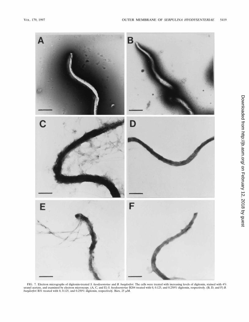
FIG. 7. Electron micrographs of digitonin-treated S. hyodysenteriae and B. burgdorferi. The cells were treated with increasing levels of digitonin, stained with 4%uranyl acetate, and examined by electron microscopy. (A, C, and E) S. hyodysenteriae B204 treated with 0, 0.125, and 0.250% digitonin, respectively. (B, D, and F) B.burgdorferi B31 treated with 0, 0.125, and 0.250% digitonin, respectively. Bars, 25 mM.
VOL. 179, 1997 OUTER MEMBRANE OF SERPULINA HYODYSENTERIAE 5419
on February 12, 2018 by guest
http://jb.asm.org/
Dow
nloaded from
OMs and HMs contained a lipid that migrated with cholesterol(Fig. 6, lanes OM, HM, and CE) and not with cholestanol (alsodetected in S. hyodysenteriae cell extracts exposed to hydrogen[32]). When the levels of cholesterol were assayed biochemi-cally in purified membrane fractions, 96% was isolated fromthe OM and HM fractions and 4% was isolated from the IMfraction (Table 1). The amount of cholesterol (380 mg/mg ofprotein) indicated that it was a major component of the OM,and the levels measured in the IM were within the limits formeasured levels of cross-contamination of IMs by OMs. Otherlipids identified were phosphatidylethanolamine and phospha-tidylserine (data not shown).
Effect of digitonin on S. hyodysenteriae. If the OM of S.hyodysenteriae contained significant levels of cholesterol, assuggested by the extraction data, this membrane should besensitive to agents that interact directly with sterols, such as thesaponin digitonin. These compounds form a complex with cho-lesterol (20) and disrupt erythrocyte membranes (27, 28). Tosee what effect digitonin had on S. hyodysenteriae, cells wereincubated with various concentrations of digitonin. The resultsare shown in Fig. 7. B. burgdorferi B31, which has an extremelylabile OM (3), was used as a control to ensure that the ob-served effects were not due to the staining technique. When S.hyodysenteriae cells were incubated with 0.125% digitonin, theOM was severely compromised (Fig. 7C) and cells were ob-served to release OM fragments and the periplasmic flagella.When the digitonin concentration was increased, the OM wascompletely removed and cells began to undergo lysis (Fig. 7E).At digitonin concentrations greater than 0.5%, lysis was essen-tially complete. Digitonin at 0.125% had no effect on B. burg-dorferi cells (Fig. 7D), and digitonin at 0.25% had a slight effect(Fig. 7F). This slight perturbation was not surprising consider-ing the labile nature of the OM of B. burgdorferi (3, 4, 25, 38).Clearly, digitonin had a very dramatic effect on the outer sur-face of S. hyodysenteriae.
DISCUSSION
The technique that we have developed to isolate OMs andIMs from S. hyodysenteriae permits the isolation of OMs withminimal contamination from the periplasmic flagella (Table 1;Fig. 4) and with little cross-contamination among variousmembrane fractions. Previous attempts by other groups toisolate S. hyodysenteriae OMs by Sarkosyl extraction wereplagued by contaminating periplasmic flagella (11, 39). In fact,the protein profile of Sarkosyl-extracted OMs most closelyresembled the FLA I and FLA II fractions from the discon-tinuous sucrose gradients used to purify IM and OM. Thus,this technique gives a realistic representation of both the OMand IM of S. hyodysenteriae. In addition, we have been able toidentify unusual characteristics of the OM of S. hyodysenteriae.First, the density of the OM was significantly lower than that ofany previously described OMs from any gram-negative bacte-ria (3, 13). Second, this represents only the second bacterialmembrane (Mycoplasma being the first) and the first bacterialOM that has been shown to contain cholesterol. Clearly, theOM of S. hyodysenteriae is unique.
Typically, a bacterial OM is more dense than the IM. This isbelieved to be due to several factors, including the types oflipids, the lipopolysaccharide, the capsule, and the proteincomposition of the OM. Spirochetal OMs have a reduceddensity compared to other bacteria with an IM and OM (3, 25).This is believed to be due to the decreased amounts of proteinin the OM (3, 4, 38) and, for T. pallidum and B. burgdorferi, thelack of lipopolysaccharide (1, 2). Walker et al. (38) haveshown, using freeze-fracture analysis, that S. hyodysenteriae has
levels of transmembrane particles that are significantly higherthan those of T. pallidum, B. burgdorferi, B. hermsii, or T.denticola (25, 26). In addition, Joens et al. (11) have identifieda LOS from S. hyodysenteriae. Taken together, these datamight suggest that the OM of S. hyodysenteriae would havegreater density than that of B. burgdorferi or T. pallidum. How-ever, we have found that the OMs of S. hyodysenteriae have thelowest density of any spirochetal OM (r 5 1.10 g/cm3) mea-sured to date. Since the S. hyodysenteriae OM we have isolatedcontained significant amounts of cholesterol, we believe thatthis is contributing to the overall density of this membranefraction.
Cholesterol is important for the healthy growth of Serpulina.In 1980, Lemcke and Burrows (14) gave the first report of thesterol requirement for the growth of S. hyodysenteriae. Inter-estingly, the addition of at least 1.25 mg of cholesterol per mlto the basal medium increased the viable counts approximately1,000-fold. Stanton and Cornell (30, 33) have studied the met-abolic fate of cholesterol and its nutritional role in S. hyodys-enteriae. Experimentally, S. hyodysenteriae can assimilate ap-proximately 4.4 mmol of cholesterol per 100 mg of cell dryweight, with .95% being incorporated into the membranefraction. Interestingly, at the membrane level, most of thesterols were in the form of cholestanol (the ratio of cholestanolto cholesterol in cellular lipids was 19:1). Furthermore, in aseparate experiment, cholestanol was found in the culture su-pernatant but none was detected in the uninoculated controlmedia. However, these authors later demonstrated that thepresence of H2 in the growth media promotes the nonenzy-matic reduction of cholesterol to cholestanol (34a). This couldoccur because S. hyodysenteriae produces significant amountsof H2 as an end product of the metabolism of glucose.
We have been able to demonstrate that the OMs of S.hyodysenteriae contain cholesterol but no cholestanol. Thiscould be because the S. hyodysenteriae cells were grown in thepresence of 5% oxygen. Stanton (31) demonstrated that cellextracts of S. hyodysenteriae produce less H2 and butyrate inthe presence of 10% oxygen than under 100% nitrogen. Thus,under the growth conditions used for cells in the membraneseparation experiments, less H2 was available for the nonen-zymatic reduction of cholesterol to cholestanol.
The role that cholesterol plays in the physiology and/orpathogenesis of S. hyodysenteriae can only be speculated. Thedifferences in the physical properties of biological membranescontaining cholesterol and those containing phospholipidsalone is well documented. These include increased rigidity ofthe overall membrane structure, decreased solute permeabil-ity, and a broadening of the phase transition temperature be-tween the gel (crystalline) and liquid (fluid) states (i.e., mem-brane fluidity is maintained over a wider temperature range)(reviewed in references 8 and 41). In addition, cholesterol mayplay a role in the uptake of fatty acids (e.g., cholesterol in-creases the uptake of oleic acid into the membrane of Myco-plasma capricolum) and in the conformation of membraneproteins (6). It is not known if some or all of these propertiesare important for the survival of S. hyodysenteriae in the intes-tinal tracts of host animals or for the pathogenesis of swinedysentery. However, because the OM directly interacts withinfected tissue, defining the components of this structure willlead to a better understanding of the complex relationshipbetween the bacterial pathogen and the host.
ACKNOWLEDGMENTS
We thank D. C. Krause and T. Hoover for critical reading of themanuscript.
This work was supported by Public Health Service grant AI33501
5420 PLAZA ET AL. J. BACTERIOL.
on February 12, 2018 by guest
http://jb.asm.org/
Dow
nloaded from
from the National Institute of Allergy and Infectious Diseases and bya University of Georgia biotechnology grant.
REFERENCES
1. Bailey, M. J., C. W. Penn, and A. Cockayne. 1985. Evidence for the presenceof lipopolysaccharide in Treponema phagedenis (biotype Reiter) but not inTreponema pallidum. FEMS Microbiol. Lett. 27:117–121.
2. Barbour, A. G., S. L. Tessier, and S. F. Hayes. 1984. Variation in a majorsurface protein of Lyme disease spirochetes. Infect. Immun. 45:94–100.
3. Bledsoe, H. A., J. A. Carroll, T. R. Whelchel, M. Farmer, D. W. Dorward, andF. C. Gherardini. 1994. Isolation and partial characterization of Borreliaburgdorferi inner and outer membranes using isopycnic centrifugation. J.Bacteriol. 176:7447–7455.
4. Brusca, J. S., A. W. McDowall, M. V. Norgard, and J. D. Radolf. 1991.Localization of outer surface proteins A and B in both the outer membraneand intracellular compartments of Borrelia burgdorferi. J. Bacteriol. 173:8004–8008.
5. Chatfield, S. N., D. S. Fernie, C. Penn, and G. Dougan. 1988. Identificationof the major antigens of Treponema hyodysenteriae and comparison withthose of Treponema innocens. Infect. Immum. 56:1070–1075.
6. Dahl, J. 1993. The role of cholesterol in Mycoplasma membranes. Subcell.Biochem. 20:167–188.
7. Dykstra, M. J. 1993. A manual of applied techniques for biological electronmicroscopy, p. 147–154. Plenum Press, New York, N.Y.
8. Gibbons, G. F., K. A. Miteoopoulos, and N. B. Myant (ed.). 1982. Biochem-istry of cholesterol. Elsevier Biomedical Press, New York, N.Y.
9. Harris D. L., R. D. Glock, C. R. Christensen, and J. M. Kinyon. 1972. Swinedysentery. I. Inoculation of pigs with Treponema hyodysenteriae (new species)and reproduction of disease. Vet. Med. Small Anim. Clin. 67:61–64.
10. Harris, D. L., and R. J. Lysons. 1992. Swine dysentery, p. 599–616. In A. D.Leman, B. E. Straw, W. L. Mengeling et. al. (ed.), Diseases of swine, 7th ed.Iowa State University Press, Ames.
11. Joens, L. A., R. Marquez, and M. Halter. 1993. Comparison of membranefractions of Serpulina (Treponema) hyodysenteriae Vet. Microbiol. 35:119–132.
12. Kennedy, M. J., D. K. Rosnick, R. G. Ulrich, and R. J. Yancey. 1988.Association of Treponema hyodysenteriae with porcine intestinal mucosa.J. Gen. Microbiol. 134:1565–1576.
13. Kotarski, S. F., and A. A. Salyers. 1984. Isolation and characterization ofouter membranes of Bacteroides thetaiotaomicron grown on different carbo-hydrates. J. Bacteriol. 158:102–109.
14. Lemcke, R. M., and M. R. Burrows. 1980. Sterol requirement for the growthof Treponema hyodysenteriae. J. Gen. Microbiol. 116:539–543.
15. Livermore, B. P. R. F. Bey, and R. C. Johnson. 1978. Lipid metabolism ofBorrelia hermsii Infect. Immun. 20:215–220.
16. Lysons, R. J., K. A. Kent, A. P. Bland, R. Sellwood, and W. F. Robinson.1991. A cytotoxic hemolysin from Treponema hyodysenteriae—a probablevirulence determinant in swine dysentery. J. Med. Microbiol. 34:97–102.
17. Markwell, M. A. K., S. H. Haas, L. L. Bieber, and N. E. Tolbert. 1978. Amodification of the Lowry procedure to simplify protein determination inmembrane and lipoprotein samples. Anal. Biochem. 87:206–210.
18. Miller, J. N. 1971. Spirochetes in body fluids and tissues, p. 3–24. In J. N.Miller (ed.), Manual of investigative methods. Charles C Thomas, Spring-field, Ill.
19. Morrisey, J. H. 1981. Silver staining for proteins in polyacrylamide gels: amodified procedure with enhanced uniform sensitivity. Anal. Biochem. 117:307–310.
20. Muhr, P., W. Likussar, and M. Schubert-Zsilavecz. 1996. Structure investi-gation and proton and C-13 assignments of digitonin and cholesterol usingmultidimensional NMR techniques. Magn. Reson. Chem. 34:137–142.
21. Muir, S., M. B. H. Koopman, S. J. Libby, L. A. Joens, F. Heffron, and J. G.Kusters. 1992. Cloning and expression of Serpulina (Treponema) hyodysen-teriae haemolysin gene. Infect. Immun. 60:529–535.
22. Nibbelink, S. K. and M. J. Wannemuehler. 1991. Susceptibility of inbredmouse strains to infection with Serpulina (Treponema) hyodysenteriae. Infect.Immun. 59:3111–3118.
23. Nuessen, M. E., J. R. Birmingham, and L. A. Joens. 1982. Biological activityof a lipopolysaccharide extracted from Treponema hyodysenteriae. Infect.Immun. 37:138–142.
24. Nuessen, M. E., L. A. Joens, and R. D. Glock. 1983. Involvement of lipo-polysaccharide in the pathogenicity of Treponema hyodysenteriae. J. Immu-nol. 131:997–999.
25. Radolf, J. D., K. W. Bourell, D. R. Akins, J. S. Brusca, and M. V. Norgard.1994. Analysis of Borrelia burgdorferi membrane architecture by freeze-frac-ture electron microscopy. J. Bacteriol. 176:21–31.
26. Radolf, J. D., L. L. Arndt, D. R. Akins, L. L. Curetty, M. E. Levi, Y. N. Shen,L. S. Davis, and M. V. Norgard. 1995. Treponema pallidum and Borreliaburgdorferi lipoproteins and synthetic lipopeptides activate monocytes mac-rophages. J. Immunol. 154:2866–2877.
27. Segal, R., and I. Milo-Goldzweig. 1978. The susceptibility of cholesterol-depleted erythrocytes to saponin and sapogenin hemolysis. Biochim. Bio-phys. Acta. 512:223–226.
28. Segal, R., P. Shatkovsky, and I. Milo-Goldzweig. 1974. On the mechanism ofsaponin hemolysis. I. Biochem. Pharmacol. 23:973–981.
29. Sellwood, R., K. A. Kent, M. R. Burrows, R. J. Lysons, and A. P. Bland. 1989.Antibodies to a common outer envelope antigen of Treponema hyodysente-riae with antibacterial activity. J. Gen. Microbiol. 135:2249–2257.
30. Stanton, T. B. 1987. Cholesterol metabolism by Treponema hyodysenteriae.Infect. Immun. 55:309–313.
31. Stanton, T. B. 1989. Glucose metabolism and NADH recycling by Trepo-nema hyodysenteriae, the agent of swine dysentery. Appl. Microbiol. 55:2365–2371.
32. Stanton, T. B. 1995. Personal communication.33. Stanton, T. B., and C. P. Cornell. 1987. Erythrocytes as a source of essential
lipids for Treponema hyodysenteriae. Infect. Immun. 55:304–308.34. Stanton, T. B., and N. S. Jensen. 1993. Purification and characterization of
NADH oxidase from Serpulina (Treponema) hyodysenteriae. J. Bacteriol.175:2980–2987.
34a.Stanton, T. B. Personal communication.35. ter Huurne, A. A. H. M., S. Muir, M. van Houten, B. A. M. van der Zeijst,
W. Gaastra, and J. G. Kusters. 1994. Characterization of three putativeSerpulina hyodysenteriae hemolysins. Microb. Pathog. 16:269–282.
36. Thomas, W., and R. Sellwood. 1992. Monoclonal antibodies to a 16-kDaantigen of Serpulina (Treponema) hyodysenteriae. J. Med. Microbiol. 37:214–220.
37. Towbin, H. T., T. Staehelin, and G. Gordon. 1979. Electrophoretic transferof proteins from polyacrylamide gels to nitrocellulose sheets: procedure andsome applications. Proc. Natl. Acad. Sci. USA 76:4350–4354.
38. Walker, E. M., L. A. Borenstein, D.R. Blanco, J. N. Miller, and M. A. Lovett.1991. Analysis of outer membrane ultrastructure of pathogenic Treponemaand Borrelia species by freeze-fracture electron microscopy. J. Bacteriol.173:5585–5588.
39. Wannemuehler, M. J., R.D. Hubbards, and J. M. Greer. 1988. Character-ization of the major outer membrane antigens of Treponema hyodysenteriae.Infect. Immun. 56:3032–3039.
40. Washerman, R. M., R. M. Phillips, and L. A. Joens. 1995. Production andcharacterization of monoclonal antibodies specific for lipooligosaccharide ofSerpulina hyodysenteriae. J. Clin. Microbiol. 33:2145–2149.
41. Yeagle, P. L. (ed.). 1988. Biology of cholesterol. CRC Press, Inc., BocaRaton, Fla.
VOL. 179, 1997 OUTER MEMBRANE OF SERPULINA HYODYSENTERIAE 5421
on February 12, 2018 by guest
http://jb.asm.org/
Dow
nloaded from